This work was conducted in the context of postmarketing biosafety assessment of genetically modified products. It presentsa systematic approach based on a chronic toxicity study on Wistar albino rats, with a range of combined parameters includingbiochemical, histopathological, and cytogenetic to evaluate the negative impact of a genetically modified (GM) diet on animal health.Histopathological and biochemical analysis procedures were performed in the liver, kidney, and testis. Cytogenetic analysis wasevaluated in germ cells and the liver. The results revealed that the laboratory diet used in our investigation was proved experimentally,using the PCR assay, to contain genetically modified components without being labeled as such. The results of all parameters evaluatedin our investigation were consistent and confirm that the GM diet fed to rats for 30, 60, or 90 days has deleterious histopathologicaland histochemical impacts. Biochemical alterations in alanine aminotransferase, aspartate aminotransferase, creatinine, uric acid, andmalondialdehyde concentrations were also observed. Genotoxicity of the GM diet was also demonstrated in germ cells as increasednumbers of cells with chromosomal aberrations and in liver cells as increased ratios of DNA fragmentation. In conclusion, the resultsof the present work indicate that there are health hazards linked to the ingestion of diets containing genetically modified components
1Cell Biology Department, Genetic Engineering and Biotechnology Division, National Research Center, Cairo, Egypt2Pathology Department, Medical Division, National Research Center, Cairo, Egypt
1. IntroductionAdvances in genetics and molecular biology have enabled the development and commercial release of genetically modified organisms (GMOs) with traits that transcend the species barriers. The development of GMOs offers the potential for increased agricultural productivity or improved nutritional values that can contribute directly to enhancing human health and development. The use of GMOs may also involve potential risks for human health and development (WHO, 2005), since many genes used in the production of GMOs have not been in the food supply before.
Most short- and medium-term studies with genetically modified (GM) foods indicate that they might cause health hazards when tested on animals. Several studies on mammals fed on commercialized GM soy and maize found consistent toxic effects on the liver and kidneys (Séralini et al., 2011). They demonstrated that long-term studies would be required to assess these effects more thoroughly.
While new types of conventional food crops are not usually subjected to safety assessment before marketing, safety assessments of GM foods were undertaken before the first crop were commercialized. However, unintended
consequences associated with the consumption of GM food by a genetically diverse population of humans and animals cannot adequately be evaluated during premarket risk assessment (EFSA, 2011). Therefore, it is important to consider applying postmarket monitoring for unexpected spread of GMOs that may pose food safety hazards (Kuiper et al., 2004). Postmarket monitoring is required to ascertain if prolonged exposure to GM food results in responses that were predicted by premarket risk assessment or to reveal the presence of side effects that were previously unpredicted.
This work was conducted in the context of postmarketing biosafety assessment of genetically modified products. It was designed to evaluate the potential impact of feeding rats with GM diets for 30, 60, or 90 days starting from weaning. Histopathological and histochemical investigations of some organs and their functions that are considered to be health indicators were performed. Blood samples were collected for biochemical analysis of alanine aminotransferase (ALT) and aspartate aminotransferase (AST) enzyme activity as an indicator of hepatocellular damage. Both creatinine and uric acid levels were measured as a test for kidney function.
Abstract: This work was conducted in the context of postmarketing biosafety assessment of genetically modified products. It presents a systematic approach based on a chronic toxicity study on Wistar albino rats, with a range of combined parameters including biochemical, histopathological, and cytogenetic to evaluate the negative impact of a genetically modified (GM) diet on animal health. Histopathological and biochemical analysis procedures were performed in the liver, kidney, and testis. Cytogenetic analysis was evaluated in germ cells and the liver. The results revealed that the laboratory diet used in our investigation was proved experimentally, using the PCR assay, to contain genetically modified components without being labeled as such. The results of all parameters evaluated in our investigation were consistent and confirm that the GM diet fed to rats for 30, 60, or 90 days has deleterious histopathological and histochemical impacts. Biochemical alterations in alanine aminotransferase, aspartate aminotransferase, creatinine, uric acid, and malondialdehyde concentrations were also observed. Genotoxicity of the GM diet was also demonstrated in germ cells as increased numbers of cells with chromosomal aberrations and in liver cells as increased ratios of DNA fragmentation. In conclusion, the results of the present work indicate that there are health hazards linked to the ingestion of diets containing genetically modified components.
Received: 20.06.2014 Accepted: 16.09.2014 Published Online: 00.00.2013 Printed: 00.00.2013
Research Article
ORABY et al. / Turk J Biol
2
Malondialdehyde (MDA) as a measurement of lipid peroxidation was assayed in liver cells as a biomarker of oxidative stress. Genotoxicity of the GM diet was evaluated using the analysis of chromosomal aberration and sperm abnormalities in germ cells. Analysis of DNA fragmentation ratio was also performed in liver cells.
2. Materials and methods2.1. Experimental dietThe experimental material consisted of a laboratory diet of mainly 60% yellow maize and 34% soybeans. The major nutritional contents of the laboratory diet were 22% protein, 3.48% fat, and 3.71% fiber. Currently, GM varieties of yellow maize and soybeans are produced for animal feed (Nowicki et al., 2010). Another well-balanced diet containing wheat, with the same nutritional value as the laboratory diet, was used as the non-GM diet.
The presence of GM components in these 2 diets was tested in our laboratory using a polymerase chain reaction (PCR) assay. DNA was extracted from samples of both diets using an extraction kit (DNeasy Mini Plant Kits, QIAGEN, CliniLab, Cairo, Egypt), following the instructions of the manufacturer. The DNA extracted from these 2 diets that were fed to the rats during the 3-month-long period of the experiment was screened for the presence of genetic modification using 2 pairs of primers specific for the Neomycin phosphotransferase II and CaMVP-35S/glyphosate-tolerant enzymes (CP4epsps). These 2 genetic elements are known to be used in the production of transformation (Hemmer, 1997). The primer sequences and PCR conditions for these 2 genetic elements are presented in Table 1.2.2. Experimental procedures Male Wistar albino rats obtained from the animal house of the National Research Center immediately after weaning were divided into 2 groups. One group of 30 animals (the GM diet group) was fed on a GM-tested laboratory diet for 30, 60, or 90 days.
The second group of rats (the non-GM diet group) was fed on the wheat-based diet for similar periods of time. Both groups of animals were housed in standard cages and
under standard conditions. Temperature was maintained at 22–25 °C and relative humidity was 55%–60% during the experiment. Exposure to light was maintained for 12 h. Diet and water were provided ad libitum. Animals were euthanized at 1 of 3 intervals: 30, 60, or 90 days. Samples were collected from their blood, liver, kidney, and testis for the subsequent analyses. The protocol applied throughout this study complies with the NRC Ethical Committee’s guidelines, and all animals received humane care. 2.2.1. Histopathological and histochemical investigations The collected specimens of liver, kidney, and testis were dissected immediately after death, washed thoroughly with saline, and fixed in 10% neutral-buffered saline for at least 72 h. All specimens were washed in tap water for 30 min, dehydrated in ascending grades of alcohol (70%, 90%, 95%, and absolute), cleaned in xylene, and embedded in paraffin wax. Serial sections were cut to a thickness of 6 µm and stained with hematoxylin (Hx) and eosin (E) (Drury and Wallington, 1980) for histopathological investigation and bromophenol blue (Mazia et al., 1953) for demonstration of protein content in the tissues. The protein materials appeared in the form of small bluish irregular particles in the cells.
Total protein content was determined by measuring the optical densities (OD) of the bromophenol blue stained sections using a Leica Qwin 500 image analyzer system. The numerical values of OD were analyzed statistically using an analysis of variance (ANOVA) test for multiple comparisons between different groups.2.2.2 Biochemical studiesAt the end of each of the experimental intervals (30, 60, or 90 days), blood samples were collected from all animals from the retro-orbital venous plexus for biochemical analyses. 2.2.2.1. Blood serum analysisThe blood sera were separated to evaluate ALT and AST enzymes activity according to Reitman and Frankel (1957). Concentrations of both creatinine (Thefeld et al., 1974) and uric acid (Haisman and Muller, 1997) were also evaluated.
Table 1. Primer sequences used for testing the presence of genetic modification in the diet used for this investigation, amplicon length, and annealing temperatures.
Target segments
Primer sequencesForward: (5´=>3´)Reverse: (5´=>3´)
NPTII Neomycin phospho-transferase II 5´-GGATCTCCTGTCATCT-3´ 5´-GATCATCCTGATCGAC-3´ 173 48
ORABY et al. / Turk J Biol
3
2.2.2.2. Measurement of lipid peroxidationMDA levels in the liver cells were determined using a spectrophotometric assay kit (Biodiagnostics) according to the manufacturer’s instructions. The lipid peroxidation values (absorbance at 534 nm) were expressed as nm MDA/mg tissue (Ohkawa et al., 1979). 2.2.3. Genotoxicity parameters Cytogenetic and DNA damage analysis were conducted in 2 different tissues (testis and liver, respectively) using different parameters.2.2.3.1. Chromosome analysis in germ cells Isolation of germ cells was performed according to Brewen and Preston (1978) from the testis. The chromosomes were spread on clean glass slides and stained with 2% Giemsa in phosphate buffered saline (pH 6.8) for 10 min. A total of 50 metaphase spreads per animal were analyzed to determine the total number of cells with chromosomal aberrations. Mitotic activity was also evaluated on the basis of the number of dividing cells (metaphase and prophase) in approximately 3000 examined germ cells per animal. 2.2.3.2. Analysis of sperm abnormalitiesSperm shape analysis from the epididymis was performed according to the published methods of Wyrobek and Bruce (1978) and Farag et al. (2002). At least 3000 sperm per group were assessed for morphological abnormalities. The sperm abnormalities were evaluated according to the standard method (Narayana, 2008). 2.2.3.3. Analysis of DNA fragmentation DNA fragmentation was determined in liver cells via colorimetric diphenylamine assay (Burton, 1968). Liver samples were collected from all rats immediately after being euthanized. Liver tissues were subjected to a lysis buffer and centrifuged for 20 min at 4 °C. The supernatant containing fragmented DNA was transferred to a new glass tube. The pellet containing intact DNA was resuspended in TE buffer (10 mM tris-HCl (pH 8), 1 mM EDTA). The fragmented DNA and intact DNA for each sample were incubated in 1.5 mL of 10% trichloroacetic acid (TCA) at room temperature for 10 min and then centrifuged. The precipitates were resuspended in 5% TCA, incubated at 100 °C for 15 min, and centrifuged. An aliquot of 2 mL of diphenylamine (DPA) solution (200 mg DPA in 10 mL of glacial acetic acid, 150 µL of sulfuric acid, and 60 µL acetaldehyde) was added to 0.5 mL of each supernatant (fragmented DNA and intact DNA) and incubated at room temperature overnight. The amounts of fragmented DNA and intact DNA were determined colorimetrically at 600 nm absorbance. The absorbance of low molecular weight DNA versus total DNA in each sample was expressed as a relative ratio (Gibb et al., 1997).
2.2.4. Statistical analysisStatistical analysis was performed with SPSS for Windows, version 11.5 (SPSS, Inc.). The data were analyzed using one-way ANOVA. The results were reported as mean ± SE and the differences were considered significant at P ≤ 0.05.
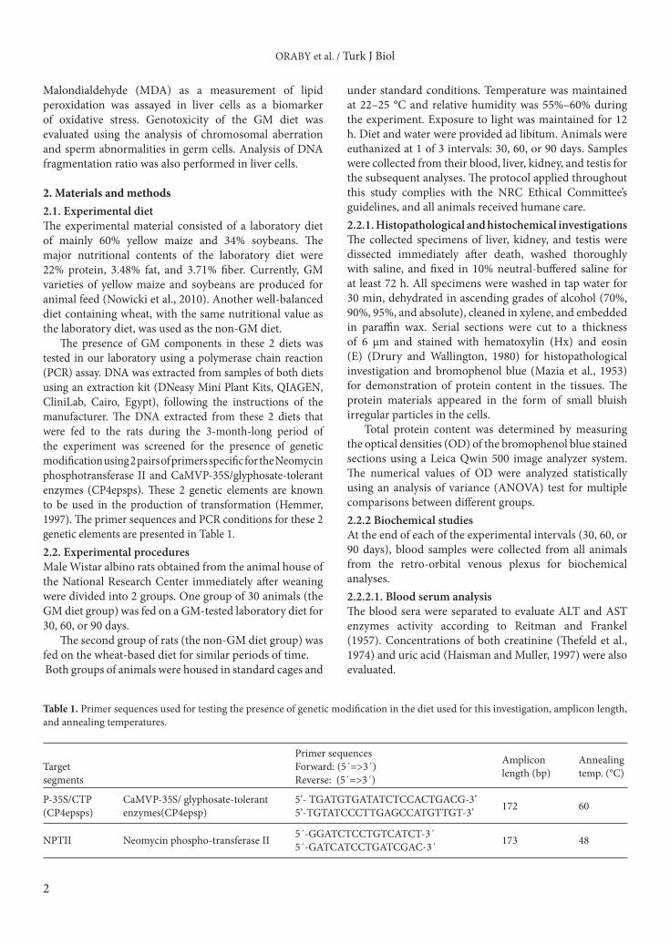
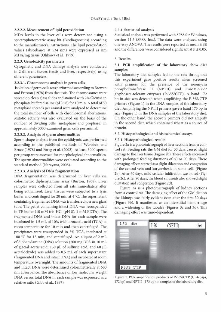
3. Results3.1. PCR amplification of the laboratory chow diet samplesThe laboratory diet samples fed to the rats throughout this experiment gave positive results when screened with primers for the presence of the neomycin phosphotransferase II (NPTII) and CaMVP-35S/glyphosate-tolerant enzymes (P-35S/CTP). A band 172 bp in size was detected when amplifying the P-35S/CTP primers (Figure 1) in the DNA samples of the laboratory diet. Amplifying the NPTII primers gave a band 173 bp in size (Figure 1) in the DNA samples of the laboratory diet. On the other hand, the above 2 primers did not amplify in the second diet, which contained wheat as a source of protein. 3.2. Histopathological and histochemical assays3.2.1. Histopathological resultsFigure 2a is a photomicrograph of liver sections from a con-trol rat. Feeding rats the GM diet for 30 days caused slight damage to the liver tissue (Figure 2b). These effects increased with prolonged feeding durations of 60 or 90 days. These damaging effects started as a slight dilatation and congestion of the central vein and karyorrhexis in some cells (Figure 2b). After 60 days, mild cellular infiltration was noted (Fig-ure 2c). After 90 days, the blood sinusoids also showed slight dilatation and congestion (Figure 2d).
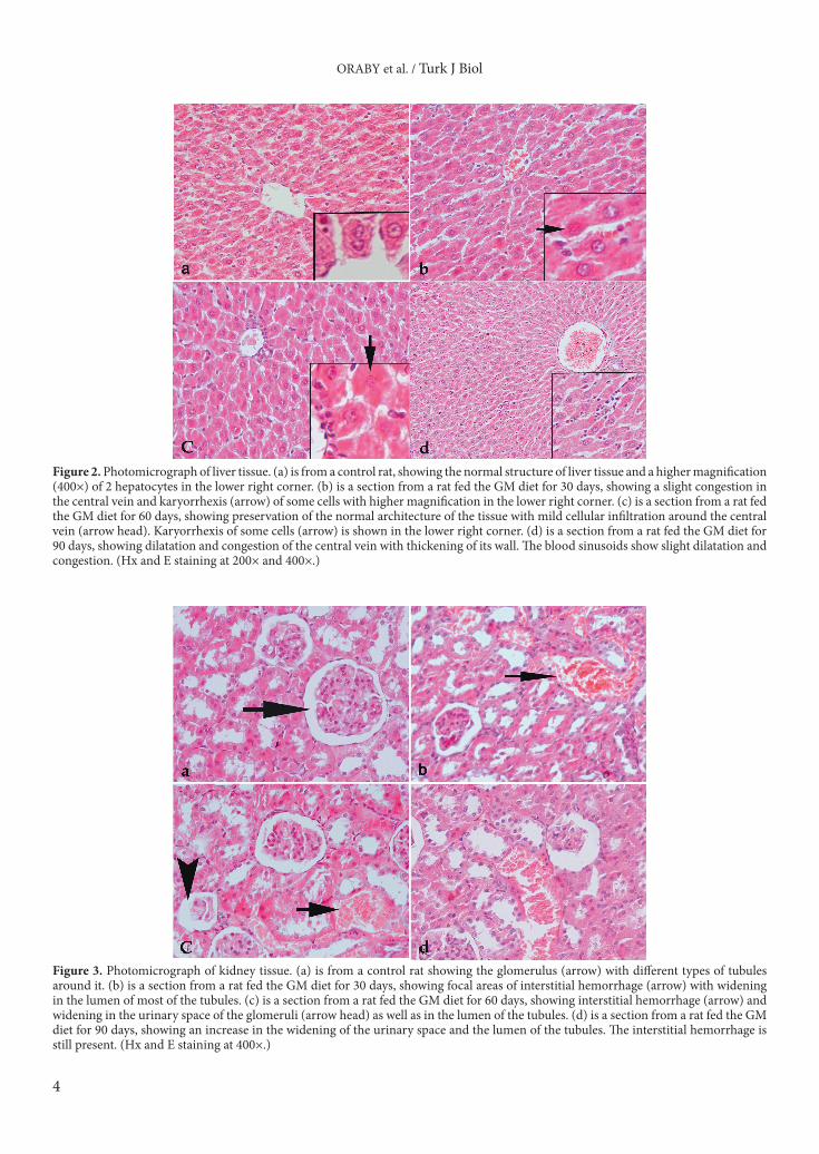
Figure 3a is a photomicrograph of kidney sections from a control rat. The damaging effect of the GM diet on the kidneys was fairly evident even after the first 30 days (Figure 3b). It manifested as an interstitial hemorrhage and a widening of the tubules (Figures 3c and 3d). This damaging effect was time-dependent.
Figure 1. PCR amplification products of P-35S/CTP (CP4epsps, 172 bp) and NPTII (173 bp) in samples of the laboratory diet.
ORABY et al. / Turk J Biol
4
Figure 2. Photomicrograph of liver tissue. (a) is from a control rat, showing the normal structure of liver tissue and a higher magnification (400×) of 2 hepatocytes in the lower right corner. (b) is a section from a rat fed the GM diet for 30 days, showing a slight congestion in the central vein and karyorrhexis (arrow) of some cells with higher magnification in the lower right corner. (c) is a section from a rat fed the GM diet for 60 days, showing preservation of the normal architecture of the tissue with mild cellular infiltration around the central vein (arrow head). Karyorrhexis of some cells (arrow) is shown in the lower right corner. (d) is a section from a rat fed the GM diet for 90 days, showing dilatation and congestion of the central vein with thickening of its wall. The blood sinusoids show slight dilatation and congestion. (Hx and E staining at 200× and 400×.)
Figure 3. Photomicrograph of kidney tissue. (a) is from a control rat showing the glomerulus (arrow) with different types of tubules around it. (b) is a section from a rat fed the GM diet for 30 days, showing focal areas of interstitial hemorrhage (arrow) with widening in the lumen of most of the tubules. (c) is a section from a rat fed the GM diet for 60 days, showing interstitial hemorrhage (arrow) and widening in the urinary space of the glomeruli (arrow head) as well as in the lumen of the tubules. (d) is a section from a rat fed the GM diet for 90 days, showing an increase in the widening of the urinary space and the lumen of the tubules. The interstitial hemorrhage is still present. (Hx and E staining at 400×.)
ORABY et al. / Turk J Biol
5
Figure 4a is a photomicrograph of testicular sections from a control rat. The testicular tissue of the rats fed on GM food for 30 days showed mild thickening of the base-ment membrane of the seminiferous tubules with the ap-pearance of gaps between the germinal epithelium of some tubules (Figure 4b). Prolonging feeding durations of 60 or 90 days caused an increase in the connective tissue com-ponent (Figures 4c and 4d, respectively) as well as in the number of Leydig cells, with a disarrangement of the ger-minal epithelium. 3.2.2. Histochemical resultsTable 2 displays the protein content values of the examined tissues (liver and kidney) from the non-GM diet group (control) and the different GM diet groups of rats. Protein
content was expressed as the mean optical density ± standard error (mean OD ± SE). Protein content in the liver tissues decreased significantly after feeding rats the GM diet for 30 or 90 days, which indicates dysfunction of some hepatocytes. Abnormal cellular activity in the kidney tissues was also confirmed by a statistically significant increase in the protein content values. 3.3. Biochemical analysis3.3.1. Blood serum analysisBiochemical analysis of AST and ALT activity revealed significant variations between the control animals and the animals fed on the GM diet (Table 3). The percent increase of AST and ALT activity ranged from 0.33% to 1.07% and from 0.33% to 0.92%, respectively.
Figure 4. Photomicrograph of testicular tissue. (a) is from a control rat, showing parts of 3 seminiferous tubules with small triangular areas containing Leydig cells between them (arrow). (b) is a section from a rat fed the GM diet for 30 days, showing mild thickening of the basement membrane of the seminiferous tubules with the appearance of gaps between the germinal epithelium of some tubules (arrow). (c) is a section from a rat fed the GM diet for 60 days, showing a noticeable increase in the connective tissue component between the tubules. In some of the tubules there is a disarrangement of the germinal epithelium (arrow). (d) is a section from a rat fed the GM diet for 90 days, showing a marked increase in the connective tissue component between the seminiferous tubules with dilatation and congestion of blood vessels. The germinal epithelium in the seminiferous tubules shows a disarrangement and detachment from the basal layer. No sperm are seen in the lumen of the tubules.
Table 2. Protein content values of examined liver and kidney tissues from the non-GM diet group and the different GM diet groups of the experimental rats.
GM diet30 days 0.272 ± 0.0123* 0.292 ± 0.0175*60 days 0.354 ± 0.0299 0.414 ± 0.0337*90 days 0.329 ± 0.0247* 0.449 ± 0.0269*
Data are presented as mean ± SE. * = the mean difference is significant at P ≤ 0.05.
ORABY et al. / Turk J Biol
6
Blood creatinine levels and uric acid concentrations were also significantly increased in animals fed the GM diet compared to the non-GM diet group (Table 3). The percent increases of creatinine levels ranged from 0.16% to 3.15%, whereas those of the uric acid concentrations ranged from 0.37% to 0.96%. 3.3.2. Measurement of lipid peroxidationMDA concentrations as a measure of oxidative stress in liver tissue are also presented in Table 3. The concentration of MDA increased significantly in all animals fed the GM diet compared to the non-GM diet group. The percent increase in MDA concentrations in the rats fed the GM diet ranged from 2.86% to 9.4%. 3.4. Genotoxicity results 3.4.1. Chromosome analysis in germ cellsTable 4 presents the mean percentages of chromosomal aberration and mitotic indices (MI) in the spermatocytes of rats
fed the GM diet or the non-GM diet for 30, 60, or 90 days. The percentage of chromosomal aberration of the spermatocytes in male rats fed the GM diet for 60 days recorded the highest value compared to the non-GM diet group and to the groups of rats fed the GM diet for 30 or 90 days.
The MI in rats fed the GM diet for 60 or 90 days were significantly linearly delayed compared to the MI of the non-GM diet group. This linear delay in the cell cycle was concomitant with a decrease in the percentage of chromosomal aberrations of the rats fed the GM diet for 90 days compared to the rats fed the GM diet for 60 days. 3.4.2. Analysis of sperm abnormalitiesMorphological sperm abnormalities in the control and GM diet groups are presented in Table 5. The results showed that the GM diet caused a significant increase in total sperm head and tail abnormalities in all experimental groups compared to the non-GM diet group.
Table 4. Total chromosomal aberrations and mitotic indices induced in the germ cells of rats fed the GM diet for 30, 60, or 90 days.
Diet Duration Groups No. of cells examined
Total chromosomal aberrations Mitotic indexM ± SENo. % M ± SE
Non-GM diet Control Control 500 4 0.8 0.4 ± 0.163 8.8 ± 0.326
GM diet
30 days 30 day GM diet 500 66 13.2 6.6 ± 0.221* 8.4 ± 0.221
60 days 60 day GM diet 500 138 27.6 13.8 ± 0.326* 6.8 ± 0.466*
90 days 90 day GM diet 500 80 16.0 8.0 ± 0.632* 6.6 ± 0.266*
Data are presented as mean ± SE. * = the mean difference is significant at P ≤ 0.05.
Table 5. Sperm head and tail abnormalities in rats fed on the GM diet for 30, 60, or 90 days.
Data are presented as mean ± SE. * = the mean difference is significant at P ≤ 0.05.
ORABY et al. / Turk J Biol
7
3.4.3. DNA fragmentation relative ratio (%)The data presented in Table 6 show that the DNA fragmentation percentage increased significantly in rats fed the GM diet for 30, 60, or 90 days compared to the control group. The DNA fragmentation percentage was higher for the group of rats fed the GM diet for 60 days than for those fed the GM diet for 90 days.
4. DiscussionAnimal experiments are important and provide valuable information regarding the safety of a GM plant for both livestock and human consumption (Alexander et al., 2007). The experimental material used in this study consisted of a laboratory diet that contained mainly 60% yellow maize and 34% soybean. Currently, GM varieties of yellow maize and soybean are produced for animal feed (Nowicki et al., 2010). The present investigated the possible toxic effects arising from feeding rats a GM diet containing both yellow maize and soybeans. This diet was experimentally proven to be genetically modified. Several products in Egypt were previously reported to be GM (Oraby et al., 2003, 2005) without being labeled as such.
The laboratory diet samples fed to the rats throughout this experiment gave positive results when they were screened with specific primers for the presence of NPTII (Neomycin phosphotransferase II) and P-35S/CTP (CaMVP-35S/glyphosate-tolerant enzymes (CP4epsps)). The presence of the regulatory sequence CaMV-35S promoter was previously detected by Oraby et al. (2003) in yellow maize grains used for animal feed. The CaMV-35S promoter is a principal component of transgenic constructs in more than 80% of GM plants (Cankar et al., 2008). A glyphosate-tolerant enzyme (CP4epsps) is also one of the main components of some GM maize and soy varieties (Zolla et al., 2008).
The first pair of the primers used in this experiment (P-35S/CTP) was chosen across the interfaces between a structural gene (glyphosate-tolerant enzyme CP4epsps) and the regulatory sequence (CaMVP-35S). The other pair
of primers amplified in the experimental diet was chosen within a single structural gene for NPTII that is used as selectable marker gene (Hemmer, 1997). The NPTII from E. coli, which confers resistance to the antibiotic Kanamycin, is most commonly used during the transformation process (Dale and Kinderlerer, 1995). The amplification of these 2 segments (CaMVP-35S/CTP and NPTII) in the diet samples used in this investigation confirms that this diet is genetically modified.
There is a growing concern that introducing foreign genes into food plants may have an unexpected and negative impact on human health. This work presents a systematic approach based on a chronic toxicity study with a range of combined parameters including biochemical, histopathological, and cytogenetic parameters. It focused on some of the tissues where histopathological and biochemical analyses were performed. Additional cytogenetic parameters were also evaluated.
Histopathological examination of liver sections showed nuclear changes in hepatocytes in the form of karyorrhexis in the hepatocytes of rats fed the GM diet for 30 or 60 days. Disturbed liver tissue as well as abnormally formed liver cell nuclei and nucleoli were observed (Ma-latesta et al., 2003; Vecchio et al., 2004) in mice fed on GM soy, which indicates increased metabolism and potentially altered patterns of gene expression. Significant modifica-tions of some nuclear features in the hepatocytes of GM-fed mice were previously reported (Malatesta et al., 2002). The authors assumed that GM food interferes with only some of the hepatocyte nuclear activities. Another study (Malatesta et al., 2008) demonstrated that GM soybean intake could influence some liver features during aging. They verified that senescence pathways were significantly activated in GM-fed mice. Others (El-Shamei et al., 2012) reported that rats fed on GM corn for 90 days showed his-topathological changes in the liver, kidney, and testis. Rats fed GM Bt maize over 3 generations suffered minor dam-age to the liver and kidneys and minor alterations in blood biochemistry (Kiliç and Akay, 2008). Furthermore, de
Table 6. DNA fragmentation in rats fed on the GM diet for 30, 60, or 90 days.
Diet Feeding durationDNA fragmentation (%)
Range M ± SE
Non-GM diet Control 7.6–14.8 11.83 ± 0.70
GM diet
30 days 16.66–24.36 19.0 ± 1.2 *
60 days 24.32–34.7 28.3 + 1.6*
90 days 21.39–27.73 24.3 + 0.7*
Data are presented as mean ± SE. * = the mean difference is significant at P ≤ 0.05.
ORABY et al. / Turk J Biol
8
Vandomois et al. (2009) reported that the modified maize varieties MON863, MON810, and NK603 had toxic effects on the liver and kidney in mammals.
The changes in the liver, an organ responsible for biotransformation and detoxification, suggest alterations in the metabolic processes, as reported by Malatesta et al. (1998). In the present investigation, biochemical changes also manifested. A marked increase in the levels of serum AST and ALT was observed in rats fed the GM diet for 30, 60, or 90 days. Increased levels of these 2 transferases are known to be an indicator of liver damage (Limdi and Hyde, 2003; Pratt and Kaplan, 2003), which was induced in the present work by the GM diet.
Significantly higher plasma activities of ALT were seen in female rats fed GM (GNA) rice (Poulsen et al., 2007). Minimal effects were also reported (Walsh et al., 2013) where ALT and AST were also slightly increased in sows fed on GM maize during gestation. ALT and AST activity was slightly altered in mice fed on GM soybean compared to a non-GM diet (Malatesta et al., 2002). Chemistry measurements including ALT activity and creatinine levels revealed signs of hepatorenal toxicity (Séralini et al., 2007) in rats fed on GM maize.
Additional data (Malatesta et al., 2008) indicated that several proteins belonging to hepatocyte metabolism and stress response were differentially expressed in GM-fed mice. In the present work, MDA concentrations as a measure of oxidative stress in liver tissue (Del Rio and Stewart, 2005) were significantly higher in all animals fed the GM diet for 30, 60, and 90 days. It has been previously suggested that Bt maize can potentiate oxidative cellular stress in immunized salmon (Gu et al., 2012). Trabalza-Marinucci et al. (2008) reported that female sheep fed GM Bt maize over 3 generations showed disturbances in the function of the digestive system, while their lambs showed cellular changes in the liver and pancreas.
Examination of kidney tissue from rats fed the GM diet revealed the presence of interstitial hemorrhage with changes in tubules. Rats fed GM Bt maize over 3 genera-tions suffered noticeable damage to the liver and kidneys and alterations in blood biochemistry (Kiliç and Akay, 2008). Renal tubules in the glomeruli of rats fed GM food for 60 or 90 days in the present study manifested widened lumina with widened urinary space. Similar results were reported by Poulsen et al. (2007), who explained that rats fed GM rice for 90 days had a higher water intake than the control group fed non-GM isogenic rice.
The histopathological results were further supported by the results of the kidney biochemical analysis. In the present investigation, blood creatinine and uric acid con-centrations increased significantly in rats fed the GM diet for 30, 60, or 90 days. Elevated creatinine and uric acid levels indicate impaired kidney function or kidney disease
(Chawla and Kellum, 2012). Increased serum creatinine levels at the end of lactation in pigs fed on Bt MON810 maize during their first gestation and lactation periods have been reported (Walsh et al., 2013). Significant chang-es in creatinine levels have also been reported in rats fed on GM Bt maize (Kiliç and Akay, 2008). Slightly increased levels of creatinine were also reported in rats fed on GM maize (Séralini et al., 2007).
The histochemical results of the protein content of the different tissues (Table 2) were consistent with those ob-tained from the histopathological analysis. Feeding rats a GM diet for 30 days caused a significant drop in the pro-tein contents of the liver and kidney specimens, which in-dicates a dysfunction in these tissues (Burkitt et al., 1993). Reductions in the concentration of protein were also re-ported (Schroder et al., 2007) in male rats fed with Bt rice.
The abnormal protein content results of the organs ex-amined in the GM-fed groups indicated abnormal cellular activity in these groups. It is known that proteins are not only a major structural component of cells but also, in the form of enzymes, mediate every metabolic process within the cells. Thus, the nature and quantity of proteins pres-ent within any individual cell determine the activity of that cell. Protein synthesis, therefore, is an essential and con-tinuous activity of all cells and in some cells it is the major function of these cells (Burkitt et al., 1993).
Very limited studies have been conducted to investi-gate the genotoxicity of diets containing genetically modi-fied plants. Results obtained by Jaszczak et al. (2008) did not reveal any difference in DNA damage between the control and experimental groups of mice fed a GM diet over 5 generations. Contrary to Jaszczak et al. (2008), in our investigation, the relative ratio of DNA fragmentation results revealed a higher ratio of DNA breaks in the liver cells of GM-fed rats.
In the present work, genotoxicity of the GM diet was also investigated in germ cells. Analysis of the spermato-genesis (Table 4) and sperm abnormalities (Table 5) re-vealed that there was a statistically significant increase in chromosomal aberrations and in the total count of abnor-mal sperm heads and tails at all experimental durations. These variations are suspected to be associated with the histopathological findings, in which the testicular tissues showed changes in their structure (thickening of basement membranes and increased connective tissue between the seminiferous tubules). These changes possibly reflect an attempt to protect the germinal epithelium from dam-age. Other researchers (Malatesta et al., 2003; Vecchio et al., 2004) reported that laboratory animals fed on GM soy showed disturbed testes function.
Results of the present work indicated signs of repair of the chromosomal aberrations (Table 5), which was driven by cell cycle delay. Cell cycle delay is a phenomenon that
ORABY et al. / Turk J Biol
9
occurs to ensure that critical events such as DNA repli-cation and chromosome segregation are completed with high fidelity (Elledge, 1996). Such induced cell cycle delay helps activate DNA repair (Zhou and Elledge, 2000). In the present work, cell cycle delay was evolved to control the genotoxic effects induced by feeding rats the GM diet for 30, 60, or 90 days, as the mitotic indices decreased linearly with increasing feeding duration, and the number of aber-rant cells in the germ cell population started to decrease in rats fed the GM diet for 90 days.
While there was a significant decrease (P < 0.003) in the number of aberrant cells of the group fed the GM diet for 90 days compared to the number of cells with chro-mosomal aberrations in the group fed the GM diet for 60 days, the decreased difference between the results of the group fed the GM diet for 90 days was still highly signifi-cant (P < 0.000) compared to the results from the non-GM diet group.
In contrast to all the above findings, other investiga-tions revealed no noticeable harmful impact on animal health. This may be attributed to the fact that most of the reported GM feeding studies incorporated a maximum of 33% GM animal feed in the test diet. No adverse effects were reported (Rhee et al., 2005) on the reproductive-
developmental ability of rats fed 5% GM potatoes for 5 generations. A diet containing 20% GM triticale (Jaszczak et al., 2008) did not induce chromosomal damage. No ap-parent adverse effect was also reported (Sakamoto et al., 2008) in rats fed on GM soy at a level of 30% in the diet for 104 weeks. Some of the studies that reported no adverse effects in animals receiving the test diet containing the Bt Cry1Ia12 protein (Guimaraes et al., 2010) were done not only at a low concentration (0.1%) in the diet but also by feeding rats for a very short duration (10 days).
4. ConclusionThe results of the present work reveal that the laborato-ry diet fed to rats throughout this work was proved ex-perimentally to contain genetically modified components without being labeled as such. The results of all the param-eters evaluated in our investigation were consistent and confirm that the GM diet fed to rats for 30, 60, or 90 days caused significant histopathological, biochemical, and cy-togenetic changes in all examined tissues.
AcknowledgmentThis experiment was funded by the National Research Center (Ninth Research Plan, 2010–2013).
References
Alexander TW, Reuter T, Aulrich K, Sharma R, Okine EK, Dixon WT, McAllister TA (2007). A review of the detection and fate of novel plant molecules derived from biotechnology in livestock production. Anim Food Sci Technol 133: 31–62.
Brewen GJ, Preston JR (1978). Analysis of chromosomal aberrations in mammalian germ cells. Chem Mutagen 5: 127–150.
Burkitt HG, Young B, Heath JW, Wheater PR (1993). Wheater’s Functional Histology: A Text and Colour Atlas. 3rd ed. Edinburgh, UK: Churchill Livingstone.
Burton K (1956). A study of the conditions and mechanisms of the diphenylamine reaction for the colorimetric estimation of deoxyribonucleic acid. Biochem J 62: 315–323.
Burton K (1968). Determination of DNA concentration with diphenylamine. Methods Enzymol 12B: 163–167.
Cankar K, Chauvensy-Ancel V, Fortabat MN, Gruden K, Kobilinsky A, Zel J, Bertheau Y (2008). Detection of nonauthorized genetically modified organisms using differential quantitative polymerase chain reaction: application to 35S in maize. Anal Biochem 376: 189–199.
Chawla LS, Kellum JA (2012). Acute kidney injury in 2011: biomarkers are transforming our understanding of AKI. Nat Rev Nephrol 8: 68–70.
Dale PJ, Kinderlerer J (1995). Safety in the contained use and the environmental release of transgenic crop plants. In: Tzotzos GT, editor. Genetically Modified Organisms: A Guide to Biosafety. New York, NY, USA: CABI, pp. 36–63.
de Vandomois JS, Roullier F, Cellier D, Seralini GE (2009). A com-parison of the effects of three GM corn varieties on mamma-lian health. Int J Biol Sci 5: 706–726.
Del Rio D, Stewart AJ, Pellegrini N (2005). A review of recent studies on malondialdehyde as toxic molecule and biological marker of oxidative stress. Nutr Metab Cardiovasc Dis 15: 316–328.
Drury RAB, Wallington EA (1980): Carleton’s Histological Technique. 5th ed. Oxford, UK: Oxford University Press.
Duijn GV, Biert RV, Bleeker-Marcelis H, Peppelmann H, Hessing M (1999). Detection methods for genetically modified crops. Food Control 10: 375–378.
EFSA (2011). Scientific opinion: guidance for risk assessment of food and feed from genetically modified plants. EFSA Panel on Genetically Modified Organisms (GMO). EFSA J 9: 2150.
Farag IM, Abdou HSA, Ayesh AM, Osfr MMH (2002). Chromosomal and sperm studies on the mutagenic effect of over heated meat and the protective role of green tea and ginseng on rats. Al-Azhar Bull Sci 13: 105–120.
El-Shamei ZS, Gab-Alla AA, Shatta AA, Moussa EA, Rayan AM (2012). Histopathological changes in some organs of male rats fed on genetically modified corn (Ajeeb YG). J Am Sci 8: 684–696.
ORABY et al. / Turk J Biol
10
Gibb RK, Taylar DD, Wan T, O’Connor DM, Doering DL, Gercel-Taylor C (1997). Apoptosis as a measure of chemosensitivity to cisplatin and taxol therapy in ovarian cancer cell lines. Gynecol Oncol 65: 13–22.
Gu J, Krogdahl A, Sissener NH, Kortner TM, Gelencser E, Hemre GI, Bakke AM (2012). Effects of oral Bt-maize (MON810) exposure on growth and health parameters in normal and sensitized Atlantic salmon, Salmo salar L. Br J Nutr 109: 1408–1423.
Guimaraes LM, Farias DF, Muchagata RCC, de Magalhaes MQ, Campello CC, Rocha TL, Vasconcelos IM, Carvalho AFU, Mulinari F, Grossi-de-Sa MF (2010). Short-term evalua-tion in growing rats of diet containing Bacillus thuringiensis Cry1Ia12 entomoxin: nutritional responses and some safety aspects. J Biomed Biotech: Article ID 630267, 8 pages, 2010. doi:10.1155/2010/630267
Hemmer W (1997). Foods Derived from Genetically Modified Organisms and Detection Methods. BATS Report 2/97. Basel, Switzerland: BATS Centre for Biosafety and Sustainability.
Jaszczak K, Kruszewski M, Baranowski A, Parada R, Bartlomiejczyk T, Zimny J, Rosochacki S (2008). Micronucleus test and comet assay on mice fed over five generations a diet containing genetically modified triticale. J Anim Feed Sci 17: 100–109.
Kiliç A, Akay MT (2008). A three generation study with genetically modified Bt corn in rats: biochemical and histopathological investigation. Food Chem Toxicol 46: 1164–1170.
Kuiper HA, Kleter GA, König A, Hammes WP, Knudsen I (2004). Safety assessment, detection and traceability, and societal aspects of genetically modified foods. European Network on Safety Assessment of Genetically Modified Food Crops (ENTRANSFOOD). Food Chem Toxicol 42: 1043–1202.
Limdi JK, Hyde GM (2003). Evaluation of abnormal liver function tests. Postgrad Med J 79: 307–312.
Malatesta M, Caporaloni C, Gavaudan S, Rocchi MBL, Serafini S, Tiberi C, Gazzanelli G (2002). Ultrastructural morphometrical and immunocytochemical analyses of hepatocyte nuclei from mice fed on genetically modified soybean. Cell Struct Funct 27: 173–180.
Malatesta M, Biggiogera M, Manuali E, Rocchi MBL, Baldelli B, Gazzanelli G (2003). Fine structural analyses of pancreatic acinar cell nuclei from mice fed on genetically modified soybean. Eur J Histochem 47: 385–388.
Malatesta M, Perdoni F, Santin G, Battistelli S, Muller S, Biggiogera M (2008). Hepatoma tissue culture (HTC) cells as a model for investigating the effects of low concentrations of herbicide on cell structure and function. Toxicol In Vitro 22: 1853–1860.
Malatesta M, Mannello F, Sebastiani M, Cardinali A, Marcheggiani F, Reno F, Gazzanelli G (1998). Ultrastructural characterization and biochemical profile of human gross cystic breast disease. Breast Cancer Res Treat 48: 211–219.
Mazia D, Brewer PA, Alfert M (1953). The cytochemical staining and measurement of protein with mercuric bromophenol blue. Biol Bull 104: 57–67.
Moore K, Roberts LJ (1998). Measurement of lipid peroxidation. Free Radic Res 28: 659–671.
Narayana K (2008). An aminoglycoside antibiotic gentamycin induces oxidative stress, reduces antioxidant reserve and impairs spermatogenesis in rats. J Toxicol Sci 33: 85–96.
Nowicki P, Aramyan L, Baltussen W, Dvortsin L, Jongeneel R, Dominguez IP, van Wagenberg C, Kalaitzandonakes N, Kaufman J, Miller D et al. (2010). Study on the Implications of Asynchronous GMO approvals for EU Imports of Animal Feed Products. Directorate-General for Agriculture and Rural Development of the European Commission.
Ohkawa H, Ohishi N, Yagi K (1979). Assay for lipid peroxides in animals tissues by thiobarbituric acid reaction. Anal Biochem 95: 351–358.
Oraby HA, Abou Mossallam AA, Hassan AA (2003). Detection of genetically modified crop plants using two structural genes (GUS and NPTII). J Genetic Eng Biotechnol (NRC) 1: 99–110.
Oraby HA, Hassan AA, Abou Mossallam AA (2005). Screening food products for the presence of CaMV 35S promoter and NOS3’ terminator. J Sci Food Agri 85: 1974–1980.
Poulsen M, Kroghsbo S, Schroder M, Wilcks A, Jacobsen H, Miller A, Frenzel T, Danier J, Rychlik M, Shu Q, et al. (2007). A 90-day safety study in Wistar rats fed genetically modified rice expressing snowdrop lectin Galanthus nivalis (GNA). Food Chem Toxicol 45: 350–363.
Pratt DS, Kaplan MM (2003). Evaluation of abnormal liver enzyme results in asymptomatic patients. N Engl J Med 342: 1266–1271.
Reitman S, Frankel S (1957). A colorimetric method for the determination of serum glutamic oxalacetic and glutamic pyruvic transaminases. Amer J Clin Pathol 28: 56–63.
Rhee SG, Dae HC, Yong HW, Ji HS, Soon SK, Seung JK, Rhee DL, Soo YC, Jae WK, Byung ML et al. (2005). Multigeneration reproductive and developmental toxicity study of bar gene inserted into genetically modified potato on rats. J Toxicol Env Health 68: 2263–2276.
Sakamoto Y, Tada Y, Fukumori N, Tayama K, Ando H, Takahashi H, Kubo Y, Nagasawa A, Yano N, Yuzawa K, et al. (2008). A 104-week study of genetically modified soybeans in F344 rats. Shokuhin Eiseigaku Zasshi (J Food Hyg Soc Jpn) 49: 272–282.
Schroder M, Poulsen M, Wilcks A, Kroghsbo S, Miller A, Frenzel T, Danier J, Rychlik M, Emami K, Gatehouse A et al. (2007). A 90-day safety study of genetically modified rice expressing Cry1Ab protein (Bacillus thuringiensis toxin) in Wistar rats. Food Chem Toxicol 45: 339–349.
Séralini GE, Cellier D, de Vendomois JS (2007). New analysis of a rat feeding study with a genetically modified maize reveals signs of hepatorenal toxicity. Arch Environ Contam Toxicol 52: 596–602.
ORABY et al. / Turk J Biol
11
Séralini GE, Mesnage R, Clair E, Gress S, de Vendomois JS, Cellier D (2011). Genetically modified crops safety assessment: present limits and possible improvements. Environ Sci Eur 23: 1–10
Thefeld W, Hffmiester H, Busch EW, Koller PU, Volmer J (1974). References value for determination of GOT (glutamic opal acetic transaminase), GPT (glutamic pyruvic transaminase) and alkaline phosphatase in serum with optimal standard methods. Deut Med Wochenschr 99: 343–351.
Trabalza-Marinucci M, Brandi G, Rondini C, Avellini L, Giammarini C, Costarelli S, Acuti G, Orlandi C, Filippini G, Chiaradia E et al. (2008). A three-year longitudinal study on the effects of a diet containing genetically modified Bt176 maize on the health status and performance of sheep. Livestock Sci 113: 178–190.
Vecchio L, Cisterna B, Malatesta M, Martin TE, Biggiogera M (2004). Ultrastructural analysis of testes from mice fed on genetically modified soybean. Eur J Histochem 48: 448–454.
Walsh MC, Buzoianu SG, Gardiner GE, Rea MC, O’Donovan O, Ross RP, Lawlor PG (2013). Effects of feeding Bt MON810 maize to sows during first gestation and lactation on maternal and offspring health indicators. Br J Nutr 109: 873–881.
WHO (2005). Modern Food Biotechnology, Human Health and Development: An Evidence-based Study. Geneva, Switzerland: WHO Food Safety Department.
Wyrobek AJ, Bruce WR (1978). The induction of sperm shape abnormalities in mice and humans. Chem Mutagens 5: 257–285.
Zhou BBS, Elledge SJ (2000). The DNA damage response: putting checkpoints in perspective. Nature 408: 433–439.
Zolla L, Rinalducci S, Antonioli P, Righetti PG (2008). Proteomics as a complementary tool for identifying unintended side effects occurring in transgenic maize seeds as a result of genetic modifications. J Proteome Res 7: 1850–1861.


![Feeding Study with Bt Corn (MON810: Ajeeb YG) on Rats ... · Egypt was GM maize[1]. “MON 810: Ajeeb YG” is a genetically modified corn that has resistance to borers, and this](https://static.documents.pub/doc/80x56/60e4ae89da78af3feb289836/feeding-study-with-bt-corn-mon810-ajeeb-yg-on-rats-egypt-was-gm-maize1.jpg)
