43
Biology 2201 Ch 9 – Control and Communication: The Nervous System 9.1 – Structure and Processes of the Nervous System (pp. 334-353)

Biology 2201 Ch 9 –Control and Communication: The Nervous System
9.1 – Structure and Processes of the
Nervous System (pp. 334-353)

Organization of the Nervous System
• nervous system - system that detects changes and responds to them; made up of the brain and spinal cord, as well as the nerves that emerge from them and connect them to the rest of the body
• The human brain alone contains over 100 billion nerve cells, and each nerve cell can have up to 10 000 connections to other nerve cells.
• The nervous system can be divided into various divisions, starting with the two broadest ones then dividing those further
• (1) Central nervous system (CNS)
• (2) Peripheral nervous system (PNS)

Divisions of the Nervous System

Divisions of the Nervous System
• central nervous system - network of nerves that includes the brain and spinal cord; integrates and processes information sent by nerves
• peripheral nervous system - network of nerves that carry sensory messages to the central nervous system (CNS) and send information from the CNS to the muscles and glands; consists of the autonomic and somatic systems• Somatic nervous system - consists of sensory receptors in the head and extremities, nerves that carry
sensory information to the central nervous system, and nerves that carry instructions from the central nervous system to the skeletal muscles. Under conscious control.
• Autonomic nervous system - controls glandular secretions and the functioning of the smooth and cardiac muscles. Not under conscious control. Divided into the sympathetic and parasympathetic divisions
• Sympathetic – prepares body for an emergency; “fight or flight response”; stress response
• Parasympathetic – calms body back down after an emergency; returns body to normal

Cells of the Nervous System
• The nervous system is composed of only two main types of cells: neurons and cells that support the neurons, which are called glial cells.
• Neuron - nerve cell; the structural and functional unit of the nervous system, consisting of a nucleus, cell body, dendrites, axons, and a myelin sheath• Neurons are the basic structural and functional units of the nervous system. They are specialized
to respond to physical and chemical stimuli, to conduct electrochemical signals, and to release chemicals that regulate various body processes.
• Glial cells nourish the neurons, remove their wastes, and defend against infection. Glial cells also provide a supporting framework for all the nervous-system tissue.

Neurons and Nerves
• Nerve - message pathway of the nervous system; made up of many neurons grouped into bundles and surrounded by protective connective tissue
• Neurons vary considerably in size and appearance, depending on their position and function in the body. Three main types of neurons, however, form the basic impulse-transmission pathway of the entire nervous system:• Sensory neurons – gather information from receptors (sense) and transmit these impulses to the central
nervous system (brain and spinal cord)
• Interneurons - Interneurons are found entirely within the central nervous system. They act as a link between the sensory and motor neurons. They process and integrate incoming sensory information, and relay outgoing motor information.
• Motor neurons - transmit information from the central nervous system to the muscles, glands, and other organs (effectors).

Types of Neurons

The Reflex Arc
• reflex arc - simple connection of neurons that results in a reflex action in response to a stimulus
• Reflexes are sudden, unlearned, involuntary responses to certain stimuli. • Examples of reflexes are jerking your hand away from a hot or sharp object, blinking when an object moves
toward your eye, or vomiting in response to food that irritates your stomach.
• Reflex arcs use few neurons, and as such tend to be fast.
• A reflex arc moves directly to and from the brain or spinal cord, before the brain centres involved with voluntary control have time to process the sensory information. • This is why, after stepping on a stone, you would not feel pain or cry out until after your foot was
withdrawn, once the brain has had time to process the information.

Reflex Arc example

Neuron Structure
• See fig 9.9 p. 340
• While neurons do vary on some structures and can vary in size, in
general they have four common features:
• Dendrites
• Cell body (soma)
• Axon
• Branching ends (terminal branches)

Neuron Structure cont…
• dendrite - short, branching terminal on a neuron that receives signals from other neurons or sensory receptors and relays the impulse to the cell body
• cell body - the main part of a neuron, containing the nucleus and other organelles and serving as the site of the cell’s metabolic reactions; processes input from the dendrites
• Axon - axon long, cylindrical extension of a neuron’s cell body; transmits impulses away from the cell body along its length to the next neuron

Neuron Structure cont…
• A neuron typically has one axon, which conducts impulses away from the cell body. Axons range in length from 1 mm to 1 m, depending on the neuron’s location in the body.
• Some nerves (white matter) have a myelin sheath around it, others do not (grey matter)• myelin sheath - the fatty, insulating layer around the axon of a nerve cell;
protects myelinated neurons and speeds the rate of nerve impulse transmission
• Schwann cell - a type of insulating glial cell that wraps around the axon of a neuron, creating a myelin sheath

Neuron Structure cont…
• axon terminals - release
chemical signals into the
space between them and
the receptors or dendrites
of neighbouring cells to
communicate with them

How Nerve Impulses are Generated
• Nerves impulses are electrical in nature. They result from a separation of electrical charges between the outside and the inside of a neuron cell membrane.
• Although neurons can conduct neural impulses from one area of the body to another, this process differs from electrical conduction along a wire. Nerve conduction is more complex and considerably slower. Unlike the movement of electrons along an electrical wire, nerve conduction depends on the movement of ions across the cell membrane of the axon.

Resting Membrane Potential and Polarization
• resting membrane potential - potential difference (voltage) across the membrane in a resting neuron
• polarization - lowering the membrane potential of the cell below its equilibrium value; in neurons, the generation of the resting potential
• When a neuron is at rest (not sending a signal or information), it has a resting potential of -70 mV (it is more negative on the inside)
• This resting potential is mainly generated by the work of the sodium-potassium exchange pump (see Fig 9.12 p. 342)
• sodium-potassium exchange pump - system involving a carrier protein in the plasma membrane that uses the energy of ATP to transport sodium ions out of and potassium ions into animal cells; important in nerve and muscle cells
• The sodium-potassium exchange pump exchanges three sodium ions for two potassium ions. As a result, an excess of positive charge accumulates outside of the cell.
• The cell membrane is not totally impermeable to sodium and potassium ions, so they leak slowly by diffusion across the membrane in the direction of their concentration gradient. However, potassium ions are able to diffuse out of the cell more easily than sodium ions can diffuse into the cell.

Action Potential
• action potential - in an axon, the change in charge that occurs when the gates of the K+ channels close and the gates of the Na+ channels open after a wave of depolarization is triggered
• When a nerve is sending a signal or message, a shift occurs in the polarization of the cell membrane
• depolarization - reducing a membrane potential to less than the resting potential of −70 mV
• threshold potential :the minimum change in the membrane potential required to generate an action potential; usually −55 mV
• If the neuron receives a stimulus of sufficient strength, special structures in the membrane called voltage-gated sodium channels open and make the membrane very permeable to sodium ions. The sodium ions on the outside of the axon suddenly rush into the axon, driven by their concentration gradient and the potential difference across the membrane. Within a millisecond or less, enough positively charged sodium ions have crossed the membrane to make the potential difference across the membrane in that tiny region of the axon +35 mV.

Repolarization
• Repolarization- return of a nerve to its resting potential following depolarization
• As a result of the change in membrane potential, the sodium channels close and voltage-gated potassium channels open. The potassium ions now move down their concentration gradient (toward the outside of the axon), carrying positive charge out of the neuron. The membrane potential overshoots to nearly −90 mV. At that point, the potassium channels close. The sodium-potassium exchange pump and the small amount of naturally occurring diffusion quickly bring the membrane back to its normal resting potential of −70 mV.
• refractory period - period following an action potential in which the membrane cannot be stimulated to undergo another action potential

Membrane Potential Throughout a Nerve Impulse

Myelinated vs. Unmyelinated Neurons
• With unmyelinated neurons, action potentials have to occur the entire length of the neuron. However, myelinated neurons are mostly coated with fat with spaces in between. With these neurons, action potentials only occur at these spaces (nodes of Ranvier) and nerve impulses “jump” from node to node.
• node of Ranvier - gap in the myelin sheath insulating the axon of a myelinated nerve cell

Myelinated vs. Unmyelinated Neurons
• saltatory conduction - situation where action potentials are forced to “jump” from one node of Ranvier to the next due to the myelin sheath,
• The transmission of an impulse along an unmyelinated axon is much slower than the saltatory conduction along a myelinated axon (about 0.5 m/s, compared with as much as 120 m/s in a myelinated axon).
• The nervous system disorder called multiple sclerosis is caused by the breakdown of the myelin sheath surrounding the axons in the central nervous system. The neurons can no longer efficiently carry electrochemical signals between the brain and the body.
• Multiple sclerosis is thought to be an autoimmune disease, in which the body’s own immune system breaks down the myelin. The symptoms of multiple sclerosis can include blurred vision, loss of balance, muscle weakness, fatigue, and slurred speech. Most people with multiple sclerosis experience periods of remission and periods of progression of the disease.
Signal Transmission Across a Synapse
• synapse - junction between two neurons or between a neuron and an effector (muscle or gland)
• Nerve cells are not directly connected to each other; rather there is a gap. The signals that cross this gap must be chemical, not electrical
• Neurotransmitter - chemical messenger secreted by neurons to carry a neural signal from one neuron to another, or from a neuron to an effector, such as a gland or muscle fibre

Neurotransmitters (see fig 9.18 p. 348)
• When an action potential arrives at the end of a presynaptic neuron, the impulse causes sacs that contain neurotransmitters to fuse with the membrane of the axon. These sacs, called synaptic vesicles, release their contents into the synaptic cleft by exocytosis. The neurotransmitters then diffuse across the synapse, taking about 0.5 to 1 ms to reach the dendrites of the postsynaptic neuron, or cell membrane of the effector.
• Upon reaching the postsynaptic membrane, the neurotransmitters bind to specific receptor proteins in this membrane. The receptor proteins trigger ion-specific channels to open. This depolarizes the postsynaptic membrane and, if the threshold potential is reached, initiates an action potential. The impulse will travel along the postsynaptic axon to its terminal and to the next neuron or an effector.
• Neurotransmitters have either excitatory or inhibitory effects on the postsynaptic membrane.
• If the effect is excitatory, the receptor proteins will trigger ion channels that open to allow positive ions, such as sodium, to flow into the postsynaptic neuron.
• If the neurotransmitter is inhibitory, the receptor will trigger potassium channels to open, allowing potassium ions to flow out. This results in a more negative transmembrane potential, resulting in hyperpolarization.
• A neuron may receive numerous both excitatory and inhibitory signals; the amounts of each will determine what occurs

Select Neurotransmitters
• Acetylcholine
• Can be excitatory or inhibitory, depending on location
• Excites skeletal muscle, inhibits cardiac muscle
• Normally, an enzyme called cholinesterase is released into a synapse, where it breaks down
acetylcholine. A nerve gas called sarin destroys this function by blocking the release of
cholinesterase into the neuromuscular junction. With the buildup of acetylcholine, critical
muscles, such as the heart and diaphragm, enter a state of constant contraction or paralysis.
Some insecticides affect insects in the same way and kill them.

Select Neurotransmitters

Biology 2201 Ch 9 –Control and Communication: The Nervous System
9.2. - The Central Nervous System
(pp. 354-364)

Grey Matter vs. White Matter
• grey matter - part of the nervous system that contains mostly cell bodies, dendrites, and short, unmyelinated nerve fibres; forms the outer areas of the brain and the H-shaped core of the spinal cord
• white matter - part of the nervous system that is made up of tracts of myelinated nerve fibres; forms the inner region of some areas of the brain, and the outer area of the spinal cord

The Spinal Cord
• The spinal cord is a column of nerve tissue that extends out of the skull from the brain and downward through a canal within the backbone
• The spinal cord is a vital communication link between the brain and the peripheral nervous system. Within the spinal cord, sensory nerves carry messages from the body to the brain for interpretation, and motor nerves relay messages from the brain to the effectors.
• The spinal cord is also the primary reflex centre, co-ordinating rapidly incoming and outgoing neural information.

Spinal Cord cont…
• contains both white matter and grey matter • The outer white matter consists of myelinated nerve
fibres. The butterfly-shaped core is made up of grey matter, which contains unmyelinated neurons as well as the cell bodies and dendrites of many spinal neurons.
• protected by cerebrospinal fluid, soft tissue layers, and the spinal column, a series of backbones (vertebrae).
• Injury to the spinal column can also damage the spinal cord, resulting in paralysis.

The Brain (fig 9.24 p. 356)
A = cerebellumB = medulla oblongataC = ponsD = midbrainE = thalamusF = hypothalamus G = cerebrum

Major Structures and Functions of Brain Components – Hindbrain
• Cerebellum - the part of the hindbrain involved in the unconscious co-ordination of posture, reflexes, and body movements, as well as fine, voluntary motor skills
• Medulla Oblongata - part of the hindbrain attached to the spinal cord at the base of the brainstem; controls automatic, involuntary responses, such as heart rate, constriction or dilation of blood vessels to control blood pressure, and the rate and depth of breathing, swallowing, and coughing
• Pons - part of the hindbrain found above and in front of the medulla oblongata in the brainstem; serves as a relay centre between the neurons of the right and left halves of the cerebrum, the cerebellum, and the rest of the brain

Major Structures and Functions of Brain Components – Midbrain
• Midbrain - part of the brain found above the pons in the
brainstem; relays visual and auditory information between areas of
the hindbrain and forebrain, and plays an important role in eye
movement and control of skeletal muscles

Major Structures and Functions of Brain Components – Forebrain
• Thalamus - sensory relay centre at the base of the forebrain that governs the flow of information from all other parts of the nervous system, mainly between the forebrain and hindbrain, and between areas of the sensory system (except for the sense of smell) and cerebellum
• Hypothalamus - region of the forebrain just below the cerebral hemispheres, under the thalamus; a centre of the autonomic nervous system responsible for the integration and correlation of many neural and endocrine functions; helps to regulate the body’s internal environment, as well as certain aspects of behaviour; coordinates the actions of the pituitary gland by producing and regulating the release of certain hormones
• Cerebrum - the largest part of the brain, divided into right and left cerebral hemispheres, which contains the centres for intellect, memory, consciousness, and language; interprets and controls the response to sensory information

Protecting the Brain
• Despite its central importance, the brain is fragile and has a gelatin-like consistency. However, it has several layers of protection
• The skull forms a protective bony armour around the brain.
• In addition, the meninges, three layers of tough, elastic tissue within the skull and spinal column, directly enclose the brain and spinal cord.
• meninges - three layers of tough, elastic tissue within the skull and spinal column that directly enclose the brain and spinal cord
• cerebrospinal fluid - dense, clear liquid derived from blood plasma, found in the ventricles of the brain, in the central canal of the spinal cord, and in association with the meninges

The Blood-Brain Barrier
• blood-brain barrier - protective barrier formed by glial cells and blood vessels that separates the blood from the central nervous system; selectively controls the entrance of substances into the brain from the blood
• The blood-brain barrier both protects the brain and supplies the brain with nutrients and oxygen. The blood capillaries that lead to the brain are made up of tightly fused epithelial cells. Thus, the capillary walls form a barrier that blocks many toxins and infectious agents.
• Some substances, such as glucose and oxygen, can still pass through the barrier by special transport mechanisms. Other lipid-soluble substances are able to pass directly through the lipid bilayer of the cell membrane. • Ex. caffeine, nicotine, alcohol, heroin
The Cerebrum
• cerebral cortex - thin outer covering of grey matter that covers each cerebral hemisphere of the brain
• The cerebral cortex is responsible for language, memory, personality, vision, conscious thought, and other activities that are associated with thinking and feeling
• Highly folded to increase surface area
• The right and left halves of the cerebrum are called the cerebral hemispheres. They are linked by a bundle of white matter called the corpus callosum.
• corpus callosum- bundle of white matter that joins the two cerebral hemispheres of the cerebrum of the brain
• Research indicates that while every cognitive function contains right-brain and left-brain components, some functions seem to have a dominant hemisphere.
• right-brain, or right cerebral hemisphere - associated with holistic and intuitive thinking, visual-spatial skills, and artistic abilities.
• left-brain, or left cerebral hemisphere - linked to segmental, sequential, and logical ways of thinking, and to linguistic and mathematical skills.
Lobes of the Cerebrum
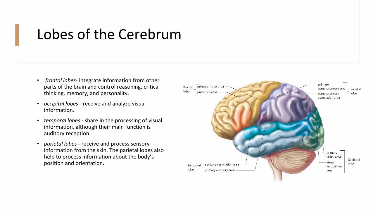
• frontal lobes- integrate information from other parts of the brain and control reasoning, critical thinking, memory, and personality.
• occipital lobes - receive and analyze visual information.
• temporal lobes - share in the processing of visual information, although their main function is auditory reception.
• parietal lobes - receive and process sensory information from the skin. The parietal lobes also help to process information about the body’s position and orientation.

Imaging Techniques Used to Study the Brain
• PET (positron- emission tomography)• A person receives an injection of radioactively labelled glucose, and a scanner monitors glucose
consumption in the person’s brain. Different colours represent different activity levels in the brain
• used to diagnose conditions such as a stroke or Alzheimer’s disease, in which the deterioration of the brain leads to memory loss and confusion, and eventual lack of conscious movement.
• MRI (magnetic resonance imaging)• A giant magnet surrounds the person’s head, and changes in the direction of the magnetic field induce
hydrogen atoms in the brain to emit radio signals. These signals can then be detected, translated, and displayed as a structural or functional image. MRI can also be used to identify various brain disorders, such as brain tumours.

Biology 2201 Ch 9 –Control and Communication: The Nervous System
9.3 – The Peripheral Nervous System
(pp. 365-369)

The Somatic System
• somatic system - division of the peripheral nervous system that controls voluntary movement of skeletal muscle
• includes 12 pairs of cranial nerves and 31 pairs of spinal nerves, all of which are myelinated. The cranial nerves are largely associated with functions in the head, neck, and face. (except the vagus nerve)
• Each spinal nerve contains both sensory and motor neurons, which service the area of the body where they are found.

The Autonomic System
• autonomic system - division of the peripheral nervous system that is under involuntary control
• The hypothalamus and medulla oblongata control the autonomic system, which has neurons that are bundled together with somatic system neurons in the cranial and spinal nerves.
• There are two opposing divisions of the autonomic system
• Sympathetic
• Parasympathetic

Sympathetic Nervous System
• sympathetic nervous system - division of the autonomic system that regulates involuntary processes in the body; typically activated in stress-related situations
• The sympathetic nervous system is typically activated in stressful situations and is often referred to as the fight-or- flight response. • The sympathetic neurons release a neurotransmitter called norepinephrine, which has an excitatory effect
on its target muscles.
• the sympathetic nerves trigger the adrenal glands to release epinephrine and norepinephrine, both of which also function as hormones that activate the stress response.
• At the same time, the sympathetic nervous system inhibits some areas of the body. For example, in order to run from danger, the skeletal muscles need a boost of energy. Therefore, blood pressure increases and the heart beats faster, while digestion slows down and the sphincter controlling the bladder constricts.
Parasympathetic Nervous System
• parasympathetic nervous system - division of the autonomic system that regulates involuntary processes in the body; typically activated when the body is calm and at rest
• The parasympathetic nervous system is activated when the body is calm and at rest. It acts to restore and conserve energy.
• Sometimes referred to as the rest-and-digest response, the parasympathetic nervous system slows the heart rate, reduces the blood pressure, promotes the digestion of food, and stimulates the reproductive organs by dilating blood vessels to the genitals. The parasympathetic system uses a neurotransmitter called acetylcholine to control organ responses.

Effects of Chemicals on the Nervous System
• Certain drugs can act as either stimulants or depressants by
directly affecting the sympathetic and parasympathetic nervous
systems.
• Ex. Caffeine would be classed as a stimulant as causit es the sympathetic
nervous system to increase heart rate and blood pressure.


