20
Biology 3.1 Looking at Cells
| Date post: | 28-Dec-2015 |
| Category: |
Documents |
| Upload: | lily-sibyl-mckinney |
| View: | 213 times |
| Download: | 0 times |
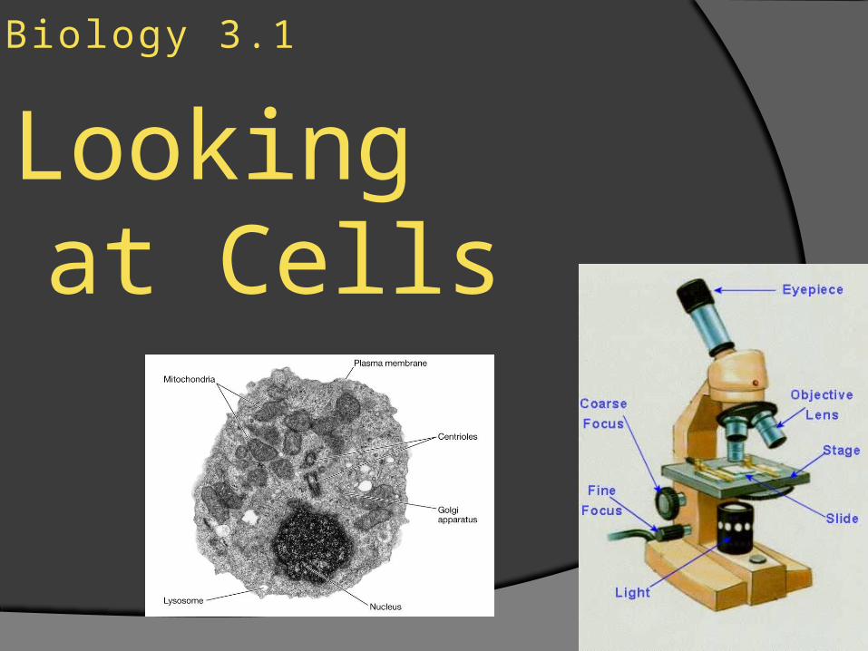
Biology 3.1
Looking at Cells
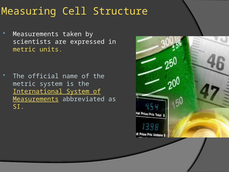
Measuring Cell Structure
Measurements taken by scientists are expressed in metric units.
The official name of the metric system is the International System of Measurements abbreviated as SI.

Measuring Cell Structure
SI is a decimal system so all units are based upon powers of 10.
A micrometer is a unit of measure in the SI system that stands for one-millionth of a meter.

Characteristics of Microscopes
Since Robert Hooke first observed cork cells, microscopes have unveiled the details of cell structure.
Biologists use different types of microscopes depending on the organisms they wish to study
Two common types of microscopes are light microscopes and electron microscopes

Light Microscope
In a light microscope, light passes through one or more lenses to produce an enlarged image of a specimen.

Electron Microscopes
An electron microscope forms an image of a specimen using a beam of electrons rather than a light.
Electron microscope image
Characteristics of Microscopes
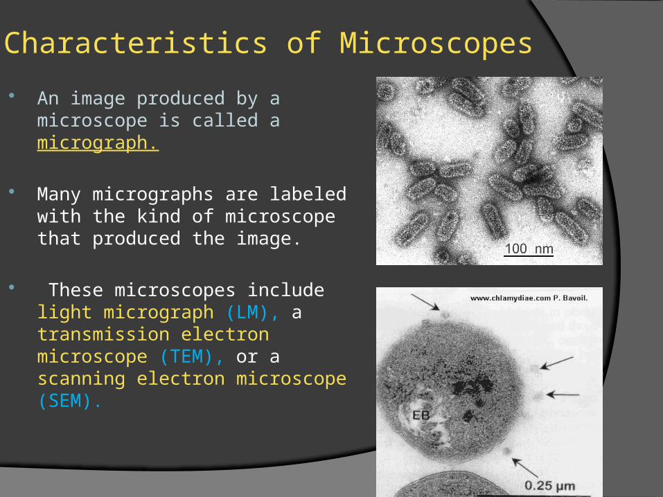
An image produced by a microscope is called a micrograph.
Many micrographs are labeled with the kind of microscope that produced the image.
These microscopes include light micrograph (LM), a transmission electron microscope (TEM), or a scanning electron microscope (SEM).
Characteristics of Microscopes
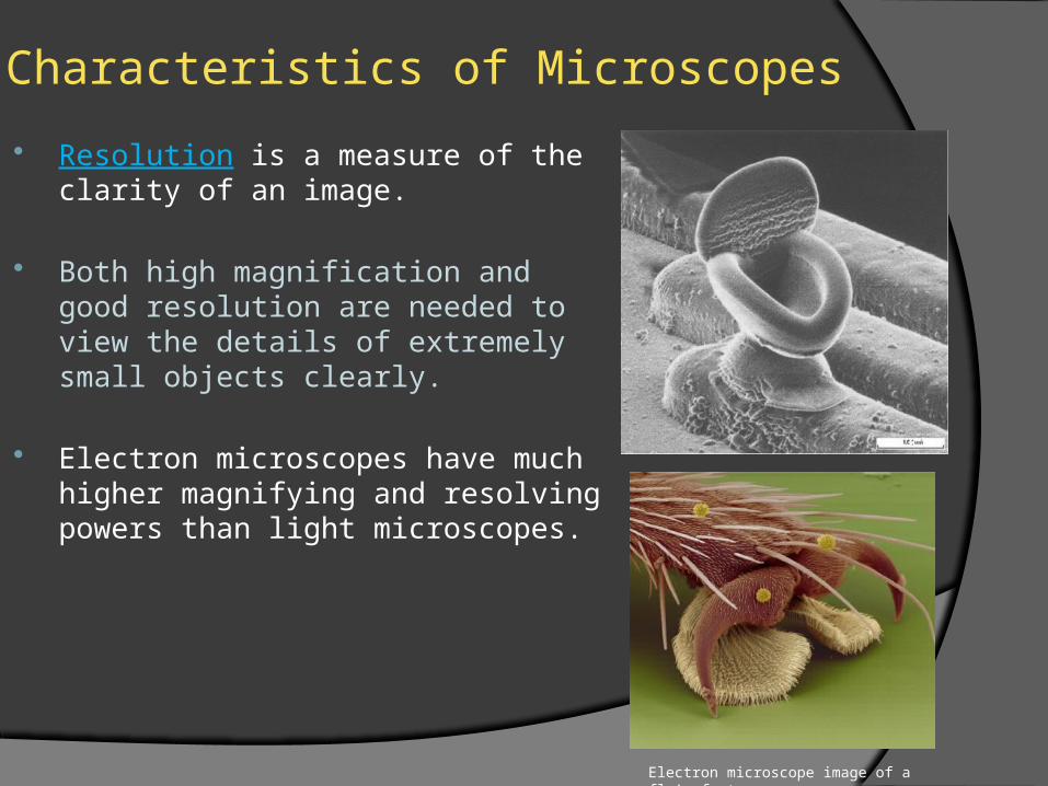
Resolution is a measure of the clarity of an image.
Both high magnification and good resolution are needed to view the details of extremely small objects clearly.
Electron microscopes have much higher magnifying and resolving powers than light microscopes.
Electron microscope image of a fly’s foot

Characteristics of Microscopes
Micrographs are often labeled with the magnification value of the image.
Magnification is the quality of making an image appear larger than it’s actual size.
For example, a magnification value of 200X indicates that an image is 200 times larger than the object’s actual size.

Compound Light Microscopes
Light microscopes that use two lenses are called compound light microscopes.
In a typical compound light microscope, a light bulb in the base shines light up through the specimen, which is mounted on a glass slide.
Compound Light Microscopes
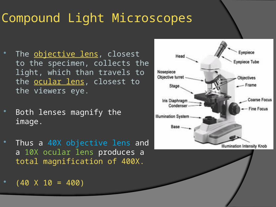
The objective lens, closest to the specimen, collects the light, which than travels to the ocular lens, closest to the viewers eye.
Both lenses magnify the image.
Thus a 40X objective lens and a 10X ocular lens produces a total magnification of 400X.
(40 X 10 = 400)

Electron Microscopes Electron microscopes can
magnify an image up to 200,000 times allowing them to study structure inside cells or on cell surfaces.
In electron microscopes, both the electron beam and the specimen must be placed in a vacuum chamber so that the electrons in the beam will not bounce off gas molecules in the air.

Electron Microscopes
Because living cells can not survive in a vacuum, they cannot be viewed using electron microscopes!!!
There are two types of primary electron microscopes; the Transmission Electron Microscope (TEM) and the Scanning Electron Microscope (SEM)

Electron Microscopes
In a Transmission Electron Microscope (TEM) the electron beam is directed at a thin slice of a specimen stained with metal ions.
Some structures in the specimen become more stained than others as they absorb more of the metal ions.

Electron Microscopes
The heavily stained parts of the specimen absorb electrons, while those that are lightly stained allow electrons to pass through.
The electrons that pass through a specimen strike a fluorescent screen, forming an image on the screen.

Electron Microscopes
In a Scanning Electron Microscope (SEM) the electron beam is focused on a specimen coated with a very thin layer of metal.
The electrons that bounce off the specimen form an image on a fluorescent screen

Electron Microscopes
A Scanning Electron Microscope (SEM) shows three-dimensional images of cell surfaces.
As with the Transmission Electron Microscope (TEM), the images produced are black and white but often they are artificially colored afterwards.
Scanning Tunnel Microscope
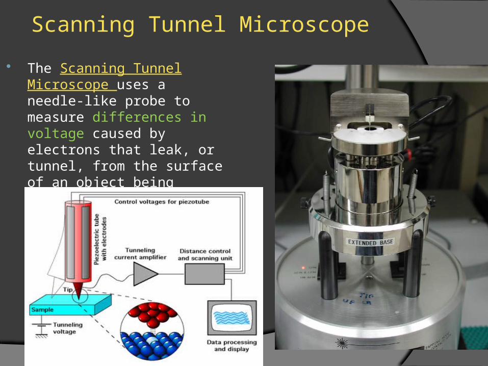
The Scanning Tunnel Microscope uses a needle-like probe to measure differences in voltage caused by electrons that leak, or tunnel, from the surface of an object being viewed.
Scanning Tunnel Microscope
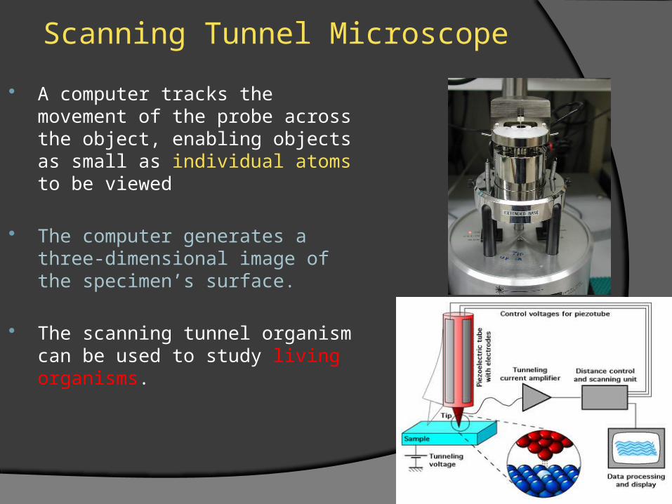
A computer tracks the movement of the probe across the object, enabling objects as small as individual atoms to be viewed
The computer generates a three-dimensional image of the specimen’s surface.
The scanning tunnel organism can be used to study living organisms.


