49
BIOLOGYOF CLONORCHIS SINENSIS b l' YOSHITAKAKOMIYA & NOR l]l SUZUKI

BIOLOGY OF CLONORCHIS
SINENSIS
bl'
YOSHITAKA KOMIYA & NORl]l SUZUKI

PREFACE
Since the dicovery of Clono7chis sinensis by MCCONNEL (1875), the biological,
pathological and clinical studies on C. sinensis have been carried out by many
workers of al1 the countries of the world
The initial description about C. sinensis in ]apan was made by ISIIIZAKA
(1878). He detected the worrn from a farmer in Okayama Prefecture (The
formal detection of C. sine耐 iswas described by BAELZ). Thereafter the study
on the worm in ]apan showed a marked advance. In 1910 K06AYASHI discovered
that cyprinoid 白shesserved as the Intermediate hosts of C1Qnorchis infection
In 1918 MUTO found the first intermediate host to be an aquatic operculate
snail. These discoveries have not only made clear the life cycle, infection
route, geographical distribution and preventive measure of the parasite, but
also contributed to the far-reaching development in the biological study of
C. sinensis
Thus, many studies about C. sinemis by wokers of ]apan occupy a
distinguished position in the parasitological society of the wor!d
Here these studies in the biological field with the life histoγy ,γe to deal with as the central theme
J. SCIENTIFIC NAME
The scientific name of this species was fixed upon C/onorchis sine肘"
(COBBOLO, 1875) LOOSE, 1907 through many troubles, and the discovery and
study of the worm in )apan played an important role in the determination of
the scientific name
The worm was obtained initially by MCCONNEL (1875) at autopsy of a
Chinese carpenter rεsiding in Calcutta
554
MCCONNBL sent it to COsBOLD to ask fOf its identi行cation,whereupon
COBsOLD (1875) gave it the name of " Disloma sinensis"
Later on BAELZ (1883), having di配 overed the ¥Vorm accidentaJly On an
autopsy of a patient suffering (rom pulmonal tuberculosis in the Tokyo Uni
versity Hospital, distinguished two species: viz. Disloma heρ'1lis innocltum
and Dislomαhepalis endemicum siι'e terniciosum. He thought that D. h. innOCUlO1f,
a larger form, had no pathogenicity and the other, a smaller form, to be of
pathological significance. This differentiation of two species by BAE!.Z met with
many oppositions. I[JIMA (1886) recognized that both剖 eidentical, and referred
the worm as Distomum endemicum BAELZ. LEUCKART (1886) and BLANCHARD
(1895) amalgamated BAELTZ'S two species into one, especially the latter creat
ing the genus Otisthorchis to include this species and referring it as OtislllOrchis
sinensis (COBsOLO)
Meanwhile, LooSE (1907) who took the distinct tWQ species over again,
suggested tnat BAELZ'S D. h. innocuum should be another species, though
BAELTZ'S D. h. endemicum and MCCONNEL'S showed the same species. And he
created the genus Clonorchis and included them in it
1. Clonorchis sinellsis (COBBOLD, 1875) LOOSE, 1907
2. Clonorcllis叩 demicus(BAELZ, 1883) L∞S芭, 1907
Later, KOBAYASHl (1912a), having made a study on the development of lhe
worm in various definitive hosts, stated that the size of the worm depends
upon the size of the host as we¥l as the number of parasite in one host, and
that other morphological differences including the shape of eggs between the
two species of LOOSE was not distinct
Thus he came to the conclusion that only one species of Clol1orchis sinensis
(COBBOLD, 1875) LOOSE, 1907 should be γecognized. This opinion is now generally
approved
11. ADULT ANATOMY
!,he structure of Clonorchis sinc1ISis has been carefully described by
KOBAYASHI (1912a, 1922)
A. Externa I Form
1. Shape, size and general apr加earance

555
Accotding lo KOBAYASHI (1912a, 1922), C. sinensis is markedly flat and the
posterior two thirds have an equa! breadth and anterior1y the body slightly
tapers. The size o( the worm varies according to its age, species of the
h051, number of the parasite in onc host and lhe type of fixation. it5
maximum and minimum length measuring 4 20mm. Generally speaking the
size of C. sinc同時 rangesfrom 8-15 mm in iength by 1.5-4 mm in width. The
living specin、enis transparent and slightly pink or brownish il1 color and has
yellowish or brownish pigment in the body. The size and color of the pigment
granulcs differ in different individuals and even in the same individual
KOBAYASIII assumed such pigment i5 due to the shell material from the vitellaria
The oral sucker lies at the anterior end and the ventral one is situated at
the level of one third or one fourth of the body length from the anterior end
The oral sucker (0.4-O.5mm) is usually a little larger than the ventral one (0.3
0.45 mm) in diameter
2. Cuticula and subcuticular tissue
The cuticula of the full grown specimen is relatively thin and has no
structure armed with no spines. The subcutaneous muscles are cons回 edof
transversal, longitudinal and diagonal layεrs. Under these muscles a layer of
subcutaneous cells is found They have a spindlc shape in younger
individuals. In full grown specimens, however, they are piliformed or star
shaped
The structure of the parenchym is of a reticular type with relatively fine
mesh and among the mesh cells of the vesicular type are found scaltered
Gianl cells of markedly large size are found in the muscles of uterus,
seminal vesicle, subcutaneous muscle, sucker and pharynx. They connect with
vanous organs 、,vithprocess
B. TnterJHlI Organ
1. Digestiv巴 organ
It initiates at the oral sucker and consists of pharynx, esophagus and
bifurcated川 testine. The intestinal ceca terminates a1 the posterior end of the
body. The pharynx is spherical with its inner surface cove問 dby cuticula. Its
wall co岡崎tsof well developed muscles. The inner wall of esophagus is covered
with a relatively thick cuticula and well develoI施 dtransversal and longitudinal
mu配 Ies. The intestine has epitherial celJs川 thewall and its exterior is
equippcd with transversal and longitudinal muscles, which are le田 developed
as compared w此hthe esophagus ones
2. Excretory system
The excretory pore opens at the posterior end. An excretory bladder is
1 shaped situating at the posterior part of the body. The main excretory canal

556
starts frorn both upper corners of the triangle of the excretory bladder
and runs upwards laterally across the intestinal caeca to the mid-body level,
wherεit divides into the anterior and posterior collecting tubes (KOBAYASHI,
1912a, 1922)
3. Nervous system
OZAKI (1960) observed the nervous system of C. sinensis by staining the
nervous celJ and fiber with mercury. He in白ltratedmodified Gilson's so¥ution
into the伺ukeby the aid of citric acid and deoxidized it by means of the
sun ray to precipitate the mercury in the nervous cell and fiber. According to
his observation the nervous system, as such is the case in trematodes in
general, consists of supra-oesophageal nerve commissure and three pairs of
longitudinal nerves (ventral, dorsal and lateral) running from both ends of the
former anteriorly as well as posteriorly. As compa同 dwith the well-developed
ventrallongitudinal nerve, the other longitudinal nerves are too poorly developed
to percelve
The huge nerve cells are arranged laterally sporadicaJly. The axial
filaments originating from them gather to forrn the longitudinal nerve. The
ring commissure connecting the longitudinal nerves was of reticulate structure
OZAKI (1960) considered that the undetermined running direction of the
nervous tissue in the reticular structure indicates the undifferentiated primitive
structure
4. Reproductive organs
The common genital atriurn opens median ventrally directly at the front
of the acetabulum and both malεand female organs unite directly before the
common openmg
a. Male genital organ
The male genital organ consists of two testes, two vasa eferentia, a
seminal vesic1e and an ejaculatory duct. The testes are dendritic, lying tandem
。nmedian line. The an担 riortestis has four branches and the posterior five
ones. These main branches a問 divided into irregulal' branchlets, but the
aspect how to branch is indefinite
The vas eferens arises from the center of the dorsal side of each testis
The two ducts from each testis run forwards for some distance and unite
together about at the middle part of the uterus winding, after forming itself
as a thick tube, opens directly into the seminal vesic1e
The seminal vesic1e runs sinuously anteriorly along the dorsal side of the
anterior portion of the uterus, turns ventral1y along the right margin of the
acetabulum, goes on 出m目指hingits diameter and forms itself as the ejaculatory
duct, which opens at the region of the anterior margin of the acetabulum

Fig. 1. Nervous system of ClonorcJ/Is $i l/e l/ú~
(OZAK1, 19略0)
1. Longitudinal nerve and nerve cell
Z. Nervous system of anterior part 3. Ne汀voussystem of posterior part
A Ventral sucker
A D N: Anterior dorsal longitudinal nerve
A L N: Anterior lateral longitudinal nerve A V N Anterior ventral longitudinal nerve
C N: Ring commi悩""o K Dorsal subcuticular surface nerve net
E: Esophagus
Inlestine
N C: Nervous cell
o S Oral sucホ"P D N Posterior dorsal longitudinal nerve
P H: Pharynx
P L N: Posterior lateral Iongitudinal nerve
P V N: Posterior ventral longitudinal ne円 e
S E N Supra.oesophageal nerve commissure
V K: Ventral subcuticular surface nerve net
557

558
along with the female genital opening
b. Female genital organ
The female genital organ is composed of one ovary, fairly、velldeveloped
vitelline glands, Laurer's canal, big seminal receptacle, ootype and uterus
(Fig. 2)
Fig. 2. Various reproductive organs which take place the formation of the egg-shell (UJlIE, 1936)
m d Meh1is' gland
, , ∞'YI" o v: ovary b y canal of KOsAYASHI'S egg-cell sucking
'P凹 ratuso d: oviduct d yolk sac
d c yolk duct r s seminal receptacle 1 C LAURIlR'S canal U t uterus
1. Vitel]ine gland and vitelline
cells
Both vitelline glands lie
exteriof to the intestinal ceca
They range from the level of
the anterior margin of the
acetabulum to the level of ovary,
and consist of many small glands
(KOBAYASHI, 1922). With a
fresh specimen they a問 slight
yellow i11 color forming an ap
pearance of a congromerate of
numerous grape-like bunches
Wはh a fixed specimen they
are dyed distinctly with various
stains, whereupon they can
be distinguished from the other
organs as an ovoid congro.
merates. The gland is a cavity
surrounded by a thin membrane
buried deeply in the body cells
and contains 8 to 20 vitelline cells
of diverse developmental stages
(UJI1E, 1936)
In山 ally the vitelline cells
are spheroid in shape attached
one another, holding in them the
evenly compact protoplasm
In the co山田 oftheir develop-
menl the light yellow minute granules are found di抽出utedevenly in the
protoplasm. Later the cell membrane has developed and the granules become
gradually bigger in size (UJ!1E, 1936)
No borders can be observed between the vitelline gland and vitelline duct
but the former gradually makes itself as a fine tube. The tubes gather into
559
two longitudinal vitelline ducts arranged on both sides of worm. They consist
of anterior and posterior branches which unite to become a transversal vitelline
duct at the region of the posterior end of the vitelline glands. The right and
left transversal vitelline ducts united in one, which swells up to make it田 lfas
a so called yolk sac. The yolk sac, with the narrow duct, bending like a bow,
opens to the oviduct at the position where it passes into the ootype (K06AYASHI,
1922; UJIIE,1936)
According to UJIIE, (1936), it has been ascertained that both the movements
of the yolk sac itself and the peristaltic movement of the oviduct transport
together the vitelline cells one by one from the yolk sac to the ootype
2. Ovary and egg-cel1
The ovary lies on the median line- anterior to the anterior testis, and
conSIS臼 ofthe mass of egg-cells of various growth stages and is surrounded
by a thin membrane. It is composed of three lobes. The egg-cel!s in the
centl'al dorsal part of the central lobe are found more developed. The oviduct
initiates from the blind duct near the dorso.central part of the central lobe
and reaches the ootype (UJIIE, 1936)
A ductus is observed on the surface of the ovarian part connecting the
oviduct and ovary. It is a called “spermatheca" of Looss (1894), or "suction
apparatus" of KOsA Y ASHI (1915). The wall of this apparatus expands and
contracts periodically to suck the matured egg-cells from the ovary and
transports them to the oviduct
The content of the oviduct is spermatozoa, which comes from the uterus
and is to be transfered to the seminal receptacJe
3. Mehlis' gland
The Mehlis' gland lies anterIor to the ovary and the gland is distributed
in the semトcirculararea dextral to the ovary with the radius about O.3mm
at the central lobe of the ovary. Around this area a large numher of glandular
cells are scattered. Their fine ducts gather at the central part and open into
the ootype found there (UJIIE, 1936)
4. Ootype
The oviduct connects itself with the ootype after uniting with the yolk duct
The ootype is a spindle-shaped cavity surrounded by Mehlis' gland. It measures
O.033mm. in length by O.023mm. in width and is connected with the uterus
(UJIIE, 1936).
5. Laurer's canal and seminal receptacJe
The Laurer's canal 凶 relativelylong and connects wIth the beginning of
the oviduct. The seminal receptacle is ovoid or piliformed (O.6.1.2xO.2-0.6mm.)
The lumen is usual!y filled with spermatozoa

560
After having a connection with the seminal receptacJe, the Laurer's canal
runs somewhat towards the abdorr】inalside a100& Its Ieft side of the latter and,
after turning inwardly backwards, opens at the anterior margin of the anterior
testis (KOsA Y ASHI, 1912a, 1922)
6. Uterus
The uterus is a curved 100g tube, occupying the area surrounded by the
tWQ intestinal caeca, the Qvary and the acetabulum, being filled with eggs
C. Histochemistry of the Fine Structure
00 the distributioll of lipid and its nature 00 C. sine出 is,MURAMATSU (1927)
reported that the sedimentation of the fat in every organ of the f1uke from
men was higher as compared with that from cats and dogs. The cholesterol
ester was seen in the parencymal ceJ1s and intestine of the fluke from men
but in small amounts in the fluke from cats, and none from dogs. In the
excretory organs the fat was found only in human parasitic cases. In the
parenchymal cells γelatively large amount of neutraI fat and lipoid was
found
SAWAOA (1926) examined the worm three years and three months after
the infection and found that in older worm配 niledegenerations were recognized
with decreased glycogen amount, whereas fat was found increased
Recently, TAKAGI (1962) examined the distribution of the fat, polysaccharide,
nucleic acid and the phosphatase in C. sinensis. The results showed that the fat
(neutral fat) was present in oesophagl1s, epithelial cells of intestine, and
parencymal cells around testis, oval"y and seminal receptacle most abundantly,
that glycogen in parencymal cells and spermatozoa, whereas RNA was present
in muscle tissues and female germ cells and DNA in male and female cells in
large amounts. Alkaline and acid phosphatase were found orominently in intestine
.and vitelline glal1d
III. THE EGG-MIRACIDIUM STAGE
A. Pormation of Egg.Shell
The detai!ed study on the formation of egg-shell by UJlie (1936) is available
According to him, one matured egg cell appears in the central part of the
surface of the middle lobe of the ovary, and arrives at the in山 alportion of the
-ootype through the oviduct 、Nhenthe egg cell passes throl1gh the

the yolk duct to enter the ootype, the central part of the yolk sac almost
suddenly contracts so as to form an arc-shaped narrow part. And subsequently
its lower part autornatically makes rhythmical movements which makes the
vItelline cells enter into the yolk duct one by one. The duct take渇 aperistaltic
movement so as to transport the vItelline cells to the ootype following one by
one after the egg cell
The formation of egg-shell takes place in the ootype. When the vItelline
cells increased by 5 to 7 in number, the yolk sac suspends its movement
for a time. In the mean time these cells attach themselves to the egg cell
lying in the initial portion of the spindle-shaped ootype and fill up the ootype
At this stage the terminal part of the oviduct contracts and its opening to the
ootype is closed. Then the ootype dillates, and makes space between these
cells and the wall of the cavity
COllsequently the shell forming
granules are released from vitelline
cells and gather around them to form
the egg-shell. When the ootype 也icontracts, a part of these granules
constituting the wall extends back-
ward along the constricted wall of
the oviduct as the fine tube, which
is reduced to become the posterior
spinal process of the egg. i. I (I01
When the ootype again dilates,ト-
a small space occurs at the anterior
portion of the egg uncovered with
the granules. Consequently the
granules adhere to its surface
secondarily and form the operculum
Then, the egg is transfered to the ι
If 11) uterus (Fig. 3). L:...:....:....
UJIIE(1936) states that the Mehlis'
gland plays an important role in the
formation of the egg shell firstly by means of its characteristic automatic
contractive and dilative movements and secondly by its secretioll
50'
¥
AMVL
/
¥
戸川比 命
A
企同wvト
Le
(仏J
Fig. 3. Formatio、。fegg-shell in the ootype (UjllE. 1936)
s. Ferti1izntion
NAKAYAMA (1912) asserted that the fertilizatioll was sometimes completed
in the oviduct and that the egg cell had, after being enclosed in the egg-shell,
already been fertilized by the spermatozoa and the egg cell kept on its original

562
state stil! after the ferti!ization. The egg-cell was enclosed in the egg-shel1
after completing the fertilization and transported to the uterus as the complete
egg
UJlIE (1936) also considered that the egg cel1 had met the spermatozoa from
the moment of its appearance at the beginning of the oviduct up to the time
when the operculum、.vasformed in the ootype, since the active spermatozoa
were found numerously in the oviduct, especially in its beginning
c. Dcvelopment of Egg
The initial study on the embryonic development was made by SA!TO (1898),
which was followed by the detailed observation of NAKAYAMA (1910), and later
by KOBAYASH! (1922). To summarize their results, the egg, after forming the
egg-sheJl at the beginning of the uterus, is usually composed of an egg cell and
5 vite11ine cells. The egg cel1, in which already one spermatozoon entered,
lies anteriorly in the egg-shell
The vite11ine ceUs attach themselves to one another after releasing the shel1
forming granules and their outline becomes indistinct. The germ cell has six
chromosomes. By the maturation of the geγm cel1 the two polar bodies are
released out and the chromosome becomes 3 in number. After the fertilization
its chromosome amount to 6 ill number by adding another 3 from spermatozoon
The fertilized egg-cel1 starts the cell division, which is equal and holoplastic
cleavage. Along with further divisions, a cel1 is separated and removes to the
portion near the posterior end of the egg, divides, and gradually forms a
vite11ine membrane. Then, the two cells detach from the embryo posteriorly
These ceJ1s divide graduaUy臼 kingthe appearance of foam, and adhere to the
vitelline membrane partially. These cells and the shell membrane perform the
function to keep the embryo in the fixed position
Then the ectoderm become differentiated from the blastula and the
miracidium is developed. It covers with several rows of ciliated fiat plates, except
the area where the cells of foam appearance attach the embryo. In the anterior
part of the embryo a primitive gut is found conical in forrn. ln the postrior
half of the body a group of cel1s which corresponds with the embryos of the
next generation is recognized
D. Shape and Size of the Egg
1. Malure egg
The morphology of the egg of C. sinensis has been observed by tnany
workers (IUIMA, 1888; LEUCKART, 1889; OOTANI, 1892; OSAFUNE, 1898a,b; SA!TO,
1898; ISHII, 1929a). It measures 0.026.0. 030x O. 015.0. 017 mm., ovoid in form,
narrowing a ¥ittle posterior to the portion of the operculum. The op町 culum
takes the shape of a watch-glass with the prorninent shoulder-¥ike rirn and is
563
found on the narrow anterior end of the shell. The shell is about 0.005-0.008 mm
thick. According to FAUST et al. (1927) and HASEGAWA (1929), the surface of
the shell shows etchings of an arabesque polygonal pattern. HASEGAWA states
that the pattern is based on ¥ines projecting to the surface of the shell. YUMOTO
(1936a) reported that the shell was composed of two layers. The inner layer
was yellowish brown and a ¥ittle thick as compared with the outer. Its thickne日
was uniform throughout. The outer one was light greenish yellow in color and
thin. I-Ie stated that the outer layer was hardly distinguished from the inner
one by the light reflexion
2. Abnormal egg
KITAMURA (1916) found the egg without the operculum and regarded it as
the unfertilized egg. FAUST el al. (1927) made a brief description on the egg
without the operculum under the name of the imperfect egg, and stated that
its contents consisted of an egg either lacking yolk cell and granules or egg cell
ls削 I(1929a) reported that he could differentiate 4 kinds of abnormai eggs in
stool, and that these eggs were produced by the worm of which vitality has
weakened
YUMOTO (1936b), who made the ob措 rvationon abnormal eggs, divided them
into the two groups, namely “abnormal" egg and "incomplete" one. Both
eggs have the shell of the same structure as normal one. The former, however,
has no operculum, though the etching of the arabesque polygonal pattern can
be found on the surface. Its content is degenerated and a drop-like substance is
recognized in it. The latter holds no miracidium, though it is possessed of the
operculum. He states that the "abnormal" egg is always produced throughout
the worm's life, but the “incomplete" one is produced l1umerously by the
initial egg laying period of the young as well as by the senile one
UjlJE (1936) considered that abnormal egg was produced when the worm
either was young or very old, or when it is affecred by an abrupt change in the
surrounding and an e仔ectof anthelminthics etc. He deduced its mechanism
as follows: 1) A temporary suspensIon of the egg cell delivery alone into
the ootype due to immaturity and senility may result in the formation of Ilon
0",,'印lated,no egg-cell eggs. 2) When one or t¥¥lO vitelline cells arrive at the
ootype in advance, non-operculated eggs containing an egg cell may be formed,
and 3) operculated or non-operculated eggs with multi-egg cells may be formed,
when more than two egg cells arrive in the ootyp巴
E. Egg-production
Egg-production by C. sinensis varies with the kind of t

564
noticed that the egg-Iaying capacity of C. sinensis in those animals remained
the same during six months after the infection. Later KAWAI (1937) ob田 rved
the egg-!aying capacity of C. sinensis in experimentally infected dogs during six
months and gained the same result of FAUST el al. above-mentioned. Prior to
KAWAI, YUMOTO (l934) observed 00 the egg-production of C. sinensis in
experimentally infected dogs by using the Wakejima's egg counting method
(WAKEj[MA, 1932), and reported that the number of eggs per day per warm was
apporoximate¥y 2,凹0
Recently WYKOFF (l959) ohsεrved its egg-production in 16 rabbits for a period
up to 55 weeks after the infection, the result of which showed that the eggs
increased steadily up to the 17th week and the cyclic variation of every 10 weeks
was noticed. The mean number of eggs per day per worm was ca. 4.000 and
that per gram of feces per worn、wasca. 100. SAITO et al. (1961) noticed also
the cyclic variation. The mean number of eggs per gram of feces per worm
in rabbits remained constant except for ca配 sin which the number of worms
was few in number. According to them, when the numbers of the worms
recovered were 19, 77 and 444, the mean number of eggs per gram of feces per
worm were 260, 216, and 231 and those of dry feces were 502, 353, and 310
respectively. They stated that a rough estimation of worm number in rabbits
could be made on the basis of the fecal counts
IV. THE FIRST INTERMEDIATE HOST OF
C. SINENSIS AND ITS DEVELOPMENT IN HOST
A. Tlte Species of the First Intermediate Uost
It was MUTO (1918) who discovered the snail intermediate host of C. sinensis
In the cour詑 ofcollecting hundreds of fresh water snails of various kinds
in Lake Biwa, he found the two species of cercariae in an unknown snail and
after infecting experimental1y Pse.udorasbora parva with tho目 cercariaehe was
able to find the Clonorchis metacercariae in the ftesh of this fish. This snail
obtained from Lake Biwa by MUTO (1918) was identified as BilJzynia sirialula var
Jaρonica (PILSBRY) by HIRASE. This snail was sent to PILSBRY previously by
HIRASE for identification and the latter recognized il as new and named it as
such (P!LSBRY, 1907)

565
According to PILBRY, its shell is light-amber Or horn-colored and glossy,
and resembled B. slrialllla from China. But it differed from the latter in the
following points: in sculpture, in the spiral striation being much stronger, in
the point that in 3 or 4 large and irregular thick rib on the periphery. It
measured 10 mm. in length, 6.5 mm. in diameter and 5 mm. in the longest axis
of aperture. PILSsRY considered that the snail from Japan was a variety of
that from China
In 1924 ANNANDALE et al. created the sl1bgenus Parafossa叩 l附 to inc¥ude
this species. But WALKER (1924) entitled it to the generic rank. As to whether
the snail from ]apan is identical with that from China or not, several authors
(KUROOA, 1913; SHlBA, 1934; OKABE, 1938; KOsA YASiII, 1950b) discus四 d. ABsOTT
(1951) suggested that the snail from )apan and that from China should be
incorporated into one species of Parafossarufus manchouricus BOURGUlGANT
SUGIHARA (I954b) carried out a morphologica¥ study on 柏崎町ai¥from various
areas in )apan, and concluded that the two kinds of snai1s above mentioned,
shou¥d be distinguished neither by the shell scu¥pture, nor by operculum form,
nor by radula formula, revealing that both snai1s should be called as
Pαrafossarulus manchouricus BOURGUIGANT
B. J~cology oC the Snail lnternもedillteHost
1. Development
SUGIHARA (1954a) observed that the oviposition took place from the middle
of May to thc middle of )une (water temperature: 20・ι24・C)under ¥aboratory
conditions. The eggs in one egg mass was apporoximately 20 to 22 in number
The egg hatched about 20 days after oviposition. Young snails developed
quickly in size until the middle of September, though the growth rate was
diminished transiently in midsummer. After November印制lscraw¥ed into the
mud and stopped its activity ur凶 1the end of March. One to two months later
a young snail developed into a slender one in shape from its former short and
thick size and took the same size of an adult about one year after hatching
2. Habitats of the snai1
JITSUKAWA (1953) reported that at the downstream area of the River Tone
the habitat of the snail was the sut"Iace of mud in water bed with the water
grass, where the water was abundant with sluggish Row. NISHIMOTO (1958)
observed in Tokushima Prefecture that the snail habilat was the Iow swampy
area belonging to the alluvial Iayer with sIuggish fresh water, with thc watcr
beds plenty with mud and organic materials, and whcre in summer watcr
grasses thrirved and evcn in wintel. no dry up of watcr obse

566
、'1atertemperature, hydrogen ion concentration, chlorine content, content of
organic materials, etc
a) Watcr temperature
00 the effect of temperalure 00 the activity of the vector snail the studies
by NAGANO (1928), KUYAMA (l938), llTsuKAwA (1953) and INATOMJ (1953a) are
available
According to KUYAMA (l938), the snail crawled into mud at the tcmperature
of 12・C,and as temperature became higher snails pre由 ntcdthemselv曲 on
mud (12. B・C: March, 28). And sl1ails, having crawled 00 mud (12.8・ι18.9.C; Mrach, 23-April, 27), adhered to the water grass or piles (18.9・C;April, 27)
when the budding of water-grass was observed. JITSUKAWA (1953) I巴portedthat
the snail began to crawl 00 mud when the watcr temperature rose to 10・CWith the rise of water temperature it adhered to the rear of grass leaves for
egg-Iaying
b) Chlorine
NAGANO (1928) statεd that the snail died in 30 %日awater as well as in 0.3
% saline water (in 100 cc, chlorine content 182 O1g.) within 24 hours
NIS旧 MOTO(1958) surveyed the content of chlorine in several water areas in
Tokushima Prefecture,副dreported that in the habitat even at the time of ebb
tide in November and December a fairly high value of chlorine content (63-351
mg.!l, in average 201 mg./l was obsεrved, This value was, however, markedly
low as compared with that (1, 825-3,495 mg.ll) of areas in the vicinity of the
river mouth, where no snails were found. SATO el al. (1959) measured the
amount of chlorine in waler of the habitat in several areas in Ishikawa
Prefecture, and noted that the maximum concentration of chlorine tolerable
for snails was 200 mgJl. INATOMI (1953a), however, was of opinion that the
temporary flowing water containing chlorine as described above had no effect
on snails, and that the amount of lO mg.ll of chlorine in water would favour
the snail
c) Hydrogen ion concentratiol1
No work was available as to hydrogen ion concentration conditioning the
snail survival. INATOMI (1田3a)observed, however, that the pH range 5.0-7.0
had no inftuence on the development of snails
d) Organic matter
INATOMI (1953a) surveyed the amount of organic matter by measuting
consumed quantities of potassium permanganate in water in Okayama Prefecture,
and reported that the snai1 showed a more favorable deve10pment in water with
dch amount of organic matlet. NISHlMOTO (1958) a1so examined the organic
mattet on the habitat of snails in Tokushima Prefecture by the same method

567
The results show巴dthat in the areas where snails were abound the consumed
Quantity of pota盟国mpermanganate in watcr was 82 to 269 mg.ll, 175.5 mg.11 in
average, while in rivers and ditches where it was 18 to剖 mgμ. 29 mg!l in
average,回ailswere found smaller in size and underdevelopcd
NAGAMQTO (1959), in the snail habitat in the downstream area of the River
Onga, observed that in the hahitats where snails were found ahundantly the
scwage watcr with the rest of foodstuff etc. Rowed from residcnccs叫 arwould
act favorably on snai1s. General1y speaking, a cerlain amounts of organic
mattcr would promote the thriving of watcr plants, which,同 turn, leads to
favor of the propagation of snails (NAGAMOTO, 1959)
e) Olhers
A certain amount of NH" NOJ and hardness had no harmful effects on the
snails (INATOMI, 19也3.)
4. Fauna and但or.
In the rivers and ditches inhabited by vector snails, SHIBANO (1933) found
。thersix species of snails; Crislariaρli回 tasta/io血, C(ρangopaludina 11Ialleala,
Corbicula leana, S初出ulcosPi目 白 河5On;,Ly11lnaea japonica and Bu/i11l叫 11Iise//us
INATOMI (1953a), JITSUKAWA (1953), NAGAMOTO (1959) and IKUYAMA (1960) found
(he same species of snails in the habitat of Parafossa四 lussnails
Water plants, in the as耳目ionby NAGANO (1928), play an important role in
the breeding and subsistence of snails
ln the snail habitat in the middle strcam area of the River Tone, the
following plants were reported by KOMIYA et al. (1950): Val/i訓 eTW噌iralis,
Myrioplzyl/u11l vertici/latum, Najas minor and Hydri//a verlicilata. Thcse watcr
plants, growing O. 3-l. 0 m. under the water surface, were adhcrcd by vector
snails
NISHIMOTO (1958) a1so rεported that snails were found attached to the root
of water p1ants such as Vallisne川柳町lis,Chara coronata, Myriophyllum
四 rticil/atll11/, A秒rioph.メIU11/spicatum, EichllOrnia crassl~抑s and Nuphar jaρonJcum
elc. which were to bud on the water bed towards the end of April. Snails
were also found adhercd on the surface of water plants like却irode如何lyrhi掴
and Trapa nalans, and reeds growing in water on lhe margin of the river
NAGAMOTO (1959) and IKUYAMA (1960) also gained the same results as those of
KOMIYA el al. and NISIIIMOTO
C. Development of C. sinensis in the Snail H08t
1. Miracidium
The morphology of the miracidium of C. sinensis was described early by
IIJIMA (1886) and SAITO (1898) briefty. Later HIGUCHI (1938) dcscribed its morphology
in detail and con自rmedthe result of the observation by FAUST el al. (1927)

568
HIGUCHI (1938) al尽ostudied 011 the development of the !arvae of C. sinensis
in P. manchQur;cu$ experimentally. Snails weγe put into an aquarium containing
numerous eggs of C. sinensis. These snails were confined in the aquarium for
18 hours. Then a specimen of snails was taken out everyday one by one and
examined
According to his observation the miracidia hatched in the intestine of snails
within 20 hou日 afterthe feeding and penetrated into the tissue of snails after
2 days. FAUST et al. (1927) found, however, that the miracidia hatched in the
oesophagus of the snail and penetrated the wall of its intestine
Regarding the time able to obtain sporocysts in the snail after feeding
with eggs, NAGANO (1925) could find a spoγocyst and several rediae in the
intestinal wall of snails 29 days after feeding. FAUST et al. (192ηwere able to
get the youngest sporocyst twenty days after feeding. But through the
observation made by HIGUCHI (1938) young sporocysts were shown in connectIve
tissues along the wall of the esophagus 5 days after feedIng. It is round in
shape and is distinguished from the host tissue by its membraneous wal1. The
germ cells in it are found divided vigorously. HIGUCHI could observe sporocysts
in almost all snai1s 7 days after feeding
2. Redia
According to HIGUCHI (1938), the redia was found in the connective tissue
around the posterior part of the esophagus, In that neiboring the wal1 of the
stomach and reproductive organ of snai1s 14 days after feeding. In these rediae
a pharynx and a primitive gut were already differentiated. And they contained
germ balls of cercariae. He found rediae in al1 snai1s 17 days after feeding
On the morphological description of redia a report by KOMIYA et al. (1940)
is availahle. The redia of Clonorchis sinensis is sausage shaped; the size varies
according to the stage of development, the smaller ones measure ahout 0.352 mm
xO.088 mm., the mature one approximately 1. 727xO.130 mm. A small suhterminal
pharynx is p町田nt,measuring about 0.022 mm. in diameter. Around its openIng
about 8 hairs are to be seen. They are supposed to be sensory hairs. A short
sacciform intestine follows the pharynx. It freQuently extends along one side
when the body of the redia is filled wIth cercariae. Very often irregularly
shaped, brownish corpuscles are observed in the intestine. Apparently they
are ingested liver tissue of the host. No glands near the pharynx can he
observed as in the case of 0か.sthorchisredia (VOGEL, 1934)
The cercariae in redia vary from 3.5 to near1y 50 in number, corresponding
to the

569
mas日 sscattered throughout the body. This fact indicates that the cercaria of
C/onorchis sinensis moves prematurely from its partl、enitato the liver tissue of
lhe host, as the other cercariae of pleurolophocerca groups do. KOMIYA et al
noticed many premature cercariae of C. sinensis are, with taiJs which have been
just forrned, present free in the liver tissue of the host. They found as well
that on examining the mature redia under the microscope sometimes such
immature cercariae were pushed out one by Qne from the redia near the end
of the intestine. The description of redia differs from that by FAUST el al
(1927) in having sensory hairs on the cephalic end of the body, and having no
pigment in the body, and containing approximately 50 cercariae when mature
KOMIYA et al. (1940) pointed out that the description by FAUST et al. was
erroneus
Concerning the excretory system of redia, KOMIYA et al. made a follow同 g
description. A pair of担 paratedexcretory systems is present extending from
the end of the intestine to the posterior extremity one on each side of the bOdy,
their main collecting tubes opening outside separately. In mature redia fifteen
or more f1ame cells forming two groups were present on the both sides of the
body
3. Cercaria
On the morphology of the younger cercaria of C. sInensis the report by
FAUST et al. (1927)・isavailable
Regarding the morphology on the mature cercaria MUTO (l920b) described
very briefly. More detailed description was made by YAMAGUTi (1935) and
KOMIYA et al. (1940). The following description is based chiefly on the studies
by YAMAGUTI and KOMIY A et al
The body is 0.216 mm. -0.238 mm. long and 0.062 mm. -0.092 mm. wide
The surface of the body is covered with minute spines. Six long and 7 short
配 nsoryhairs are pr回 目ton the body laterally. Throughout the bOdy, masses
of brownish pigment are scattered and a pair of eyespots is present in the
anterior one third of the body
The oral sucker measures 0.040 x O. 022 mm. -0.045 x O. 031 mm. in diameter
It has small teeth like appendages on the dorsal part of the mouth opening
They are arranged in four rows; the number of the most ventrally located
being 4. The acetabulum lies between the penetration glands and the excretol'y
vesicle. lt is laterally ellipsoid in shape and is about one third of the oral
sucker in size. The pharynx lies a little below the eye-spot and measures 0.011
• The description by FAUST el af. (1927) on the CloJlo/'chIs cercaria was erroneus in several pomls

570
mm. in diameter. Seven pairs of penetration gIands
0, are present, occupying the part of the anterior body
f. below the pharynx. Their ducts run up to the lower
'" 内 rimof the Ofa¥ sucker, where they afe divided into
雷、
c.t
Fig. 4. Cercaria of ClonQrchis si1/fnsis (KOMIYA el al., 1940)
Bg: penetration gJand C: cyslogen gJand Df: dorsal fin Ep: excrelory pore Esp: eyespot Exb: excretory veside Fc: flame cell Ga: genital "AnJage" 101; intestine 09: oTal sucker Ph: pharynx Sh 鶴岡οryhair Vf: venlral fin Vs: ventral sucker
two, !eft and right bundle and each bundle is diverged
into 2 bundles, the internal Qne containing 4 ducts
and the external 3. They open at the anterior margin
of the oral sucker
The genital anlage can be recognized as a compact
cell mass dorsal to the accetabulum. The nerve
commissure is observed beneath the pharynx. The
cystogenous cells are l4 pairs in number and are
arranged in a row dorsolateral1y on the both sides of
the body
The tail, with double length of bOdy, measures
0.374-0.488 mm. in length and 0.045-0.053 mm. 111
diameter and is covered with a transversely lined
cuticula over the whole surface. The cuticula swel1
conspicuously 00 the upper one thiγd of the taillength
00 its ventral side. A fin-like membrane can be seen
dorso-ventrally. Dorsally it begins from about one
third of the length of the tail from the joint, while
on the ventral side it commences a little more
distally (KOMIYA et al., 19哨)
On the excretory system of the cercaria, KOMIY A
et al. (1剖 0)made a detailed description
The excretory vesicle occupies the greater part of
the posterior body. In shape it is a blunt cornered
inverted triangle or roughly speaking bluntly oval
Its wall is thick and consists of a layer of cells. The
main excretory canal starts from the upper corner of
the triangle and runs laterally upwards to the mid
body level, where it divides into an anterior and a
posterior collecting tubes. The anterior collecting tube
divid田 again;nto the two tertiary cOllecting tubes,
and posterior one into three, each of which is connected
with three flame c沼IIsby their respective collecting
capillaries. Thus the flame cel1 pattern of the cercaria
of Clcnorchis sinensis is 2 x ((3+3) + (3+3+3)) (KOMIYA

571
.el al., 1940)
KOMIYA e/ a/. (1940) noticed that the dctail of the excretory system of the
cercaria of Clonorcllis sinensis is very di侃cultto lrace because of its fine structure
as well as of the existence of pigment masses scattered throughout the body
.Contrary to YAMAGUTI (1935)'5 figure Clonorchis cercaria, in which the mature
cercaria had an excretory canal in the tail stem, KOMIYA el al. pointed out
that 110 canal entering the tail can be recognized in the mature one in spitε
of the fact that in the tail no pigment mass is present. Recently INATQMI et
a/. (196ι) confirmed KOMIYA et al:s observation above mentioned using an
.electron microscope tcchnique
D. Shedding of the Cercaria frol1l Snails
Accord川 gto F AUST et 01. (1927), the cercaria escapes from the redia in its
immature state and completes its development in the interhepatic Iymph space
of the host. The enveloping membrane of this region becomes burst because
of the mature cercariae in it and the cercariae remove匝 tweenthe bOdy of the
snail proper and its shell and after fully developed they emerge free into water
in which the host lives. KOMIYA el al. (1940) also confirmed such a fact
Prior to their observation above mentioned, ITO (1926) noticed that cercaria
shedded from snails at the temperature of 20・C-27・C,1. 5 hours after putting
them in water, and at 14・C-19・C,4-5 hours after putting, but no cercaria shedding
from snails at the temperature of 7・C-80C. He showed that the shedding of
cercariae was influenced by temperature. The shedding of cercariae was
observcd during several days hereafter
He slso described the activity of the cercarIa. It proceeds forwards rapidly
by mcans of the vigorous movement of its tail. After standing still for a while
it makes a similar jump again. At the time of standing still the cercaria
hangs in water with their body downward, bending its tail like a pipe. The
same behaviour was observed by YAMAGUTI (1935) and KOMIYA el a/. (1940)
The longevity of the cercaria of Clono町 hisin water is for 24 hours at the
lemperature of 12・じ270Cin average and for 28-29 hours at the temperature of
8・C-9・Cat the longest (ITo, 1926)

V. THE SECOND INTERMEDIATE HOST OF
CLONORCHIS SINENSIS AND THE
DEVELOPMENT OF THE LATTER
A. Diseovery and Species of Second Intermediate Host
1. The species of the second intermediate host
In spite of the discovery of the adult Clonorc1ds sinensis in relatively old
times (MCCONNEL, 1875) its life history has been little known till 1909
In 1910, KOsA YASHI found a kind of metacercariae in the muscles of fresh
water fishes, Leucogobio guntheri and Pseudorasbora parva. He fed cats with
these metacercariae and one month after feeding he found C/Qnorchis eggs in
feces of experimental animals, which were sacrified later and adult Clonorcllis
¥VQfmS were recovered from the liver
Later, KOBAYASHI (1912a) made a survey on the second intermediate host
naturalJy infected with Clonorchis metacercaria in Miyagi, Shiga and Okayama
Prefecture in Japan, and demonstrated that the following 12 species of fresh
water fishes belonging to the family of Cyprinidae as the second intermediate
host of C. sinensis. Pseudorasboraρarva (SCH.), Le叫 ')gobiaelangaia (T. & S.)
(Syn. L. guniherj ISHIKAWA), L. jaρanica (SAUVAGE), Sarcachei/ichthys variegatus
(SCH.), Pseudoteri!amtus ty.ρus BLEEKER, Parachei[ogna(us rhombea SCII.,
Acheilognatus lan曲 olala (SCH.), A. limbola (SCH.), A. cyanostigma J. & F.,
Pseudogobio rivularis (BAS.),品ωio:zezera (ISHlt【AWA)and Corossi町四町四川(L.)
Since then many studies on the second intermediate hosts of C. sinensis were
performed by many Japanese woγkers in Japan, Korea, China, and Formosa
(KOBAYASHI, 1910a,b, 1912a, 1923, 1924a; OHOI, 1919; MUTO, 1917, 1919b; KOGA,
1922; ANDO et al., 1924; K060町, 1927; ISHII, 1929b; lCHIOKA, 1930; SATOMI, 1931;
lZUMl, 1935; IDE, 1935a,b; KOMIYA el 01., 1936, 1942; ASADA, 1937, 1940; NISHIMURA,
1938; OKABE, 1938b; MUTO, 1938; MIYANAGA, 1939, 1944; KUBO et 01., 1941; SAKAI,
1953 ;日ORIUCHl,1956)
A list of the histhero recorded sεcond intermediate hosts of C. sinensis is
given in Table 1 and 2
It is to be noticed here that several older reports had mentioned names of
fishes in Japanese commonly used locally with no scientific names. Thus some

Scientific name
Table 1. The list 01 the second intermediate hosts 01 Clolwrchis suwllsis in Japan
573
Abゆ切れ1/(/ rivularis (BAS!LEWSKY)
Author
AcheifoglJa/lIs cyollQs/igma (jordan et Fowler) KOBAYASHI
KOBAYASHI
A. lal1ceola/a Im/ceola/a (T. et 5.)
A. 1. limba/a (T. et 5.)
A. 1. lIIoriokae (Jordan et Thompson)
A. rhombea (T. et 5.)
siw;a zeura (ISHIKAwA)
Carassius ca/'(/ss;us (L.)
KOBAYASHJ
KOBA YASIIJ
I-IORJUCHI
KOBAYASHI
KOBAYASHI
KOBAYASHI
Cytrinus cartio L 込1UTO
G即 r加ρogollelol1gaflls caemfesel1s(5AUVAGF.) ANDO el (1/
G. e. elOl申山IS(T. et 5.) KODAYASIII
G. e. gracilis (T. et 5.) OKABE
G. c. jatol1iws (5AUVAGE)
Hemibarb出向rb"s(T. et 5.)
Hemigra川 IIIOCY,ρrisrasborella FOWLER
HytOmeslls ofidlls (PALLAS)
Mogllruudo obSClfro (T. et 5.)
。ρsariichfh)'sIIncirosfris (T. et 5.)
p,制 dogobioesoci削 IS(T. et 5.)
PselldOterifamtus tytllS BLEEKER
PselldOlasbo/'(/ρr/rva (T. et 5.)
RhodeU5 ocel(llllS (KNER)
$orcocheilich/hys !/ariega/Ils (T. et 5.)
5i吋 obiobi附 eυORDANet臼 YDER)
Tribolodoll h(lkollellS;s GUNTHER
ZaccoρlolyρIIS (T. et 5.)
Z. lemlll作品cki(T. et 5.)
KOBAYASII!
10' Izu~!1
10'
KOCA
5ATOMI
KOsORJ
K08AYASHI
KOBAYASIII
OKABE
KOBAYASHI
5AKAl
I印 IOKA
MUTO
Izu~1I
幻
nmwm一旬
町
的幻山町川町叫期間同
町
閉
め
り
η問
的
問的勾仙川
η間
9
9
9
9
9
9
9
9
倒的U9999mm9mm田
mM99広
引
防
犯
引
航
t
1
1
1
1
1
1
1
1
1
1
1
1
1
1
1
1
1
1
1
1
l
u
r
-
:
1
(
(
(
(
{
{
(
{
{
{
(
{
{
(
(
{
(
{
{
(
(
{
{
{
(
(
(
Lo四 lity
Okayama
Okayama
5higa, Okayama
5higa, Okayama
Akita
5higa
Okayama
5higa
Okayama
5higa. Aにhi
Okayama, 5higa
Fukuoka
Okayama
Ibaragi
Hyogo
Ibaragi
Kumamoto
00担 k.
Aichi
Miyagi
Okayama, Shiga
Fukuoka
Shiga, Okayama
5higa
Toyama
Shiga
Hyogo
of these fishes were not able to be identified. Such fishes were discarded from
the table. On seeing the table it is clear that most of fish田 servedas the
second intermediate host of Clonorchis sinensis belongs to the Family Cyprinidae.
except those belonging to the Family Gobiidae, Osmeridae and Ophicephalidae.
KOGA (1922) found the metacercaria in Mogurunda obscura (Fam. Gobiidae)
from Kumamoto Prefecture. Later no report on this fish as the second
intermediate host was recorded. It is uncertain whether this fish is incriminated
as the second intermediate host of C. sInensIs
IOE (1935b) found the metacercaria in 的向mesusolid町 (Fam口smeridae).No
workers recorded this fish as such later
The records of FAUST et al. (1927) and KUYAMA (1938) were discarded from
the tab!e because of the possibility of their taking another metacercaria for that
of C. sinensis

574
Table 2. The list of lhe second intermediate hosts of
ClOllorchis si"ellsis in China, Formosa and Korea-
Sdenlific name Author Locality
Abbotina riV/floris (sAS1I.EWSKV) KOIIAYASIIl (1923) China:品位how
KOBAYASIII (1924) Korea
ACOlltf,obr(llll(l simoni BLEEKER KOM!YA (1944) China: Hankow
Acanlhorhodeus aS/JI!lssi DVBOWSKl ASADA (1倒的 China: Mutan-kiang
A. alranalis GUNTHER KOBAYASI¥I (1923,24) China: 50田 how
A, grot:ilis REGAN KOBAYA5Hl (1924) Korea
A. faCIII(wa{is GUNTIIER CHUNG et aλ (1960) China: Peiping, Tientsin
Athyocytris chimlllsis (GUNTIIF.R) ASADA (1940) China: Tiehling
ε。rassillscarassius (L.) ISHII (1929) China: Canton
ε'ltmoplwryngodon idelllls (C. el V.) 01101 (1919) Formosa
ISIlH (1929) China: Canton
ClIller albllnws BAS11.F.WSKY KOM1YA et al. (1936) China: 5hanghai
N!SH!MURA (19羽) Korea
C. mOllgoliclIs BASILWSKY ASADA (1940) China : Tiehling
ClllleriClIIlIS hwri (WARPACIIOWSKI) MUTD (1938) Formo姐
CytrInllS cortio L. ISHlI (1929) China: Canlon
EleotrIs swi.thollis GUNTHt:R CHUNG cl 01, (1960) China: Peiping
ElotichthJ's bombllso (R!CHAROSON) KOMIYA et 01. (1936) China: 5hanghai
Cnothotogoll coreOlIl<S BERG KOBAYASIII (1924) Korea
G. herZClIslcini GUNTHER Hsu el 01 (1930) China: Peiping
G. polytaellio (N!CIIOLS) KOMlYA cl 01. (1942) China: 5hanghai
C. slrigoll<s REGAN KOBOYASIII (1924) Korea
ASAOA (1940) China: Tiehllng
Cobio gobio (L,) ASAOA (1940) China; Mutan.kiang
fI. lOllgirosl,.is REGAN NIS!IIMURA (19謁) Korea
H刊 liborbllS川 oClIlatusBI.EEKER Hsij el 向上 (1936) China: Peiping
HcmiC/IlIer C/IIρcoidcs NlCIIOLS HsO el al (1936) China: Peiping
fI. lellciswllls (BAS!LEWSKY) Hsu cl al. (1936) China; Peiping
l1y釦'thlftal川 ichthys問。lilrix(C咽 etV,) ISHIl (1929) China; Canlon
fI. IIobi/is (RlCIIARDSON) TSIIl! (1929) China: Canton
Lobeo col/oris NJCIIOLS el POPE HsO el (.d, (1931) China: C且nton
L. kOlltills jERDON ISIIl! (1929) China: Canton
品1ylotltary"godolloethiots (BAS1LEWSKY) Hsu cl al. (1934) China: Canton
0ρIliceρJtafllS orgllS CANTOR ASADA (1931) China: Harbin
ParaμleclIs eigellll101l11i JORDAN el METZ KODAYASIJ! (1928) Korea
P. Il1IIgcJlowe>lsis TCIIANG CIIUNG el 01. (1960) China: Peiping
P. orgelltclIs GijNTIIER KOM!YA el 01, (1936) China: 5h且白ghai
Parabramis broJ/llllo (ιel V.) HSO el 01 (1934) China: Canton
Porac/tdlogllotlws ,.llOmbea (T. et 5.) NtSHtMURA (1938) Korea
Psetulorasboro tor/Jo (T. et 5.) KOBA YASllt (1923) Chsm。oachShanHgahn ai, ow, Hangchow
KOBAYASHJ (1924,25) Korea

51.
l'utlJungia Iltrzi HERZI!NSTEIN NISHJMURA (19羽) Korea Rhodeu$ nO'"II/$ NrCHOLS 島11YANAGA (1939) China: Mukden R. OCellalllJ KNI!R H吉tiel al. (19謁} China: Peiping R. str;ceu$ (PA1.LAS) ASADA (1940) China: Mutan-kiang Sarcocheilichrh)'j kQ白川sM;MORI KOBAYASHI (1928) Korea S‘ laC/ulris DYsDWSKI ASADA (1937) China :HaMrbu in,
lan-kiang S. 1110/"; jOIl.OAN et Hun白E KOBAYASHl (1924) Kore且
S. I,;grip;,wis (GUNTHI!R) KOM1YA 61 01. (1936) China: Shanghai S. sinelUis BI,F.I!KI!R KOBAYAS!II (1924) China:ShaSn剖gEchhaoi,w
S. soldalovi BI!RG ASADA (1937) China: Harbin S. variegall/S (T. et 5.) KOBAYASHI (1923) China: Shanghai
KOBAYA5Hl (1924) Korea S削 'Irog()/)iodabri BLI!I!KI!R KUBO r;f al. (1941) China: Chahar Squtlliob(ll'blfS cllrrIcufns (RICHARDSQN) H50 el (/1. (1936) China: 50侃 howTilatIa IIIossamoica (PETERS) CUQW (19伺} Form(拘昌
• After the World War 11, following fishes were newJy recorded as the second intermediate h。針。fC. ulle旧日 by LEE el 01. (19泊)and KIM (l鏑1)川 Korea,but with neither figure nor photograph of hosts and metacercaria in texts: lIisllo elongola 田ennet)(L田 elof., 1958), PselldOterifam帥 S帥 lollls B1~ker (LEE el 01., 19剛,AcheilogllOlllS slKni!er Berg(K川 ,19611,A. )"omolslS/lloe MORE{KIM, 1961), Core,拍 1rgllsbrerII印巾叫C.etV.)(拍 M,1961), Hemi加rbllSlobeo PALLAS (KtM, 1961), Microtlt)"sogobil 削 reensisMORI (K刷, 1961) and So何 oclleiliclltll)"swoki)"oe (KtM, 1961)
ZacCQ tlalytus was recorded as the intermed凶 tehost of C. sinensis by MUTO
(1917), SAKAcucm (1956) and HAYASHI et al. (1957). But according to the records
of many olhcr川 vestigatorsthis fish was never been found nalurally harboured
ClQ110rchis metacercaria (lZUl¥1I, 1935; OKAsE, 1938b; KANF.MITSU el al., 1953;
KOMIYA et al., 1950; SAKAI, 1953; OOTSURU el (11., 1963). Whether this fish can
serve as a natural intermediate host of C. sinensis is to be studied further
Regarding Otllicethalus argus, ASADA (1937) found ClO1!orcltis metacercaria in
it from Harbin. Later KUBO el al. (1941) examined a large number of Q. argus
in Manchuria, but no凱 ngleca田 wasfound harboured the Clo1!orcltis metacercaria
Al about the same time ASADA (1941) recorded another case of Oþhic~ρhalus
naturally infected with ClOllorchis metacercaria from the River Sungari in
Manchuria. KOMIYA el al. (1949) examined a number of O. argus in Shanghai
and found only one case infected with ClOllorchis metacercaria. But they
mentioned that the metacercariae found in this fish was small in size and p∞dy
developed. At any rate the natural infectioll of Clonorcllis metacercaria in this
species of fish appears to be extremely rare
2. The density of metacercariae among various kind of fishes
KOBAYASHI 0912a, 1922) noriced that among species of fishes obtained from
the same area, the average number of Clonorchis metacercariae varied remarkably

576
He considered that the reasons might be attributed particular1y to the extent
of cercarial invasion and development in fishes
MUTO (l919b) experiロentallyInfected Clonoychis cercat-ia to P. parva and C
cα叩 ssiusunder the same condition. The result showed that P ρarva had be叩
heavily Infected with its metacercaria, whereas in C. carassius ClQnorchis
metacercariae had been found very few in number. MIKI (1922) took off a part
of dorsal skin of C 回叩'"附,Cypri附 scarpio and Pρarva and put them together
with vector snails shedding ClQlIorchis cercaria. The results showed that many
Clonorchis metacercariae were found in P ρ'arv.α, whereas in C. carα'ssius and C
mrρio no metacercariae were found. The same results were obtained among the
control group of normal I1shes. Then he injected ClonorcJzis cercariae into the
suhcutaneous lissue of C. carassius and C. carρIo. All cercariae were found a¥ive
in flesh of these I1shes just after injection. But, in the course of time, in these
fishes they were prominently reduced in number and only a few metacercariae
were able to be recovered. In gold fish, 00 pr田 enceof the metacercaria were
observed, wheras in P. parva used as a control many metacercariae were found
infected. From the ahove reslIlts he conc¥l1ded that the penetration and the
development of Clonorchis cercaria in fishes would depend upon the specific
nature of fishes
B. 'l'he Developl1H':nt of Clonorchis l¥1etacercaria in 日開 SecondIntermediate
Host
1. Infection experiment of the second intermediate host with ClOllorchIs
cercanae
According to the results of KOM1YA el al. (1940), the cercaria hanging in
water does not attack the I1sh host by its own accord. In the course of repeating
its characteristic movement, when the cercaria come into contact with the body
of the fish, it attaches itself instantaneously to the body of the fish with the
penetration organ, raising its tail perpendicularly to the body sllrface of the
fish host or extending it parallel to the latter. Then it adheres to the body of
the host with the whole ventral sl1rface, elevating somewhat the dorsal part of
the body, and making a sort of creeping movement, and tries to enter the host
body. The tail falls off spontaneouly in most cases two or three minutes after
the cercaria attaches itself to the body of the host. The place most commonly
invaded appeared to be the trunk of the host. The cercaria which iovaded
the fins removed centripetally along the long axis. Usually it takes about ten
minutes or more for a distance of some 5 mm. ln ordinary cases it la
577
within the fish body and pointed out that the most favorab!e area was its
muscl田 suceededby the soft tissue of the head. They considered that the high
incidence of Clonorchis metacercariae in the latter would be attributed to the
fact thal the cercariae were sucked into the mouth of白shwith water and
invaded the surrounding tissues through the oral mucosa
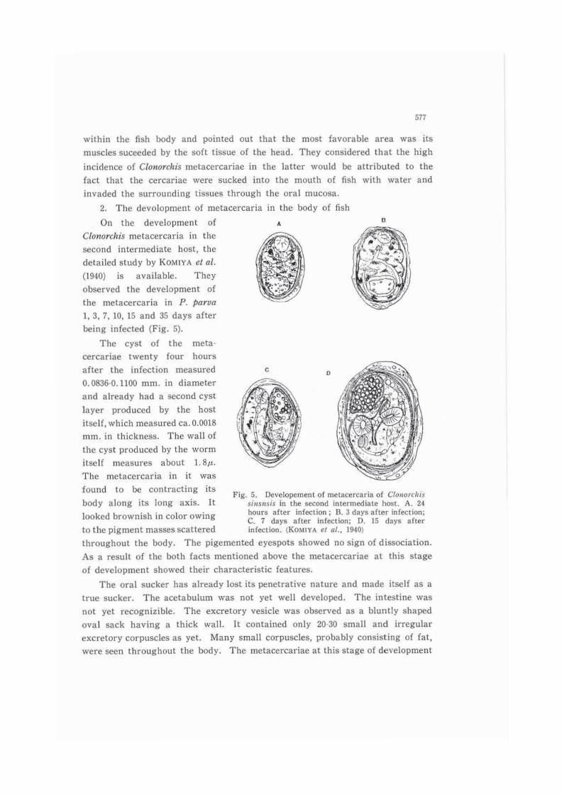
2. The devo!opment of metacercaria in the body of fish
On thc devc!opment of
Clonarchis metacercaria in the
間 cQnd intermediate host, the
delai1ed study by KOMIY A el al
(1940) is availab!e. They
observed the development of
the metacercaria in P ρarva
1,3,7, 10, 15 and 35 days after
being infected (Fig. 5)
The cyst of the meta
cercariae twenty four hours
after the infection measured
0.0836-0.11曲 mm.in diameter
and already had a second cyst
!ayer produced by the host
itse!f, which measured ca. 0.0018
mm. in thickncss. The wall of
the cyst produced by the worm
ilseJf mcasures about 1. 8μ
The mctaccrcaria in it was
found to be contracting its
body along its long axis. It
looked brownish in color owing
A e
c D
Fig. 5. Developement of rnetacercaria of CI()lIorcllis "肘nsisin the second interslediate host. A. 24 hours after infection; B. J da)'8 aher inr~割ct lOn:C. 7 days after infection: D. 15 days aher
to the pigment masses scattered infection. (Ko州 YAtl al., 1940)
throughout the body. The pigemented eyespots showed no sign of di田 ociation
As a result of the both facts mentioned above the metacercariae at this stage
of developmenl showed their characteristic features
The oral sucker has already lost its penetrative nature and made 比目!fas a
true sucker. The acetabulum was not yet well develo問 d. The intestine was
Ilot yet recogn山 ble. The excretory vesicle was obscrved as a bluntly shaped
oval sack having a thick wall. It contained only 20-30 small and irrcgular
excretory corpuscles as yet. Many smaU corpuscles, probably consisting of fal,
were seen throughout the body. The me回目rcariaeat this stage of development

578
were in the process of metarnorphosis
The elliptica! cyst three days after the infection measured approximately
0.114-0.084 mm. X O. 13CドQ.108mm. in diameter. The pigmented eyespots were
sti1l present in the metacercaria at this stage of development. The pharynx,
intestine, and acetabulum became more c1early v山 ble.The excretory corpuscles
in the bladder increased in number as well as in size. However, on the whole,
the worm was stil1 in the process of metamorphosis
The cyst seven days after infection measured 0.119-0.092 mm. xO. 141-0.101
mm. The metacercaria in it was found folding its body two times. The
eyespots began already to di日 ociate.The excretory corpuscles削除dthe vesicle
almost completely. In the intestine many round disc shaped corpusc¥es began to
appear. H。、vever,the oral sucker as 、vellas the acetabulum were not yet
perfectly developed. At this stage of development the former was still larger
than the latter
The cyst ten days after infection measured about 0.132-0.097 mm.xO.141-
0.110 mm. Now both suckers were fairly well developed, and the excretory
corpusc¥es large as well as small,自lledthe vesic¥e completeJy. Metacercariae
in alJ the stages of development above mentioned died when acted upon by
artificial intestinal juice for twenty minutes at 37・cThe metacercariae fifteen days after infection measured 0.123-0.092 mm. x
0.141-0.097 mm. Now the metacercaria in the cyst was found foJding its bOdy
two or three times. (t made a vigorous rotatary movement intermittently. At
this stage of development the characteristic structure of the metacercaria was
complete. The eyespots were found compJetely dissociated; the acetabulum
became larger than the oral sucker (the former being 0.04-5 mm. and the Jattel"
0.0436 mm. in diameter respectively); the pharynx measured 0.0252 x O. 0216 mm
The larger excretory corpuscles attained to 7.2μin size. The disc like corpusc¥es
above mentioned appeared numerously now in the intestine. After acting
act同cialintestinal juice on cysts at this stage of development for thirty minutes
at 37・c.5 among 36 were found liberated from cyst and alive
Thirty five days after infection the metcercaria was found well developed,
its acetabulum became larger than the oral sucker. Now the former measured
about 0.066 mm. in diameter, while the latter m田 suredsome 0.048 mm. in
diameter. It should now be regarded as a mature metacercaria. However, when
the melacercariae were fed to a mouse, no adult ¥Vorms、,vererecovered thirty
days after feeding (KOMIYA et al., 1940)
It

579
in the beginning of August. NAGANO (1936), after suc!:essive experiments
confirmed that within a period of 23 days after infection of P. tarva with
cercaria no adult worms were recovered
The de日screpancybetwecn the results above mentioned would be attributed
to the different temperature of the environment during the course of experiment
3. The distribution of metacercariae in the fish host
KOBA Y ASHI (1922) found the majority of encysted metacercariae in the
connective tissue and in the muscle near the skin. In muscle layers far apart
from the skin they were found less in number. KAWAI et al. (1935) investigated
the distribulion of Clonorcltis metacercariae in six P. tarva which harboured
them 1,939 in average number and reported that they were found mostly in
muscles of fishes. His results showed that 47.4 % of their total number was
found in muscles, 26.6 9杉山 heads,16.8 % in skin and subcutaneous tissues, 5.2
% in fins and 0.7 % in scales. IWATA (1937), divided fish body into the
subcutaneous tissue, superficial muscle and inner muscle, and recovered Clonorchis
metacercariae from each portion. The result showed that the rate of the
Table 3. The distribution of CIOl1orchis metacercariae in Ihe fish body (KOMIYA et al., 19凶0,19014)
F時 h(No. examined) P. par四 (40) P. pan.・.(j (15) s. 5iJte"sis (15)
Lxation No. of metacercai日 No. of metacercaria No. of metacercaria found (弱} found (%l found (%)
Scales 5 ( 0.2) I ( 0.3) 169 ( 1.6)
Gilles 16 ( 3.6) 13 ( 3.6) 125 ( 1.2) Tail fin 185 (10.自〉 3 ( 0.8) 651 ( 6.2)
D"回 1fio 12 ( 0咽 7) o 300 ( 2.9)
Breast fins 11 ( 0.6)
Ventral fins 3 ( 0.2) 1 ( 0.3) 324(3.1) Other fin 。Muscles 1. 328 (77. OJ) 320 (87. 9) 6,616 (63.4) Head 155 ( 9.0) 26(7.1) 2.250 (21.6) Inner organ 。 o 。Total J, 715 総4 10,435
incidence in the subcutaneous tissue was found highest (71 %), followd by those
m super白cialmuscle (15 %)
KOMIYA et al. (1940, 1944)examined the distribution of Clonorchis metacercariae
in bodies of various fishes (Table 3). As seen in the table, metacercariae were
found mostly in their muscles and particularly in the posterior bOdy following
the tail白n,as pointed out by HsO et a/, (1936)

580
c. 'l'he Metacercaria of ClQlIQrchis sinensis
1. Morphology of the mature metacercaria
The metacercaria of ClonQTchis sinensis has been described briefly by
KOBAYASHI (1912a, 1922) and more in detail by HsO el al. (1937) and KOMIYA el
al. (1940)
The cyst of C. sinensis is oval in shape, measuring aboul 0.135-0.145 mm. x
0.09-0.10 mm. (KOBAYASHI, 19叩)
According to KOM1YA et al. (1940), the liberated metacercaria is spatula-like
in form, tapel'川gslightly towards the posterior part of the body. lt measures
apporoximately 0.406 x 0.121 mm. and makes vigorous leech-like movements
When extended, the anterior half of the body attains about twice its length of
the posterior part of the body. The body surfac巴 isbeset transversally with
the small sp目的, which extend all over the body surface except for a smal1 area
on the dorso-anterior part of the body, mouth opening and acetabulum. These
spines become finer at the posterior part of the body. The sensory organs are
small papillary projections, surmour】tedby a short hair, and are fo山ldlaterally
on the side of the body, usually 14-16 in number
HsO et al. (1937a) observed that 12 papillae were found around the oral
sucker forming two circl回目milarpapillae, nine in number, were found on the margin around the acetabulum. They regarded these as sensory organs
Numerous masses of brownish yellow pigments are found scattered
throughout the body. Both suckers are round in shape. The oral sucker measures
about 0.049 mm. in diameter and is smaller than the ventral one, which measures
about 0.062 mm. in diameter. Following the oral sucker a short prepharynx
and an oval-shaped pharynx are present, the latter measuring 0.020xO.018mm
The oesophagus is relatively long. lt bifurcates medianly at a point between
the pharynx and the anterior margin of the acetabulum into intestinal branches,
which run along the side of the body up to its posterior end. Many'disc-shaped
corpusc¥es are observed in the intestine. They move to and fro as the worm
moves. Two kinds of glands, skin glands and cephalic glands, are present
The skin glands are found, arranged in a row, on the surfaces of the anterior
part of the body, numbering about 24 in all. Ordinarily their arrangement is
as follows; one lateral to the oral sucker; one lateral to the pharynx; six
laterally, in the area between the pharynx and acetabulum; three near the
oesophagus and one on the upper margin of the acetabulum, respectively, on
each side of the body. They all open ventrally to the exterior. The cephalic
glands are found

where they open externally. The transversal
nerve commissllre is found medianly under the
pharynx. lt rlll1S up to its posterior end. The
genital primordia call be seen in stained
speclInens as 、vell in living materials. The
testes primordia are recognized as small bal1s
obliquely sitllated, QneS on each side of the
excretory vesicle. The ovarian complex is
fOll¥ld in the midline between the acetablllllm
and the excretory vesicle The uterus
primordillm is recognized as a cord extending
from the ovarian complex to the mediall upper
margin of the acetablllum
The excretory vesicle occllpies the greatel
part of the posterior part of the body. lt is
blllntly oval in shape in any instance and is
ftlled wilh numerous excretory corpuscles
They have compact outlines and are spherical
or bll1ntly oval in shape, ranging from 0加9
0.0018 mm. in diameter. Owing to their
refractive nalure they appcar almost black
under the ordinary microscope. WATANABE
(1942) reported that thesc corpllsc!es consist
of calcium carbonate and calcium phosphate
As for the excretory system of Clonorchis
metacercaria, KOM1YA el al. (1940) also made
a detailed description. The main excretory
canal starts from the lIpper lateral corner of
the excretory vesicle and proceeds upwards
with several coils, a!ong the ol1ter margin of
the intestine, and on attaining the level of
approximately the uppcr half of the acetabllll1m
it is divided into the anterior and posterior
collecting tubes. The former proceeds upwards
and then is divided into two branches, each of
which has three flame cells with their respective
collecting excretory capillaries, The posterior
collecting tllbe proceeds downwards parallel to
the main excretory canal, and after scnding
581
Fig. 6. Metacercaria of CloJ/ol"chis simJ1lsis {KOMJYA el (11., 1940)
Cc: central nerve commissure Cg: cephal犯 glandEp: excretory pore Exb: excretory v(!sic1e Exc: excretory canal Fc: Hame cel1
Ga: genital "Anlage' In1: intestine C回目鈎phagusOs: oral sucker Pg: pigment mass Ph: pharynx Sg: skin gland 50: sensory organ T: testis Vs: acetabulum
582
one branch upwards at the level of the upper end of the main excretory canal
it is divided into branches. Each of these three branches has three f1ame cells
with their re日spective excretory capillaries. Thus the ftame cell pattern is
represented by the following formula: 2x (3+3)+(3+3+3) . which is just the
same as that of the cercaria
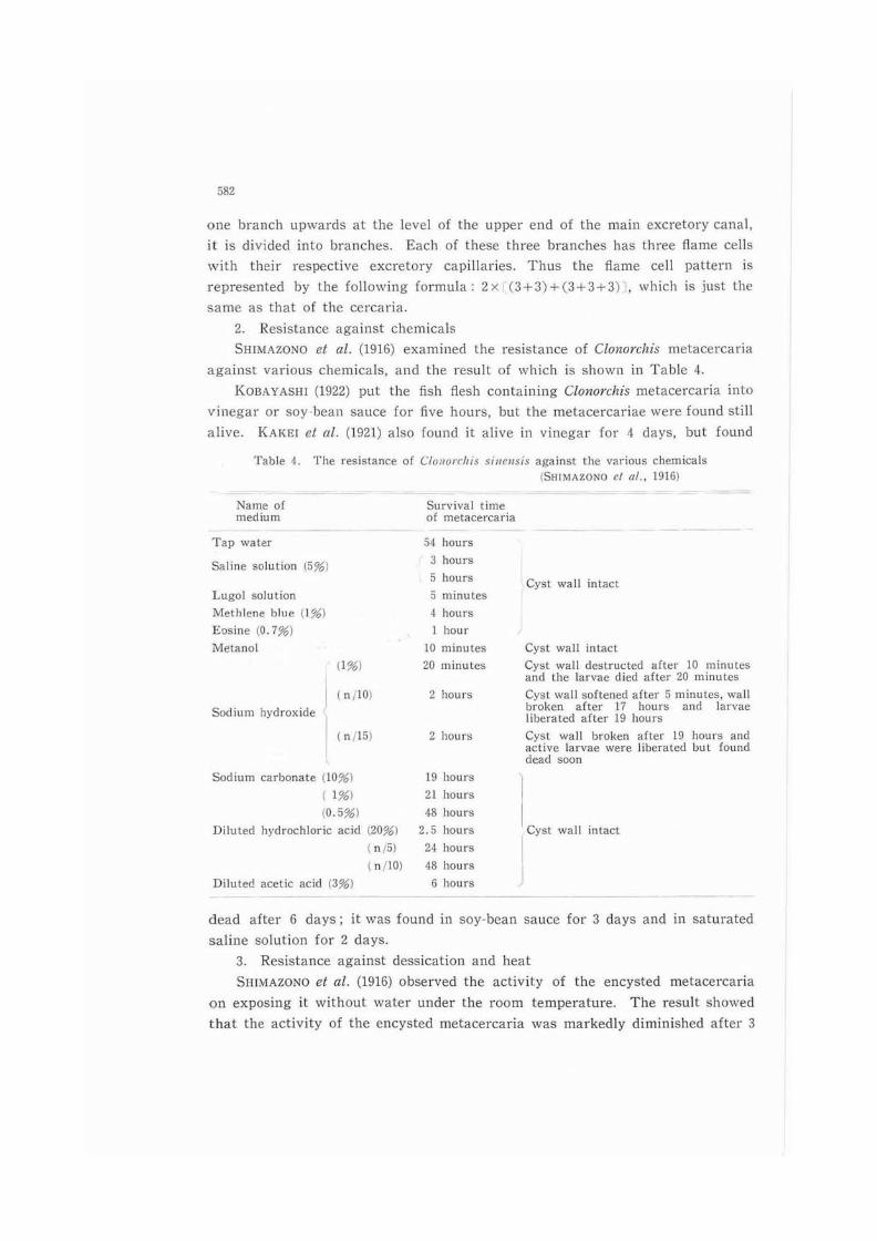
2. Resistance against chemicals
SH1MAZONO a al. (1916) examined the resistance of Clonorchis metacercaria
against various chemicals, and the result of which is shown in Table 4
KOB.....YASHl (1922) put the fish ftesh containing CIQnQrchis metacercaria into
vinegar or soy bean sauce for five hours, but the metacercariae were found still
alive. KAKEl et al. (1921) also found il alive in vinegar for 4 days. but found
Table 4. The陀 sistanceof CloJlorrhis silll!lIsis againSl the various chemicals (SHIMAZONO CI 01.. 1916)
Name of Survival tlm世
medium 。fmetacercaria Tap water 54 hours
Saline solution (5%) 3 hours
5 hours
Lugol solution :) mmutes
Methlene blue (l~副 4 hours
Eo宮ine(0.7%) 1 hour
Me:tanol 10 mInut出
(1%) 20 minutes
( n{lO) 2 hours S剖liumhydroxide
( n/15) 2 hours
Cyst wall intact
Cyst wall intact
Cyst wall deslructed after 10 minutes and the larvae died after 20 minutes
CySI wall softened after 5 minutes. wall broken after 17 hours and lar四 eliberated after 19 hours Cyst wall broken after 19 hours and active larvae were liberated but found dead叩
sゆdiumcarbonate {lO%l 19 hours
( 1%) 21 hours
(0.5%) 48 hours
C
a
n
a
v
s
y
c
-
-
Diluted hydrochloric acid (20%) 2.5 hours (nβ24 hours
(n/10) 48 hours
Dilutec! acetic acid (39的 6hours
dead after 6 days; it was found in soy.bean sauce for 3 days and in saturated
saline solution for 2 days
3. Resistance against dessication and heat
SHI:-VIAZuNO el al. (1916) observed the aClivity of the encysted metacercaria
on exposing it without water under the room temperature. The result showed
that the activity of the encysted metacercaria was markedly diminished after 3

583
hours and no activity was recogr羽田dafter 7 hours' exposu:-e. They also e治:amined
the effect of heat 011 its viability and indicated that it was killed for 3 minutes'
exposure al a teη1perature of 65・Cand for 2.5 hours' exposure at 39・c.0・cMUTO (1921) showed that in certain cases the cysts of C/QnorcJtis, after
日 paratingfrom the flesh of decomposcd fishes, fell into water and remained
viable for 5-12 days. Is11lI el al. (1935) domenstrated thal, within 24 hOU,.5 after the
cyst became separated from the decomposed fiesh of fishes in water, the
metacercaria remained viable for 48 hours. And they found thal the encysted
metacercariae survived for 3 to 7 days in the lissues of the dead I1sh. They
could obtaiI、theadu¥l ¥VQrm from the rabbils experimentally fed with slIch fish
4. Effect of digestive juice on metacercaria
SHIMAZONO el 01. (19l6) put fish flesh containing Clonorchis metacercaria into
gastnc J山田 obtainedfrom men for 17-18 hours. The results showed that the
activity of the eilcysted metacercariae. after separating from the flcsh, slightly
diminishcd. NAGANO (1927) applied an artificial gastric juice (HCI ; Icc, watcr;
I回 cc,pepsin; 0.5 gr.) for 3 hours undcr the temperature 39・C for obtaining
the encys1ed metacercariae from the fish 何回h,and he reported that the
metaccrcariae wcre able to 町 coverstill alive. KOMIYA ct al. (1944) also applied
an artificial gastric j山田 (dilutedHC1; 3.0 cc. distil1ed water; 1曲 cc.pepsm;
0.3 gr.) for 3-4 hou同 atthe temperature ca. 37 C for collecting the viable
maturc encystcd metacercariae from the flesh, and gained a good result
SIIlMAZONO el a/. (1916) kept the encysted mctacercariae in rabbits' bile juice
(or 1he bilc juicc dilulcd with physiological salinc solu1ion 01' with water) for
2 -20 hours in an incubator at 37・C. His reslllt showed that a few cysts were
softened and metacercariae were liberaled, bu1 the majority of metacercariae
was no1 able 10 be liberated from the cysts. At the same time, they fed dogs with the fish ficsh containing encysted metacercariae and disscctcd them 5 hours
l.¥fter feeding. They found the excysted metacercariae in its in1estine, but not
in the slomach. From this fact, they considered tha1 no excystation would take
place in the slomach of animals. but the metacercaria wOllld be liberated in
the intestine
KOMIYA el 01. (1941) applied, after exposing them to the effect of the gastric
juice above mentioncd, the artiticial intest‘nal iuice (Nalrium bicarbonate; 0.2 gr trypsin; 0.5 gr.. physiological NaCl solution; 50.0 cc.) for ahOllt 30 minutes
at the

VI. DlFINITIVE HOST
A. 1'he kind of definitive host
Soon after the discovery of the臼ukein men in ]apan, ir was found in dogs
(KATSURADA, 1892), cats (llJ1MA, 1886), pigs (the discovery from the pig in ]apall
by ]ANSON was described by LOOSE in 1907), and Raflus norvegicus norvegiCIIs
(MUTO, 1920c). KOsAYASHI (1912a) demOl1strated that cats, dogs, rabbits and
guinea pigs were experimentally slIsceptible. Later EcuclII (1925) reported that
a camel bred in a zoo lVas positive for Clonorchis eggs
The infectiotl of C. sinensis in birds was first observed by ASADA (1920). He
examined 6 Nycticorax nic/i,叩 raxand found in one of them 13 specimens of C
sinensis harbouring in its gall bladder. He fed N. nycticorax and Anas dome山田
町..ithPρarva and C. carαssius harbouring CfonOTchis metacercariae and the adult
ClonoTchis was found in them though small sized. But he reported that phcasants,
crows and ducks experimentally infected with Clonorchis metacercariae were not
able to harbour the adu¥t自uke
KOM1YA el al. (1949) eventlla!ly fOllnd in one of seven Anas domestica 17
specimens of C. sinensis in the gall bladder. But those A.ukes were smal!er in
size than those naturally found in cats and dogs. An experimental infection of
A. domeslica with C/o削 Tclzismetacercaria proceeded by them, however, was not
successful. In view of the above mentioned findings they concluded that A
domesfica was not considered as an suitable definitive host of C. sineげlSIS
The work of KAWAl et al. (1935) is worth to be mentioned here. They fed
rabbi旬、 dogsal1d cats with the same number of ClonOTchis metacercariae and
found cats showing the highesl incidence. According to Wn<OFF (1958), rabbits
and guinia pigs were found to be equally susceptible with this A.uke, about one
third of the orally administered metacercariae developing to matul"Ity, but rats
are less susceptible, showing only 6 % of the fed metacercariae became adult
B. Invllsion into Defi・nitiveHost
1. Route of migration of larva
In order to ascertain the route to はshabitat in the host, KOB,-¥YASHl (1912a)
fed cats with fish 何回hα川 tainingencysted metacercariae and on dissecting
them 15 itnd 24 hours after feeding, free yOllng A.ukcs were fOllnd both in the

585
gall bladder and the bile duct
MUKOYAMA (1921, 1922) fed rabbits with raw fish containing viable encysted
metacercariae and di田 ectedthem at certain intervals after feeding. He found
young flukes in the bile passage after 6 hours in case of rabbits. With the
lap田 oftime, the flukes made an increase in number. Ten to fourty hours aftel
feeding a large number of flukes were noticed in the liver and bile passage
He also confirmed the location of nukes in rabbits at given intervals after
feeding. The results arc shown in Table 5. He also gained the same results
in the casc of dogs and guinea pigs
TabJe 5. The site of C 刊 'leIlS;Sin rabbits al intervaJs foJJowing infection (MUKOYAMA, 1921)
Time after feeding
Within 5 hou悶
10-16 hours 20 hours
35 hours
Sile where found価ukesm伺'"In inteslIne, a porlion about 50 Cffi. be10w pyJoric region 1"・ntestme,a p。同胞nabout 30-40 cm. below pyloric regi削
Tn int舗 tine,a portion near the opening of the bile duct and the duodenum In intestine. a回 rtiona回以 10-20cm出lowpyloric region
Eventually the larvae, which excysted in the intestine. were found in the
fairly lower part of the intestine, but the majority of the young伺ukemigrated
from the duodenum through the common duct directly into the bile duct. He
then ligated the ductus choledochus of rabbits immediately after feeding them
with metacercaTIae and examined their thoracic and abdominal cavities, bile
passages, duodenum, and livers for flukes twenty hours to nine days after feeding
A large number of young larvae remained in the duodenum, while no larvae
were recovered from the other organs. Thus he concJuded that after liberation
Clono町 hismetacercaria is incapable of penet悶 tingthe intestinal wall of the host
Furthermore he encJosed liberated metacercariae in the abdominal cavity of
rabbits. but 110 fiukes were observed in the liver, bile passage, thoracIc cavity
and intestine from 46 hours [0 24 days after enclosing. From the result oblained
he concluded that the metacercariae migrated from the duodenum through the
common duct direclly into the bile duct
WYKOFF et al. (l田7)'sob盟 rvation,howev町 isnot in agreement with tho担
of MUKOYAMA (1921). They ligated the ductus choledochus in one group
of eXT泥Tlmen回 1rabbits and in another group applied a tube 針。m the gall
bladder outside for drainage. Then both groups of rabbit were fed with Clonorchis
metacercaria. After dis担 ctionyoung fiukes were obtained from the liver in both
cases. Their results indicated that Clonorchis metacercariae、、.'erecapable of
I問、etratingthe intestinal wall of rabbits and making their way to the liver

586
2. Site of worms in the defin山 vehost
The usual habitat of the f1uke in the host is the hepatic ducts, occasional1y
the gall bladder and the hile duct can serve as its habitat. 10 回目 ofhaving
fairly numerous fluk目。 mostof them are found in the perpherial fine hepatic
duct and only a few in the gall bladder. In heavy infections in the pancreas
and in the duodenum flukes could be found. They were apparently in good
condition (KOsAYASHI, 1912a)
1n the survey of the site of ¥VQrms in the bOdy of the rabbits, IWATA (1937)
showed that in heavy infectiol1s 77.4 % of a¥l自ukeswere found in the livcl
21.5 % in the bile duct, 1.8 % in the gall bladder, and in mild infections 96.3
% of flukes in the liver, 3.4 % in the bile duct and 0.3 % in the gall bladder
In all cases the majority of worms was found in the ¥iver. Among 100 cases of
the rabbits infected experimentally, ARIYOSHl (1930b) observed in 22 cases f1ukes
in the pancreas, and in 4 cases of dogs found them in the pancreas in all
cases. YOSHIDA (1931) observed that the young flukes actively invaded the
pancreas to deve!op up to adult in it. And he confirmed that the young flukes
showed positive chemotaxis to the pancreatic juice, but they were attt'acted by
the bile juice more intensively. YOSHIDA (1931) utilized the urinary bladder of a
rabbit previously prepared for an experiment and made an observation on the
effect of bile juice and pH for the behabiour of the fluke. The result showed
that the majority of flukes proceeded to the ureter with the bile juice (pH 7.4-7.6)
5 hours later. But when sodium carbonate was added to the saline walel'
(changing pH to 7.8-8.0) many youn[! flukes pos山 velyproceeding into medium
were observed. From these γ'esults YOSHIDA concJuded that the hydrogen ion
concentration had an impor回 nteffect on the invasion of the fluke, provided
that pH was under 9.0
Regarding the distribution of flukes in the liver, lTO et al. (1925) autopsied
21 infected rabbits and examined Clonorclus adu¥ts. The result showed that the
highest incidence was observed in the right lobe. When dividing the liver into
the anterior and posterior part, the higher percentage of flukes were found in
the anterior one. In the case of dividing the liver into the central and
peripherial parts, the majority of ClonorchIs adults was found in the latter. On
the other hand he found a large number of fl.ukes in the central part of the
liver after the death of the host. The reason could be explained as follows;
After the death of the host, the temperature in the peripheral part would fall
Quicker than that in the central 0
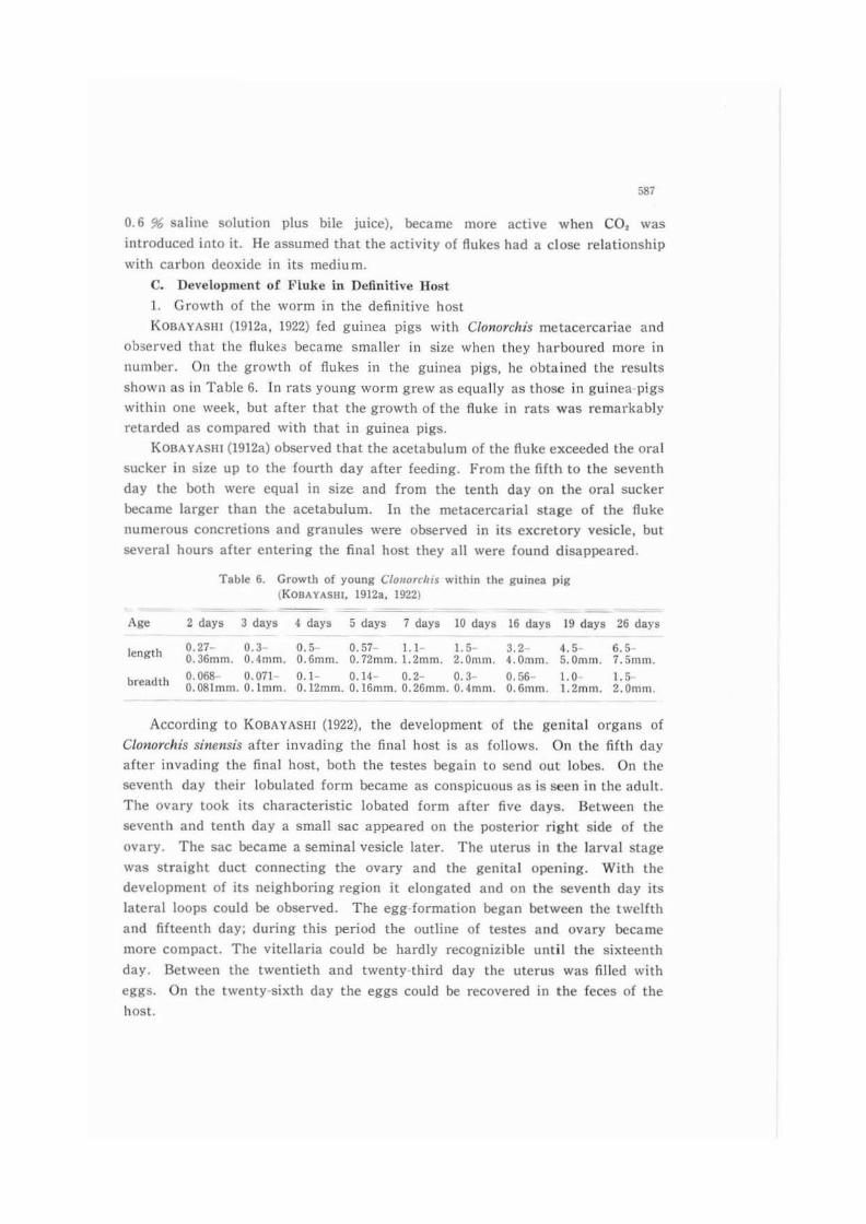
58'
0.6 % salinc solution plus bile juice), became more actIve when CO, was introduced into it. He assumed that the activity of伺ukeshad a closc relationship
with carbon deoxide in its mediu m
C. Development o( F¥uke in Definitive Host
1. Growth of the worm in the definitive host
KOsAYASHl 0912a, 1922) fed guinca pigs with Clonorchis metacercariae and
observed tl1at thc fiukcs became smaller in size when they hal'boured more in
number. On the growth of f1ukes in the guinea pigs. hc obtained thc results
shown as in Tahle 6. In rats young worm grew as equalJy as those in guinea-pigs
within one week, but after that the growth of the fluke in ra阻 wasremarkably
retarded as comparcd with that in guinea pigs
KOsAYAS川(1912a)ob田 rvedthat the acetabu!um of the伺ukeexceeded the oral
sucker in sIze up to the fourth day after fecding. From the fiflh to lhe seventh
day the加thwere cqual in size and from the lenth day on the oral sucker
became larger than the acetabu¥um. ln the metacercaria¥ stage of the伺uke
numerous concretions and granu¥es were observed in its excrelory vcsicle. but
日 veralhours after cntering the final host出eyal1 were found disappcared
Table 6. Growth of young CIOllorcflis w川、inthe guinea pig lK08AYASIII. 1912a, 1922)
~ 2_3_._._'_w_w_m_u_ length 0.27- 。.3 0.5- 0.57- 1.1- 1. 5- 3.2 ι5 ι.-
0.36IOm. 0.4mm. 0.61010. 0.72mm.1.2mm. 2‘Omm. 4.0mm. 5.0mm咽 7.5mm.
breadth 0.0SSO,O710.1- 014- OE- 0。3- O,54- 1+01 .5-0.081mm.0.1mm. 0.12mm. 0.161010. 0.26mm. 0.4mm. 0.6mm. 1.2mlll. 2.0mm.
According to KOBAYASHI (1922), the development of thc genital organs of
CIOllorcllis sillcnsis after invading the白nalhost is as follows. On the白fthday
after illvading the final host, both the testes begain to田 ndout lobes. 011 the
seventh day their lobulatcd form became as conspicuous as is seen in the adult
The ovary took its characteristic lobated form aftCI 自vedays. setween the
seventh and tenth day a smal1 sac appeared on the posterior right side of the
ovary. The sac became a時間nalvesicle later. The uterus in the larval stage
was straight duct connecting the ovary and th巴 genitalopening. With the
developme川 ofi1S r泥沼hboringregi聞 は elongatedand on the日 venLhday its
lateral loops cou¥d be ob田 CV国 The egg formation began between the twelfth
and fifteenth day; during出isperiod the outline of testes and ovary becamc
more compact. The vitellaria could be hardly recognizible until the sixtee川h
day. setween the twentieth and twenty-third day lhe uterus was filled with
eggs. On the twenty-sixth day the eggs could be recovered in the feces of the
host

'88
2. Devε]opment of excretory system
011 the deve]opment of the excretory system of C. sinellsis in its definitive
host. a detailed description by KOl¥HYA et (11. (1953) is available
They fed tnice with metacercariae and aIler a certain period of time they
¥¥'ere dissecled and examined the development of excretory system in every
developmental stage of the伺uk,The data obtained are as foJ]ows
One day after infection: The warrn was already in the liver. In most
cases, neither excretory corpuscles nor granules were found already In the
excretory vesicle. The flame part of the ftame cell ¥Vas 8. Iμlong. The main
excretory canal became considerably larger in diameter. The伺amecell formula
remained as the same as that of the cet'caria and metacel'caria: 2x [(3+3)+(3
+3+3)1
Two days after infection: The body of the worm was 0.56 mm. long and
0.15 111m. wide. The main exc陀 tol'ycanal became larger, measuring 5.4μm
its diameter. The flame cell formula remained as the same as before
Three days after infection: The body of the worm became 0.73 mm. long
and O. 19 mm. wide. The flame cell formula remained st川 asthe same as before
Four days after infection: The body of the worm became 0.91 mm. long
and 0.24 mm. wide. The flame part of the flame cell wasεnlarged to 9x4.5μ
and the diameter of the mail1 excl'etory canal measured 7.2μ ln this stage,
the flame cell formula stiU remained unchanged as before
Ten days after infection: The body of the worm became 1.4 mm. long and
0.28 mm. wide. The both suckers were still of the same size. The flame cells
川 thepos句riorsecondary collecting tube remained unchanged in number and
arrangement. but a new small flame ce¥l was seen on the foremost original one
of both first and second tertiary excretory tubes of the anterior secondary
collecting tube (Fig. 8, a). Sometimes, such a feature was found only in the
first tertiary collecting tube. The new flame cell was about 12 in its size of
the original flame cell; the flame part of the latter measured 9 x 4. 5μTh, flame cell formula in this stage was principally 2x 1(4+4)+(3+3+3)]
Twelve days after infection: The bOdy of the worm became conspicuously
larger. measuring 2.9 mm. long and 0.45 mm. wide. Particularly, the posterior
part of the body was found markedly elongated: it was about th問 etlmes
as long as the anterior part of the body. In this stage, the flame cells in each
tertiary collecting tube increased cOllsiderably in nurr】ber. Each of the three
original flame cells of the first tertiary collecting tube was added with 3 to 4
new flame cells which were usually smaller in size, about 1 2 of

Fig. 7. The excrelOry systen、。fClollor(hi$ sillr"sis in cercaria and metacercaria stage. lKo .¥IIYA tl 01., 19531
a: m cercar‘a b: in metacercaria
日-
:'1¥0'
•
Fig.8. TI時 excrεtorysystem of C/Ollord,;s sim!nsi5 in its definitive host. (KOMIYA el al 1953) a: ten days after infecti加
b: Iwelve days after infection c: twenty four days after infection. d: Ibi(f
""
rhc newly added flame cells were found grouped around the original one. On the
second lCl'tiary collecting tube, two anterior original Aame cells werc added with
Ilo5ncw 伺amecel1s respcctively, while the most posterior original onc 7 to
a new f1ame cells. These new flame cells wcre about the same as those on lhe
first tertiary collecting tu困 insize and arrangement
Each original flame cell of the third阻"叩ycollecting tube was added with
2 to 5 new何amecells. The Increase of ftame cells in the fourth tertiary collecting
tube was the same as that of the third one, whi1e each original ftame cell of
the fifth tertiary collecting tube was added with 2 to 4 l1ew ftame cells
respectively. Fig. 8, b re叩 resentsan example of the l1umber and arrangement
of the flame cells in the worm in this slage. Here, the ftame cell formula 011
刷 leside was represented as ¥(4+4+ 5)+(6+5+9)1 + [(3+6+3)+(3+3+5) +(3+6+5)j
Fifteen days after infection: The general as回目 of th巴 increase and
arrangt"ment of the伺amecells was almost the same as those twelve days aftel
川 fCC1IOll. Each original ftame cell in the metacercaria stagc was found increased
by 3 to 9 in IlUmbel.. The number of the flame cclIs on each tertiary collecting
tube was not always constant
Twenty four days after infectioll: The body of the worm was 5.5 mm. long

590
3nd L 7 mm. wide. The diameler of the main excretory canal attained to 7.5μ
Bllt the flame part of the flame ccJl was oot so large as compared with that
ten to twelve days after infectioo. The largest size of the flame part of the
伺amecell remained 9x4.5μas yet. The size of the flame cell田 emedto show no
fllrther increase. ln this slage the number of the flame cells was found
considerably increased, bul it was very difficllll to elucidate the exact feature
of the number and arrangemenl of伺amecells 011 every tertiary col!ecting tllbe.
p31.ticularly on that locating in the middle parl of the body, because here the
extensive development of the uterus became an obstacle to the exact observation
With great effort. KOM!YA et al. were able to make clear the aspect of the numbel
and arrangement of the flame cells 00 the first, second and fifth tertiary collecting
tubes. Fig. 7 and 8 show an example of the arrangement of flame cells 01 this
stage. As seen in the Figures. each of the original three flame cells on the first
tertiary collecting tube increased by 6 to 14 in number respectively, and those
of the second and third ones by 10 to 14 in number rcspectively
From the observations mentioned above, KOM!YA el al. (1953) stated that no
de自oitefactor of division or no regularity in division of flame cells was rccognized
in the COUl配 ofthe iocrease of flame cells in young Clonorchis in its final host
3. Longevity of the C. sinensis in the definitive host
KATSURADA (1916) cited a case of clonorchiasis which was believed to have
harboured an infection for 3 years and 11 months. KOBAYASH! (1922) took the
melacercariae himself in 1910, and hc noticed eggs in his feces one month latcl
ln 1919, he found a marked decrease in egg number in fcces and concluded that
the 1ife span of Clonorchis in human bodies may possibly be wider than 8 years.
On the 3 dogs experimentally infected with the fluke, MUTO (1922) noted the
decrease of egg number in feces 2 years and 8 month$, 3 years and 4 month$
<lnd 3 years and 6 months after infection. On their autopsy, he observed
degenerated atrophy of the fluke from them
In this regard, MUTO considered that, in the fluke, with the atrophy of the
body the gradual degenerating changes of the reproductive organs were occurred
<lnd subsequently the reduction of the egg number in the uterus took place
until they eventually died
'1. 1n vilro cuJture
fo this point no 1'eports、.¥'ereavailable recently. In εarly times SH!I¥1AZONO
e/ al. (1916) cultured young worms (5 hours 01' 11 days after infection) in variou

591
a cloth plus 9 volumes of 0.6.9百 sterilizedsaline solution), was most efficient i11
maintaining their survival (6-37 days)
REFERENCES
1. ANDO, A. & K. KOBORI (1924): On an abnormal modc of infection of CIO!lorchis SilICIW~
1. Oral infe<:tion and thc relation between C. sIllclIsis and birds. Cflllg削 ljiShiJ/to
(1057), 523-535. (in japanese) 2. ARJYOSHI, C. (1930a); A study of the liver of the rabl川 experimentally infected with
CIQ!wrcllis si川町Î.~. sJorぽ刊/1 Ki)'o, 6 (2),277-362. (in Japanese with English summary)
3. ARIYOSHl, C. (l930b): The infestation of thc pancreas with CIQ//Qr,ιfus sillclIsis 3nd
its histological study. Byorigak/l Kり0,6(2), 363-372. (in japanese with En兄lish凹 mmaryl
'1. ASADA, J. (19四 ): Study on trematodes in birds. Nitf/O!I 8yori G叫加iZasshi. 1日 347
350. (in Japanese)
5. ASAQA. J. (1937): Study on trematodes in Manchuria the intermediate hosts of which
are fresh water fishes. particularly recovered frorn dog experirnentally. Ni,仲 011KiseIl:11II
Gakkni Kiji, 9, 103-108. (in japanese)
6. ASAOA. J. (1940): Clonorchias凶 in M主nchuria. 1. Particularly On the prevalence of
clonorchia引 sin Mukden. ,/,(iirikll Kngakllill 1110. 4 (6), 932-956. (in japanese)
7. ASAOA, j. (1942): 00 pHasites, particularly on helminths in Manchuria. NゆpOl/!US('ir"i1ll
Gllkkai KIji. 14, 22-44. (in ja凹"'溜)
8. BAELZ.巴 (1883): Uber einige neue Parasiten des 1¥1enschen. 8erl. Klin. Wochenschr
1883, 23.1 238
!). sLANCHARD, R. (1891): Notes sur quelque渇 versparasites de I"homme. Dislolllfl SiIlCIIS.
Comtl, rClld. $0'. biof., p. 604
10. BLAトαIARD. R. (1895): 0ρ stllorcflis sil1clIsis. "in Traite de zoologie m剖 icale et
argricole" 2d ed., Paris 11. BLANCltARD, R. (1896): Parasites animaux. Jn Traite de pathologie generale. Bonchard
Paris. 2,“9-810 12. Braun, M. (1920): Die Tierischen Parasiten des Menschen 11 teiL Wiirzburg
13. CIIOW. L. P. (1960): Epidemiological studies of clonorchiasis at meinung township in
southern Taiwan. FOI'IIIOSOJ/ Sci., 14 (3), 134-166 14. CHUN. S. K. (1962): Studies on some trematodes whose intermediate hosts are fishes in
the Naktong River. 8111f. Fishe,.ics Colll!gc. P/lS叩 Nol.Ullill.. 4 (1, 2), 21-38. (in Korean
with English summary)
15. CUUNG,日 L., S. j. HUANG, T. H. LI, H.C. Hsu, Y. M. Hu & F. N. Hsu (1959): Studies
。nclonorchia引 ssinensis in Peking and Tientsin. Occurrence of clonorchiasis sinensis at Yungfeng Hsiang. L山 lit'unof Ch'angp'iog district, Peking, with observations on
intermediary and definitive hosts and detectωn of clinical cases by means of intradermal
test with CI0I10Tc/lis antigen. ScieJ/cc Rccon/, New Ser., J (10),49ゅーる03
16, CUUNG, H. L. T. C. I-Iou, S. J. HUANG, P. S. 1¥10, L. Y. Ho & C. H. KUANG (1960): Stud悶
on clonorchiasis sinensis i:J Pi:!dng and Tientsin. Occurrence of clonorchiasis sinensis
in Peking and its suburbs, wI!h observations on secondary intermediary and definitive
hosts. Ibid., 4 (1), 19-25 17. CHUNG, H. L.. T. C. I-Iou. H. C. Hsu, P. S. MO & H. C. WENG (1960) : Studies on clonorchiasis
sinensis in Peking and Tientsin. Occurrence of clonorchiasis sinensis at Palit'al, Tientsin, with observations on intrermediary and de自nitivehosts, Ibid.. 4 (1), 26-32
18. COBBOLD, T. S. {1815 a}: The new human fluke. Lnllcel, 187も (2),423
19. COBBOLD, T. S. {l815 b}: The new ¥iver tluke. Vele,.illarioll (London), 46, 780-781
10. U)BsOLD, T. S. (18日S4): Flukes in man. LlIII'el, 1884 (2), 976
21. EGUCHI, S. {I925}: ]>arasiles of wild animals, parlicu1ar1y lhose of fierce animals and
the inte問 lationthose human parasites. Aichi Igak如 iZaS$lzi, 32 (6), 176-189. (in ja四 nese)
22. FAUST, E. C. (1925): Somc recenl as凹ctsof the叩 demiologyof Clol1o/"cllis infection in

592
China, CIIiIU/ MI'II. J., 39 (4), 287 -295
23. FAUST, E. C. & Q. K. KIIAW (1927); 5IUd附加 C!flllorrliissiIlCIIU,' (Co帥 01.0. .-¥:11. J Hyg., MonogげaphicSeries, No. 8. 284 pp
24. Hi¥SEG.¥WA, T. (1929): A morphological characteristic of CIυIlfIrdus egg. An ilrabesque
p)lygonユ1pattern on the surface of tl憎 shcll. Ok(IJ"l1川 fI Ij.fflklmi Zfls.IIJi. 41 (101, 2424
(in 1apanese)
25. HA$EGAWA. T. (1934): Uber die enzystienen Zerkarien in P~clldornsboJ"(l 何町'(1. lbid.,ω (6), 1397-1434. (in japanese with German summary)
26. HASU[, N. (1917a): Biological study on Clollorrllis sillclIsis. Ibid., (325), 33-40. (in Japanesel
27. HAYASII[, 5., K. 5ATO, M. 5mGA & K. YAMAOKA (19喝7):The area along Lake Suwa
area. Nagano Prefecture as an endermic area of C/flllordlis siJ/cllsIs. KISl'ICJlIIgflkll ZflS$l1l
6 (3, 4), 361. (in japanese) 28. HIGU乙11[,R. (1938): A contribution on tl惜 morphology of Clol/on';'is 5Illrllsis in
Para[()s目7ntlusIIwllchQlllic"S, Kcio Igllkll, 18 (12), 1289-1305. (in japanese with English
summ同y)29. HOR1GUCIII, T. (1941): The effects of Vitamin C upon the development of CIOIIOl"clli,<
Silll:llsIS in the final hos1. Nゅρ0111 ka Daigukll Z,ω叫ん 13(12), 952-953. (in 1a凹 nesel
30. HOR1UCIIl, K. (1956): Epidemiological studies on c 。、orchiasisin Noshiro are<l, Akita Prefecture. Akilakell NosolI Igak如 iZa$$hi, 3 (2), 32 68. (in Japanese)
31. HOZUMI, K. (1925); On donorchiasis in Niigata Prefecture and on Melagol/imus J'okoga川 Mand larval form of Ligllla formed DipJt)"/{oゆolltl"illll/. Nip,仰 115110村知byoGak加 i7.asshi
24 (4), 254-266. (in Ja凹 nese)
32. Hsu, H. F. (1939); 5tudies on certain problems of CIQJ/on;his si>lc叫 isV[]J. Further
advance in the study of the life cyde of CIOl/orrflIs silwlIsis. Chillese Metl. J.. 55. 542
545 33. HsU, H. F. & C. y, CIIOW (1937): Studies on certaln problems of C/ol/ol'chis sil/rllsis 11
Investigation in the chief endemic center of China, the CanlQn area. Cltillese Mr(l. J 51,341-156
3,1. Hsu, H. F. & C. Y. CIIOW (1939): Development of CloJ/orc/lIs si/Jellsis eggs 10 cercaria
民agein laboratory bred snails, silh)"l,;a [uchsiall(/. Pr,ω 50c. E.ゅ Bio/.Mrd., 41. 158
160 35. Hsu, H. F. & O. K. KIIAW (1936); Studies on certain problems of Clollo/"chis sil/ellsis 1
On lhe cysls and second intermediate h田旬。fClollorr.ltis sil!ellsis in the Peiping area
Chi'le/Jse Med. J., 50, 16心9-162036. Hsu, H, F. & O. K. KIIAW (1937): Studies on certain problems of CIOIIQ/"eltis Silll'>lSIS 111
On the morがIOlogyof the metacercaria. Feslsrll川 flBl'rllllflrfl NOrlll, HOII山 11"1{, 216-220
37, Hsu, H. F. & S. Y. LI (1940): Studies on certain problems of CIQII(}I"';';$ silll'lIsis. 1X
The migration route of its early larval stages in the snail, silhJ'llIa [lIrll針。1/(/. Cflilll'sl'
l¥IIed. J., Suppl., 3, 244-254
38. Hsu, H. F. & L. S. WANG (1938): Studies 0
593
(470), 253-256. (in Japane氾叫43. !!JJMA, L (1886): Dislo川 aflldemicuJII, BAHLZ. J. Colf. Sci. Imt. UllilJ. TokYQ, 1. 47
59 “hJIMA. 1. (1888): The aoimal para叫 esof man. .190 pp. Tokyo. (in Japanese)
45. lKUYAMA, T. (1960): Studies on CIQIIQrcllis sillclIsis in the lower basin of the River
Chikugo J. Results of the survey on cJono閃 h・asisin Saga Pref.田ture. I¥rI/"IIlIIf! IgaA'A'ai
Zasshi, 23 (7), 2730-2753. (in Japanese with English summary)
46. IKUVAMA. T. (1960b): Studies 00 Cfollorchis Sillf!II$IS in the lower basin of the River
Chikugo 11. Results of the surv町 onclonorch・"・sin Fukuoka Prefecture. Jbid.. 23 (7).
2754-2776. (in ]apanese with English sumrnary)
47. INAG .~削 M. (1952): The prevalence of Clollorcltis metacercaria during 1951_1952. ][
Investigatio田 00the incidence in Tokyo and Ibaragi Prefecture. Ni.ρ仰 uKisci叫 11 Cakklli
Kiji, 21, 62-63. (in japanes叫48. INATOMI, S. (1953a): Studies on trematodes in Okayama Prefecture. 1. The development
。fPara!ossarllfllS 1II11IIc/lOlIriCIIS and water Quality. Okll)"alllll 19l1kんaiZa.ulti, 65(1), 31
39. (in japanese)
49. INAT.)MI, S. (1953b): Studies on tremato:les in Okayama PrefeC¥ure. IV, The seasonal
、lariatぬnof the cercaria in Parafo.崎市1r1l!IISmauchOllriws snail. Ibu!., 65 (1), 43-45. (in japanese)
50. INATOMI, S. (1953c): Studies on trematoヨesin Okayama Prefeclure. V. The distribution
of ParafossarlllllS m(lI/cI!OIlJ'iCIIS in the coast of the Kojima Bay. Ibul., 65 (1), 45-49. (in japanese)
51. ISHlI, S. (I929a): Studies on clonorchiasis in Kwangtung. 11. On the Clol/oJ"cllIs egg in
feces. jikkclI 19akll Zasshi, 12 (2), 39-59, (in Japanese)
52. ISHII, S. (1929b): Studies on clonorchiasis in Kwangtung. IV. On the second intermediate
host of CIOl/Orchis sincIIsit. /bid., 13問。 111-183.(in japanese)
53. ISIIII. S. & F. MATSUOKA (1935). Study on the relationship betwccn ClonoYl:hi.. infcctioll
and water. Tok)"o lji SflInslti, (2912), 80-回(inJapanese)
54. ISHIZAKA, K. (1818): On the results of凹stmortemexamination of liver trouble disease
and its clinica! symptoms. Igllkll Z,“5slli. (40), 20-26. (in japanese)
55. ISODA, M. (19日 ): Experimenta! studies on clonorchiasis 1. On the distribution of
metacercarb in the body of PJCII/lorasbora t{/r叩 and experiments on the artifidal
infection with metacercll.ria in rabbits. Nゆ仰11 jlligakll ZlIsshi, 14 (2), 105-114. (in
japanese w・thEnglish summary) 56. ITO. K. (1926): Bblogical studies on the cercaria of Clollorr:!,;s sine出 is. Aichi IgfiNNai
Zassfl人 33(2), 389-402. (In Japanese)
57. ITv, K. & M. MUTO (1925): The dislribulions of the worm in rabbil!l of the exper・mental
donorchiasis. 8J吻rigakll 1<i)'o, 15,565-571; Aichi Ig/lkklli lf/S♂lii, 32 (6), 1013-1081. (in
Japanese)
58. lwATA, S. (1937): Experi

594
par1'_sito!ogical z回 logy.Nissltill fgaklt, 1 (3), I ~ IO. (in Japanese)
64. KAWAI, T. (1937): Study on animal on anthelmintic effect of gentian violet. Suppl
The evalualIon of WAK~;SHJMA '5 egg counting method as an assay technic of anthelmintic
eITect of drugs against CloIIOI',IIi$ sinClIsis. TII;W(II/ Igakkoi Zasshi, 36 (5). 927-934
(in Ja凹 nesewith English summary)
65. KAWAI, T. & Y. YUMOTO (1935): 00 the distribution of the encysted metacercariae of
C/oJlol'chis siJlellsis in P;;emIQI"osbQI'(Iρan'o as the鑓 condintermediate host. and the r3te
of their infections to the marnmalian hosts. Jbid.. 35 (4), 880-887. (in Japanese wilh EngJish summary)
66. KAWANA. H. (1936): 5tudy on CIOJ1Qrchis sinclIsIs in the district of Shanghai. 111. Dogs
cats and ralS as reservoir hosts of C10lIorchis. J. SfwlIghai Sci. Il/sl., l$ect. 4). 2. 75-
" 67. K1M, J. W. (1961): ClOllorchis si阿川崎川 varioussecond intermediate hosts and th創 r
seasonal variations in Dog.chon area of the River Kumho. Ncw IIIcd. J.. 4 (10), 1221-
1223. (in Korcan wilh English summary)
68. K1TAMURA, K. (1916): An unknown helminthic eggs found in man. JikkcII 1110, 2 (16)。{in Jap:mese}
69. KOsAYAS1I1, H. (191011.): A study of liver distoma. The first preliminary report
DobllfslIgakll Zassfli, 22 (264), 1-4. (in Japane副
70. KOIlAYASHJ, H. (l910b): A study of liver distoma (Preliminary陀凹rt). SaikiJlg(lkll
/σsshi, (178). 49-51; (180), 1-6. {in Japane則
71. KOIlA YASIt1, H. (191Ia): A preliminary re回目 on the source of the human liver distome
CJonorchis elldemicus (BALZ) (-D;SIO/ll1l川 sρ71fwlalllll1 LEUCKART). Allllol. Zool. Jap
1. 271-277 72. KOsAYASH1, H. 0911b): A study of liverdistoma出 condaryre凹 rt).SlIikillgakll ZassfII
(185), 1 2. (in Japanese)
73. KOsAYAS1H. H. (l911c): First intermediate host of CIOIIQrchis. Ibid., (188). 1-4. (in
Japanese)
74. KOBAYASH1, H. (1912a): A study of CIOlIorcllis SIlltilsis (Final re凹 rt). Ibid., (20). 1-66
(in Japanese) 15. KOsAYAS1H 日 (l912b):00 the life h凶 toryand morphology of the liver distome(Clollo,.c/tis
SiIlCtlSis). Cetllmlbl. B叫 1.Pamsil. 01'ぽ 75(4). 299-318.
76. KOsAYASIt1, H. (1915): Studies on the enaoparasitic trematodes. D,ωIIlslIgaku Zasshi. 21
(320), 1-13. (in Japanese) 77. KO也AYASIII,H. (1917): On the 1ife history and morpho1ogy of the liver distome (CIOIIOrChIS
S/Jwns叫 Mμleil.Mcdiz. Hodtscflllle ut Keljo, 1, 251-284
78. KOsAYAStlT, H. (1922): The由。logicalas開ctsof c1onorchiasis. Nisshill Ig(lkll, Suppl.,
1-56. (in Japanese) 79. KOsAYASIlt, H. (1923): On clonorchiasis in China. CItOSCII Igakkaiノ'assh人 42,80-87. (in
h問 n目的
80. KOsA YASHI, H. (1924a): On the human I1ver fluke in Korea and a note on the intermediate hosts of the liver lluke in China. Millcif. Mcdiz. Hocllscfmle ZII J(cijo
595
{21. Especially the relation between C. SUlc.,sis and COz. Ibld.. 34 (10), 1513-1525. (in 片岡田健}
86. KOOA. 1. (19陪2):On human parasites in Kumamoto Pref.民 ture.esp舵 iallyon t問 matodesNittoll B)'ori Gfl帥 fliKfliho, 12, 120-149. (in japanese)
87. KOOA. T. (1936): On the study technic of the distribution of ClOl/Orcltis SillC11悶
SntklllgflN/I Zflsshi. (479), 15-21. (in japanese)
88. KOMIYA, Y. (1955): CIOIIOI叫 IssillcII$Is. - The endemic disease in japan ShIZcJ/
1日間, 2&-33. (in Japanese)
89. KOMIYA, Y. (1961): The excretory syslem of digenetic trematocles, its deveJopment and
relation 10 laxonomy. K信仰dlllgakll Zasshi, I日(3),369-378. (in Japanese with EngJish
lIummary)
90. KOMIYA, Y. & H. KAWANA (1935): Study on Clollorcflis sinc/lsis in the dislricl of
Shanghai 111. Dogs, ca匂 andralS踊 thede1initive host of CfOllorchis引川町is.S加 "g加,
5111uII Kagtlkll Ke"kyujo 1110, 5 (9), 59-70. (in Japane鍵}
91. KOMJYA, Y. & H. KAWANA (19361: Study on Cf01lorchis SillClI$IS in the district of Shanghai
IV. The 館四nd intermediate host of CI削 orchisSIIICIISis. Ibid.. 5 (l4), 205-217. (in
Japane鎗):J. SIwIIglwi Sci. IIISI., (Sect. 4), 2. 155-167 92. KOMJYA, Y. & S. KONOO (1949): Am1S domcslica as a definitive host and OpllicepJ/flllls
tlrgus回 a 町田町d intermediate host of CI側 orcll;ssi川町is. Seiltli 110 Kagoku, 1 (1),
31 33. (in ]apanese); Joρ Med. j., 4問 .157-161. (1951)
93. KOMIYA. Y. & T. KAWANA.TAJIMI (1953): The development of the ex(;retory s)'Stem of c/.岬 rcJ/lSs;lIellsis in its de自nitivehost. Jaρ J. Med. Sci. & 8;01.. 6 (6), 571-575
94. KO~IIYA. Y. &K. MURASE(l944): Onthedistributionof various metacercariae from the
1ish body. S加 ngJwiSIIiu!II Ktlgakll Kellk)'ujo 11/0, 14 (6), 4'9-434. (in Ja回目鍵}
95. KOMIYA. Y. & K. MURASE(l951): E侃(;a(;yof digestion method for collecling metatercariae
from the fish以xly. Jaρ Med. J., 4 (1)。拍-<8
96. KOMtYA. Y. & K. MURASE (1952): On the distribution of variou8 metacercariae of
trematodes within the fish body. Jaρ j. Med. Sci. 8iol., 5 (5). 277・29也
97. KOMIYA, Y.. N.SUZUKI, M. KUMADA, T. FUKUSHIMA & 1. KOZAI (19唱。);On the distribu tion of C. SIlle"sIs around Lake Biwa areas, Shiga Preh淀川re. KIsucllugakll Zasslli, 9
(2). 162・166.(in japanese with English summary)
98. KOMIYA, Y.. N. SUZUKI, M. KUMADA. M.SIIlGA & E. OBI (1957): On the prevalen目。f
c/.岬 rchisumwsis in the districl of Lake Suwa, Naga叫 Prefccture. NitρOJI KOlls11It
Eisci Zasslti. 4 (4). 200-202. (in ]a岡田.,)
99. KOMIYA. Y., H. TAKANO & Y. OGURA (1950): Studies on CIOlIortllis simwsis in Oura
diSlrict. Gunma prefecLUre. KOlIshll Eisei, 8 川 198-199.(in japane削l曲 KO~IIYA. Y. & Y. T叫 IMI(1942): Fresh water fishes as the関田ndintermediate h曲,,'
trematocles in Central China. NiptoJ/ KiseIcll/l G(lk加 ;K明 14. 87一関 (in ja田 nese)
101. KO~HYA , Y.&Y.TAJ IMI (1 940 ) : SLU

59o
106. LF.UCKART, H. (1876): Oie menschlichen Parasiten. sd. 11., Leipzig u. H削除lberg田2pp
107. LWCKART, R. (1889): Oie Parasiten des Menschen 11. Au伺 2Abt., Leipzig u. Heidelberg
440 pp 108. Looss. A. (1907): On some parasites in the museum of the School of 1'ropical Medicine,
Lh'erpooJ. AIIII. 1'rop. Ml!d. Parasil.. 1, 123-152
109. MCCONNI::I. (1875): Rernarks on the anatomical and pathologica 1 relations of a new spec陪 S。fliver fluke. Lancel, 1875 (2); 271-274
110. MIKI, T. (1922): The 民 :cond intermediate host of Clonorcf山 silll!lIsisand i凶 relation
"山 cercaria. .Niptoll Byori Gak加 ishi,12, 116-118. (in japanese) 111. MI¥'ANAGA. S. (1939): The second intermediate host of Clol1orchis slllcl1sis in Mukden
and its vicinity. Mtll/slm Igakll Z.仔sshi.31 (3),565-568. (in Japanese with English summary)
112. MIYOSHI, H. (19~8) : Studies on the encysted metacercariae wh釦ck takes fresh water
fishes as their second intermediate host in Katayama d凶trict. 50go Igakll, 5 (11), 474 476. {in japanesel
113. MORIS!l!TA, K. (1951): The paras川icdisea鍵 sin Japan. 5引 shinKiseicflllbyogaku, L 96
pp. Igaku Shoin, Tokyo. (In japanese) 114. Mo恥"制TA,K. (1954): The study on Clollo/"chis sillclI5Is in Japan. 1'ok.¥"o lji 5f1i1lSIIl,
71 (6), 343-347. (in lapanese)
115. MORISI¥lTA, K. (1959): The post-mortem examination of Mr. A. SUZUKI. A page of the
history of the study on C 刊 /ClIsisin la問 n. Niptoll Iji 511ill向 (1811),79-83. (in Japane明 }
116. MUKOYAMA. T. (1921): The experiment on the route migration of the larva of C. Sinl!lISis
in the final host. NiP.向 118yori Gak加 ishi,11, 443-445; Aichi 19ak加 iZasshi, 29 (札88-103. (l922) (in Japanese with German summary)
117. MURAKAMr. S. (1937): The egg shell formation of trematodes. Okayama Igaklmi Zasslt人
49 (4), 705-768. (in Japanese)
118. MURAMATSU, K. (1927): On the distribution of fat in the worm body of human helminths
Hoklll!lSIf 19akkai Zasslli, 42 (5), 531→553. (・nJapanese)
119. MURASE, K. (19巧0):Studies on trematodes凹 rasiticin fresh water fishes as the second
intermediate host in Kaibe-gun, Aichi Prefecture. Aiclli-kl!lI Eisei Keulり,リ'0Hokokll. 2
15-17. (in Japanese)
120. MUTO, M. (l917): Studies on heJminths whose intermediate h田 tsare fresh water fishes
from Lake副wa,especial1y on jr4elllgollimlls yokogall'ai whose intermediate hosts are
Cyprill川町rpioand Carassi目印rassills. SflOkt1kibyogak/f Zflsshi, 16 間 135-166. (in
Japanese)
121. MUTO, M. (l918): On the first intermediate host of ClolI()l"chis SIII♂IIsis. {First report)
Ni仲0118yori G付加ishi, B. 22渇 230.(in lapanese)
122. MUTO, M. (l919a): On the first intermed剛 ehost of ClonorchIS sillellsis (Second rep。川
Cfl/lQ Igllku Z(lsshi, 26 (3), 1-28. (in Japanese)
123. MUTO, M, (l919b): Experimental stud同swhether Clollol"chis silll!llsis can be infected
through water and the significance of its 民団ndintermediate host. lji 511illρ。{10251769-788. (in japanese)
124. MUTO, M. (i919c): On the infection r
(in Japanesel
127. MUTO, M. (1921): Experiments on the filtration of water containing encysted larvae of Clollorchis sIllclIsis. Niptoll 110 Ikai, 1 (1), 8-10. {in japanesel
128. MUTO, M. (1922): Duration of life of ClollorcflIs sillclIsis in the body of the mammalian host. 1"0勾o/ji SfliIlShi, (2292), 1656-1660. (in Japanese)
129. MUTO, l¥'1. & K. KODAMA (1918): Stud四加 theroute of infection of Clollorcftis sillcJlsIs

59;
11. Can the回 rpand the gold fish serve as Ihe SE回 nd;ntcrmediate hO$11l of Clollorrllu ? Iji $11111111111, (10261制8-858.{in Ja田町鑓}
130. MUTO, S. {l9381: 011 ClllluirltlllS Imcri (KREYENBERG)抽 the縛 cond inlermec!聞.h曲【
。fClo!/l}l'(/lIs SIIIt!IIS1s in Formosa. 1"11111"(11/ Ig(/H"(/I Z(/SSIIl, 31 (10), 1537-1539
131. NAGAMOTO. K. (1959): Studies 011 clonorchia!lIs 1. A sur¥'cy of乙'/ollorc!ti,s 5111/111S13 in
the basin or the River Onga. !UtrIIlIlC IKflklulI 7.IIH/II. 22 (41, 13制 1398.(in Japanese wIth l~ngl Î sh summary)
JJ2. NAGANO. K. (19271: The geographical rlistribuい011of the fil'st intermediate host. 81111)'1川
.<1";(/111111 var. ja仰Jlifa.in O:.cayama PrefeclU問 ο加 1'11山 '1IKflkkll1 7.1I$shl. {44'1I, 124 132 {in Jap11llωe with Engli!lh summa円YI
1:1:1. NACANO. K. (19281: Studies 011 the eradic担lionof c.:. ullclI..is 1. The tecJ川トcof the
eradication of由民rst1I11erm同協同 h師 1,!J1/l1)'11If1 #1'川 t巾 bymean!l of removing、yaterweed and by inserting bamboo into waler. '/υkyo Ijl Shlllslt;. (2563). 557 558. (in Japanese
l訓 NAGANO, K. (1950: Pre¥'ent;on and therapy of donor出削凶恥川111 Ki.wi(/lIIb)'ogflkn
1V. 43pp. Igaku 5hoin. Tokyo. (in Japallese)
135, NAKAYAMA. H. (19101: The developmem of the cgg of Cltmρ/,(/11.( .'1/IICIUI.". 7'okJ'o Igρ"仰,
7.lIsslll. 24 (12), 1-51. lin Ja凹 nese)
1:16. NAKAYAMA, K. 11912): The structure of the vilelline gland訓nrluterus of l'lIrngOIllIllIl.<
and Cfol/orrll/S. Ibid" 26 (2), 1 13; (31. 18 65; (4). 10 48. (in Ja岡"'鍵}
日7. NISIUMOTO, M. (19581: 5todies on Clollor('llI.c sil/f'l/sis in Tokushima I'refecture. particularly
.h.行rstintermediate h田t. Sltikokll Igllkl/ Zlm;!,;. 12 0). 580-595. (in Japanese with
ト~ngli曲!iiUlllmary
138. NJSHIMUIIA. 5. (1938): Human intestinaJ parasiles In Kyung sang Puk 00, Korea. Tnikylt IglJklt S~IJIIIQI/ G岬加 Zns.shi.4 (1). 40 50. (in Ja問問剖
139. OKAB区 K.(1937): On the町 currellcesof cJonorchiasIs in Yanagawa district, Fukuoka Prefecwre 升Itkml加 Ikn /)a;gnklf ZflS5lti, 30川 151 155 川 Japane田 with Eng1ish sumlllary)
140. OKABE. K. (1938a): On 1'lIm/03Sf/rl/{l/.c IIl1l11rJwlI,.;nls in Fukuoka Prefecwre. ¥'1'11115. 8
(2), 97 102. (In Japanesel
141. OKAs払 K.(l938b): 00 the second intermediate hosts of Clllllo/,('ltis 511/1'11315 (COB箇0'"in Fukuoka Prefecture. Fukllo加 Jkll Dω'gllkll 7.11ω111. 31 (7), 1217 1229. (in Japanese
with English summary)
1'12. OKAsR, K. (1939a): The 1削 ofmelace陀 ariaein fish h曲凶 inNOl'lh Manchuria. Fllkl/oHtI
Jgllkll 7.•ω $$111. 32 12). 289 296. (in Japanese)
1.13. OKAsE. K. (1939b): Observation 011 Ihc habitats of 011岬1111'1(1 11附 1/Q.~fJρ加印刷d the infection
rate with S. 1111101';('1/111 in Katayama distriCl, Hiroshima Prefecture. Supplement, The
occurrence of l'al'm!osslIn山 SlII(lII('!tollr;rll.c. K)'IIIIII; 11/0, 13 (3). I 4. (in Ja岡田叫
144. OKAsE, K. (1942): Clonorchiasis in North China. f)ojillk,u 211純 111.16 (5), 219-225. (in
Japanese with Chine鑓 刷
145. OHOI, T. (1919): Clollo/'r1l1s infection among the Formosans. With notes on the add山。n
01民間ndintermediate h帽 1of C. Silll'IISIS. Tokl-o Iji SI/IIlSlti. (21111, 6 13. (in Ja問問鍵
146. OsAFUNE, 5. (1898a): On the si:ze of shell of (.'Iol/o,-('Im egg. Oknyalllfl Il(akkfli 2nsslll
(103), 195 224. (In J叩,l.ne!iie)
141. OSAFUNI!. S. (l898b): On the de¥'elopmenl of C. SII/('Im.(. 11111/.. 11071, 347 356; (108
8 12. (in JapJ.nesel 148. OoTA訓.S. (!89也): f¥目録。fhuman infection wilh Cfollorrltis $1111'附附 To旬。 IgnkkOl
Znsslll. B (9), 381 384. ¥In Ja田"'制
119. OZAKI. H. (19601: NelY technic 10 demonstrate the nervous system of Glallorl'll" SlIItllSll
and on the nervous fibrils of CIOIlOrdlls demonstrated by thIs technic. 111/'0.<11111111 D(//g"kfl
K(IIbゆ19(jkllKyoslulslI /)o;,('IIi KoZlt Gyosl'klslm. (91, 27 36. (in Ja同 nese)
150. SAITO, S. (189刷 Theintemal struclUre of CloIlIIl'chi.< elZg 3n(( its l11rva. 1'1}k.lo 19nkk(/i
7.11$#11. 12 1131, 579 587. lin 片岡田副
598
151. SA1TO, 5, & M, HORI (1961): Egg production of C101IO/'d1l5 sil印刷IS in feces of the 。:開riment;¥llyinfected rabbits. KIseIrllI情。1mZ(lsshI, 10 (4), 503. (in jap:me舘 1
152, SAITO. S. & M. HOR! (1962): Egg producing c;¥pacily of CIOllo/'rhis .~illellsis. IMd.. 1 t
(4). 272-213. (in japane明}
153. SAKAGUCI!I, K. (1956): The sludy on CIOIw/'dlIs Sillf'IISIS in the basin of the River
Onga. Ig(/kll KCllkyu, 26 (5). 1139-1164. (in japanesc with English summary)
154. SAKAI, K. (1953): On the metacercariae 回目別ticin fresh water fishes in Lake siwa
Kyolo FllrilSIl Ik(l /)a iga~' 1I Z,同 shi,56 (3), 409-418. (in japanese with English制 mmary)
155. SATO, A., H. SUGIHARA, K. MATSUO & L MIYASIHTA (1959): Sludy on C, sille"sis in
!shikawa Prefecture. 11. Niρ向 11 KiseicJlII G'akk仰 Nishiuiρ仰 ItShibll 1'(lihli, 15.62-64. (In
Japanese)
156. SATOMI. K, (193n: The prevalence of cJonorchiasis in 00銅 kaPrefecture. Nisshill Igaku
23 (16), 3010 3022. (in ja開間関}
151, SAIVAOA. T. (192.')): Glycog四 and fat in trernatodes at四 riousdeveloprnental stages Alchi Igakklliノ'(lssM,32 (6), 1088-1133. (in japanese)
158. SAWAOA. T. (1926): Glycogen and fal in senile CIOllordtis $i ;tf'lI si.~. 1'"わυ1)1Shiil叫ん
(2454), 231-234. (in japanese)
159, SEKIGUCHI, G" S. OZU, C. AlDA & R. URYU (1963): Studies on ClolIQrdlis Silll'lIsis in
Saitarna Prefecture. 1. On the human infection, distrihution of its hosl snail and occur-
rence of its metacercariae in the fresh water fishes in the easl lowland areas. /(iscirllllJ!akll
乙附shi,12 (5), 426-435. (in japanese with Engl同h剖 mmary)
160, SH1BA, N, (1934): The Genus slllil!llls in Korea and Manchuria, VCIIIIS, 4 (4), 241-257
(in 1apanese)
161. SlHBANO, 1. (1933): The distribution of the first intermediate host of じ'10 110 1'(/';5 則!lf' II$I.~ ,
POr(lfOSS(/f'IIl川 II/OIIChOllriclI!';, in the vicin町。fLake Kahoku-gata, Ishikawa Prefecture
Ibul.. 38 (11), 3351. (in japanese)
162. SHIBASONO,I. & N. HASUI (1916): A biological study on Clo/l(J/ dtis sIllcusis (First re凹 n)
Okayama Ig(lkkai Zasshi, (314), 202-241. (in Japanese)
1日 SUGIHARA,H. (1954a): An observation on the growth of p,作m!os:mr/ll川 !ll(IlIdtOllri"tsin
the laboratory. Igakll 10 SeibulsIIgakll. 33 (6), 265-269. (in 1apanese)
164. SUGIIIARA, H, (l954b): Taxonomical study on Parafossμ/'Illlls l!IallchQllriclIS in japan
/(yolo FurilSII I伽 JωigakllZasslti, 56 (3), 512-516. (in 1apanese wI¥h English制附¥ary)
165. SUZUKI, M. (1935): A consideration on the disε:Qvery of CfollQl'chis siu印 sisin Japan. 1
11. 1'0旬。 IjiSltillslti, (2943), 2164-2161: (2944), 2209-2214. {in 1ap!ln俗的
166. TAKAGI, K. (I~渇O) : Studies on the detailed morphology of Clol/o/'cltis ullclIsis. Kiseichllgakll
Zasslti, 9 (4). 361-368. (in 1apanese)
161. TAKAG!, K. (1962): Studies on histochemical changes in adult worms of CI
1-31. (in japanese with English sumrnary)
168. TAKAGI, K. (1962): Morphological studies on Clollo/'cltis sillcIIsh. Tokllshilllll J. Hxt Med., 9 (1), 60-66
169. TAKAYA, Y. & D. AOKl (1936): Ear!y record of liver fluke disease in Kiushi. Miyagi
Prefecture. CllIIgai Iji Shil/to, (1235), 398-413: (1236), 47ト473:(1231), 5曲 一516.(in 1a岡田,<)
110. TAKAYAMA, H. (1941): The relationship between the liver ar干dthe parasite (1) The development of Clollorchi,~ SII/打開Isby pancreas brockade. Niρρ011 K isridw Gflkkai Kリt
13, 65-66. (in japane叫
111. TAKAYAMA. H. (1942): The dcvelopment of CIOlIorc/lI's SiJlI'IISis by pancreas brockade (continued). lbid., 14. 86-81, (in japanese)
112. UjllE, N. (1936): On the process of egg-shell formation of Clollol'chis UIlf'II$lS. '/'lIil<'(11/
Igakkfli 7.flSS“35 (8), 18田-1896.(in japanese with English Slll¥¥mary) 173. VERDUN, P. & L. sRUYANT (1908¥: Sur la dualite spec焔quede la douve de Chine
tι'''I/o/'l'hi,. ,<iltf'IISis (COBBOLO)). !lrcltitJ. r!c 1仇 1'(I$il" 12. 99-124

599
174. VOCEL, 1-1. (1934): Der Entwicklungszyklus '1011 0ρiSlhurrhis felill e ll.~ (Riv.) nebst
Bernerkungen uber die Syslematik und Ep凶emiologie. ZQolQgicu, 33 175. WATANAllE. H. (1942): The histochemical nature of the excrelOry corpuscles in
metace閃 arieof trematodes. SI/(lIIg!ltu ShiulII Kagakll /(elll.')'ujo 1110, 12 (4), 289-294.(ill
片岡nesewith English sumnlary)
176. WATANAsι11. (19同3):011 the size of cysts and its variation ofωme metacercaria of
"叩latod田 Ibul" 13 (1). (ill japanese with English summary)
177. WVKQFf" D. E. (1958): Studies 011 Clollorcllis sincmis IIJ. The host-parasite relations in the rabbit and observations 011 the relative susceptibility of certain Jaboratory hosts
j. P(lr(lsil., 44 (5), 461-466 178 、VYKO~'P. D. E. (I959): Studies 011 ClollorrnIs sIncnsIs IV. Production of eggs in
experimentaJly infeaed rabbits. J. I加川il.,45 (1), 91-94
179. WVKQFF, D. E. & LEPES. T. J. (1957): Studies on CIOllorchIs山 'I"'$;SI. Observations
。nthe route of migration in the de1initive host. Am. J. Troρ Mcd. H)"g.. 6. 1061-1065 180. YAMA(;U引。 S.(¥935): Ober die Cercaria von CIOlIQrrh;s sill仰附$ (COsIlOLD). Z. Parllsil
8 (2), 183-187. (in German)
l山8剖1. YOS!!川l川IωDA,T. (19幻30的) υ ", d由"凶凶}
das Mensche朗,. 0ん臼,的Bり')"11仰仰m附11r匂g'叫kk加0仰;z,ο旧"幼h;九, 42(川1川).2勾泡加0肝7.(in Jap凹温""問"聡ew刷"山hGerman s凹u山mma町Y叩y))
1凶82 、。s剖}“川l1DA.l'、(1931): Experimentel】11怜eUnlersuchung uber die B目dingungenfur die Einwan
derungen von Clollol"chis sillclIsis In den Ductus Pancre泡ticus. fhid., 43 (4), 920-934. (in
Japanese with German summary)
183. YOSHINO, K. (1940): Die Untersuchungen uber die enzystierten Zerkarien von Trematoden
in CarflSsills carassills仏 ). Ober die jahrzeitJichen verandrungen der enzystierten
zerkarien von Trematoden in Camssills caJ"(Jssills (L.). lbid., 52問。 274-308;52 (3), 548-592;日 (5),1144-1188; 52 (6), 1355-1390. (in Japanese with German summary)
18~. YUMOTO, Y. (1934).; The egg-production of C 針 lIellsisand its clin比almeaning. Taiμ'(111
1"帥 aiZasslli, 33 (12), 1851-1852. (in Japanese with English summary)
185. YUMOTO, Y. (1936a): On the mi削除 structureof the egg-shells of Clollorr.flIs sintllsis and on its abnormal eggs. Taiz"1II1 Ig(lkklli ZlIsshi, 35 (8), 1836-1846. (in Japane港e with
English summary) 186. YUMOTO, Y. (l936b): On the abnormal eggs of CloJ/u,.dlis si"clIsis. TaiwfllI 19ak帥 J
211s山hi,35 (8), 1908. (in Japanese)
187. ANNANDALE, N. & B. PRASIIAD (1924); Rop::>rt Ofl a small collection of !llollusca 針。m
the Chekiang Province of China. Proc. l¥1al. Soc. Lomf., 16. 27-<19
188. INATOMJ, 5., D. 5AKUMOTO, K. ITANO & T. TSUBOTA (19“): The ultrastructure of the cercaria of CIOlIor(/lis SIIII!IISi.f (COBHOLD, 1875) LOOSE, 1907. I<iseirll/lgak/l Zasshi, 13
(3), 337-338. (in Japanese)
189. KAKE1, S., K. MAEKAWA & T. FUKASE (1921): Study on prevention of clonorchiasis
NippolI Nllik(fg(fklf Znsshi, 9 (6), 375-381. (in Japanese)
190. KATSURADA, F. (1892): Pathology of c1onor山田崎1. TQk)"o Ig.叫ん。iZ(lsshi. 6 (問 9ー
32. (in Japanese) 191. KOGA, 1. (1922): The diffe閃 ntiationof various encysted metacercariae in the body of
the second川 termediatehost. Tokyo lji SIIiIlShi, (2286), 1370-1375. (in Japanese)
192. MUTO, M. (1920): Invωtigation on helminth parasitωof dogs, cats, and mice in the
endemic area of cl加 orchiasis. T,仲間 ljiShillShi, (2188), 1-10. (in Japanese)
193. NA(;ANO, K. (1925a): The development of CloJ/ol"chis siJleJ/sis. Tr. Biellll. COIぽr.,(6th
Tokyo), 281
l例 NAGAND,K. (1925b): Studies on the problems of CIOIlQrchis SIJIC./US. -rl". BieJ/lI. COlIgr (6th Tokyo), 375-385
195. NAGANO, K. (1936): Some experiments on ClOJlorchis metacercaria. Nゅρ011Kise;cJllrg(lkk(lI Kiji, 8, 67. (in Japanese)

6回
196. OoTSU削 M.,S. SA1TO, K. HASeGAWA & ~.1. 1I0Rl (1963); Epidε'niological surveys of
ClOllorclli5 $;'/('11$;5 in Niigata Prefectllre, Japan. Kis('lcllII/t"kllノ055111.12 (3). 196-202
lin Japanese with English slImmary)


![BMC Evolutionary Biology BioMed Central · The ascomycete Ophiocordyceps sinensis (Berk.) Sung, Sung, Hywel-Jones and Spatafora (syn. Cordyceps sinensis) [1], ... The singleton sites](https://static.documents.pub/doc/80x56/60b6ad30fc316b4a363742fd/bmc-evolutionary-biology-biomed-central-the-ascomycete-ophiocordyceps-sinensis-berk.jpg)


