Page 1
1
Biomaterials Original Research
The development of a gene vector electrostatically assembled with a polysaccharide capsule
Tomoaki Kurosakia, Takashi Kitaharaa, Shigeru Kawakamib, Koyo Nishidac, Junzo Nakamurac,
Mugen Teshimaa, Hiroo Nakagawaa, Yukinobu Kodamaa, Hideto Toa, Hitoshi Sasakia*
a Department of Hospital Pharmacy, Nagasaki University Hospital of Medicine and Dentistry,
1-7-1 Sakamoto, Nagasaki 852-8501, Japan; b Department of Drug Delivery Research,
Graduate School of Pharmaceutical Sciences, Kyoto University, 46-29
Yoshidashimoadachi-cho, Sakyo-ku, Kyoto, 606-8501, Japan; c Course of Medical and Dental
Sciences, Nagasaki University Graduate School of Biomedical Sciences, 1-14 Bunkyo-machi,
Nagasaki 852-8521, Japan.
Fax +81-95-819-7251
Telephone +81-95-819-7245
e-mail [email protected]
Correspondence: H Sasaki, Department of Hospital Pharmacy, Nagasaki University Hospital
of Medicine and Dentistry, 1-7-1 Sakamoto, Nagasaki 852-8501, Japan.
Page 2
2
Abstract
The purpose of this study was to develop a gene vector electrostatically assembled with a
polysaccharide capsule. We used pDNA/polyethylenimine (PEI) complexes as efficient
non-viral vectors. The pDNA/PEI complex was electrostatically encapsulated with various
polysaccharides such as fucoidan, λ-carrageenan, xanthan gum, alginic acid, hyaluronic acid,
and chondroitin sulfate (CS). The pDNA/PEI complex was shown as nanoparticles with
positive ζ-potential, although the ternary complexes encapsulated with polysaccharides were
shown as nanoparticles with negative ζ-potential. The pDNA/PEI complex showed high
agglutination activity and cytotoxicity, although the ternary complexes encapsulated with
polysaccharides had no agglutination activities and lower cytotoxicities. The pDNA/PEI
complex showed high uptake and high transgene efficiency in B16-F10 cells. On the other
hand, most of the ternary complexes show little uptake and gene expression. The ternary
complex encapsulated by CS, however, showed comparable transgene efficiency to the
pDNA/PEI complex. The uptake and gene expression of the ternary complex encapsulated
by CS were significantly inhibited by hypothermia and the addition of CS, suggesting that the
ternary complex was taken by CS-specific receptor-mediated energy-dependent process.
Page 3
3
1. Introduction
Polysaccharides consist of monosaccharides linked together by O-glycosidic linkages,
and diversification of their monosaccharides yield a variety of properties. They are found in
abundance, widely available, inexpensive, and able to select some properties according to
their monosaccharides [1]. Furthermore, they are also highly stable, safe, nontoxic,
hydrophilic, and biodegradable, which suggests their use in targeted drug delivery systems [2,
3].
Some glucosides, such as asialoglycoproteins and galactosides, were known to be
suitable for the receptor mediated drug delivery systems. It was reported that the
mannosylated, fucosylated, and galactosylated liposomes showed high accumulation in the
liver via each specific receptor [4]. The polysaccharides also have the potential to be taken
by the cells via specific receptors. Actually, it was also reported that hyaluronic acid (HA)
was taken by hyaluronic acid-specific receptor mediated endocytosis, and HA was suitable for
the targeted drug delivery systems via their specific receptor [5, 6].
In the present study, we investigated the ternary complexes of pDNA/PEI encapsulated
by some polysaccharides such as fucoidan, λ-carrageenan (CGN), xanthan gum (XG), alginic
acid (AA), HA, and chondroitin sulfate (CS) for the effective and safe gene delivery.
Among them, we firstly discovered that the ternary complex encapsulated by CS taken in the
cells via CS-specific receptor and showed highest gene expression without cytotoxicity and
agglutination of erythrocytes.
Page 4
4
2. Materials and Methods
2.1. Chemicals
PEI (branched form, average molecular weight of 25,000) and rhodamine B
isothiocyanate were purchased from Aldrich Chemical Co. (Milwaukee, WI, USA).
Fucoidan, xanthan gum from Xanthomonas campestris, alginic acid, hyaluronic acid, and
chondroitin sulfate A sodium salt were obtained from Sigma (St. Louis, MO, USA). The
λ-carrageenan was purchased from Wako Pure Chemical Industries, Ltd. (Osaka, Japan).
Fetal bovine serum (FBS) was obtained from Biosource International Inc. (Camarillo, CA,
USA). RPMI 1640, Opti-MEM I, antibiotics (penicillin 100 U/mL and streptomycin 100
μg/mL), and other culture reagents were obtained from GIBCO BRL (Grand Island, NY,
USA). The 2-(4-iodophenyl)-3-(4-nitrophenyl)-5-(2, 4-disulfophenyl)-2H-tetrazolium,
monosodium salt (WST-1) and 1-methoxy-5-methylphenazinium methylsulfate (1-methoxy
PMS) were obtained from Dojindo Laboratories (Kumamoto, Japan). YOYO-1 and Hoechst
33342 were purchased from Molecular Probes (Leiden, The Netherlands). Rhodamine-PEI
(Rh-PEI) was prepared in our laboratory. Briefly, PEI and rhodamine B isothiocyanate were
dissolved in dimethyl sulfoxide (DMSO) and stirred overnight at room temperature in the
dark. Rh-PEI was purified by gel filtration. Almost 1.5% of PEI nitrogen was labeled with
rhodamine B. All other chemicals were of the highest purity available.
2.2. Preparation of pDNA and Ternary Complexes
Page 5
5
pCMV-Luc was constructed by subcloning the Hind III/Xba I firefly luciferase cDNA
fragment from the pGL3-control vector (Promega, Madison, WI, USA) into the polylinker of
the pcDNA3 vector (Invitrogen, Carlsbad, CA, USA). Enhanced green fluorescence protein
(GFP) encoding the pDNA (pEGFP-C1) was purchased from Clontech (Palo Alto, CA, USA).
The pDNA was amplified using an EndoFree® Plasmid Giga Kit (QIAGEN GmbH, Hilden,
Germany). The pDNA was dissolved in 5% dextrose solution and stored at -80 oC until
analysis. The pDNA concentration was measured at 260 nm absorbance and adjusted to 1
mg/mL. For fluorescent labeling, pDNA was mixed with the intercalating nucleic acid stain
YOYO-1 using a molar ratio of 1 dye molecule per 300 base pairs for 30 minutes at room
temperature in the dark.
For the preparation of ternary complexes, pDNA solution and PEI solution (pH 7.4) were
mixed by pipetting thoroughly and left for 15 min at room temperature, and then each
polysaccharide was mixed with pDNA/PEI complex by pipetting and left for another 15 min
at room temperature. In this study, we constructed ternary complexes at a theoretical charge
ratio: phosphate of pDNA: nitrogen of PEI: sulfate or carboxylate of polysaccharide = 1: 8: 6.
2.3. Physicochemical Property of Ternary Complexes
The particle sizes and ζ-potentials of ternary complexes were measured using a Zetasizer
Nano ZS (Malvern Instruments, Ltd., United Kingdom). The number-fractioned mean
diameter is shown.
Page 6
6
To determine complex formations, 10 µL aliquots of ternary complex solution containing
1 µg of pDNA were mixed with 2 µL of loading buffer (30% glycerol and 0.2% bromophenol
blue) and loaded onto a 0.8% agarose gel containing 0.5 µg/mL of ethidium bromide.
Electrophoresis (i-Mupid J®; Cosmo Bio, Tokyo, Japan) was carried out at 35 V in running
buffer solution (40 mM Tris/HCl, 40 mM acetic acid, and 1 mM EDTA) for 80 min. The
retardation of the pDNA was visualized using a FluorChem Imaging Systems (Alpha Innotech,
CA, USA).
2.4. Agglutination study
Erythrocytes from mice were washed three times at 4 °C by centrifugation at 5,000 rpm
(Kubota 3700, Kubota, Tokyo, Japan) for 5 min and resuspension in PBS. A 2% (v/v) stock
suspension was prepared. Various complexes were added to the erythrocytes (complexes:
stock suspension = 1: 1). The suspensions were incubated for 15 min at room temperature.
The 10 μL suspensions were placed on a glass plate and agglutination was observed by
microscopy (400× magnification).
2.5. WST-1 Assay
The mouse melanoma cell line, B16-F10 cells, was obtained from the Cell Resource
Center for Biomedical Research (Tohoku University, Japan). B16-F10 cells were maintained
in RPMI 1640 supplemented with 10% FBS and antibiotics (culture medium) under a
Page 7
7
humidified atmosphere of 5% CO2 in air at 37 oC. Cytotoxicity tests of various complexes
on B16-F10 cells were carried out using a WST-1 commercially available cell proliferation
reagent. WST-1 reagent was prepared (5 mM WST-1 and 0.2 mM 1-methoxy PMS in PBS)
and filtered through a 0.22 μm filter (Millex-GP, Millipore Co, Bedford, MA, USA) just
before the experiments. B16-F10 cells were plated on 96-well plates (Becton-Dickinson,
Franklin Lakes, NJ, USA) at a density of 3.0 × 10³ cells/well in the culture medium. Ternary
complexes containing 1 µg of pDNA in 100 μL Opti-MEM I medium were added to each well
and incubated for 2 h. After incubation, the medium was replaced with 100 μL culture
medium and incubated for another 22 h. Medium was replaced with 100 μL culture medium
and 10 μL of the WST-1 reagent was added to each well. The cells were incubated for an
additional 2 h at 37 °C, and absorbance was measured at a wavelength of 450 nm with a
reference wavelength of 630 nm, using a microplate reader (Multiskan Spectrum, Thermo
Fisher Scientific, Inc., Wyman Street Waltham, MA, USA). The results are shown as a
percentage of untreated cells.
2.6. Transfection Experiments
B16-F10 cells were plated on 24-well plates (Becton-Dickinson, Franklin Lakes, NJ,
USA) at a density of 1.5 × 10⁴ cells/well and cultivated in 0.5 mL culture medium. In the
transfection experiment, after 24 h pre-incubation, the medium was replaced with 0.5 mL
Opti-MEM I medium and each complex containing 1 µg of pDNA was added to the cells and
Page 8
8
incubated for 2 h. After transfection, the medium was replaced with culture medium and
cells were cultured for a further 22 h at 37 oC.
2.7. Determinations of Uptake of Ternary Complexes and Gene Expressions
To visualize the uptake of the ternary complexes and gene expressions, B16-F10 cells
were transfected by various complexes constructed with pEGFP-C1, Rh-PEI, and
polysaccharides as described above. After 22 h incubation, the relative levels of Rh-PEI and
GFP expression were characterized using fluorescent microscopy (200× magnification).
To determine the uptake of ternary complexes, B16-F10 cells were transfected by various
complexes containing Rh-PEI as described above. After 22 h incubation, cells were washed
with PBS and then lysed in 300 μL of lysis buffer (pH 7.8 and 0.1 M Tris/HCl buffer
containing 0.05% Triton X-100 and 2 mM EDTA). The lysates were placed onto 96-well
plates, and the fluorescence of Rh-PEI was measured at an emission wavelength of 590 nm
with an excitation wavelength of 540 nm, using a fluorometric microplate reader (Fluostar
OPTIMA, BMG LABTECH, Offenburg, Germany).
To determine gene expression, B16-F10 cells were transfected by various complexes
containing pCMV-Luc, PEI, and polysaccharides as described above. After 22 h incubation,
the cells were washed with PBS and then lysed in 100 μL of lysis buffer. Ten microliters of
lysate samples were mixed with 50 μL of luciferase assay buffer (Picagene, Toyo Ink, Tokyo,
Japan) and the light produced was immediately measured using a luminometer (Lumat LB
Page 9
9
9507, EG & G Berthold, Bad Wildbad, Germany). The protein content of the lysate was
determined by a Bradford assay using BSA as a standard. Absorbance was measured using a
microplate reader at 570 nm. Uptake of Rh-PEI was indicated as µg per mg protein, and
luciferase activity was indicated as relative light units (RLU) per mg protein.
2.8. Evaluations of Intracellular Distribution of Ternary Complexes
To evaluate the intracellular distribution of complexes, B16-F10 cells were transfected as
described above with the pDNA/PEI complex and pDNA/PEI/CS complex containing
YOYO-1 labeled pCMV-Luc and Rh-PEI. At 21.5 h after transfection, cells were incubated
with culture medium containing Hoechst 33342 for 30 min to visualize nuclei and then
medium was replaced with PBS, and fluorescence distributions of YOYO-1 labeled
pCMV-Luc, Rh-PEI, and Hoechst 33342 were observed with fluorescent microscopy (400×
magnifications). The tone of each image was adjusted and overlapped to give a merged
picture by digital processing.
2.9. Inhibition Study
For hypothermal experiments, B16-F10 cells were plated as described above and
pre-incubated for 23.5 h, and the cells were incubated at 4 oC for 30 min in Opti-MEM I
medium prior to the addition of complexes to the cells. After incubation, pDNA/PEI/CS
complex was added to the well and incubated for a further 2 h at 4 oC. After transfection,
Page 10
10
medium was replaced with culture medium and the cells were cultured for a further 22 h at 37
oC. For experiments using CS as inhibitors, the cells were transfected by pDNA/PEI/CS
complex in Opti-MEM I medium containing various concentrations of CS. After
transfection, medium was replaced with culture medium and cells were cultured for a further
22 h at 37 oC, and then the uptake of Rh-PEI and luciferase activities were determined as
described above.
2.10. Statistical Analysis
Statistical significance among groups was identified by Dunnett's pairwise multiple
comparison t test.
Page 11
11
3. Results
3.1. Physicochemical Characteristics and Electrophoresis Assay
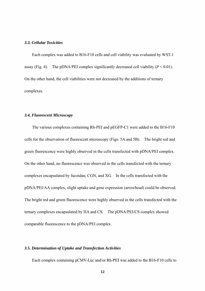
We determined the ζ-potentials of ternary complex at various charge ratios (Fig. 1). The
addition of polysaccharides to the pDNA/PEI complex decreased its ζ-potential and reached a
plateau at a charge ratio 1:8:6 of phosphate of pDNA: nitrogen of PEI: sulfate or carboxylate
of polysaccharide; therefore, the ternary complex at a charge ratio of 1:8:6 was used
throughout this study. The sizes and ζ-potentials of various complexes at a charge ratio of
1:8:6 were shown in Table 1. The pDNA/PEI complex showed 72.0 ± 11.1 nm particle size
and +48.6 ± 0.6 mV ζ-potential. Ternary complexes, however, showed anionic charges and
had significantly lower ζ-potentials than the pDNA/PEI complex (P < 0.01), although
addition of polysaccharide did not greatly affect the size of the complex.
A gel retardation assay was performed for determination of the complex formations (Fig.
2). In the lane of naked pDNA, the pDNA was migrated in the gel, and a band was showed.
The other lanes loaded various complexes, however, pDNA was not migrated in the gel and
stayed in the wells.
3.2. Agglutination Study
Agglutination activities of various complexes were evaluated (Fig. 3). The pDNA/PEI
complex agglutinated a lot of erythrocytes. On the other hand in the ternary complexes,
any agglutination was not observed.
Page 12
12
3.3. Cellular Toxicities
Each complex was added to B16-F10 cells and cell viability was evaluated by WST-1
assay (Fig. 4). The pDNA/PEI complex significantly decreased cell viability (P < 0.01).
On the other hand, the cell viabilities were not decreased by the additions of ternary
complexes.
3.4. Fluorescent Microscopy
The various complexes containing Rh-PEI and pEGFP-C1 were added to the B16-F10
cells for the observation of fluorescent microscopy (Figs. 5A and 5B). The bright red and
green fluorescence were highly observed in the cells transfected with pDNA/PEI complex.
On the other hand, no fluorescence was observed in the cells transfected with the ternary
complexes encapsulated by fucoidan, CGN, and XG. In the cells transfected with the
pDNA/PEI/AA complex, slight uptake and gene expression (arrowhead) could be observed.
The bright red and green fluorescence were highly observed in the cells transfected with the
ternary complexes encapsulated by HA and CS. The pDNA/PEI/CS complex showed
comparable fluorescence to the pDNA/PEI complex.
3.5. Determination of Uptake and Transfection Activities
Each complex containing pCMV-Luc and/or Rh-PEI was added to the B16-F10 cells to
Page 13
13
quantify uptake and gene expression (Figs. 6A and 6B). The ternary complexes
encapsulated by fucoidan, CGN, XG, AA, and HA showed significantly lower uptakes than
pDNA/PEI complex (P < 0.01, Fig. 6A). On the other hand, the pDNA/PEI/CS complex,
however, showed significantly higher uptake than the pDNA/PEI complex (P < 0.01).
Gene expression of each complex was evaluated by luciferase activity (Fig. 6B). The
pDNA/PEI complex showed extremely high gene expression exceeded 1010 RLU/mg protein.
On the other hand, all ternary complexes were significantly lower than the pDNA/PEI
complex (P < 0.01). The pDNA/PEI/CS complex, however, showed high gene expression
exceeded 1010 RLU/mg protein; it dramatically higher than the commercial transfection
reagent, lipofectin, showed only 2.52×10⁸ RLU/mg protein under the same conditions.
3.6. Intracellular Distribution of Ternary Complexes
To visualize the intracellular distributions of pDNA/PEI and pDNA/PEI/CS complexes,
YOYO-1 labeled pCMV-Luc, Rh-PEI, and Hoechst 33342 were applied (Figs. 7A and 7B).
In the both complexes, green dots of YOYO-1-labeled pCMV-Luc were located mainly in the
cytoplasm of most cells together with red dots of Rh-PEI. Synchronized green dots with red
dots were seen as orange dots in merged images as shown in Figs. 7Av and 7Bv. On the
other hand, a few dots were observed in nuclei.
3.7. Inhibition Study
Page 14
14
Hypothermia and addition of CS were performed for the inhibition study. Figure 8A
shows the uptake of the pDNA/PEI/CS complex. Uptake of the complex significantly
decreased at 4 oC incubation (P < 0.05), and CS reduced the uptake of pDNA/PEI/CS
complex concentration-dependently. Figure 8B shows the gene expression of the
pDNA/PEI/CS. The same inhibitions were shown in gene expression experiment.
Page 15
15
4. Discussion
The gene delivery vector is categorized into viral and non-viral vector. The viral vector
is highly effective and has been used in clinical trials, although some severe adverse events
such as immunogenicity and their oncogenic potential were of great concern for its safety
[7-9]. In light of these concerns, non-viral gene delivery has emerged as a promising
alternative, because non-viral vectors have advantages such as much lower immunotoxicity, a
clear structure, and easy modeling [10, 11].
Polyethylenimine (PEI) is a popular cationic polymer as shown by high gene expression
in in vitro and in vivo gene delivery, because of their specific mechanisms such as condensing
pDNA by electrostatic interaction, binding to the cell surface, take up by the endocytotic
pathway, and release of pDNA into the cytoplasm, via the so-called ‘proton sponge
mechanism’ [12-16]. On the other hand, PEI was known to cause some severe adverse
effects such as cytotoxicity and agglutination by its strong cationic charge [17].
In the present study, we prepared some ternary complexes of pDNA/PEI complex
encapsulated by anionic polysaccharides for overcoming those disadvantages of PEI.
Because of their highly stable, safe, nontoxic, and biodegradable, polysaccharides are useful
biomaterials in targeted drug delivery systems. Some polysaccharides such as fucoidan,
CGN, XG, AA, HA, and CS were used in this experiment. Among them, we newly
discovered that a ternary complex encapsulated by CS was taken by the CS-specific receptor
mediated pathway.
Page 16
16
Physicochemical properties such as the sizes and ζ-potentials of these complexes were
determined and are shown in Table 1. The polysaccharides changed the surface charge of
pDNA/PEI complex cationic to anionic without much effect on the particle size. The
concentrated capsules of the anionic polysaccharides on the complex surface were suggested,
because those ternary complexes showed apparently anionic surface charge regardless of their
total charge ratio +1. It was reported that the ternary complex, encapsulated pDNA/PEI
complex with HA and AA, showed much large particle sizes [18, 19]; however, large particles
often lead adverse effect such as microinfarctions. In this experiment, the ternary complexes
showed less than 150 nm particle size, and such nano-particles should show high
biocompatibility.
Some anionic polysaccharides, such as heparin sulfate and heparan sulfate, have known
to release pDNA from pDNA/PEI complex [20, 21]. We performed the agarose gel
electrophoresis experiment. In the any complexes, the band of pDNA was not detected,
when the naked pDNA was detected as a band on agarose gel as shown in Fig. 2. These
results support that stable self-assembled nano-particles were formed.
Many cationic non-viral gene vectors have been reported to show agglutination activities
and cytotoxicities [17, 22]. We evaluated agglutination activities and cytotoxicities of those
complexes (Figs. 3 and Fig. 4). The pDNA/PEI complex agglutinated a lot of erythrocytes,
and a huge agglomeration was observed. In contrast, all ternary complexes showed no
agglutination activities (Fig. 3). The basic cytotoxicities of complexes were evaluated in
Page 17
17
the B16-F10 cells by WST-1 assay. The pDNA/PEI complex showed extremely high
cytotoxicity, as shown in Fig. 4. On the other hand, all ternary complexes showed little
cytotoxicity. In the previous reports, the ternary complexes, pDNA/PEI and
pDNA/protamine coated with anionic polymer such as alginic acid and anionic PEG
derivatives, were demonstrated to reduce agglutination and cytotoxicity [19, 23]. In the
same way, the negative charges of the polysaccharides on the surface must abate the toxicities
of their cationic core.
Generally, anionic complexes can not be taken up well by cells because they repulse the
cellular membrane electrostatically. We evaluated the uptake of these complexes and their
gene expression efficiency; the complexes loading Rh-PEI and pEGFP-C1 were added to
B16-F10 cells, and the fluorescent microscopy was performed (Figs. 5A and 5B). Any
uptake and gene expression were not shown in the ternary complexes encapsulated by
fucoidan, CGN, and XG. Amazingly, uptake of the complex and GFP expression were
observed in the ternary complexes encapsulated by AA, HA, and CS even if it had anionic
surface charge. Furthermore, uptake of these complexes and their gene expression efficiency
was quantified in B16-F10 cells by using the complexes loading pCMV-Luc and/or Rh-PEI
(Figs. 6A and 6B). The pDNA/PEI/CS complex showed extremely high uptake and gene
expression in the cells, although, the other ternary complexes showed significantly lower
uptake and gene expressions than the pDNA/PEI complex (P < 0.01). The cationic
pDNA/PEI complex was reported to be taken by cells through endocytosis according to
Page 18
18
electrostatic interaction with cell membrane [24], however, anionic complex could not be
taken by cells in the same manner. These results indicate that the specific mechanism
participates in the uptake of the pDNA/PEI/CS complex with a negative charge. So, the
intracellular distribution and complex dissociation of the pDNA/PEI/CS complex and the
pDNA/PEI complex were visualized by using Rh-PEI, YOYO-1-labeled pDNA, and Hoechst
33342 (Figs. 7A and 7B). We were able to confirm that pDNA/PEI/CS complexes were
located in the cytoplasm without dissociations and a few complexes were observed in the
nuclei. It was also reported that some efficient non-viral vectors were reported to be mostly
located in the cytoplasm, not nuclei [25].
It is valuable to note that pDNA/PEI/CS complex have strong gene expression regardless
of its anionic surface charge. Therefore, we evaluated the inhibition effects of the
hypothermia and CS on the uptake and gene expression of pDNA/PEI/CS complex (Figs. 8A
and 8B). Hypothermia and the addition of CS significantly inhibited the uptake and gene
expression of pDNA/PEI/CS by the cells. These results strongly indicate that an
energy-dependent process and CS-specific receptor-mediated pathway concerned the uptake
of pDNA/PEI/CS complex. It was reported that clearance of CS from lymph and blood in
mammals is mediated by the HA receptor for endocytosis (HARE), which is present in the
sinusoidal endothelial cells of liver, spleen, and lymph nodes [26, 27]. Furthermore, it was
also reported that HARE was substantially colocalized with clathrin [28]. In the present
study, the pDNA/PEI/CS complex might be recognized by HARE and taken by the cells via
Page 19
19
clathrin-mediated endocytosis and released pDNA to cytoplasm from endosome by proton
sponge mechanisms of PEI; and then the complex showed high gene expressions. On the
other hand, the ternary complex encapsulated with HA showed lower uptake and gene
expression than the complex with CS. The particular mechanisms may exist, and the further
study should be performed.
Page 20
20
5. Conclusion
We developed a non-viral vector of pDNA/PEI complex encapsulated by CS. This vector
consisted of stable particles with apparently negative ζ-potential and low toxicities. On the
other hand, pDNA/PEI/CS complex was highly taken by the cells via CS-specific
receptor-mediated pathway and showed high gene expressions. Further studies are
necessary to examine the in detailed uptake mechanism.
Page 21
21
Figures
Fig. 1.
Page 29
29
Figure captions and tables
Fig. 1. Effect of each polysaccharide on ζ-potentials of pDNA/PEI complex.
Each polysaccharide was added to the pDNA/PEI complex at various charge ratios, and ζ-potentials of the
complexes were evaluated. Each value represents the mean ± S.E. of three experiments.
Table 1. Particle size and ζ-potential of the complexes.
pDNA/PEI 72.0 ± 11.1 +48.6 ± 0.6pDNA/PEI/fucoidan 71.9 ± 3.2 -27.4 ± 0.4**
pDNA/PEI/CGN 92.3 ± 33.2 -35.9 ± 0.4**pDNA/PEI/XG 132.7 ± 35.3 -44.2 ± 0.6**pDNA/PEI/AA 26.7 ± 6.1 -29.0 ± 1.9**pDNA/PEI/HA 95.0 ± 8.3 -29.9 ± 0.9**pDNA/PEI/CS 77.0 ± 3.2 -29.2 ± 0.2**
**; P < 0.01 vs pDNA/PEIEach data was represent the mean ± S.E. (n=3).
Size ζ-Potential
Fig. 2. Effect of each polysaccharide on electrophoretic migration of pDNA through an agarose gel.
Each complex was loaded onto agarose gel, and electrophoresis was carried out. Retardation of pDNA was
visualized using ethidium bromide.
Fig. 3. Agglutination with erythrocytes.
Each complex was added to erythrocytes, and agglutinations were assessed. Agglutination was observed by
phase microscopy (400× magnification).
Page 30
30
Fig. 4. Cytotoxicity tests of various complexes on B16-F10 cells.
Cell viability of cells treated with each complex was measured by WST-1 assay. Cells were incubated with each
complex for 2 h and cell viability was measured at 24 h after treatment. Data represent the percentage to
untreated cells. Each bar represents the mean ± S.E. of sixteen experiments. **: P < 0.01 vs control.
Fig. 5. Fluorescent microscopy images of B16-F10 cells transfected with each complex.
Cells were transfected with each complex containing pEGFP-C1 and Rh-PEI. Twenty-four hours after
transfection, the uptake of Rh-PEI (A) and the expression of GFP (B) were monitored (200× magnification). (a):
pDNA/PEI; (b): pDNA/PEI/fucoidan; (c): pDNA/PEI/CGN; (d): pDNA/PEI/XG; (e): pDNA/PEI/AA; (f): pDNA/PEI/HA;
(g): pDNA/PEI/CS.
Fig. 6. Uptake efficiency (A) and transgene efficiency (B) of each complex.
B16-F10 cells were transfected with each complex containing pCMV-Luc and/or Rh-PEI. Twenty-four hours after
transfection, fluorescence of Rh-PEI (A) and luciferase activity (B) were evaluated. Each bar represents the
mean ± S.E. of three experiments. **: P < 0.01 vs pDNA/PEI complex.
Fig. 7. Intracellular distribution of pDNA/PEI complex (A) and pDNA/PEI/CS complex (B).
Cells were transfected with each complex containing YOYO-1-labeled pDNA and Rh-PEI. Twenty-four hours after
transfection, phase contrast image (i), nuclei staining with Hoechst 33342 (ii), YOYO-1-labeled pDNA (iii), Rh-PEI
(iv), and merged image (v) are indicated (400× magnification).
Page 31
31
Fig. 8. Effect of Inhibitors on uptake efficiency (A) and transgene efficiency (B) of pDNA/PEI/CS complex.
pDNA/PEI/CS complex was transfected in medium which was at 4 °C or contained various concentrations of CS.
Twenty-four hours after transfection, fluorescence of Rh-PEI (A) and luciferase activity (B) were evaluated. Each
bar represents the mean ± S.E. of three experiments. *: P < 0.05, **: P < 0.01 vs control.
Page 32
32
References
[1] Hovgaard L, Brondsted H. Current applications of polysaccharides in colon targeting.
Crit Rev Ther Drug Carrier Syst 1996;13:185-223.
[2] Sinha VR, Kumria R. Polysaccharides in colon-specific drug delivery. Int J Pharm
2001;224:19-38.
[3] Coviello T, Matricardi P, Marianecci C, Alhaique F. Polysaccharide hydrogels for
modified release formulations. J Control Release 2007;119:5-24.
[4] Kawakami S, Wong J, Sato A, Hattori Y, Yamashita F, Hashida M. Biodistribution
characteristics of mannosylated, fucosylated, and galactosylated liposomes in mice.
Biochim Biophys Acta 2000;1524:258-65.
[5] Coradini D, Pellizzaro C, Miglierini G, Daidone MG, Perbellini A. Hyaluronic acid as
drug delivery for sodium butyrate: improvement of the anti-proliferative activity on a
breast-cancer cell line. Int J Cancer 1999;81:411-6.
[6] Luo Y, Bernshaw NJ, Lu ZR, Kopecek J, Prestwich GD. Targeted delivery of
doxorubicin by HPMA copolymer-hyaluronan bioconjugates. Pharm Res
2002;19:396-402.
[7] Sun JY, Anand-Jawa V, Chatterjee S, Wong KK. Immune responses to
adeno-associated virus and its recombinant vectors. Gene Ther 2003;10:964-76.
[8] Donahue RE, Kessler SW, Bodine D, McDonagh K, Dunbar C, Goodman S, et al.
Page 33
33
Helper virus induced T cell lymphoma in nonhuman primates after retroviral mediated
gene transfer. J Exp Med 1992;176:1125-35.
[9] Gore ME. Adverse effects of gene therapy: gene therapy can cause leukeaia: no shock,
mild horror but a probe. Gene Ther 2003;10:4.
[10] Tang MX, Szoka FC. The influence of polymer structure on the interactions of
cationic polymers with DNA and morphology of the resulting complexes. Gene Ther
1997;4:823-32.
[11] Filion MC, Phillips NC. Major limitations in the use of cationic liposomes for DNA
delivery. Int J Pharm 1998;162:159-70.
[12] Godbey WT, Wu KK, Mikos AG. Poly(ethylenimine) and its role in gene delivery. J
Control Release 1999;60:149-60.
[13] Kircheis R, Wightman L, Wagner E. Design and gene delivery activity of modified
polyethylenimines. Adv Drug Deliv Rev 2001;53:341-58.
[14] Boussif O, Lezoualc'h F, Zanta MA, Mergny MD, Scherman D, Demeneix B, et al. A
versatile vector for gene and oligonucleotide transfer into cells in culture and in vivo:
polyethylenimine. Proc Natl Acad Sci USA 1995;92:7297-301.
[15] Kichler A, Leborgne C, Coeytaux E, Danos O. Polyethylenimine-mediated gene
delivery: a mechanistic study. J Gene Med 2001;3:135-44.
[16] Itaka K, Harada A, Yamasaki Y, Nakamura K, Kawaguchi H, Kataoka K. In situ single
cell observation by fluorescence resonance energy transfer reveals fast
Page 34
34
intra-cytoplasmic delivery and easy release of plasmid DNA complexed with linear
polyethylenimine. J Gene Med 2004;6:76-84.
[17] Lv H, Zhang S, Wang B, Cui S, Yan J. Toxicity of cationic lipids and cationic
polymers in gene delivery. J Control Release 2006;114:100-9.
[18] Ito T, Iida-Tanaka N, Niidome T, Kawano T, Kubo K, Yoshikawa K, et al. Hyaluronic
acid and its derivative as a multi-functional gene expression enhancer: protection from
non-specific interactions, adhesion to targeted cells, and transcriptional activation. J
Control Release 2006;30:382-8.
[19] Jiang G, Min SH, Kim MN, Lee DC, Lim MJ, Yeom YI. Alginate/PEI/DNA
polyplexes: a new gene delivery system. Yao Xue Xue Bao 2006;41:439-45.
[20] Ruponen M, Yla-Herttuala S, Urtti A. Interactions of polymeric and liposomal gene
delivery systems with extracellular glycosaminoglycans: physicochemical and
transfection studies. Biochim Biophys Acta 1999;1415:331-41.
[21] Moret I, Esteban Peris J, Guillem VM, Benet M, Revert F, Dasi F, et al. Stability of
PEI-DNA and DOTAP-DNA complexes: effect of alkaline pH, heparin and serum. J
Control Release 2001;76:169-81.
[22] Sakurai F, Nishioka T, Yamashita F, Takakura Y, Hashida M. Effects of erythrocytes
and serum proteins on lung accumulation of lipoplexes containing cholesterol or
DOPE as a helper lipid in the single-pass rat lung perfusion system. Eur J Pharm
Biopharm 2001;52:165-72.
Page 35
35
[23] Maruyama K, Iwasaki F, Takizawa T, Yanagie H, Niidome T, Yamada E, et al. Novel
receptor-mediated gene delivery system comprising
plasmid/protamine/sugar-containing polyanion ternary complex. Biomaterials
2004;25:3267-73.
[24] Demeneix B, Behr JP. Polyethylenimine (PEI). Adv Genet 2005;53:217-30.
[25] Hama S, Akita H, Ito R, Mizuguchi H, Hayakawa T, Harashima H. Quantitative
comparison of intracellular trafficking and nuclear transcription between adenoviral
and lipoplex systems. Mol Ther 2006;13:786-94.
[26] Weigel JA, Weigel PH. Characterization of the recombinant rat 175-kDa hyaluronan
receptor for endocytosis (HARE). J Biol Chem 2003;278:42802-11.
[27] Zhou B, McGary CT, Weigel JA, Saxena A, Weigel PH. Purification and molecular
identification of the human hyaluronan receptor for endocytosis. Glycobiology
2003;13:339-49.
[28] Zhou B, Weigel JA, Saxena A, Weigel PH. Molecular cloning and functional
expression of the rat 175-kDa hyaluronan receptor for endocytosis. Mol Biol Cell
2002;13:2853-68