10/25/2016
1
Biomedical EngineeringElectrophysiology
Dr. rer. nat. Andreas Neubauer
Andreas Neubauer I Slide 2 I 18.11.2014
Sources of biological potentials and how to record them
1. How are signals transmitted along nerves?
• Transmit velocity
• Direction
• Intensity
• Frequency
2. How can measurements be standardized?
• Electrode position
• Data visualization
10/25/2016
2
Andreas Neubauer I Slide 3 I 18.11.2014
The nervous system
Andreas Neubauer I Slide 4 I 18.11.2014
Organization of the nervous system
• brain, nerves and muscles are the major components of the nervous system
Brown, Medical Physics
• sensory/afferent nerves deliver information to the brain
• information is passed along motor/efferent nerves by the brain
• the nervous system is highly parallel
Synapses
• synapses allow reflex loops via the spinal column⇒ can be suppressed by the brain
10/25/2016
3
Andreas Neubauer I Slide 5 I 18.11.2014
Neurons
Brown, Medical Physics
• basic concept of nerves• dendrites can be considered as the
means of information input• axons are the channels for output
information• cell bodies may be considered to be
located in the brain/spinal cord• axons supply muscles or carry
information to the brain
http://www.biotele.com/research.htm
Andreas Neubauer I Slide 6 I 18.11.2014
Neural communication I
• electrical signals in the body are constant in amplitude and vary in frequency
⇒ pain intensity is regulated by the frequency of the signals
• normal frequency � 1��� (pulse per second)
• relation of frequency and intensity is approx. logarithmic
⇒ ��� �log���� � �
Brown, Medical Physics
10/25/2016
4
Andreas Neubauer I Slide 7 I 18.11.2014
Neural communication II
• Example:
• Dynamic range of the ear: min 10�/1
⇒ 120��
⇒ The eye is sensitive to a similarly wide range of intensities
• Assume a linear relationship: � � � � ���
• Maximum transmission frequency: 100 pps
⇒ min. sensory input would correspond to 10�����
⇒ impractical!
⇒ with a logarithmic scale a dynamic range of 10�/1 is compressed to 25/1
⇒ recognition of different amplitudes is much worse
Andreas Neubauer I Slide 8 I 18.11.2014
Why is smooth movement possible?
• increasing contraction is achieved be an increase in frequency
• not all muscle fibers twitch simultaneously
Brown, Medical Physics
10/25/2016
5
Andreas Neubauer I Slide 9 I 18.11.2014
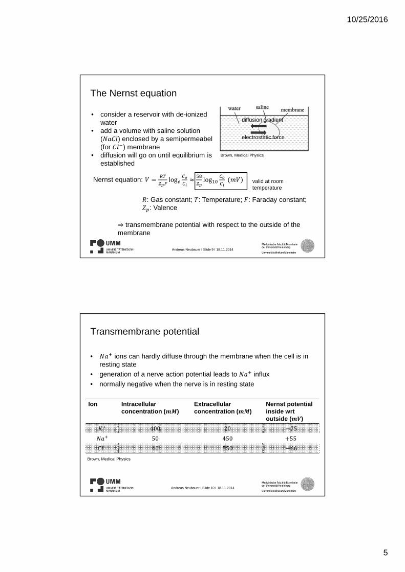
The Nernst equation
Brown, Medical Physics
• consider a reservoir with de-ionized water
• add a volume with saline solution (����) enclosed by a semipermeabel(for ���) membrane
• diffusion will go on until equilibrium is established
diffusion gradient
electrostatic force
Nernst equation: � !
"#$log%
&'&(�
)*
"#log+,
&'&(�-�
.: Gas constant; /: Temperature; 0: Faraday constant; 12: Valence
⇒ transmembrane potential with respect to the outside of the membrane
valid at roomtemperature
Andreas Neubauer I Slide 10 I 18.11.2014
Transmembrane potential
• ��4 ions can hardly diffuse through the membrane when the cell is in resting state
• generation of a nerve action potential leads to ��4 influx
• normally negative when the nerve is in resting state
Ion Intracellular concentration (56)
Extracellular concentration (56)
Nernst potential inside wrtoutside (57)
84 400 20 :75
��4 50 450 �55
��� 40 550 :66
Brown, Medical Physics
10/25/2016
6
Andreas Neubauer I Slide 11 I 18.11.2014
Membranes and nerve conduction
• electrical impulses can travel along the nerve with a velocity of 50-/�
• high/low intracellular potassium/sodium concentration is established by the membrane ⇒ polarization i.e. resting potential
Brown, Medical Physics
• stimulation leads to an efflux/influx of potassium/sodium⇒ change in transmembrane potential⇒ avalanche effect
⇒ DEPOLARIZATION!
=4>?4
Andreas Neubauer I Slide 12 I 18.11.2014
Transmission of Nerve Action Potentials (NAPs) I
Brown, Medical Physics
• impulse of depolarization which travels along a nerve
• muscle fibers can also transmit action potentials (MAPs)
• ionic currents will flow from depolarized to polarized parts⇒ source of bioelectric
signals!• myelinated fibers transmit
APs 10 times faster than non-myelinated fibers
10/25/2016
7
Andreas Neubauer I Slide 13 I 18.11.2014
Transmission of NAPs II
• speed of transmission depends on:• Membrane
capacitance • Myelin • Axon resistance Brown, Medical Physics
• assume a cylindrical membrane with diameter @ and length A:
⇒ . �BC
DEF; G: resistivity HΩmK
⇒ � LMDL; L: dielectric constant of neural membrane RS
TU
⇒ �. BV �WDE
DEF GL
�DWF
X; time constant of the membrane H�K
• typical values: Membrane capacitance: 1YZ
[UF , @ 10]-, A
10--, ^ � 1Ω-⇒ � � 3 �10�`]F, . � 1.3 �10*Ω,time constant � 0.4�⇒ .[c22%d � 2.14Ω
Andreas Neubauer I Slide 14 I 18.11.2014
Muscle Action Potentials (MAPs)
Smooth muscle Striated muscle
intestines and blood vessels skeletal muscle
intrinsically active voluntarily active
Brown, Medical Physics
10/25/2016
8
Andreas Neubauer I Slide 15 I 18.11.2014
Volume conductor effects I• electrical potential: Φ
+
�Dfghid��
• assumptions:
• Potential at infinity equal zero
• Tissue is homogeneous
⇒ � j k gB
�DdF�j
l
d
Bh
�Dd
• cylindrical nerve fiber:
⇒ � j Bmnop
�Dq
• contribution made to the potential field at r�s‘, u‘, v‘
⇒ j s– s‘x� u : u‘ x � v : v‘ x
yF
⇒ Φ s, u, v gBhn p
�D p�pz F4{zF4|zFyF
�s
Brown, Medical Physics
Andreas Neubauer I Slide 16 I 18.11.2014
Volume conductor effects II
• connection of kU to the transmembrane potential
⇒ kU s k} –kc HT p � T p�op ~
Bop:
HT p4op � T p ~
Bop
x~
B
oFT
opF
FT
Brown, Medical Physics Brown, Medical Physics
Brown, Medical Physics
10/25/2016
9
Andreas Neubauer I Slide 17 I 18.11.2014
Detection and analysis of ECG/EKG
Andreas Neubauer I Slide 18 I 18.11.2014
ECG/EKG characteristics
Brown, Medical Physics
• electrical events can be recorded from the body surface⇒ complex relation to the source
• lighthouse analogy• recording is only possible when
potentials are changing⇒ record of the changing activity
of the heart
10/25/2016
10
Andreas Neubauer I Slide 19 I 18.11.2014
Electrocardiographic planes
• standardization of recorded signals is needed
Brown, Medical Physics
Andreas Neubauer I Slide 20 I 18.11.2014
The frontal plane ECG/EKG – lead configurations
• electrical activity of the heart can be described as movement of an electrical dipole
⇒ cardiac vector is the line joining the charges of the dipole
• Einthoven‘s triangle: triangle between RA, LA and LL
⇒ lead configurations:
⇒ Lead I: RA �: to LA ��
⇒ Lead II: RA �: to LL ��
⇒ Lead III: LA �: to LL��
• plotting the measured signal in the three leads at any time of the cardiac cycle on Einthoven‘s triangle leads to the cardiac vector
• body build and age influence the cardiac vector
10/25/2016
11
Andreas Neubauer I Slide 21 I 18.11.2014
The transverse plane ECG/EKG
Brown, Medical Physics
• recorded unipolarly wrt an indifferent electrode (LA + RA + LL)• usually with six electrodes in a line round the chest
Andreas Neubauer I Slide 22 I 18.11.2014
The sagittal plane ECG/EKG
http://www.cardiocommand.com/research_cathinsert.html
• also recorded with an indifferent electrode
• catheter with electrode is placed down the oesophagus
• rarely used in practice
10/25/2016
12
Andreas Neubauer I Slide 23 I 18.11.2014
Electrodes and amplifiers
• good skin preparation leads to an electrode impedance � 10�Ω
⇒ amplifier input impedance of 1�Ω is adequate
⇒ electrodes do not have the same impedances
⇒ common-mode voltage is produced
⇒ 80�� common-mode rejection with 10�Ω difference impedance between electrodes requires a common-mode input impedance of 100�Ω
• normally the majority of EMG spectra lies above the ECG spectra
⇒ apply bandpass filter
Andreas Neubauer I Slide 24 I 18.11.2014
Detection of EEG signals
10/25/2016
13
Andreas Neubauer I Slide 25 I 18.11.2014
Sources of the EEG signal
• electroencephalographic signals were first recorded in 1929 (ECG/EKG in 1895)
• electroencephalograph means graph of electrical changes from the enkephalos (Greek for brain)
• sources of the EEG signals are the neuronal potentials of the brain
⇒ attenuation by bone, muscle and skin
⇒ electrocorticography (ECoG) records signals directly from the cortex
• EEG signals are between 10 and 300]V
• Ag-AgCl discs are best to record an EEG
⇒ time consuming
⇒ skullcaps are much more convenient in use
Brown, Medical Physics
Andreas Neubauer I Slide 26 I 18.11.2014
EEG equipment and settings
• differential amplifiers are used for signal amplification
• min. eight channels at the recorder
• assume 16 differential amplifiers ⇒ 32 input connections plus one earth connection
• „standard“ EEG settings:
• Chart speed 30--/�
• Gain setting: 100]V/�-
• Time constant: 0.3� (corresponds to a :3��point of 0.531/�)
• Filters: High frequency response is a -3��at 751/�
• electrode impedance � 10�Ω
10/25/2016
14
Andreas Neubauer I Slide 27 I 18.11.2014
Normal EEG signals
Brown, Medical Physics
• a quiet environment is required
• only one person should be in the room with the patient
• wide-awake „normal“ persons produce an unsynchronized high-frequency EEG
• rhythmic activity at8– 131/� is produced if a „normal“ person closes the eyes
Andreas Neubauer I Slide 28 I 18.11.2014
Artifacts
• electrode artifacts
⇒ electrode impedances
⇒ interference
⇒ movement of the cables
⇒ perspiring of the patient
• potential difference of several -�between the back and front of the eyes
• ECG may be seen if recording electrodes are spaced a long way
• dental fillings may produce artifacts
http://www.psychologie.uzh.ch/fachrichtungen/plasti/Labor.html
http://bipolaraspiemom.wordpress.com/2011/07/15/are-you-still-awake-our-eeg-story/
10/25/2016
15
Andreas Neubauer I Slide 29 I 18.11.2014
Detection of EMG signals
Andreas Neubauer I Slide 30 I 18.11.2014
Sources of electromyographic (EMG) signals
• record signals of nerves and muscles
• needle and surface electrodes can be used
⇒ examine shape and sound of the signal with needle electrodes
⇒ overall activity of the muscle is recorded with surface electrodes
• functional unit of a muscle is one motor unit
Brown, Medical Physics
10/25/2016
16
Andreas Neubauer I Slide 31 I 18.11.2014
EMG equipment
• recording of an EMG is possible between two surface electrodes
⇒ reducing the distance below 4 mm leads to a significant signal drop
• surface electrodes will always record signal from multiple muscles
⇒ needle electrodes are more accurate but uncomfortable⇒ fine wire electrodes are excellent for long term EMG recording
• surface electrodes record less high-frequency content than needle electrodes
• signals up to 2-� are typical
Brown, Medical Physics
Andreas Neubauer I Slide 32 I 18.11.2014
EMG settings
Standard settings for the pre-amplifier
Amplification 100
Input impedance 10�Ω
Noise with input shorted 2]V� : �
Common-mode rejection ratio 80��
Bandwidth (:3��points) 101/�–10`1/�
• equipment testing⇒ short circuit the inputs of the amplifier and set maximum gain
⇒ only noise should be visible⇒ check leads and plugs
Brown, Medical Physics
10/25/2016
17
Andreas Neubauer I Slide 33 I 18.11.2014
Normal EMG signals
• voluntary EMG pattern is recorded with a needle electrode
⇒ several points must be observed
⇒ „normal“ EMG sounds like gunfire
• normal APs last few milliseconds and contain two/three deflections
• myopathic muscles produce smaller APs with more deflections
Brown, Medical Physics
Andreas Neubauer I Slide 34 I 18.11.2014
Neural stimulation III
10/25/2016
18
Andreas Neubauer I Slide 35 I 18.11.2014
Nerve conduction velocity measurement
• measurement of time between stimulus and response ⇒ conduction time (average of myelinated fibers: 50-/�)
• myelination is not complete at birth
⇒ nerve conduction increases over the first years of life
Brown, Medical Physics
Andreas Neubauer I Slide 36 I 18.11.2014
Motor nerve conduction velocity
• measure latency of proximal/distal stimulation⇒ calculate velocity from the values of obtained latencys