77
Biomembrane and Cell signalling BCH 452(IV) Transport across the membrane Dr. Samina Hyder Haq Assistant professor Dept of biochemistry Collage of Science King Saud university
| Date post: | 14-Dec-2015 |
| Category: |
Documents |
| Upload: | silvester-walton |
| View: | 215 times |
| Download: | 0 times |

Biomembrane and Cell signalling BCH 452(IV)Transport across the membrane
Dr. Samina Hyder HaqAssistant professor Dept of biochemistry
Collage of ScienceKing Saud university

Transport across the membrane
Biological membrane is a supramolecular structure = many molecules
ordered into a higher level of organizationwith emergent properties beyond those ofthe individual molecules.The supramolecular structure of themembrane results in selective permeability

•Lipid bilayer is permeable to small nonpolar molecules such as CO2 and O2.
•Not permeable to ions and nonpolar molecules such as glucose, succrose. Na+. Cl_.
•Special transport proteins makes the cell permeable to specific ions and polar molecules and water.
•The selective nature of membrane comes from both the discriminating nature of phospholipids and the specific transporter proteins.


Three ways of Transport 1. Passive Transport: Diffusion across the
membrane down the concentration gradient.2. Osmosis: Passive transport of water3. Facilated Diffusion: Specific protein helps
in transport of water and other nutrients2. Active Transport: Pumping of solutes
against the concentration gradient. Co transport: membrane protein helps in
transport of one solute to another 3. Exocytose and Endocytosis Transport of large molecules.

Simple diffusion
•The movement of ions from higher concentration to lower concentration.

Simple Diffusion• <150 Da, uncharged
species,• going down their• concentration gradient
▫ O2
▫ CO2
▫ Alcohol▫ Anaesthetics▫ Pesticides
Extracellular
Cytosol
Concentration Gradient

Osmosis is the passive transport of water. •Water moves from hypotonic solution to hpertonic solution.

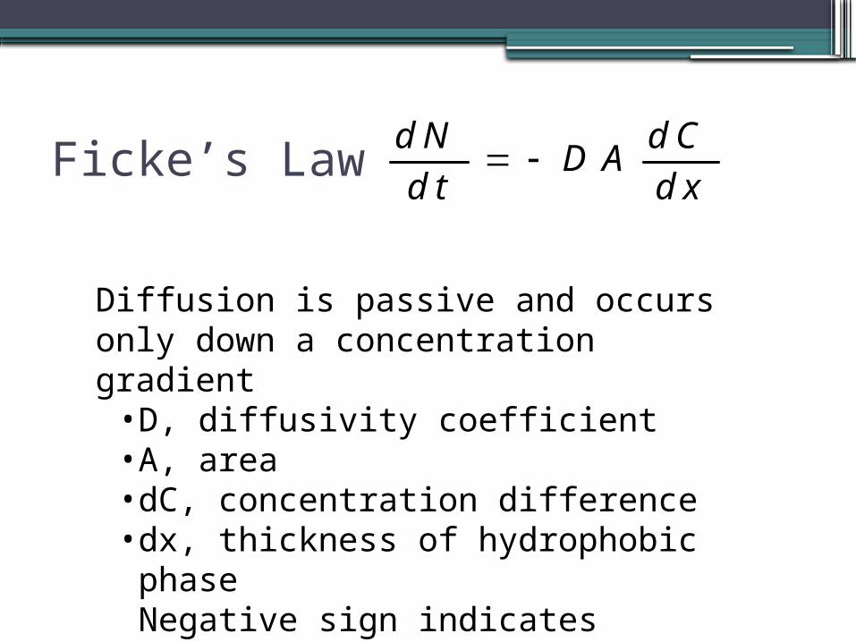
Ficke’s LawdN dC
DAdt dx
Diffusion is passive and occurs only down a concentration gradient• D, diffusivity coefficient• A, area• dC, concentration difference• dx, thickness of hydrophobic phaseNegative sign indicates directionDiffusion also depends on the solubility.

Passive Transport•Specific ions and polar molecules can cross
the lipid bilayer by passing through transport proteins that span the membrane. Some transport proteins, called channel proteins, have a hydrophilic channel that certain molecules or ions can use as a tunnel through the membrane. For example, the passage of water through the membrane can be greatly facilitated by channel proteins known as aquaporins.

•Free energy of transporting materials across the membrane depends on concentration gradient across the membrane. Where C1 and C2 are the two concentration gradient.

▫Solutes move spontaneously ( Gt < 0) from compartment of higher concentration to compartment of lower concentration
▫ G = 0 when C1 = C2 (equilibrium) oDiffusion "down" the concentration gradient (from
region of greater concentration to region of lower concentration, toward equilibrium of equal concentrations) is a manifestation of the 2nd law of thermodynamics -- molecules tend spontaneously to assume the distribution of greatest randomness, i.e., entropy increases until system is maximally randomized.

•A transport process must be active when G is positive, whereas it can be passive when G is negative. For example,consider the transport of an uncharged molecule from c 1 = 10-3 M to c 2 = 10-1 M. At 25°C (298 K),
• G is + 2.7 kcal mol-1 (+11.3 kJ mol-1), indicating that this transport process requires an input of free energy. It could be driven, for example, by the hydrolysis of ATP, which yields -12 kcal mol-1 (-50.2 kJ mol-1) under typical cellular conditions. If G is negative, the transport process can occur spontaneously without free-energy input

▫where Z = charge on the ion, F = the Faraday constant (96.5 kJ/(V•mol) and = the membrane potential (the charge gradient across the membrane, in volts, or millivolts, etc.)
•If Y is negative, going from outside to inside, then the transport of cations into the cell is favored over anions. The opposite would be true if Y were positive

Facilated Diffusion


Facilitied Transport
•Other transport proteins, called carrier proteins, bind to molecules and change shape to shuttle them across the membrane. Each transport protein is specific as to the substances that it will translocate.
•For example, the glucose transport protein in the liver will carry glucose into the cell but will not transport fructose, its structural isomer.
•Some transport proteins do not provide channels but appear to actually translocating the solute-binding site and solute across the membrane as the transport protein changes shape.
•These shape changes may be triggered by the binding and release of the transported molecule.

•In certain inherited diseases, specific transport systems may be defective or absent.
•Cystinuria is a human disease characterized by the absence of a protein that transports cysteine and other amino acids across the membranes of kidney cells.
•An individual with cystinuria develops painful kidney stones as amino acids accumulate and crystallize in the kidneys.

Active transport•Pumping of solutes against the
concentration gradient, which requires energy. Plants usually takes up nutrients in this way.

Active Transport•Special transport proteins, electrogenic
pumps, generate the voltage gradient across a membrane.
•The sodium-potassium pump in animals restores the electrochemical gradient not only by the active transport of Na+ and K+, setting up a concentration gradient, but because it pumps two K+ inside for every three Na+ that it moves out, setting up a voltage across the membrane.
•The sodium-potassium pump is the major electrogenic pump of animal cells.

Comparison of different transport mechanism

Proton Pump in Plants•In plants, bacteria, and fungi, a proton
pump is the major electrogenic pump, actively transporting H+ out of the cell.
•Proton pumps in the cristae of mitochondria and the thylakoids of chloroplasts concentrate H+ behind membranes.
•These electrogenic pumps store energy that can be accessed for cellular work.

Endocytosis = the process which cell takes in macromolecules and particulate matter by forming new vesicles from the plasma membrane.
3 types of endocytosis:1.phagocytosis = celular eating2.pinocytosis = cellular drinking3.receptor-mediated endocytosis

phagocytosis•occurs only in specialized cells such as
macrophages and granulocytes. Phagocytosis involves the ingestion of large particles such as viruses, bacteria, cells, or debris.
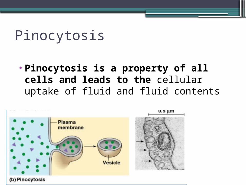
Pinocytosis
•Pinocytosis is a property of all cells and leads to the cellular uptake of fluid and fluid contents

Receptor mediated endocytosis
•primarily responsible for the uptake of macromolecules for which there are a finite number of binding sites on the plasma membrane. These high-affinity receptors permit the selective concentration of ligands from the medium, minimize the uptake of fluid or soluble unbound macromolecules, and markedly increase the rate at which specific molecules enter the cell.

Comparison of simple endocytosis and receptor mediated endocytosis.


Receptor Mediated Endocytosis•Human cells use this process to take in
cholesterol for use in the synthesis of membranes and as a precursor for the synthesis of steroids.
•Cholesterol travels in the blood in low-density lipoproteins (LDL), complexes of protein and lipid.
•These lipoproteins act as ligands to bind to LDL receptors and enter the cell by endocytosis.
• In an inherited disease called familial hypercholesterolemia, the LDL receptors are defective, leading to an accumulation of LDL and cholesterol in the blood.
•This contributes to early atherosclerosis.

Exocytosis.
•Most cells release macromolecules to the exterior by exocytosis. This process is also involved in membrane remodeling, when the components synthesized in the Golgi apparatus are carried in vesicles to the plasma membrane. The signal for exocytosis is often a hormone which, when it binds to a cell-surface receptor, induces a local and transient change in Ca2+ concentration. Ca2+ triggers exocytosis.

A comparison of endocytosis and exocytosis .

Ionophores can carry charged ions across the membrane.
Ionophores are agents that enter the cell membrane and change its permeability.
They make the ions move inward and outward of the cell membrane, so the ion concentrations will become equal in both sides.

Valinomycin an antibiotic acts as Ion Ionophore
•Carries 105 K+ per second across membranes

Mechanism of action of Gramicidin

Different transport system
•Uniport: Molecule travel only in one direction.•Symprt: Both molecule travel in same direction•Antiport: Two molecule travels in opposite
direction.
Examples of symport (amino acid Transport across the membrane)
Amino acid transport across the plasma membrane is mediated by both sodium-dependent and sodium independent transport mechanisms
•All the amino acids can be actively transported, for example out of the kidney tubules and into the blood
•the reuptake of Glu from the synapse back into the presynaptic neuron by sodium-driven symport pumps.
•The Na+/iodide transporter. This symporter pumps iodide ions into the cells of the thyroid gland (for the manufacture of thyroxine) and also into the cells of the mammary gland (to supply the baby's need for iodide).
•The permease encoded by the lac operonof E. coli that transports lactose into the cell.
A

Transport of a. Acids by gamma glutamyl cycle.
• Kidney contains a high concentration of y-glutamylcysteine synthetase and also of glutathione synthetase. The presence of these enzymes in kidney thus makes possible a series of catalytic events involving the synthesis and degradation of glutathione, and the coupled uptake and release of free amino adids from •§-glutamyl linkage.
I= y-glutamylcysteine synthetase; II= glutathione synthetase; III= y-glutamyl transpeptidase; IV= 'y-glutamylcyclotransferase; V, peptidase. PCA = pyrrolidone carboxylic acid; AA= amino acid.

Reabsorbtion of electrolytes in kidney

Major transport pathways in the proximal tubule. Identify symport, antiport, and uniport pathways for ions.

Glucose Transport
•The Na+/glucose transporter. This transmembrane protein allows sodium ions and glucose to enter the cell together. The sodium ions flow down their concentration gradient while the glucose molecules are pumped up theirs. Later the sodium is pumped back out of the cell by the Na+/K+ ATPase.

Transport of glucose • More complex than channels are carrier proteins
such as glucose permease in erythrocytes. At least 4 carrier protein are identified for the transport of glucose. A family of glucose transporters in various tissues (GluT1, GluT2, etc.) facilitate glucose movement DOWN its concentration gradient
• Erythrocytes depend on constant supply of glucose from blood plasma, where [Glc] = ~4.5-5 mM, to use as energy source (fuel) via glycolysis
• GluT1 increases rate of glucose diffusion across membrane by factor of about 50,000.
• The transported molecule (glucose) moves down its concentration gradient. Once inside the cell, the molecule is transformed into another, impermeant species, thus lowering the inside concentration and maintaining the concentration gradient.

Glucose transport as uniport
Glucose permease
GluGlu Glu GluGlu Glu
Glu
Glu-P
Glu-PP + Glu ---> Glu-P
glucose phosphorylation
erythrocyte
blood
cytosol

Glucose Na symport in Intestine
glucose-Na symport
Naglucose
intestinal lining

Glucose transport from intestinal lumen

Symport (Cotransport): coupled transport
•Two molecules travel together, one as a passenger, the other as a driver. The driver diffuses down its electrochemical gradient, but it cannot do so unless it has the passenger.
•ATP is not directly involved, but it sets up the electrochemical gradient used to propel the driver.
•Sucrose/ H+ antiport in mitochondria and chloroplast.

Antiport
The driver and passenger travel in opposite directions.
Ca-Na antiport takes place in cardiac muscle.
Very popular are proton-driven pumps: H-lactose symport in E. coli, Na-H antiport, Ca-H antiport, sucrose-H antiport in plant vacuoles.


the inner membrane
Its lipid bilayer contains a high proportion of the "double" phospholipid cardiolipin, which has four fatty acids rather than two and may help to make the membrane especially impermeable to ions.

Inner membrane- contains a variety of transport proteins that make it selectively permeable to those small molecules that are metabolized or required by the many mitochondrial enzymes concentrated in the matrix. - enzymes of the respiratory chain, are essential to the process of oxidative phosphorylation, which generates most of the animal cell's ATP.


The proton Pump in Mitochondria
• H+ is actively pumped out by hydrolyzing ATP • H+ accumulated outside the membrane, generating a
concentration and electrochemical gradient
This is a common means to store energy in cells Used in mitochondria & chloroplasts


Bacterial Cell membrane
•A well-characterized exception is provided by the porins—a class of proteins that form channels in the outer membranes of some bacterial cell membrane. Porins typically control the diffusion of small metabolites like sugars, ions, and amino acids.

The H+/ succrose pump in plants cells. • The H+ cannot cross the membrane of
chloroplast, it has to bind to a carrier protein for H+, but sucrose must also bind. When both are bound, the configuration changes and the protein opens to the membrane interior. ▫This is known as cotransport as two molecules
are pumped across a membrane, one "downhill" (with its gradient) coupled with one "uphill" (against its gradient)
▫It is also known as a symport as both molecules are crossing in the same direction

Na+/K+ Pump
•Cellullar [K+] is low and [Na+] is high - must pump K+ in and pump Na+ out. K+ and Na+ transport require ATP energy

Experimental evidence has shown that this pump will only work if [K+] is high on outside and [Na+] is high on inside.
This pump works independent of concentration gradient The pump is an integral membrane protein. It Has Two conformation state E1 and E2• In E1( high affinity for Na+) state it Binds to 3 Na+
inside cell which results in phosphorylation. when the pump is phosphorylated, its configuration changes and it opens up the Na+ to the outside of the cell . The Na+ are released (the altered configuration does not favor the binding of Na+)
• In E 2 state (high affinity for K+) binds to 2 K+ which results in dephosphorylation. the phosphate group is gone, the altered protein reverts back to its original shape, which was open to the inside of the cell . The original shape does not favor the binding of K+, so these are released. Na+ then binds to the protein and the process is repeated
How Na+/K+ Pump Work?


Ca2+ ATPase in Sarcoplasmic Reticulum
• Ca2+ ATPase found in the sarcoplasmic reticulum (SR Ca2+ ATPase) of muscle cells is an enzyme, which constitutes 80% of the sarcoplasmic reticulum membrane protein, plays an important role in muscle contraction, which is triggered by an abrupt rise in the cytosolic calcium level. Muscle relaxation depends on the rapid removal of Ca2+ from the cytosol into the sarcoplasmic reticulum, a specialized compartment for calcium storage, by the SR Ca2+ ATPase. This pump maintains a Ca2+ concentration of approximately 0.1 mM in the cytosol compared with 1.5 mM in the sarcoplasmic reticulum.

Mechanism of P-Type ATPase Action
• The binding of Ca2+ and the phosphorylation of the ATPase (steps1 and 2), illustrated here for the Ca2+ ATPase, lead to the conversion of the binding sites (step 3) and the release of Ca2+ to the luminal side of the membrane (step 4). Hydrolysis of phosphoaspartate (step 5) and eversion (step 6) reset the enzyme to its initial state.

Mechanism of action1.The postulated reaction cycle begins with the binding
of ATP and two Ca2+ ions to the E1 state.2. The enzyme cleaves ATP, transferring the g-
phosphoryl group to the key aspartate residue. Calcium must be bound to the enzyme for the phosphorylation to take place. Phosphorylation shifts the conformational equilibrium of the ATPase toward E2.
3. The transition from the E1 to the E2 state causes the ion-binding sites to "evert" so that the ions can dissociate only to the luminal side of the membrane.
4. In the E2-P state, the enzyme has low affinity for the Ca2+ ions, so they are released.
5. With the release of Ca2+, the phosphoaspartate residue is hydrolyzed, and the phosphate group is released.
6. The enzyme, devoid of a covalently attached phosphoryl group, is not stable in the E2 form. It everts back to the E1 form, completing the cycle.

Endoplasmic ReticulumMembrane
• The endoplasmic reticulum (ER) is a eukaryotic organelle that forms an interconnected network of tubules, vesicles, and cisternae within cells.
• These structures are responsible for several specialized functions: protein translation, folding and transport of proteins to be used in the cell membrane
• (e.g. transmembrane receptors and other integral membrane proteins), or to be secreted (exocytosed) from the cell (e.g. digestive enzymes); sequestration of calcium; and production and storage of glycogen, steroids, and other macromolecules.
• The endoplasmic reticulum is part of the endomembrane system.
• The basic structure and composition of the ER membrane is similar to the plasma membrane.


Glycosylation takes place in ER and Golgi Complex•Most polypeptides synthesised on ER , are
Glycosylated . Sugar residues are added to it and transported to its target location .

Rough ER•The membrane of the RER is continuous
with the outer layer of the nuclear envelop. Although there is no continuous membrane between the RER and the Golgi apparatus, membrane-bound vesicles shuttle proteins between these two compartments. Vesicles are surrounded by coating proteins called COPI and COPII. COPII targets vesicles to the golgi and COPI marks them to be brought back to the RER. The RER works in concert with the Golgi complex to target new proteins to their proper destinations.


Membrane of nerve tissues
•Central nervous system consist of brain and spinal cord.
•Functional unit is neuron.


Myelin Structure and Function
•Myelin is the insulating sheath surrounding nerve cells…the white matter coating our nerves, enabling them to conduct impulses between the brain and other parts of the body. It consists of a layer of proteins packed between two layers of lipids.
•Myelin is produced by specialized cells: oligodendrocytes in the central nervous system, and Schwann cells in the peripheral nervous system. Myelin sheaths wrap themselves around axons, the threadlike extensions of neurons that make up nerve fibers. Each oligodendrocyte can myelinate several axons.
• .

Types of neurons
•Functional Classification: Sensory. Motor and inter-neurons
•Structural classification:unipolar
Bipolar
Multipolar

•Demyelination is the act of demyelinating, or the loss of the myelin sheath insulating the nerves, and is the hallmark of some neurodegenerative autoimmune diseases, including multiple sclerosis, acute disseminated encephalomyelitis, transverse myelitis, Alexander's disease, chronic inflammatory demyelinating polyneuropathy, Guillain-Barré Syndrome and central pontine myelinosis.

Multiple Sclerosis


Synapse

Action potential (see handouts for detail)


