Bionic balance organs: progress in the development
of vestibular prosthesesPaul F Smith
ABSTRACTThe vestibular system is a sensory system that is critically important in humans for gaze and image stability as well as postural control. Patients with complete bilateral vestibular loss are severely disabled and experience a poor quality of life. There are very few e� ective treatment options for patients with no vestibular function. Over the last 10 years, rapid progress has been made in developing artificial ‘vestibular implants’ or ‘prostheses’, based on cochlear implant technology. As of 2017, 13 patients worldwide have received vestibular implants and the results are encouraging. Vestibular implants are now becoming part of an increasing e� ort to develop artificial, bionic sensory systems, and this paper provides a review of the progress in this area.
The fi rst cochlear implant was per-formed in 1961 by William House and John Doyle of Los Angeles, Califor-
nia.1 This was followed by stimulation tests conducted by Blair Simmons and Robert White at Stanford in 1962.1 Graeme Clark of Melbourne, Australia, implanted the fi rst multi-electrode cochlear implant in 1978, which became the fi rst successful commer-cially available multi-electrode cochlear im-plant.1 Although the concept was met with considerable scepticism, over the last sever-al decades the procedure has been refi ned and it is estimated that nearly half a million people had received cochlear implants by 2016.2 The success of cochlear implant tech-nology has naturally encouraged research-ers and clinicians to consider other forms of bionic implants that can substitute for lost sensory function, and considerable progress has now been made in the development of retinal and other cortical implants for humans. Another kind of sensory implant that is developing rapidly is the ‘vestibular implant or prosthesis’, which aims to replace a missing or dysfunctional vestibular sys-tem. Groups of patients have been receiving such implants since approximately 2007 and considerable progress has been made in this area, which this paper aims to review.
It is often said that the vestibular system is a sensory system that is not appreciated until something goes wrong with it. More primitive than the visual and auditory systems, it is estimated to have evolved over 500 million years ago, and exists in a rudimentary form in animals such as jellyfi sh, where it functions to indicate gravitational vertical, ie, which way is up. In mammals it is more sophisticated and consists of three semi-circular canals and two otolithic organs, in each inner ear.3 The horizontal, anterior and posterior semi-circular canals sense rotation of the head in three dimensions and the otoliths, the utricle and saccule, sense translation in three dimensions. It is important to appre-ciate that, strictly speaking, the stimulus for all of these sensory organs is ‘acceleration’ or change in angular or linear velocity, not velocity itself, and therefore one of the stimuli they detect is linear acceleration by gravity, which is critical for survival. Together, these vestibular end organs sense acceleration during head movement as well as gravitational vertical. The vestibular system contributes to the control of posture through the vestibulo-spinal refl exes and even to autonomic function, but in humans one of its most important roles is
Figure 1: The push-pull organisation of the horizontal (yaw) rotational vestibulo-ocular refl ex (VOR).
The direct pathway comprises a short tri-synaptic reflex consisting of the vestibular nerve (VIIIth nerve), the vestibular nuclei (VN), motoneurons in the VI (abducens) and III (oculomotor) nuclei, and the lateral rectus (LR) and medial rectus (MR) muscles. When the head rotates to the right (arrows), the right horizontal semi-circular canal a� erents increase their firing rates, whereas the le� a� erents decrease their firing rates (orange versus blue colours, respectively). The excitation/inhibition is transmitted to the contralateral/ipsilateral abducens (VI) nucleus through neurons in the right/le� VN, respectively. Internuclear neurons in the abducens nuclei cross the midline and terminate in the contralateral medial rectus motoneurons (III). Thus, motoneurons in the le� VI and right III nuclei fire at a higher frequency, whereas those in the right VI and le� III nuclei fire at a lower frequency. As a result, the le� LR and right MR muscles contract, whereas the le� MR and right LR muscles relax and both eyes rotate le� ward. Reproduced from Angelaki (2008)6 with permission.
in the generation of rapid compensatory eye movements during unexpected head movement—the ‘vestibulo-ocular refl exes (VORs)’.3 These refl exes generate equal and opposite eye movements during unex-pected head movement and keep the visual image of the world stable on the retina, thereby preventing blurring of the visual image or ‘oscillopsia’ (see Figures 1 and 2).3–7 It is natural to think that the vestibular system should not be necessary for this purpose, because the visual system should be able to compensate for this kind of head movement. However, this is not the case. The visual system is too slow to adequately correct for unexpected, high acceleration head movements, because visual feedback is required before the eye movements can be generated. The vestibular system responds
to acceleration of the head directly (ie, it is an ‘open-loop system’), generating VORs in approximately 9ms, and does not require any visual feedback, although feedback regarding the success of the VORs does modulate the refl ex.4 Furthermore, the vestibular system is exquisitely sensitive to head acceleration, generating VORs even in response to the small degree of head movement caused by the pulse beat. Although we are not usually aware of it, we are exposed to high accelerations of the head when we walk down a street, and without adequate compensatory eye movements from the VORs, we would see the world as blurred and bouncing around (‘oscillopsia’), as in footage taken from a video camera while walking.
For these reasons, the consequences of dysfunction of the vestibular system in humans can be severe and extremely debil-itating. In the worst case scenario, people without effective vestibular systems become housebound and lead very restricted lives, with high rates of anxiety disorders and depression.8 Recent evidence also indi-cates that such people develop cognitive defi cits as a result of the effects of the loss of self-motion information generated by the vestibular system, on the brain’s ability to construct mathematical maps of the spatial environment.9 Vestibular dysfunction of various sorts is now thought to be quite common. Although there are no reliable statistics available in New Zealand, in the US it is estimated that approximately 35% of people aged 40 and over have suffered from some form of vestibular dysfunction (based on a sample of 5,086).10 The incidence of vestibular defi cits increases with age, reaching almost 50% over the age of 60, with a 12-fold increase in the risk of falling.10
There are, of course, many different types of vestibular disorders and not all of them would be candidates for a vestibular implant. Vestibular disorders can be acute, chronic, paroxysmal, unilateral or bilateral and lesions partial or complete. The most common vestibular disorders are generally
agreed to be benign paroxysmal posi-tional vertigo (BPPV), vestibular migraine (a relatively recent diagnostic category) and Meniere’s Disease. Less common are conditions such as vestibular neuritis and labyrinthitis, aminoglycoside antibiotic vestibulo-otoxicity (eg, from gentamicin), vestibular schwannomas (a tumour on the vestibular nerve), perilymph fi stulas, idio-pathic vestibular loss and central vestibular disorders.11 To date, vestibular implants have been investigated and trialled mostly in cases of complete bilateral vestibular loss. In this case very few therapies have been successful and the quality of life for patients with this condition is generally very poor.8 Mammalian vestibular hair cells do not readily regenerate, therefore despite substantial attempts to stimulate their re-growth using drugs and gene therapy, this option remains unavailable.12 Unfor-tunately, no drug treatment can substitute for the sensory functions of the vestibular system. Non-vestibular sensory substitution has been used with some success, such as electrical stimulation of the tongue or torso during head movement, and vestibular rehabilitation delivered by physiothera-pists does help, but ultimately the unique functions of the vestibular system cannot be replaced.12 Therefore, implanting an
Figure 2: Computer generation of an acuity chart in original unfi ltered format (A) and blurred (B) to take into account the change in spatial response brought about by whole body oscillation for patient P3 (ie, with an absent vestibulo-ocular refl ex).
The lines of the acuity chart correspond to 6/9, at the top, followed by 6/6, 6/5, and finally 6/4 at the bottom. Reproduced from Morland et al (1998)7 with permission.
artifi cial vestibular system that senses head acceleration electronically, using miniature three-dimensional accelerometers, and translating those signals into patterned elec-trical stimulation of the vestibular nerve, becomes an enticing possibility.13
The first vestibular implantsThe idea for vestibular implants arose
from the original studies of Cohen, Suzuki and Bender in the 1960’s, who demonstrated that electrical stimulation of individual semi-canal nerves could elicit VORs in the plane of the corresponding canal.14 The fi rst experi-mental vestibular implants in animals were performed in 2001 by the Harvard/Mass Eye and Ear (MEE) research group15–18 and then later by the Johns Hopkins research group.19,20 Based on cochlear implant tech-nology, the prototype MEE VI transduced angular head acceleration around one rotational axis using an accelerometer and transmitted this information to the brain by modulating the rate of electrical impulses delivered to the corresponding semi-cir-cular canal nerve.15–18 All of these studies focused on the semi-circular canals rather than the otoliths, due to the more complex nature of the latter. Whereas the direction of excitation during angular acceleration—the ‘polarization vector’—is the same for each hair cell in the cupula inside the ampulla of each individual semi-circular canal (but different for different semi-circular canals), in the utricle and saccule the pattern of polarization vectors reverses halfway across the sensory epithelium.3 This division, known as the ‘striola’, means that each otolithic structure is capable of responding with depolarization for either direction of translational acceleration in each plane.3
The actual transduction of head accel-eration could be performed by modifi ed accelerometers. Accelerometers have been used in engineering for a long time but they have been miniaturised to the point where they can occupy very little space. However, in this case the acceler-ometers needed to mimic the biophysical properties of real semi-circular canals.21,22 There was a clear anatomical and phys-iological framework for the conception of the vestibular implant.23 The cochlear implant-style processor and its electrode design provided the means of delivering
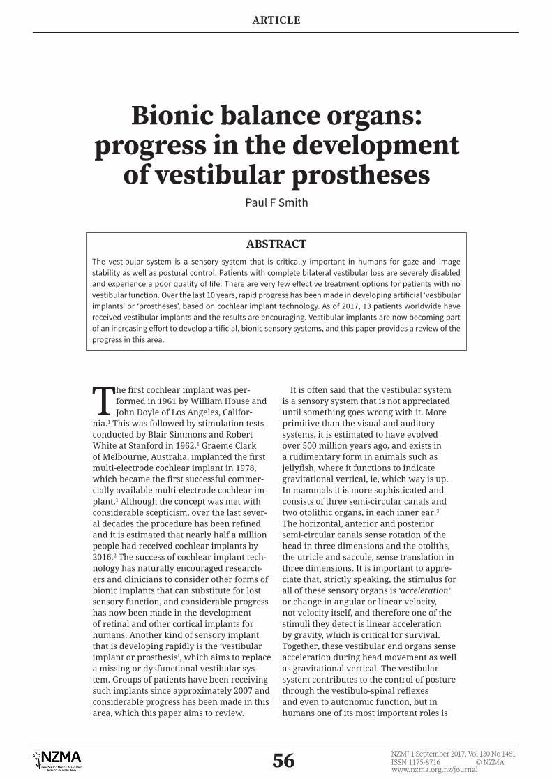
the patterned electrical stimulation (see Figure 3).24–27 In many cases, patients with vestibular implants also received cochlear implants.12 So the remaining question for the vestibular system was: what should be the nature of this electrical stimulation?28 Decades of neurophysiological research in animals had provided detailed information on the nature of the electrical activity in the primary afferent neurons of the vestibular nerve, which takes information from the vestibular hair cells and transmits it to the brainstem vestibular nucleus complex (VNC) and the cerebellum (see Figure 1).3,29 It was well established that there were two main kinds of vestibular afferents, regular and irregular, and that their basic pattern of activity was a high baseline fi ring rate (approximately 90 spikes/sec on average) that was modulated up or down during stimulation of the semi-circular canals.3,29 During head acceleration, the dynamic range of these neurons extended to over 300 spikes/sec.29 It was also well known that these afferents synapsed on ‘posi-tion-vestibular-pause (PVP) neurons’ in the VNC—so-called because they carry both vestibular and eye movement signals—and ‘fl occular target neurons (FTNs)’, which project to motor neurons innervating the eye muscles and driving the VORs (see Figure 3).3,4 Therefore, theoretically, if an electrode positioned on a semi-circular canal ampullary nerve delivered a baseline fi ring rate of approximately 250 pulses/sec and this was modulated by an acceler-ometer during rotational head acceleration, it would be possible to modulate the afferent fi ring rate up to approximately 450 pulses/sec during angular rotation that would normally excite that canal, and down to approximately 100 pulses/sec during angular rotation that would normally inhibit it. The pulses were biphasic and in the amplitude range of 160–300µA.30 The electrodes could be positioned accurately in one of the ampullary nerves by extending the mastoidectomy normally used for a cochlear implant procedure.30 Studies were initially carried out in guinea pigs and then in monkeys.15,16,31,32 The results showed that electrodes implanted in any of the three semi-circular canal nerves could elicit quasi-normal VORs in the appropriate plane.15,16,32
Figure 3: Early vestibular processing and the sensory coding of self-motion: central neurons.
Neurons in the vestibular nuclei that receive direct input from the vestibular a� erents can be categorised into two main categories: (i) neurons that control and modulate the vestibulo-ocular reflex to ensure gaze stability during everyday life (ie, PVPs and FTNs), and (ii) neurons that control posture and balance, and also project to higher order structures involved in the estimation of self-motion (ie, VO neurons). Reproduced from Cullen (2012)4 with permission.
Human trialsThe fi rst trials of the vestibular implant
were carried out in humans in Geneva in 2007.30,33 To date, all of these trials have been conducted at the University Hospital of Geneva, Geneva, Switzerland, and University of Maastricht Medical Centre, Maastricht, the Netherlands,30 and the University of Washington, Seattle.34 Thirteen patients have received implants to date, mostly for complete bilateral vestibular loss, and they were also deaf in the implanted ear.12,35 Most patients have received a single implant in one of the semi-circular canal nerves. Some have also received implants in all of them (but not the otoliths).12,36
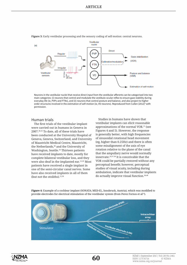
Studies in humans have shown that vestibular implants can elicit reasonable approximations of the normal VOR.12 (see Figures 4 and 5). However, the response is generally better, with high frequencies of sinusoidal rotational head movement (eg, higher than 0.25Hz) and there is often some misalignment of the axis of eye rotation relative to the plane of the canal that the ampullary nerve would normally innervate.24,37,38 It is conceivable that the VOR could be partially restored without any perceptual benefi t; however, perceptual studies of visual acuity, including during ambulation, indicate that vestibular implants do actually improve visual function.36,40
Figure 4: Example of a cochlear implant (SONATA; MED-EL, Innsbruck, Austria), which was modifi ed to provide electrodes for electrical stimulation of the vestibular system (from Perez Fornos et al24).
A key issue to be resolved is whether pulse amplitude modulation or pulse rate modulation of the vestibular nerve is more effective.41,42 The original vestibular implants in animals employed pulse rate modulation (PRM), in which the electrical stimulation of a canal nerve is modu-lated only by varying the rate of pulses. An alternative, which is used in cochlear implants, is pulse amplitude modulation (PAM), in which the frequency of pulses remains the same but the amplitude varies.
Theoretically, PAM should recruit activity in more neurons than PRM, whereas PRM should recruit the same number of neurons but with a higher fi ring rate than PAM.42 So far it appears that PAM evokes higher peak eye velocities in the VORs than PRM; however, the eye movement responses are better aligned with the canal axes using PRM.42 Nguyen et al42 have suggested that the larger peak eye velocities obtained with PAM are more advantageous.
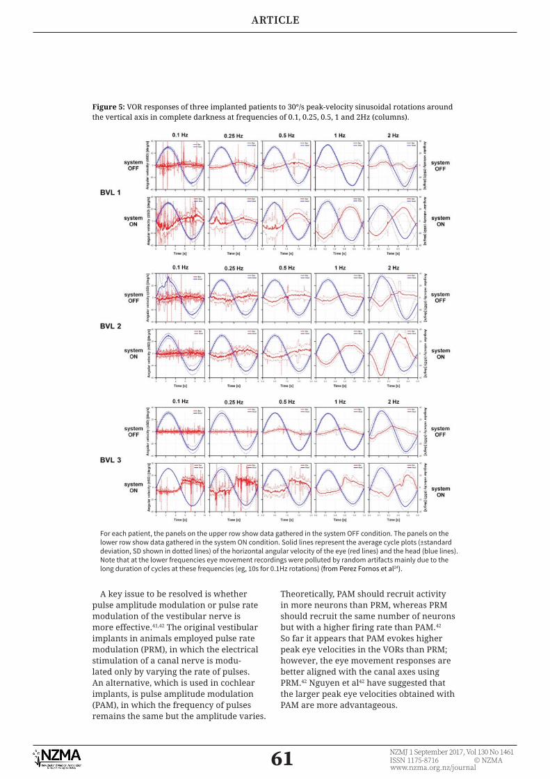
Figure 5: VOR responses of three implanted patients to 30°/s peak-velocity sinusoidal rotations around the vertical axis in complete darkness at frequencies of 0.1, 0.25, 0.5, 1 and 2Hz (columns).
For each patient, the panels on the upper row show data gathered in the system OFF condition. The panels on the lower row show data gathered in the system ON condition. Solid lines represent the average cycle plots (±standard deviation, SD shown in dotted lines) of the horizontal angular velocity of the eye (red lines) and the head (blue lines). Note that at the lower frequencies eye movement recordings were polluted by random artifacts mainly due to the long duration of cycles at these frequencies (eg, 10s for 0.1Hz rotations) (from Perez Fornos et al24).
One critical issue is what will be the effect of stimulating the canal nerves to generate VORs in the absence of otolithic signals that indicate gravito-interial force (GIF). The otoliths normally integrate linear acceler-ation by gravity with linear acceleration due to translation, so that they calculate a vector sum of linear acceleration signals. These are important to indicate to the brain whether the head is tilted relative to gravity. If the head is rotated off the vertical axis, otolithic signals would normally provide the brain with information about the degree of head tilt, so that it can integrate this with information from the canals about rotational acceleration.17,18 Without this additional information, it is possible that the brain will be confused about the plane of rotation of the head relative to space. At present it is unclear what the long-term effects of this may be.
Although most patients to have received vestibular implants to date have had bilateral hearing loss from disease,12,30 there is evidence from case reports that auditory function can be maintained despite the surgery on the vestibular system.43
It is clear that, as with cochlear implants,44 the brain adapts to the artifi cial stimu-lation provided by vestibular implants over time. When the initial baseline stimulation is turned on, the brain adapts to that high rate of stimulation, over several days.45–50 One concern is that many of the stimulated afferents are likely to be entrained at the same fi ring rate, while normal afferent fi ring rates vary between fi bres.17,18 It has been suggested that the phase or timing of the VORs does not adapt because the noise in the afferent fi bres is coherent rather than random.17,18 It is evident that brain plasticity in response to stimulation by the vestibular implant can actually enhance its effi cacy, so that the brain attempts to compensate for the shortcomings of the artifi cial stimulation.51
Although there is relatively little infor-mation available on the chronic effects of vestibular implants on neuronal activity
itself, some very recent studies have recorded from both the vestibular nerve and the VNC in monkeys with implants. Mitchell et al52 reported that while the primary afferent fi ring rate does not adapt with repeated stimulation, the PVP neurons in the VNC which receive input from them and which drive the VORs do undergo some habituation. Nonetheless, the artifi cial VOR responses are relatively preserved.52 Neural network simulation studies are also underway to predict the effects of vestibular implants on the central vestibular system.53
ConclusionsArtifi cial vestibular implants have been
developed and several groups of patients around the world have received them. Although the technology is in its infancy, it is developing rapidly.12,17,18 To date, adverse side effects appear to be minimal,12,17,18 although it might be predicted that as more patients with intact hearing receive implants, complications related to the spread of current to the cochlea will arise, just as with cochlear implants in relation to the vestibular end organs.54 However, there is too little information available at present to speculate on the impact of vestibular implants on auditory function.43 The long-term effects of vestibular implants on the brain, especially on higher cognitive and emotional functions, remain to be determined, but the fi rst studies have been conducted analysing the effects of chronic electrical stimulation on primary afferent and VNC neurons that are part of the VOR pathways. Whether vestibular implants may evolve as a treatment possibility for patients with other kinds of vestibular disorders, not involving complete vestibular loss, remains to be seen; however, they have been considered for the treatment of Meniere’s Disease.12 As the vestibular implant moves closer to becoming a clinical reality, the bioethical issues surrounding this kind of intervention are increasingly being discussed. 18,55,56
1. Mudry A, Mills M. The early history of the cochlear implant. JAMA Otolaryngol. Head Neck Surg. 2013; 139:446–453.
2. Wilson BS. The modern cochlear implant: A triumph of biomedical engineering and the fi rst substantial restoration of human sense using a medical intervention. IEEE Pulse. 2017; 8(2):29–32.
3. Vidal PP, Cullen K, Curthoys IS, Du Lac S, Holstein G, Idoux E, Lysakowski A, Peusner K, Smith PF. The vestibular system. In G. Paxinos (Ed): The Rat Nervous System, 4th Edition, (2014) Academic Press, San Diego. Chap-ter 28, pp. 805–864.
4. Cullen KE. The vestibular system: multimodal integration and encoding of self-motion for motor control. Trends Neurosci. 2012 ;35(3):185–96.
5. Bartl K, Lehnen N, Kohlbecher S, Schneider E. Head impulse testing using video-oculogra-phy. Ann. NY Acad. Sci. (2009); 1164:331–333.
6. Angelaki DE. The VOR: A model for visual-motor plasticity. In: The Senses: A Comphrensive Refer-ence. Chapter 2.22. (2008) pp. 359–370. Elsevier.
7. Morland AB, Bronstein AM, Ruddock KH, Wooding DS. Oscillopsia: Visual function during motion in the absence of vestibulo-ocular refl ex. J. Neurol. Neurosurg. Psychiat. 1998; 65:828–835.
8. Strupp M, Feil K, Dieterich M, Brandt T. Bilateral vestibulopathy. Handb Clin Neurol. 2016; 137:235–40.
9. Smith PF. The vestibular system and cognition. Curr. Opin. Neurol. 2017; 30(1):84–89.
10. Agrawal Y, Carey JP, Della Santina CC, et al. Disorders of balance and vestibular function in US adults: data from the National Health and Nutrition Examination Survey, 2001–2004. Arch. Intern. Med. 2009; 169(10):938–944.
11. Brandt T, Dieterich M. The dizzy patient: don’t forget disorders of the central vestibular system. Nat
Rev Neurol. (2017) doi: 10.1038/nrneurol.2017.58.
12. Perez Fornos A, Cavuscens S, Ranieri M, van de Berg R, Stokroos R, Kingma H, Guyot JP, Guinand N. The vestibular implant: A probe in orbit around the human balance system. J Vestib Res. 2017; 27(1):51–61.
13. Della Santina CC. Regaining balance with bionic ears. Sci Am. 2010; 302(4):68–71.
14. Cohen B, Suzuki JI, Bender MB. Eye movements from semicircular canal nerve stimulation in the cat. Ann Otol Rhinol Laryn-gol. 1964; 73:153–69.
15. Gong W, Merfeld DM. System design and performance of a unilateral horizontal semicircular canal prosthesis. IEEE Trans Biomed Eng. 2002; 49(2):175–81.
16. Gong W, Merfeld DM. Prototype neural semi-circular canal prosthesis using patterned electrical stimulation. Ann Biomed Eng. 2000; 28(5):572–81.
17. Lewis RF. Vestibular implants studied in animal
Competing interests:Nil.
Acknowledgements:This review is an extension of an invited lecture to the ‘Brain and Technology: A Catalyst for Collaboration Symposium’, a joint meeting of the MedTech and Brain Research New Zealand
Centres of Research Excellence, held at the University of Auckland, in April 2017.Author information:
Paul F Smith, Dept. of Pharmacology and Toxicology, School of Biomedical Sciences, and Brain Health Research Centre, University of Otago, Dunedin; Brain Research New Zealand
Centre of Research Excellence, and the Eisdell Moore Centre for Hearing and Balance Research, University of Auckland, Auckland.
Corresponding author: Paul F Smith, Department of Pharmacology and Toxicology, School of Biomedical Sciences,
models: clinical and scientifi c implications. J Neurophysiol. 2016; 116(6):2777–2788.
18. Lewis RF. Advances in the diagnosis and treatment of vestibular disorders: psychophysics and prosthetics. J Neurosci. 2015;35(13): 5089–96.
19. Della Santina CC, Migliaccio AA, Hayden R, Melvin TA, Fridman GY, Chiang B, Davidovics NS, Dai C, Carey JP, Minor LB, Anderson IC, Park H, Lyford-Pike S, Tang S. Current and future management of bilateral loss of vestibular sensation - an update on the Johns Hopkins Multichannel Vestibular Prosthesis Project. Cochlear Implants Int. 2010; Suppl 2:2–11.
20. Rahman MA, Dai C, Fridman GY, Davidovics NS, Chiang B, Ahn J, Hayden R, Melvin TA, Sun DQ, Hedjoudje A, Della Santina CC. Restoring the 3D vestibulo-ocular refl ex via electrical stimula-tion: the Johns Hopkins multichannel vestibular prosthesis project. Conf Proc IEEE Eng Med Biol Soc. 2011; 2011:3142.
21. Ciaravella G, Laschi C, Dario P. Biome-chanical modeling of semicircular canals for fabricating a biomimetic vestibular system. Conf Proc IEEE Eng Med Biol Soc. 2006; 1:1758–61.
22. Phillips JO, Bierer SM, Ling L, Nie K, Rubin-stein JT. Real-time communication of head velocity and acceleration for an externally mounted vestibular prosthesis. Conf Proc IEEE Eng Med Biol Soc. 2011; 2011:3537–41.
23. Highstein SM, Holstein GR. The anatomical and physiological framework for vestibular prostheses. Anat Rec (Hoboken). 2012; 295(11):2000–9.
24. Perez Fornos A, Guinand N, van de Berg R, Stokroos R, Micera S, Kingma H, Pelizzone M, Guyot JP. Artifi cial balance: restoration of the vestibu-lo-ocular refl ex in humans with a prototype vestibular neuroprosthesis. Front Neurol. 2014; 5:66. doi: 10.3389/fneur.2014.00066.
25. Valentin NS, Hageman KN, Dai C, Della Santina CC, Fridman GY. Development of a multichannel vestib-ular prosthesis prototype by modifi cation of a commercially available cochlear implant. IEEE Trans Neural Syst Rehabil Eng. 2013; 21(5):830–9.
26. Shkel AM, Zeng FG. An electronic prosthesis mimicking the dynamic vestibular function. Audiol Neurootol. 2006; 11(2):113–22.
27. Wall C 3rd, Merfeld DM, Rauch SD, Black FO. Vestibular prostheses: the engineering and biomed-ical issues. J Vestib Res. 2002–2003; 12(2–3):95–113.
28. Rubinstein JT, Della Santina CC. Development of a biophysical model for vestibular prosthesis research. J Vestib Res. 2002–2003; 12(2–3):69–76.
29. Goldberg JM, Fernandez C. Physiology of periph-eral neurons innervating semicircular canals of the squirrel monkey. I. Resting discharge and response to constant acceleration. J. Neurophysi-ol. 1971; 34(4):635–660.
30. Guinand N, van de Berg R, Cavuscens S, Stokroos RJ, Ranieri M, Pelizzone M, Kingma H, Guyot JP, Perez-Fornos A. Vestibular Implants: 8 years of experience with electrical stimulation of the vestibular nerve in 11 patients with bilateral vestibular loss. ORL J Otorhinolaryngol Relat Spec. 2015; 77(4):227–240.
31. Nie K, Ling L, Bierer SM, Kaneko CR, Fuchs AF, Oxford T, Rubinstein JT, Phillips JO. An experi-mental vestibular neural prosthesis: design and preliminary results with rhesus monkeys stimulated with modulated pulses. IEEE Trans Biomed Eng. 2013; 60(6):1685–92.
32. Dai C, Fridman GY, Della Santina CC. Effects of vestibular prosthesis electrode implantation and stimulation on hearing in rhesus monkeys. Hear Res. 2011; 277(1–2):204–10.
33. Guinand N, Guyot JP, Kingma H, Kos I, Pelizzone M. Vestibular implants: the fi rst steps in humans. Conf Proc IEEE Eng Med Biol Soc. 2011; 2011:2262–4.
34. Phillips JO, Shepherd SJ, Nowack AL, Ling L, Bierer SM, Kaneko CR, Phillips CM, Nie K, Rubinstein JT. Longitudinal performance of a vestibular prosthesis as assessed by electrically evoked compound action potential recording. Conf Proc IEEE Eng Med Biol Soc. 2012; 2012:6128–31.
35. Guinand N, van de Berg R, Ranieri M, Cavuscens S, DiGiovanna J, Nguyen TA, Micera S, Stokroos R, Kingma H, Guyot JP, Perez Fornos A. Vestib-ular implants: Hope for improving the quality of life of patients with bilat-eral vestibular loss. Conf Proc IEEE Eng Med Biol Soc. 2015b; 2015:7192–5.
36. Della Santina CC, Migli-accio AA, Patel AH. A multichannel semicircular canal neural prosthesis using electrical stimulation to restore 3-d vestibular sensation. IEEE Trans Biomed Eng. 2007; 54(6 Pt 1):1016–30.
37. van de Berg R, Guinand N, Nguyen TA, Ranieri M, Cavuscens S, Guyot JP, Stokroos R, Kingma H, Perez-Fornos A. The
vestibular implant: frequency-dependency of the electrically evoked vestibulo-ocular refl ex in humans. Front Syst Neurosci. 2015; 8:255. doi: 10.3389/fnsys.2014.00255
38. Dai C, Fridman GY, Chiang B, Davidovics NS, Melvin TA, Cullen KE, Della Santina CC. Cross-axis adaptation improves 3D vestibulo-oc-ular refl ex alignment during chronic stimulation via a head-mounted multichannel vestibular prosthesis. Exp Brain Res. 2011; 210(3–4):595–606.
39. Pelizzone M, Fornos AP, Guinand N, van de Berg R, Kos I, Stokroos R, Kingma H, Guyot JP. First functional rehabilitation via vestibular implants. Cochlear Implants Int. 2014; Suppl 1: S62–4.
40. Guinand N, Van de Berg R, Cavuscens S, Stokroos R, Ranieri M, Pelizzone M, Kingma H, Guyot JP, Pérez Fornos A. Restoring visual acuity in dynamic condi-tions with a vestibular implant. Front Neurosci. 2016; 10:577. doi: 10.3389/fnins.2016.00577
41. Davidovics NS, Fridman GY, Della Santina CC. Co-modulation of stimulus rate and current from elevated baselines expands head motion encoding range of the vestibular prosthesis. Exp Brain Res. 2012; 218(3):389–400.
42. Nguyen TA, DiGiovanna J, Cavuscens S, Ranieri M, Guinand N, van de Berg R, Carpaneto J, Kingma H, Guyot JP, Micera S, Fornos AP. Characterization of pulse amplitude and pulse rate modulation for a human vestibular implant during acute electrical stimulation. J Neural Eng. 2016; 13(4):046023.
43. van de Berg R, Lucieer F, Guinand N, van Tongeren J, George E, Guyot JP, Kingma H, van Hoof M, Temel Y, van Overbeeke J, Perez-Fornos A, Stokroos R. The vestibular implant: Hearing preservation during intra-labyrinthine electrode insertion-A case report. Front Neurol. 2017; 8:137. doi: 10.3389/fneur.2017.00137
45. Guyot JP, Sigrist A, Peliz-zone M, Kos MI. Adaptation to steady-state electrical stimulation of the vestib-ular system in humans. Ann Otol Rhinol Laryngol. 2011; 120(3):143–9.
46. Saginaw MA, Gong W, Haburcakova C, Merfeld DM. Attenuation of eye movements evoked by a vestibular implant at the frequency of the baseline pulse rate. IEEE Trans Biomed Eng. 2011; 58(10):2732–9.
47. Merfeld DM, Haburcakova C, Gong W, Lewis RF. Chronic vestibulo-ocular refl exes evoked by a vestibular prosthesis. IEEE Trans Biomed Eng. 2007; 54(6 Pt 1):1005–15.
48. Merfeld DM, Gong W, Morrissey J, Saginaw M, Haburcakova C, Lewis RF. Acclimation to chronic constant-rate peripheral stimulation provided by a vestibular prosthesis. IEEE Trans Biomed Eng. 2006; 53(11):2362–72.
49. Lewis RF, Gong W, Ramsey M, Minor L, Boyle R, Merfeld DM. Vestibular adaptation studied with a prosthetic semicircu-lar canal. J Vestib Res. 2002–2003; 12(2–3): 87–94.
50. Thompson LA, Haburcako-va C, Gong W, Lee DJ, Wall C 3rd, Merfeld DM, Lewis RF. Responses evoked by a vestibular implant provid-ing chronic stimulation. J Vestib Res. 2012; 22(1):11–5.
51. Dai C, Fridman GY, Chiang B, Rahman MA, Ahn JH, Davidovics NS, Della Santina CC. Directional plasticity rapidly improves 3D vestibulo-ocular refl ex alignment in monkeys using a multichannel vestibular prosthesis. J Assoc Res Otolaryngol. 2013; 14(6):863–77.
52. Mitchell DE, Della Santina CC, Cullen KE. Plasticity within non-cerebellar path-ways rapidly shapes motor performance in vivo. Nat Commun. 2016; 7:11238. doi: 10.1038/ncomms11238
53. DiGiovanna J, Nguyen TA, Guinand N, Pérez-Fornos A, Micera S. Neural network model of vestibular nuclei reaction to onset of vestibular prosthetic stimulation. Front Bioeng Biotechnol. 2016; 4:34. doi: 10.3389/fbioe.2016.00034
54. Abouzayd M, Smith PF, Moreau S, Hitier M. What vestibular tests to choose in symptomatic patients after a cochlear implant? A systematic review and meta-analysis. Eur Arch Otorhinolaryngol. 2017; 274(1):53–63.
55. Poppendieck W, Hoffmann KP, Merfeld D, Guyot JP, Micera S. Ethical issues in the development of a vestibular prosthesis. Conf Proc IEEE Eng Med Biol Soc. 2011; 2011:2265–8.
56. Guyot JP, Gay A, Kos MI, Pelizzone M. Ethical, anatomical and physiolog-ical issues in developing vestibular implants for human use. J Vestib Res. 2012; 22(1):3–9.