Department of Biotechnology, Sapthagiri College of Engineering, Bangalore, Karnataka, India *Corresponding author: [email protected]
Received November 12, 2014; Revised December 04, 2014; Accepted December 07, 2014
Abstract The term biosensor is often used to cover sensor devices used in order to determine the concentration of substances and other parameters of biological interest even where they do not utilize a biological system directly. This review discusses recent advances in biosensor technology which draw on the disciplines of physics, chemistry, biochemistry and electronics. This article states that a biosensor consists of three components, a biological detection system, a transducer and an output system. Biological receptors are briefly reviewed, followed by a detailed discussion of immobilization procedures for the efficacious attachment of receptor molecules to a transducer surface. Widely used in the fields of research and development in this field is wide and multidisciplinary, spanning biochemistry, bioreactor science, physical chemistry, electrochemistry, electronics and software engineering.
Cite This Article: Shruthi GS, Amitha CV, and Blessy Baby Mathew, “Biosensors: A Modern Day Achievement.” Journal of Instrumentation Technology, vol. 2, no. 1 (2014): 26-39. doi: 10.12691/jit-2-1-5.
1. Introduction BIOSENSORS are defined as any measuring device
that contains a biological element. It combines the exquisite selectivity of biology with the processing power of modern microelectronics and optoelectronics to offer powerful new analytical tools with major applications in the field of medicine, environmental studies, food and processing industries [1]. These analytical devices are based on the union between biological and physio-chemical components. Biological components include macro-molecules such as antibodies, enzymes, tissue slices which are used to recognize and interact with a specific analyte [1]. Physiochemical components are
usually referred to as transducers which converts the interactions into signals; it is later amplified with respect to its concentration of analyte [1]. The transducer may use potentiometric, amperometric, optical, magnetic, colorimetric devices [2]. A target analyte in the external membrane must be able to enter the biosensor [2]. The external membrane of the biosensor must be permeable to the analyte where the biosensor is sensitive to it. The biological element inside the biosensor then interacts with chemical species through a biochemical reaction which in turn produces another chemical product and characterized by change in mechanical, electrical properties. The output signal may be a conventional electrochemical signal depending on the type of transducer it uses.
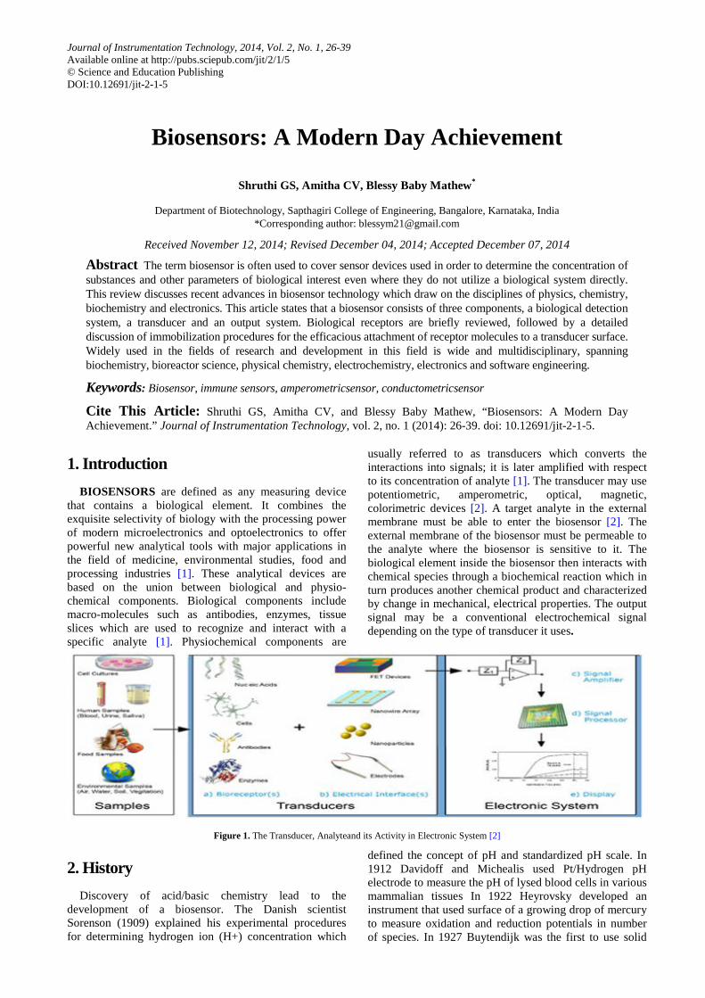
Figure 1. The Transducer, Analyteand its Activity in Electronic System [2]
2. History Discovery of acid/basic chemistry lead to the
development of a biosensor. The Danish scientist Sorenson (1909) explained his experimental procedures for determining hydrogen ion (H+) concentration which
defined the concept of pH and standardized pH scale. In 1912 Davidoff and Michealis used Pt/Hydrogen pH electrode to measure the pH of lysed blood cells in various mammalian tissues In 1922 Heyrovsky developed an instrument that used surface of a growing drop of mercury to measure oxidation and reduction potentials in number of species. In 1927 Buytendijk was the first to use solid
Journal of Instrumentation Technology 27
metal antimony pH electrode for medical purposes. In 1935 Muller and Baumburger measured oxygen in biological fluids which were more and accurate than all the previous discoveries. Later, In 1938 Baumburger used the dropping electrode polarography to determine the oxyhemoglobin equilibrium curve for O2 and Petering and Daniels used the mercury polarography to measure oxygen consumption rates for algae, yeast and blood cells. In 1956, Clark published his definitive paper on the oxygen electrode. In 1959 Heyrovsky was awarded Nobel Prize for the recognition of his pioneering work by Stanford University. In 1962, at a New York Academy of Sciences symposium he described to construct electrochemical sensors (pH, polarographic, potentiometric, or conductometric) in an intelligent way by adding enzyme transducers as membrane enclosed. In
1975 Divies suggested that bacteria could be harnessed as the biological element in microbial electrodes for the measurement of alcohol. In 1976, Clemens et al incorporated an electrochemical glucose biosensor in a bedside artificial pancreas this was later marketed by Miles (Elkhart) as the Biostator. In 1982 Shichiri et al described the first needle-type enzyme electrode for subcutaneous implantation. In 1970, Liedberg et al. issued a paper which included the idea of building direct immunosensors by fixing antibodies to a piezoelectric or potentiometric transducer and also described the use of surface plasmon resonance to monitor affinity reactions in real time. In 1984, a much-cited paper on the use of ferrocene and its derivatives as an immobilized mediator for use with oxidoreductases was published. [2]
Table 1. History of Biosensors [2] DATE EVENT 1909 Explanation of hydrogen ion by Sorenson 1912 Davidoff and Michealis used Pt/Hydrogen pH electrode to measure the pH of lysed blood cells in various mammalian tissues 1916 First report on the immobilization of proteins: adsorption of invertase on activated charcoal 1922 First glass pH electrode 1956 Invention of the oxygen electrode 1962 First description of a biosensor: an amperometric enzyme electrode for glucose only 1969 First potentiometric biosensor: urease immobilized on an ammoniaelectrode to detect urea 1970 Invention of the ion-selective field-effect transistor (ISFET)
1972–75 First commercial biosensor: Yellow Springs Instruments glucose biosensor
1975 First microbe-based biosensor, first immunosensor: ovalbumin on a platinum wire Invention of the pO2/pCO2 optode
1976 First bedside artificial pancreas (Miles) 1980 First fiber optic pH sensor for in vivo blood gases 1983 First surface plasmon resonance (SPR) immunosensor 1984 First mediated amperometric biosensor: ferrocene used with glucoseoxidase for the detection of glucose 1987 Launch of the Medi Sense Exac Tech blood glucose biosensor 1990 Launch of the Pharmacia BIACore SPR-based biosensor system 1992 i-STAT launches hand-held blood analyzer 1996 Glucocard launched 1998 Launch of Life Scan Fast Take blood glucose biosensor and Merger of Roche and Boehringer Mannheim to form Roche Diagnostics 2001 Life Scan purchases Inverness Medical’s glucose testing business for $1.3 billion 2003 i-STAT acquired by Abbott for $392 million 2004 Abbott acquires Therasense for $1.2 billion
3. Principle The basic principle of a biosensor includes a
bioreceptor that is an immobilized sensitive biological element such as enzyme, DNAprobe, antibodyrecognizing the analytes such as enzyme substrate, complementary DNA, antigen. They also include antibodies, whole cells, including microbial, plant, and animal cells, subcellular organelles, tissue slices, lectins, and numerous synthetic molecules with affinity or catalytic properties extending to those obtained through parallel synthesis and imprinted polymers Although antibodies and oligonucleotides are widely employed, enzymes are by far the most commonly used biosensing elements in biosensors. A transducer is
used to convert biochemical signal resulting from the interaction of the analyte with the bioreceptor into an electronic one. The intensity of generated signal is directly or inversely proportional to the analyte concentration. Electrochemical transducers are often used to develop biosensors. These systems offer some advantages such as low cost, simple design or small dimensions. Biosensors can also be based on gravimetric, calorimetric or optical detection [1]. Biosensors is categorized according to the basic principles of signal transduction and biorecognition elements. According to the transducing elements, biosensors can be classified as electrochemical, optical, piezoelectric, and thermal sensors. Electrochemical biosensors are also classified as potentiometric, amperometric and conductometric sensors [3,4].
Figure 2. Principle behind the movement of the analyte
28 Journal of Instrumentation Technology
4. Characteristics of a Biosensor Selectivity is probably the most important feature of a
biosensor. Selectivity means that sensor detects a certain analyte and does not react to admixtures and contaminants. Antigen-antibody interaction has the highest selectivity, it is analyte-specific. Precision is a characteristic of any scientific device that makes quantitative measurements. It is usually characterized in terms of the standard deviation of measurements. Signal error in measured concentration. Signal stability influences the precision of sensor. It is an important characteristic of a sensor that performs continuous monitoring. Sensitivity shows the minimal
amount or concentration of analyte that can be detected. Working range is the range of analyte concentrations in which the sensor can operate. Working range of sensor should correlate with the range of possible concentrations analyte in the assay. Response time is time required to analyze the assay. Regeneration time is the time required to return the sensor to working state after interaction with the sample. Number of cycles is the number of times the sensor can be operated. Degradation of biological material is inevitable and it needs to be replaced. In some sensors (e.g. hand-held commercial glucose sensors) transducers are disposable, they need to be changed after each measurement [5].
Table 2. Types of Biosensors [10] TYPES OF TRANSDUCERS MEASURED PROPERTY
Electrochemical Potentiometric, Amperometric, Conductometric, Nanotechnology and Bioelectronics Protein Immunosensor
Mass sensitive Resonance frequency of piezocrystals Light Bioluminescence
5. Construction of Biosensors In order to construct a successful biosensor certain
conditions must be met, such as the biocatalyst must be highly specific for the purpose of the analysis, be stable under normal storage conditions and show a low variation between assays. The reaction should be as independent as manageable of such physical parameters as stirring, pH and temperature. This will allow analysis of samples with minimal pre-treatment. If the reaction involves cofactors or coenzymes these should, preferably, also be co-immobilized with the enzyme. The response should be accurate, precise, reproducible and linear over the concentration range of interest, without dilution or concentration. It should also be free from electrical or other transducer induced noise. If the biosensor is to be used for invasive monitoring in clinical situations, the probe must be tiny and biocompatible, having no toxic or antigenic effects. Furthermore, the biosensor should not be prone to inactivation or proteolysis. For rapid measurements of analytes from human samples it is desirable that the biosensor can provide real-time analysis. The complete biosensor should be cheap, small, portable and capable of being used by semi-skilled operators [6].
6. Working of Biosensors
6.1. Electrochemical Biosensors Typically in (bio-) electrochemistry, the reaction under
investigation would either generate a measurable current (amperometric), a measurable potential or charge accumulation (potentiometric) or measurably alter the conductive properties of a medium (conductmetric) between electrodes. References are also made to other types of electrochemical detection techniques, such as impedimetric, which measures impedance (both resistance and reactance), and field-effect, which uses transistor technology to measure current as a result of a potentiometric effect at a gate electrode. All of these
measurement techniques will be introduced here, as well as some devices that employ variations of these techniques. Since reactions are generally detected only in close proximity to the electrode surface, the electrodes themselves play a crucial role in the performance of electrochemical biosensors. Based on the chosen function of a specific electrode, the electrode material, its surface modification or its dimensions greatly influence its detection ability. Electrochemical sensing usually requires a reference electrode, a counter or auxiliary electrode and a working electrode, also known as the sensing or redox electrode. The reference electrode, commonly made from Ag/AgCl, is kept at a distance from the reaction site in order to maintain a known and stable potential. The working electrode serves as the transduction element in the biochemical reaction, while the counter electrode establishes a connection to the electrolytic solution so that a current can be applied to the working electrode. These electrodes should be both conductive and chemically stable. Therefore, platinum, gold, carbon (e.g. graphite) and silicon compounds are commonly used, depending on the analyte [6]. Nanotechnology and Bioelectronicshave revealed new possibilities to miniaturize and to optimize existing microscale devices at the nanoscale. It is becoming possible to more accurately measure specific electrical properties in combination with various electrochemical transducers. The higher surface-to-volume ratio of nano-objects makes their electrical properties increasingly susceptible to external influences, especially as these structures continue to shrink toward the atomic limit. Since the nanometer dimensions of these objects are comparable to the size of the target biomolecules, higher measurement sensitivity may result and sensitivity may also increase due to higher capture efficiency. Nanostructures already represent important new components in recently developed electrochemical biosensors, such as the use of nanoparticles as electrochemical labels for DNA sensing. Nanowires, carbon nanotubes, nanoparticles and nanorods are merely some of the familiar objects that are emerging as candidates to become crucial elements of future bioelectronic devices and biosensors [6].
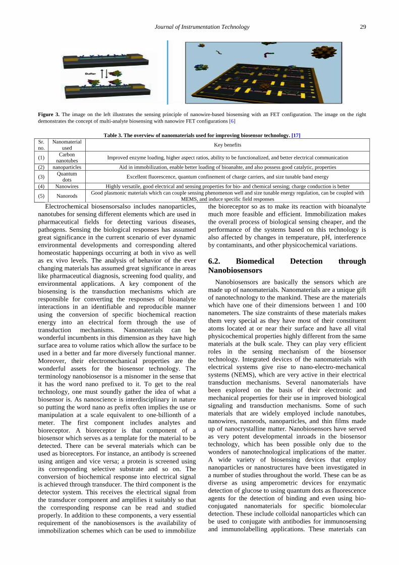
Journal of Instrumentation Technology 29
Figure 3. The image on the left illustrates the sensing principle of nanowire-based biosensing with an FET configuration. The image on the right demonstrates the concept of multi-analyte biosensing with nanowire FET configurations [6]
Table 3. The overview of nanomaterials used for improving biosensor technology. [17] Sr. no.
Nanomaterial used Key benefits
(1) Carbon nanotubes Improved enzyme loading, higher aspect ratios, ability to be functionalized, and better electrical communication
(2) nanoparticles Aid in immobilization, enable better loading of bioanahte, and also possess good catalytic, properties
(3) Quantum dots Excellent fluorescence, quantum confinement of charge carriers, and size tunable band energy
(4) Nanowires Highly versatile, good electrical and sensing properties for bio- and chemical sensing; charge conduction is better
(5) Nanorods Good plasmonic materials which can couple sensing phenomenon well and size tunable energy regulation, can be coupled with MEMS, and induce specific field responses
Electrochemical biosensorsalso includes nanoparticles, nanotubes for sensing different elements which are used in pharmaceutical fields for detecting various diseases, pathogens. Sensing the biological responses has assumed great significance in the current scenario of ever dynamic environmental developments and corresponding altered homeostatic happenings occurring at both in vivo as well as ex vivo levels. The analysis of behavior of the ever changing materials has assumed great significance in areas like pharmaceutical diagnosis, screening food quality, and environmental applications. A key component of the biosensing is the transduction mechanisms which are responsible for converting the responses of bioanalyte interactions in an identifiable and reproducible manner using the conversion of specific biochemical reaction energy into an electrical form through the use of transduction mechanisms. Nanomaterials can be wonderful incumbents in this dimension as they have high surface area to volume ratios which allow the surface to be used in a better and far more diversely functional manner. Moreover, their electromechanical properties are the wonderful assets for the biosensor technology. The terminology nanobiosensor is a misnomer in the sense that it has the word nano prefixed to it. To get to the real technology, one must soundly gather the idea of what a biosensor is. As nanoscience is interdisciplinary in nature so putting the word nano as prefix often implies the use or manipulation at a scale equivalent to one-billionth of a meter. The first component includes analytes and bioreceptor. A bioreceptor is that component of a biosensor which serves as a template for the material to be detected. There can be several materials which can be used as bioreceptors. For instance, an antibody is screened using antigen and vice versa; a protein is screened using its corresponding selective substrate and so on. The conversion of biochemical response into electrical signal is achieved through transducer. The third component is the detector system. This receives the electrical signal from the transducer component and amplifies it suitably so that the corresponding response can be read and studied properly. In addition to these components, a very essential requirement of the nanobiosensors is the availability of immobilization schemes which can be used to immobilize
the bioreceptor so as to make its reaction with bioanalyte much more feasible and efficient. Immobilization makes the overall process of biological sensing cheaper, and the performance of the systems based on this technology is also affected by changes in temperature, pH, interference by contaminants, and other physicochemical variations.
6.2. Biomedical Detection through Nanobiosensors
Nanobiosensors are basically the sensors which are made up of nanomaterials. Nanomaterials are a unique gift of nanotechnology to the mankind. These are the materials which have one of their dimensions between 1 and 100 nanometers. The size constraints of these materials makes them very special as they have most of their constituent atoms located at or near their surface and have all vital physicochemical properties highly different from the same materials at the bulk scale. They can play very efficient roles in the sensing mechanism of the biosensor technology. Integrated devices of the nanomaterials with electrical systems give rise to nano-electro-mechanical systems (NEMS), which are very active in their electrical transduction mechanisms. Several nanomaterials have been explored on the basis of their electronic and mechanical properties for their use in improved biological signaling and transduction mechanisms. Some of such materials that are widely employed include nanotubes, nanowires, nanorods, nanoparticles, and thin films made up of nanocrystalline matter. Nanobiosensors have served as very potent developmental inroads in the biosensor technology, which has been possible only due to the wonders of nanotechnological implications of the matter. A wide variety of biosensing devices that employ nanoparticles or nanostructures have been investigated in a number of studies throughout the world. These can be as diverse as using amperometric devices for enzymatic detection of glucose to using quantum dots as fluorescence agents for the detection of binding and even using bio-conjugated nanomaterials for specific biomolecular detection. These include colloidal nanoparticles which can be used to conjugate with antibodies for immunosensing and immunolabelling applications. These materials can
30 Journal of Instrumentation Technology
also be used to enhance the electron microscopic detections. Further, metal based nanoparticles are very excellent materials for electronic and optical applications and can be efficiently used for detection of nucleic acid sequences through the exploitation of their optoelectronic properties [17].
6.3. Selection, Optimization and Working of Nanomaterials in Sensor Technology
There is a multitude, factors which govern or decide the use of a particular kind of nanomaterials for biosensing applications. These factors are the chief ingredients of their physical and chemical properties along with their energy sensitive and selective responses. Before exactly implementing or adding a nanomaterial for the sensing applications, we first focus on their desired manufacturing which is a part of experimental design known as “Nanofabrication.” The technique of nanofabrication targets two vital operations, namely, the manufacturing and design of nanoscale adhesive surfaces via the technology of integrated circuits and the engineering of nanomaterial surfaces through the process of micromachining. This technique, thus developed for biosensing, uses the variations of four basic processes, namely, photolithography, thin film etching/growth, surface etching strategies, and chemical bonding parameters [17].
Nanoscale electrodes which have come into picture as a result of lithography technique have enhanced the biosensing accuracy by providing much better and greater surface areas that in turn enable the immobilization to be achieved with greater precision. Glucose biosensors, making use of enzyme glucose oxidase, have been developed using these innovations. The strategies involving the use of active nanoparticles of platinum over the sheets of carbon nanotubes have significantly enhanced the immobilization of enzyme systems required for the detection of the analyte materials. These systems have significantly much wider applications to biosensing technology, enabling the detection of glucose from several sources other than blood. In a similar manner, couples of immunosensors have also been developed which involve coating of thin films over the sensing surface that enables faster and better detection of the corresponding analytes. Nanostructured semiconductor crystals can be efficiently used to improve the detection of neurological responses via coupling through the sensing molecule of biological nature. These can be coupled with peptide assembly of a range of nanomaterials so that efficient interaction can be generated by means of self-assembly and this also saves a lot of time that is being involved in the currently available technologies and methods. Moreover, these can rapidly detect the biological stimulus such as that of a DNA segment or a characteristic nucleotide sequence pertaining to proteins or even RNA. [17]
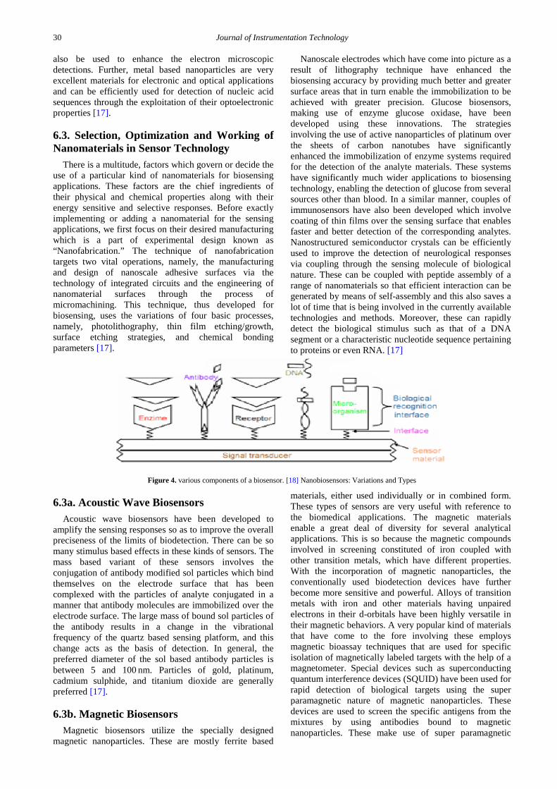
Figure 4. various components of a biosensor. [18] Nanobiosensors: Variations and Types
6.3a. Acoustic Wave Biosensors Acoustic wave biosensors have been developed to
amplify the sensing responses so as to improve the overall preciseness of the limits of biodetection. There can be so many stimulus based effects in these kinds of sensors. The mass based variant of these sensors involves the conjugation of antibody modified sol particles which bind themselves on the electrode surface that has been complexed with the particles of analyte conjugated in a manner that antibody molecules are immobilized over the electrode surface. The large mass of bound sol particles of the antibody results in a change in the vibrational frequency of the quartz based sensing platform, and this change acts as the basis of detection. In general, the preferred diameter of the sol based antibody particles is between 5 and 100 nm. Particles of gold, platinum, cadmium sulphide, and titanium dioxide are generally preferred [17].
6.3b. Magnetic Biosensors Magnetic biosensors utilize the specially designed
magnetic nanoparticles. These are mostly ferrite based
materials, either used individually or in combined form. These types of sensors are very useful with reference to the biomedical applications. The magnetic materials enable a great deal of diversity for several analytical applications. This is so because the magnetic compounds involved in screening constituted of iron coupled with other transition metals, which have different properties. With the incorporation of magnetic nanoparticles, the conventionally used biodetection devices have further become more sensitive and powerful. Alloys of transition metals with iron and other materials having unpaired electrons in their d-orbitals have been highly versatile in their magnetic behaviors. A very popular kind of materials that have come to the fore involving these employs magnetic bioassay techniques that are used for specific isolation of magnetically labeled targets with the help of a magnetometer. Special devices such as superconducting quantum interference devices (SQUID) have been used for rapid detection of biological targets using the super paramagnetic nature of magnetic nanoparticles. These devices are used to screen the specific antigens from the mixtures by using antibodies bound to magnetic nanoparticles. These make use of super paramagnetic
Journal of Instrumentation Technology 31
effect of magnetic materials which is particularly observed in the nanoscale particles [17].
6.3c. Nanotube Based Sensors Carbon nanotubes are one of the most popular
nanomaterials known right now in the world of material science and optoelectronic applications. Since their discovery in 1990’s they are been used more widely. The most vital of which are the electronic conductivity, flexible physical geometric features, and the ever dynamic physic mechanical properties ranging from high aspect ratios to very good functionalization abilities along with having high mechanical strength and folding abilities. Because of these attributes, both single walled nanotubes as well as multi-walled nanotubes have been used in designing biosensors for better and better performances [17].
The most popular sensing advances that have come to the fore are the developments in the design of glucose biosensors that involve the use of nanotubes as immobilizing surfaces for enzyme glucose oxidase; this enzyme is used for estimation of glucose from the several body fluids. In convention, the sensors using enzymes predicted the presence of glucose from major body tissues but the use of nanotubes as assemblies for immobilization has led to the estimation of glucose from even scarce body fluids such as tears and even saliva. In one such arrangement, single walled nanotubes have been wonderfully employed for enzymatic detection of glucose, and this innovation has also yielded significant increase in the enzyme activity. The enhanced performance of the biosensor was analyzed and found so, largely due to the high enzyme loading and better electrical conductivities of the nanotubes. Not only with their structural flexibilities, carbon nanotubes have also been used for enhancing the electrical detection of the sensing phenomenon, owing to their better and smoother electron transfer flow characteristics. The significant improvements in the catalytic biosensors have been widely exploited in several studies, and in one such study, this innovation resulted in better oxidoreductase performance in both glucose oxidase and flavin adenine dinucleotide precursors binding to their substrates more efficiently and in a much more controllable. [17]
6.3d. Nanowire Based Sensors Nanowires are cylindrical arrangements just like those
of carbon nanotubes, having lengths in the order of few micrometers to centimeters and diameters within the nanorange. Nanowires are the one-dimensional nanostructures with very good electron transport properties. Significantly, the motion of charge carriers in the nanowires is vigorously improved and different from those in bulk materials. Semiconductor nanowires have been exploited in a great detail and have also been used for coupling a number of biomolecules for identifying their specifically linked substrates. Silicon nanowires coated with biotin have been used for the detection and isolation of streptavidin molecules from a mixture. The small size and capability of these nanowires make them ideal candidates to be used for biodetection of pathogens and many other real time analysis of a wide range of biological and chemical species, thus vastly improvising
the current accuracies of presently used in vivo diagnostic procedures. As these sensing materials work on very precisely defined dimensions, they can also be used to accomplish in vivo applications and operate in the smallest environments within the living cells. Nanowires are very versatile in their performance and are significantly better than nanotubes in two major ways. First, they allow a range of modifications in their design by control of operational parameters during their synthesis. Secondly, they possess a lot much more scope for the development of functionalized assemblies by virtue of the existence of compatible materials on their surfaces. [17]
In this way, nanomaterials have proved to be highly prosperous for brightening the sensing technology and have improved the diagnostic and detection procedures by leaps and bounds. The faster and quicker diagnosis enabled by still faster analysis and evaluation protocols through the nanomaterials has just revolutionized the biosensing mechanism. There are many other nanomaterials except those mentioned above that have been capitalized upon and made use of in biosensing applications. Nanodots resembling the morphology of quantum dots, nanosheets, and many other structures of altered geometries such as nanocombs, nanobelts and nanoribbons have been used for improving the conventional procedures of sensing. The coupling of piezoelectric and cantilever systems has further added a new charm to this technology. Nanomaterials like quantum dots have been added as labels coupled with sensitive dyes, and they have yielded thermochromic, photochromic, and electro chromic materials which can show highly sensitive detections that can be monitored easily. They have significantly helped in improving electron transport mechanisms and also in the development of much more efficient actuating mechanisms to impose a particular state of observation on a system [17].
6.3e. Biomedical Applications of Nanobiosensors in Pharmaceutical Field
Detection of diabetes Patients with diabetes must take small blood samples many times a day to control glucose levels. Such procedures are extremely uncomfortable and inconvenient. To avoid this problem, the level of sugar in the body can be observed via constant glucose monitoring using medical nanorobotics. To envisage how actual and upcoming stages of nanotechnology can be applied in medicine, numerical analysis and computational nanotechnology, to illustrate the proposed nanorobot performance in the bloodstream, using a 3D vessel as test bed for diabetes control. The nanorobot sensor activation used proteomic-based information to detect biochemical changes associated with hyperglycemia [18].
Immunoassay (detection of Ab-Ag reaction) The peak extinction wavelength of the localized surface plasmon resonance (LSPR) spectrum is reliant upon the size, shape and interparticle spacing of the nanoparticles as well as its own dielectric properties and those of its local environment including substrate, solvent and adsorbates. The high sensitivity of the LSPR spectrum of non spherical nanoparticles to adsorbate induced changes in the local dielectric constant (viz., refractive index) are now being used to develop a different class of nanoscale
32 Journal of Instrumentation Technology
chemosensors and nanobiosensors. This sensor detects changes in the refractive index induced by molecules near the surface of noble metal thin films [18].
Detection of cancer Telomerase is a specialized reverse transcriptase, which is composed of an essential catalytic subunit and an RNA component that is held together with telomere-associated proteins, maintains telomere length and function. In normal cells, a critical telomere length is eventually reached, thereby inducing cellular senescence and finally leading to apoptosis. Elevated levels of telomerase activity are found in the majority of malignancies and are believed to play a critical role in tumor genesis [18].
A novel nanobiosensor (based on magnetic nanoparticles) has been developed for rapid screening of telomerase activity in biological samples. The technique makes use of nanoparticles which, upon annealing to telomerase-synthesized telomeric repeats (TTAGGG), change their magnetic state (a phenomenon readily detectable by magnetic readers). A high throughput version of this technique and the use of magnetic resonance imaging for the purpose of detection allow processing of hundreds of samples within tens of minutes with ultrahigh sensitivities. Nanoparticle assembly formation leads to change in the relaxation time (T2) of surrounding water, which can be readily measured by
bench top magnetic resonance (MR) relaxometers or imaging systems. The developed magnetic nanosensors can be utilized to determine telomerase activity in a variety of applications. The sensitivity of the method ranges from potential single molecule detection (e.g., magnetic force microscopy) to 10-100 attomole levels using benchtop read-outs (relaxometers). The assay permitted the detection of ca. 10 attomoles of telomerase-synthesized DNA by MR imaging, which competes well with other PCR (polymerase chain reaction) independent assay methodologies [18].
An optical fiber nanobiosensor has been constructed to detect efficiently a general cancer biomarker, telomerase at single cell level with its nanoscale tip. The number of significant advantages shown by developed technique has over other methods are as follows: (i) assay is quantitative, (ii) method is easy and fast (approximately 150 min for an entire determination and only a minute for actual measurements), (iii) does not requires solid phase, (iv) method can be extended to a high-throughput screening format and (v) achieves high degree of sensitivity without PCR and therefore avoids PCR-related artifacts and difficulties in quantification [18].
6.4. Immunosensor
Figure 5. Different types of immunoassays used in immunosensors
Immunosensor is constructed with an array of interdigitated electrodes and to monitor antibody-antigen reactions in between the gaps of the electrodes as illustrated. Binding events of complementary antibody-antigen components alter the electrical properties in the gap between two electrodes, where changes in gap conductivity correspond to changes in the real impedance component and changes in the gap capacitance correspond to changes in the imaginary impedance component. The use of impedimetric sensing to detect the binding state of DNA a microarray configuration of interdigitated electrodes was used. And show their intent to demonstrate the potential of EIS for low-density DNA microarrays. Single-stranded DNA (ssDNA) was immobilized to a modified surface between the electrodes to act as the
biorecognition element. The immobilized ssDNA is associated with counter cations, which initially support ionic conductivity. However, upon hybridization with the complementary DNA strand to the ssDNA there is a reduction in the density of these cations. This reduction further inhibits the free displacement of ions near the surface and leads to a corresponding increase in the overall electrical impedance between the interdigitated electrodes. DNA biosensing, electrolyte-insulator-semiconductor systems have been used to measure changes in capacitive impedance as a result of DNA hybridization. Detection occurs when the thickness of the dielectric layer increases as a result of target DNA hybridization at the electrode interface [6].
Figure 6. Immunosensor showing the principle behind the change in network impedance upon hybridization of ssDNA probe to its complement interdigitated micro-sensorelectrode [6]
Journal of Instrumentation Technology 33
6.5. Potentiometric Devices They are commonly used to measure glucose
concentrations greater than 10−5 M, which is in the physiological range in most cases. The potential difference between the reference electrode and the indicator electrode is measured at zero current flow. The ideally nonpolarizable reference electrode provides a constant potential, while the indicator electrode shows an erratic potential depending on the concentration of the analytes. The zero current potential applied between those two electrodes are recorded as a function of the concentrations of target analytes in a logarithmic manner [6].
6.6. Amperometric Devices
They are a type of electrochemical sensor, since they continuously measure current resulting from the oxidation or reduction of an electroactive species in a biochemical reaction Clark oxygen electrodes perhaps represent the basis for the simplest forms of amperometric biosensors, where a current is produced in proportion to the oxygen concentration. This is measured by the reduction of oxygen at a platinum working electrode in reference to Ag/AgCl reference electrode at a given potential. Typically the current is measured at a constant potential and this is referred to as amperometry. If a current is measured during controlled variations of the potential, this is referred to as voltammetry the peak value of the current measured over a linear potential range is directly proportional to the bulk concentration of the analyte, i.e. the electroactive species [7].
Figure 7. A sample amperometric measurement: showing the change in current response is proportional to the ATP concentration as glucose is consumed at the glucose oxidase and hexokinase modified electrode surface [7]
6.7. Conductometric Devices They measure the ability of an analyte (e.g. electrolyte
solutions) or a medium (e.g. nanowires) to conduct an electrical current between electrodes or reference nodes. Although conductometric devices can be considered as a subset of impedimetric devices, techniques for measuring capacitance changes is reviewed later in combination with electrochemical impedance spectroscopy. In most cases conductometric devices have been strongly associated with enzymes, where the ionic strength, and thus the conductivity, of a solution between two electrodes changes as a result of an enzymatic reaction. Thus, conductometric devices can be used to study enzymatic reactions that produce changes in the concentration of charged species in a solution. The variable ionic background of clinical samples and the requirement to measure small conductivity changes in media of high ionic strength limit the applicability of such enzyme-based conductometric devices for biosensing. Another approach is to directly monitor the changes in conductance of an electrode as a result of the immobilization of e.g. enzymes, complementary antibody-antigen pairs, etc. onto the electrode surface. The construction of multi-analyte conductance biosensors and conductive polymer-based devices has been made possible by the rapid development of semiconductor technology and sensor integration with microelectronic devices, such as FET devices. Now there is an increased interest in conductometric immunosensors in combination with nanostructures, and especially nanowires, for biosensing. These devices are used for practical application, such as drug detection in human urine and pollutant detection in environmental testing. Whole cells have also been used as a biorecognition
element in conductometric biosensors for toxicity analysis by immobilizing the cells to a transducer of interdigitated electrodes [7].
6.8. Piezoelectric Biosensors These type of biosensors has two components: a
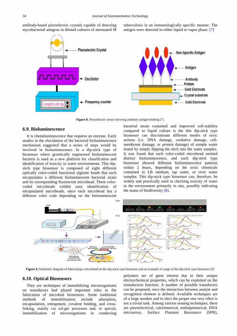
receptor and a detector. The receptor is responsible for the selectivity of the sensor. Examples include enzymes, antibodies, and lipid layers. The detector, which plays the role of the transducer, translates the physical or chemical change by recognizing the analyte and relaying it through an electrical signal. The detector is not selective. For example, it can be a pH-electrode, an oxygen electrode or a piezoelectric crystal. Figure 8 describes a typical biosensor configuration that allows measurement of the target analyte without using reagents. The device incorporates a biological-sensing element with a traditional transducer. The biological-sensing element selectively recognizes a particular biological molecule through a reaction, specific adsorption, or other physical or chemical process, and the transducer converts the result of this recognition into a usable signal, which can be quantified. Common transduction systems are optical, electro-optical, or electrochemical; this variety offers many opportunities to tailor biosensors for specific applications. For example, the glucose concentration in a blood sample can be measured directly by a biosensor (which is made specifically for glucose measurement) by simply dipping the sensor into the sample. The objective of the research described here is to use biosensor technology to develop a rapid method for the diagnosis of tuberculosis and other infections caused by mycobacteria. The work encompassed here describes the construction of
34 Journal of Instrumentation Technology
antibody-based piezoelectric crystals capable of detecting mycobacterial antigens in diluted cultures of attenuated M
tuberculosis in an immunologically specific manner. The antigen were detected in either liquid or vapor phase. [7]
6.9. Bioluminescence It is chemiluminescence that requires an enzyme. Early
studies in the elucidation of the bacterial bioluminescence mechanism suggested that a series of steps would be involved in bioluminescence. In a dip-stick type of biosensor where genetically engineered bioluminescent bacteria is used as a new platform for classification and identification of toxicity in water environments. This dip-stick type biosensor is composed of eight different optically color-coded functional alginate beads that each encapsulates a different bioluminescent bacterial strain and its corresponding fluorescent microbead. These color-coded microbeads exhibit easy identification of encapsulated microbeads, since each microbead has a different color code depending on the bioluminescent
bacterial strain contained and improved cell-stability compared to liquid culture is the this dip-stick type biosensor can discriminate different modes of toxic actions (i.e. DNA damage, oxidative damage, cell-membrane damage, or protein damage) of sample water tested by simply dipping the stick into the water samples. It was found that each color-coded microbead emitted distinct bioluminescence, and each dip-stick type biosensor showed different bioluminescence patterns within 2 hours, depending on the toxic chemicals contained in LB medium, tap water, or river water samples. This dip-stick type biosensor can, therefore, be widely and practically used in checking toxicity of water in the environment primarily in situ, possibly indicating the status of biodiversity [8].
Figure 9. Schematic diagram of fabricating a microbead on the dip-stick type biosensor and an example of usage of the dip-stick type biosensor [8]
6.10. Optical Biosensors They are techniques of immobilizing microorganisms
on transducers had played important roles in the fabrication of microbial biosensors. Some traditional methods of immobilization include adsorption, encapsulation, entrapment, covalent binding, and cross-linking, mainly via sol-gel processes and, in special, immobilization of microorganisms in conducting
polymers are of great interest due to their unique electrochemical properties, which can be exploited on the transduction function. A number of possible transducers can be proposed, once the interaction between analyte and recognition element is defined. Available techniques are of a large number and to elect the proper one very often is not a trivial task. Among various sensing techniques, there are piezoelectrical, calorimetrical, enthalpimetrical, DNA microarray, Surface Plasmon Resonance (SPR),
Journal of Instrumentation Technology 35
Impedance Spectroscopy, Scanning Probe Microscopy (SPM), Atomic Force Microscopy (AFM), Quartz Crystal Microbalance (QCM), Surface Enhanced Raman Spectroscopy (SERS), but electrochemical and optical techniques are the widest used in the development of microbial biosensors, due to their numerous possibilities, which turn possible the construction of a number of selective sensors. Electrochemical biosensors are classified as amperometric, potentiometric, conductometric, voltammetric, depending on which detection principle is employed in the biosensor. In a quick overview on these important devices, an amperometric biosensor is the one that operates at a given applied potential between the working and the reference electrodes. A current signal, related to analyte´s concentration in the sample, is then generated, due to the reduction or oxidation process suffered by an electroactive metabolic product. A conductometric biosensor is that in which a conductivity change is observed upon production or consumption of ionic species involved in the metabolic process. It became a very attractive device due to its enhanced sensitivity and fastness brought about through
sophisticated modern analytical techniques. Additionally, they are suitable for miniaturization once it requires no reference electrode in the system. Its disadvantage lies on that all charge carriers lead to a change of conductivity, which directly affects the device selectivity and is known as relatively poor. The potentiometric biosensor is based on the potential difference between working and reference electrodes. In these biosensors, the measured species is not consumed, as it is in the amperometric biosensor. Its response is on the activity of the species in comparison to the reference electrode, with the output signal recorded in voltage units and, independently of the sensor size, the signal is proportional to natural the analyte concentration. Its great advantage lies on sensitivity and selectivity, if the working electrode is species-selective. However, a highly stable and accurate reference electrode is always a requirement. The most versatile electrochemical technique applied to biosensors is voltammetry, since both current and potential difference combined; consist on a reasonable system response. Its major advantage its successfully application as a multi-component detector. [8]
Figure 10. General Scheme of an optic biosensor [8]
6.11. Potentiometric Biosensor They are based onion-selective electrodes (ISE) and
ion-sensitive field effect transistors (ISFET). The primary outputting signal is possibly due to ions accumulated at the ion-selective membrane interface. Current flowing through the electrode is equal to or near zero. The electrode follows the presence of the monitored ion resulting from the enzyme reaction. For example, glucose oxidase can be immobilized on a surface of the pH electrode. Glucose has only minimal influence on pH in the working medium; however, the enzymatically formed gluconate causes acidification. A biorecognition element is immobilized on the outer surface or captured inside the membrane. In the past the pH glass electrode was used as a physicochemical transducer. Semi-conductor based physico-chemical transducers are more common. ISFETs and LAPS (light addressable potentiometric sensor) based systems especially are convenient for biosensor construction. The ISFET principle is based on a local potential generated by surface ions from a solution. This potential modulates the current flow across a silicon semiconductor. The transistor gate surface in ISFET is covered by a selective membrane; for pH detection this could be made from compounds such as silicon nitride (Si3N4), alumina (Al2O3), zirconium oxide (ZrO2) and tantalum oxide (Ta2O5).The LAPS principle is based on
semiconductor activation by a light-emitting diode (LED). The sensor is made from n-type silicon typically coated with 30 nm of silicon oxide, 100 nm of silicon nitride, and indium-tin oxide. The LAPS measures a voltage change as a function of medium pH in the LED activated zone. This opens the way for multiposition sensing and construction of an array of biorecognition zones. A potentiometric biosensor with a molecularly imprinted polymer constructed for the herbicide atrazine assay allows detecting from 3×10⁻⁵ to 1×10⁻³ M molecularly imprinted polymer was also used for tracking the level of neurotransmitter serotonin. Another potentiometric biosensor with co-immobilized urease and creatinase on the poly (vinylchloride) ammonium membrane was used for creatine analysis. ISFET with immobilized butyrylcholinesterase was employed for the glycoalkaloids assay. A simple pH electrode modified with acetyl cholinesterase (AChE) was used for the detection of organophosphate pesticides. The LAPS biosensor was used for the Escherichia coli assay allowing detection as low as 10 cells/ml when the specific primary capture antibody is immobilized on the LAPS flow-through cell, and the secondary antibody labeled by urease for sandwich complex formation was used. A commercial device Bio-Detector (Smiths Detection, Warrington, UK) based on the LAPS type biosensor is found in mobile laboratories for automated 8-channel analysis of biological agents [10].
36 Journal of Instrumentation Technology
Figure 11. Block diagram of the light addressable potentiometric sensor with biorecognition component bound into membrane and with buffered reaction cell [10]
6.12. Amperometric Biosensor They consist of a working electrode of the
amperometric biosensor is usually either a noble metal or a screen-printed layer covered by the biorecognition component. Carbon paste with an embedded enzyme is another economic option. At the applied potential, conversion of electroactive species generated in the enzyme layer occurs at the electrode and the resulting current (typically nA to μA range) is measured. Example is,
( ) ( )Glucose GOD FAD Gluconolactone GOD FADH2+ → + (1)
( ) ( )2 2 2GOD FADH2 O GOD FAD H O+ → + (2)
2 2 2H O O 2H 2e−→ + + (3)
The reactions (1) and (2) are catalyzed by glucose oxidase (GOD) containing FAD as a cofactor. The last reaction (3) is the electrochemical oxidation of hydrogen peroxide at the potential of around +600 mV. Amperometric biosensors can work in two or three-electrode configurations. The former case consists of reference and working (containing immobilized biorecognition component) electrodes. The main disadvantage of the two-electrode configuration is limited control of the potential on the working electrode surface with higher currents and because of this the linear range could be shortened. To solve this problem, a third auxiliary electrode is employed. Now voltage is applied between the reference and the working electrodes and current flows between the working and the auxiliary electrodes. A common screen-printed three electrode sensors [10].
Figure 12. Amperometric biosensors showing a 3-electrode screen. The sensor body is made from ceramics. A gold working electrode (a) is surrounded by an Ag/AgCl reference electrode (b) and gold auxiliary electrode (c) and (d) means silver output contacts. The ruler in the bottom is in millimeter scale [10]
6.13. Pharmaceutical &Clinical Biosensors Blood glucose monitoring has been established as a
valuable tool in the management of diabetes. Since maintaining normal blood glucose levels is recommended, a series of suitable glucose biosensors have been developed. Diabetes mellitus is the most common endocrine disorder of carbohydrate metabolism. It is a leading cause of morbidity and mortality and a major
health problem for most developed societies. The prevalence of diabetes continues to increase. Multiple laboratory tests are used for the diagnosis and management of patients with diabetes. The blood glucose concentration is the major diagnostic criterion for diabetes with HbA1c level [5] and is a useful tool for patient monitoring. Self-monitoring of blood glucose (SMBG) has been established as a valuable tool for the management of diabetes [6-12]. The goal of SMBG is to
Journal of Instrumentation Technology 37
help the patient achieve and maintain normal blood glucose concentrations in order to delay or even prevent the progression of microvascular (retinopathy, nephropathy and neuropathy) and macro vascular complications (stroke and coronary artery disease). Due to such recommendations for maintaining normal blood glucose levels, a series of suitable glucose-measuring devices have been developed. Biosensor technology has developed rapidly and can play a key role providing a powerful analytical tool with major applications particularly in medicine [15].
Figure 13. Cross-sectional schematic representation of the multi-layer amperometric glucose sensor used in the numerical simulations [16]
There are three main parts of a glucose biosensor includes: (i) the biological recognition elements that differentiate the target molecules in the presence of various chemicals, (ii) a transducer that converts the sensors biorecognition event into a measurable signal, and (iii) a signal processing system that converts the signal into a readable form [19]. The molecular recognition elements include receptors, enzymes, antibodies, nucleic acids, microorganisms and lectins [20,21]. The majority of the current glucose biosensors are of the electrochemical type, because of their better sensitivity, reproducibility, and easy maintenance as well as their low cost. Electrochemical sensors include enzymatic amperometric glucose biosensors which are the most common devices commercially available. Generally, glucose measurements are based on interactions with one of three enzymes: hexokinase, glucose oxidase (GOx) or glucose-1-dehydrogenase (GDH) [24,25]. The hexokinase assay is the reference method for measuring glucose using spectrophotometry in many clinical laboratories [26]. Glucose biosensors for SMBG are usually based on the two enzyme families, GOx and GDH. These enzymes differ in redox potentials, cofactors, turnover rate and selectivity for glucose [25]. GOx is the standard enzyme for biosensors; it has a relatively higher selectivity for glucose. GOx is easy to obtain, cheap, and can withstand greater extremes of pH, ionic strength, and temperature than many other enzymes, thus allowing less stringent conditions during the manufacturing process and relatively relaxed storage norms for use by lay biosensor users [15].
The basic concept of the glucose biosensor is based on the fact that the immobilized GOx catalyzes the oxidation of β-D-glucose by molecular oxygen producing gluconic acid and hydrogen peroxide [27]. In order to work as a catalyst, GOx requires a redox cofactor-flavin adenine dinucleotide (FAD). FAD works as the initial electron acceptor and is reduced to FADH2 [15].
+2Glucose+GOx-FAD Glucose+GOx-FADH→
Hydrogen peroxide is oxidized at a catalytic, classically platinum (Pt) anode. The electrode easily recognizes the number of electron transfers, and this electron flow is proportional to the number of glucose molecules present in blood [20,28].
2 2 22 2H O H O e+→ + +
Three general strategies are used for the electrochemical sensing of glucose; by measuring oxygen consumption, by measuring the amount of hydrogen peroxide produced by the enzyme reaction or by using a diffusible or immobilized mediator to transfer the electrons from the GOx to the electrode. The number and types of GDH-based amperometric biosensors have been increasing recently. The GDH family includes GDH-pyrroquinolinequinone (PQQ) [29,30,31] and GDH-nicotinamide-adenine dinucleotide (NAD) [32]. The enzymatic reaction of GDH is independent of the dissolved oxygen. The quinoprotein GDH recognition element uses PQQ as a cofactor [15].
Glucose+PQQ(ox) gluconolactone+PQQ(red)→ This mechanism requires neither oxygen nor
NAD+.GDH-PQQ is a particularly efficient enzyme system, with a rapid electron transfer rate, but it is relatively expensive [17]. GDH with NAD as a cofactor produces NADH rather than H2O2. NAD is a major electron acceptor in the oxidation of glucose, during which the nicotinamide ring of NAD+ accepts a hydrogen ion and two electrons, equivalent to a hydride ion. The reduced form of this carrier generated in this reaction is called NADH, which can be electrochemically oxidized [15].
+
+ +
Glucose+NAD gluconolactone+NADH
NADH NAD +H +2E
→
→
7. Advantages High specificity of the biological elements used in the
system. Such is the case for a number of enzymes which are able to catalyze only the reaction of a specific isomer or the extremely specific binding of antibody with antigen. Fast movement is obtained in a direct way and based on an electrical or optical signal. This also has the additional advantage that a large number of samples can be analyzed in a relatively short period of time and therefore cost can be reduced. The method is simple as sample preparation and manipulation are reduced to a minimum. Very low reagent usage as these are only necessary for calibration, maintenance of optimum conditions and, when necessary, sample dilution. The biological element can be re-used as this is usually immobilized. On line measurements are possible, an indispensable requirement when continuous recording or automation of a process is the goal. Many processes could be rationally optimized if sensors measuring certain crucial parameters could be configured such that they permitted automatic process control. Enzyme based sensors are highly selective and fairly fast acting. Have catalytic activity, thus improving sensitivity.
38 Journal of Instrumentation Technology
Antigen/Antibody and Nucleic acids they are highly selective. They are ultra-sensitive They bind very powerfully. Tissue materials sensors have longer lifetime. enzymes more stable as they exist in their natural environment so less subject to degradation cheaper than purified enzymes. Cheaper than purified enzymes [9].
8. Disadvantages Enzyme based transducers are expensive, it’s cost of
source, extraction, isolation and purification is very high. Activity may be lost when immobilised on a transducer. Tend to lose activity, due to deactivation after a relatively short period of time (unless stored under appropriate conditions). Tissue materials used contains a multiplicity of enzymes so may not be as selective as purified enzymes decreased substrate specificity. Response time is slower. More tissue material for substrate to diffuse through; this may also dilute effect of enzymes. Whole cells sometimes have longer response times but the substrate needs to be transported into cytoplasm. Longer recovery times (cells need to be re-energized). Less selective (contain many enzymes like tissues) antibody/antigen and nucleic acids have no catalytic effect (only a binding reaction occurs on contact with its antigen. Binding reaction is very strong and very harsh conditions needed to reverse the reaction and so the biosensor can be used only once (disposable strips) [9].
9. Applications Glucose monitoring is essential in diabetes patients. A
common example of a commercial biosensor is the blood glucose biosensor, which uses the enzyme glucose oxidase to break blood glucose down. In doing so it first oxidizes glucose and uses two electrons to reduce the FAD (a component of the enzyme) to FADH2. This in turn is oxidized by the electrode in a number of steps. The resulting current is a measure of the concentration of glucose. In this case, the electrode is the transducer and the enzyme is the biologically active component. Recently, arrays of many different detector molecules have been applied in so called electronic nose devices, where the pattern of response from the detectors is used to fingerprint a substance. In the Wasp Hound odor-detector, the mechanical element is a video camera and the biological element is five parasitic wasps that have been conditioned to swarm in response to the presence of a specific chemical. Current commercial electronic noses, however, do not use biological elements. Many of today's biosensor applications are similar, in that they use organisms which respond to toxic substances at much lower concentrations than humans can detect to warn of their presence. Such devices can be used in environmental monitoring, trace gas detection and in water treatment facilities. Light of a fixed wavelength is reflected off the gold side of the chip at the angle of total internal reflection, and detected inside the instrument. The angle of incident light is varied in order to match the evanescent wave propagation rate with the propagation rate of the surface plasmon plaritons. This induces the evanescent wave to penetrate through the glass plate and some distance into
the liquid flowing over the surface. The refractive index at the flow side of the chip surface has a direct influence on the behavior of the light reflected off the gold side. Binding to the flow side of the chip has an effect on the refractive index and in this way biological interactions can be measured to a high degree of sensitivity with some sort of energy. The refractive index of the medium near the surface changes when biomolecules attach to the surface, and the SPR angle varies as a function of this change. Many optical biosensors are based on the phenomenon of surface plasmon resonance (SPR) techniques. This utilizes a property of and other materials; specifically that a thin layer of gold on a high refractive index glass surface can absorb laser light, producing electron waves (surface plasmons) on the gold surface. This occurs only at a specific angle and wavelength of incident light and is highly dependent on the surface of the gold, such that binding of a target analyte to a receptor on the gold surface produces a measurable signal. Surface plasmon resonance sensors operate using a sensor chip consisting of a plastic cassette supporting a glass plate, one side of which is coated with a microscopic layer of gold. This side contacts the optical detection apparatus of the instrument. The opposite side is then contacted with a microfluidic flow system [11,12,13,14]. The contact with the flow system creates channels across which reagents can be passed in solution. This side of the glass sensor chip can be modified in a number of ways, to allow easy attachment of molecules of interest. Normally it is coated in carboxymethyl dextran or similar compound. Other optical biosensors are mainly based on changes in absorbance or fluorescence of an appropriate indicator compound and do not need a total internal reflection geometry. For example, a fully operational prototype device detecting casein in milk has been fabricated. The device is based on detecting changes in absorption of a gold layer. A widely used research tool, the micro-array, can also be considered a biosensor. Nanobiosensors use an immobilized bioreceptor probe that is selective for target analyte molecules. Nanomaterials are exquisitely sensitive chemical and biological sensors. Nanoscale materials demonstrate unique properties. Their large surface area to volume ratio can achieve rapid and low cost reactions, using a variety of designs. Biological biosensors often incorporate a genetically modified form of a native protein or enzyme. The protein is configured to detect a specific analyte and the ensuing signal is read by a detection instrument such as a fluorometer or luminometer [24,25,32].
10. Conclusion An example of a recently developed biosensor is one
for detecting cytosolic concentration of the analyte cAMP (cyclic adenosine monophosphate), a second messenger involved in cellular signaling triggered by ligands interacting with receptors on the cell membrane. Similar systems have been created to study cellular responses to native ligands or xenobiotics (toxins or small molecule inhibitors). Such "assays" are commonly used in drug discovery development by pharmaceutical and biotechnology companies. Most cAMP assays in current use require lysis of the cells prior to measurement of
Journal of Instrumentation Technology 39
cAMP. A live-cell biosensor for cAMP can be used in non-lysed cells with the additional advantage of multiple reads to study the kinetics of receptor response. Environmental applications e.g. the detection of pesticides and river water contaminants such as heavy metal ions. Also used in the detection of pathogens using sensors. Determining levels of toxic substances before and after bioremediation Detection and determining of organophosphate. Routine analytical measurement of folic acid, biotin, vitamin B12 and pantothenic acid as an alternative to assay Determination of drug residues in food, such as antibiotics and growth promoters, particularly meat and honey. Used in drug discovery and evaluation of biological activity of new compounds. Protein engineering is used in biosensor technique. Also used in the detection of toxic metabolites such as mycotoxins.
References [1] Khandpur, R. S. Handbook of biomedical instrumentation. Tata
McGraw-Hill Education, 1992, 15-75. [2] Buerk, D. G. Biosensors: Theory and applications. Crc Press,
1995. [3] Newman, J. D., & Setford, S. J. (2006). Enzymatic biosensors.
Molecular biotechnology, 32 (3), 249-268. [4] Koyun, A., Ahlatcıoğlu, E., & İpek, Y. K. Biosensors and Their
Principles and Kahraman C. (2008). Fuzzy multi-criteria decision making: theory and applications with recent developments (Vol. 16) Springer.
[5] Eggins, B. R. (2008). Chemical sensors and biosensors (Vol. 28). John Wiley & Sons.
[6] Grieshaber, D., Mac Kenzie, R., Voeroes, J., & Reimhult, E. (2008). Electrochemical biosensors-Sensor principles and architectures. Sensors, 8 (3), 1400-1458.
[7] Kumar, A. (2000). Biosensors based on piezoelectric crystal detectors: theory and application. JOM-e, 52 (10).
[8] Martins, T. D., Ribeiro, A. C. C., de Camargo, H. S., da Costa Filho, P. A., Cavalcante, H. P. M., & Dias, D. L. (2013). New insights on optical biosensors: techniques, construction and application.
[9] Strehlitz, B., Nikolaus, N., & Stoltenburg, R. (2008). Protein detection with aptamer biosensors. Sensors, 8 (7), 4296-4307.
[10] Pohanka, M., & Skládal, P. (2008). Electrochemical biosensors–principles and applications. J Appl Biomed, 6 (2), 57-64.
[11] da Costa Silva, L. M., Melo, A. F., & Salgado, A. M. Biosensors for Environmental Applications.
[12] Allain, L. R., Stratis-Cullum, D. N., & Vo-Dinh, T. (2004). Investigation of microfabrication of biological sample arrays using piezoelectric and bubble-jet printing technologies. Analytica chimica acta, 518 (1), 77-85.
[13] Newman, J. D., & Setford, S. J. (2006). Enzymatic biosensors. Molecular biotechnology, 32 (3), 249-268.
[14] Blum, L. J., & Coulet, P. R. Biosensor principles and applications, 1991.
[15] Yoo, Eun-Hyung, and Soo-Youn Lee. "Glucose biosensors: an overview of use in clinical practice." Sensors 10.5 (2010): 4558-4576.
[16] Croce RA, Jr, Vaddiraju S, Papadimitrakopoulos F, Jain FC. Theoretical Analysis of the Performance of Glucose Sensors with Layer-by-Layer Assembled Outer Membranes. Sensors. 2012; 12 (10): 13402-13416.
[17] Parth Malik, Varun Katyal, Vibhuti Malik, Archana Asatkar, Gajendra Inwati, and Tapan K. Mukherjee, “Nanobiosensors: Concepts and Variations,” ISRN Nanomaterials, vol. 2013, Article ID 327435, 9 pages, 2013.
[18] Rai, Mahendra, Gade, Aniket, Gaikwad, Swapnil, Marcato, Priscyla D., & Durán, Nelson. (2012). Biomedical applications of nanobiosensors: the state-of-the-art. Journal of the Brazilian Chemical Society, 23 (1), 14-24.
[19] http://www.scielo.br/scielo.php?script=sci_arttext&pid=S0103-50532012000100004&lng=en&tlng=en.10.1590/S0103-50532012000100004 (Retrieved on December 03, 2014).
[20] Yoo, E. H., & Lee, S. Y. (2010). Glucose biosensors: an overview of use in clinical practice. Sensors, 10 (5), 4558-4576.
[22] Chambers, J. P., Arulanandam, B. P., Matta, L. L., Weis, A., & Valdes, J. J. (2008). Biosensor recognition elements. TEXAS UNIV AT SAN ANTONIO DEPT OF BIOLOGY.
[23] Kawai, T., & Akira, S. (2010). The role of pattern-recognition receptors in innate immunity: update on Toll-like receptors. Nature immunology, 11 (5), 373-384.
[24] Zhang, X., Ju, H., & Wang, J. (Eds.). (2011). Electrochemical sensors, biosensors and their biomedical applications. Academic Press.
[25] Valgimigli, F., Mastrantonio, F., & Lucarelli, F. (2014). Blood Glucose Monitoring Systems. In Security and Privacy for Implantable Medical Devices (pp. 15-82). Springer New York.
[26] Heise, H. M., Marbach, R., Koschinsky, T., & Gries, F. A. (1994). Multicomponent assay for blood substrates in human plasma by mid-infrared spectroscopy and its evaluation for clinical analysis. Applied Spectroscopy, 48 (1), 85-95.
[27] Park, S., Boo, H., & Chung, T. D. (2006). Electrochemical non-enzymatic glucose sensors. Analytica Chimica Acta, 556 (1), 46-57.
[28] Vasylieva, N. (2013). Implantable microelectrode biosensors for neurochemical monitoring of brain functioning (Doctoral dissertation, ETH Zurich).
[29] Henry, C. (1998). Getting under the skin: implantable glucose sensors. Analytical chemistry, 70 (17), 594A-598A.
[30] Abad, J. M., Vélez, M., Santamaría, C., Guisán, J. M., Matheus, P. R., Vázquez, L., Fernández, V. M. (2002). Immobilization of peroxidase glycoprotein on gold electrodes modified with mixed epoxy-boronic acid monolayers. Journal of the American Chemical Society, 124 (43), 12845-12853.
[31] Huang, S. (2011). Glucose Biosensor Using Electrospun Mn2O3-Ag Nanofibers.
[32] Toghill, K. E., Compton, R. G. (2010). Electrochemical non-enzymatic glucose sensors: a perspective and an evaluation. Int. J. Electrochem. Sci, 5 (9), 1246-1301.