Ozansoy Kasap et al. Nanoscale Research Letters (2017) 12:162 DOI 10.1186/s11671-017-1943-x
NANO EXPRESS Open Access
Biosensors Based on Nano-Gold/Zeolite-Modified Ion Selective Field-EffectTransistors for Creatinine Detection
Berna Ozansoy Kasap1, Svitlana V. Marchenko2, Oleksandr O. Soldatkin2,3, Sergei V. Dzyadevych2,3*
and Burcu Akata Kurc1,4
Abstract
The combination of advantages of using zeolites and gold nanoparticles were aimed to be used for the first timeto improve the characteristic properties of ion selective field-effect transistor (ISFET)-based creatinine biosensors.The biosensors with covalently cross-linked creatinine deiminase using glutaraldehyde (GA) were used as a controlgroup, and the effect of different types of zeolites on biosensor responses was investigated in detail by usingsilicalite, zeolite beta (BEA), nano-sized zeolite beta (Nano BEA) and zeolite BEA including gold nanoparticle(BEA-Gold). The presence of gold nanoparticles was investigated by ICP, STEM-EDX and XPS analysis. The chosenzeolite types allowed investigating the effect of aluminium in the zeolite framework, particle size and the presenceof gold nanoparticles in the zeolitic framework.After the synthesis of different types of zeolites in powder form, bare biosensor surfaces were modified bydrop-coating of zeolites and creatinine deiminase (CD) was adsorbed on this layer. The sensitivities of the obtainedbiosensors to 1 mM creatinine decreased in the order of BEA-Gold > BEA > Nano BEA > Silicalite > GA. The highestsensitivity belongs to BEA-Gold, having threefold increase compared to GA, which can be attributed to thepresence of gold nanoparticle causing favourable microenvironment for CD to avoid denaturation as well asincreased surface area. BEA zeolites, having aluminium in their framework, regardless of particle size, gave higherresponses than silicalite, which has no aluminium in its structure. These results suggest that ISFET biosensorresponses to creatinine can be tailored and enhanced upon carefully controlled alteration of zeolite parametersused to modify electrode surfaces.
BackgroundCreatinine is one of the most important analytes usedfor determination of kidney and muscular dysfunctionand control of patients receiving hemodialysis. Its con-centration in serum can rise from 35–140 μM to 1 mMduring nephron malfunction. However, it can fall below40 μM due to decreasing of muscle mass [1, 2]. It is usu-ally determined by a spectrophotometric method basedon the Jaffe’s reaction despite the high volume of sample
* Correspondence: [email protected] of Biomolecular Electronics, Institute of Molecular Biology andGenetics, National Academy of Sciences of Ukraine, 150 Zabolotnogo Str.,03680 Kyiv, Ukraine3Institute of High Technologies, Taras Shevchenko National University of Kyiv,64 Volodymyrska St., Kyiv 01003, UkraineFull list of author information is available at the end of the article
and the poor selectivity of this method. HPLC andcapillary electrophoresis have been also used for clinicaldetermination of creatinine [3, 4]. These methods re-quire expensive instruments, time-consuming samplepre-treatment and skilled persons to operate them. Inaddition, sensors and biosensors for creatinine determin-ation have been proposed to reduce cost, time andcomplexity of routine analysis of biological fluids bybeing fast, reliable and portable methods allowing hometesting. Creatinine biosensors developed are mostlybased on either amperometric or potentiometric detec-tion methods [5, 6]. The sensing performance of thesebiosensors is greatly affected by the immobilization ofthe bioselective element onto the transducer surface.The conventional methods of enzyme immobilization
is distributed under the terms of the Creative Commons Attribution 4.0rg/licenses/by/4.0/), which permits unrestricted use, distribution, ande appropriate credit to the original author(s) and the source, provide a link tochanges were made.
Ozansoy Kasap et al. Nanoscale Research Letters (2017) 12:162 Page 2 of 11
are physical adsorption, covalent binding, cross-linkingand enzyme entrapment with polymers. These methods,however, may suffer from low reproducibility and poorspatially controlled deposition. Recently, inorganic mate-rials such as clays, sol-gels and zeolites have been inves-tigated to solve these problems.Zeolites are inorganic solids with large surface areas
and well-defined internal structures of uniform cages,cavities or channels of monodisperse dimensions [7]. Inthe field of biosensors, zeolites are promising materialsfor enzyme immobilization since they have a large sur-face area, thermal/mechanical stabilities, ion exchangecapacity and controllable hydrophilicity/hydrophobicity.Recently, there have been an increasing number of re-searches on zeolite containing biosensors such as con-ductometric biosensors based on zeolite immobilizedurease [8], natural clinoptilolite [9] and amperometricbiosensor based on dealuminated zeolite [10]. Addition-ally, Phadare et al. presented that conjugation of goldnanoparticle and zeolite leads to enhanced biocatalyticactivity compared to the free enzyme in solution. Theybelieved that the interaction of gold nanoparticles andoxide zeolite particles have an inductive effect on thebioactivity of protein. This study also resulted in en-hanced pH and temperature stability [11].Gold nanoparticles have different properties from its
bulk size such as biocompatibility, high specific surfacearea, high surface energy and high conductivity. They alsooffer numerous adsorption sites to enzymes, antibodiesand proteins, which make them an ideal choice for biosen-sors [12]. Crumbliss et al. used colloidal gold as animmobilization matrix for the development of ampero-metric biosensor to detect glucose. They concluded thatenzymes are tightly adsorbed onto gold nanoparticles andthese nanoparticles provide a biocompatible surface that issuitable for immobilizing active enzymes onto electrodes.The same enzymes were shown to denature on bulk metalsurfaces [13, 14]. Despite these advantages, there are onlya few articles using gold nanoparticles in the field-effecttransistor-based biosensors [15–19].We have previously shown the development of new
methods for potentiometric [20, 21], amperometric [22]and conductometric [8, 23] transducers based onimmobilization of enzymes on zeolites. In our recentstudy, owing to the unique advantages such as easiness,fastness, inexpensiveness and ability to analyse the bio-logical content as electrical signal of electrochemicalbiosensors with an additional advantage of miniaturizedsilicon-based semiconductor nature of field-effect tran-sistor (FET)-based sensors, we have developed a novelapproach for constructing an ion selective field-effecttransistor (ISFET) device for creatinine monitoring usingsilicalite [24]. The objective of this study was to combinethe advantages of using zeolites with different properties
(i.e. structure and particle size) with an additionalbenefit of the ability to use zeolites as hosts for goldnanoparticles to be used for the first time to improvethe characteristic properties of ISFET-based creatininebiosensors.
MethodsMaterialsTetrapropylammonium hydroxide (TPAOH; 25% inwater), tetraethyl orthosilicate (TEOS; 98%) from AcrosOrganics, sodium aluminate (anhydrous, Fisher), so-dium hydroxide (97%, J.T. Baker), tetraethylammoniumhydroxide (TEAOH; Aldrich, 35% in water), Ludox HS-40 colloidal silica (SiO2, Sigma-Aldrich, 40% in water)and aluminium isopropoxide (98%, Aldrich) were usedfor zeolite synthesis. For the ion exchange procedure,gold(III)chloride (Aldrich) and sodium borohydride(Sigma-Aldrich) were used.For biosensor studies, enzyme creatinine deiminase
(EC 3.5.4.21) with the activity of 36 U/mg, bovine serumalbumin (BSA; fraction V), glycerol 25% aqueous solu-tion of glutaraldehyde (GA) and creatinine were pur-chased from Sigma-Aldrich. Diethylaminoethyl dextran(DEAE-Dextran) was from Fluka Biochemica and lactitolwas from Fluka. The working buffer solution (KH2PO4-NaOH), pH 7.4, was prepared from reagents of Helicon.
Design of Potentiometric Transducer and MeasuringSystempH-sensitive field-effect transistors used in the currentstudy (Fig. 1) were fabricated at Lashkarev Institute ofSemiconductor Physics of National Academy of Sciences ofUkraine (Kyiv, Ukraine) and the detailed information canbe found in [25] and [26] respectively. Basically, the po-tentiometric biosensor presented has a differential pair oftwo identical p-channel field-effect transistors placed on asingle crystal with the total area of 8 × 8 mm2. Usage of two
Ozansoy Kasap et al. Nanoscale Research Letters (2017) 12:162 Page 3 of 11
transistors allows working in differential mode to avoid thechanges associated with the fluctuations in temperature,environmental pH and electrical noise. The gate of the di-electric layer was formed from SiO2 and Si3N4 films. Thetransconductance of the ISFETs measured in phosphatebuffer with Ag/AgCl reference electrode was 400–500 μA/V and pH sensitivity of the transistor was approximately40 mV/pH, thus providing pH sensitivity of the transistorchannel current of 15–20 μA/pH.
InstrumentsThe synthesized zeolites were characterized by powder X-ray diffraction (XRD) using Rigaku-Ultima IV. Scanningelectron microscopy (SEM) analyses were performed in a400 Quanta FEI. Particle sizes of silicalite and nano BEAwere estimated from SEM images whereas for zeolite BEA,Malvern Mastersizer 2000 was used. Cross-sectional SEMimages were taken from zeolite-coated silicon wafers. High-angle annular dark-field (HAADF) STEM images of gold-BEA zeolite was obtained using a JEM JEOL 2100F electronmicroscope equipped with a field emission gun and oper-ated at 200 kV with a STEM detector. This observationwas coupled with EDX investigations for elemental analysisusing Oxford Energy Dispersive Spectroscopy. The nitrogenadsorption/desorption experiments were carried out at by aAutosorb 6 series (Quantachrome Instruments) instrument.The surface area of the samples was obtained by MultipointBET, while external surface area was obtained by a t-plotmethod. Sample preparation method includes outgassingsamples under vacuum at 300 °C for 4 h before analysis.The elemental analysis of ion-exchanged and reduced sam-ples was determined using Perkin Elmer Optima 4300DVICP-OES. X-ray photo-electron spectroscopy (XPS) analysiswas carried out on a PHI 5000 VersaProbe spectrometerwith an Al-Kα radiation source. The binding energies werereferenced to the internal standard C 1s binding energy at284.5 eV. Contact angles, Ɵ, were measured from electrodesurfaces using static sessile water drop method with anAttension Theta goniometer. For each sample, at least fivemeasurements were made and the measurements weretaken immediately after the drop had been deposited onthe surface. The average angle was calculated by using theOneAttension software from both the left and right sides ofthe droplet. The standard error in Ɵ was approximately±2°. Atomic force microscopy was performed in air on aVeeco MultiMode V AFM operated in tapping mode. Asilicon tip was used with a scan rate of 1–2 Hz. For AFMmeasurements, zeolite-coated silicon wafers were used.
Preparation of Biosensors for Creatinine DetectionSynthesis of SilicaliteThe optimized molar composition of the gel used forsynthesis of silicalite-1 is 1 TPAOH : 4 TEOS : 350 H2O.The alkali source used was TPAOH and the silica source
was TEOS. TEOS was added to TPAOH solution undervigorous stirring. The mixtures were aged at roomtemperature for 6 h under stirring. The gel was intro-duced into Teflon-lined autoclaves for crystallization.Static synthesis was carried out for 1 day at 125 °C.Product was centrifuged three times at 7500 rpm anddried overnight at 50 °C. The particle size of silicalite isapproximately 450 nm.
Synthesis of BEAOptimized molar composition of the gel used for thesynthesis of zeolite beta (BEA) is 1.92 Na2O : 1 Al2O3 :60 SiO2 : 444 H2O : 4.6 (TEA)2O. The mixture of so-dium aluminate, sodium hydroxide and distilled waterwas stirred for 40 min then placed in an oven at 100 °Cfor 50 min to form alumina precursor solution. TEAOHas a structure directing agent was added to the cooledmixture and stirred for 15 min. Finally, Ludox HS-40colloidal silica as a source of silica were added into theprepared precursor solution and mixed for 15 min. Theresulting mixture was introduced into Teflon-lined auto-claves for crystallization. Static synthesis was carried outfor 7 days at 120 °C. Product was centrifuged three timesat 7500 rpm and dried overnight at 50 °C. The averageparticle size of zeolite beta is 1–1.5 μm.
Synthesis of Nano BEAMolar composition of the nano beta is 0.25 Al2O3 : 25SiO2 : 490 H2O : 9 TEAOH. Silica source was TEOS.Aluminium isopropoxide, TEAOH and distilled waterwere used as the other reactants. Aging was continuedunder static conditions for 4 h with a clear solution.Crystallization was completed within 14 days understatic conditions at 100 °C in Teflon-lined autoclaves.Product was purified by using centrifugation three timesat 7500 rpm [27]. The particle size of nano beta is ap-proximately 100 nm.All zeolites used were calcined at 550 °C for 6 h at a rate
of 1 °C/min to remove the templates and clear the pores.
Modification of BEA to BEA-Gold by Ion Exchange andReductionThe ion exchange and reduction of zeolite beta were doneby slight modification of the procedure found in the litera-ture [28]. The synthesized zeolite beta was calcined at 500 °C in air for 6 h before ion exchange procedure. Four hun-dred milligrams of calcined BEA was added to 2.33 mMAu(III)chloride solution for obtaining gold ion-exchangedBEA samples (Au(III)BEA)) according to maximum theor-etical loading of 4 wt.% Au at 50 °C with stirring. After24 h, obtained Au(III)BEA samples were washed and cen-trifuged for four times at 7500 rpm. Following this, thesamples were dried at 50 °C under ambient air. Au(0)BEAnanoclusters were produced from gold ion reduction at
Ozansoy Kasap et al. Nanoscale Research Letters (2017) 12:162 Page 4 of 11
50 °C in sodium borohydride (3.4 mM) suspension andcalled as BEA-Gold. The reduction step was terminatedwhen the hydrogen gas formation was finished. BEA-Goldsamples were washed and centrifuged for four times at7500 rpm and dried at 50 °C. The gold content was mea-sured as 0.65 wt.% using ICP-OES.
Immobilization of Enzymes in Glutaraldehyde VapourWorking solution was prepared using 10% CD and 10%BSA in 20 mM phosphate buffer (pH 7.4) containing10% glycerol, 4% lactitol and 0.4% DEAE-Dextran. Inreference solution, instead of enzyme, 20% BSA wasused to hold the protein concentration constant. BSAwas used both for providing amino groups for cross-linking and stabilization of enzymes. Lactitol and DEAE-Dextran were used as enzyme stabilizers. The latter andglycerol were also used to prevent the membrane frombeing cracked with a view to providing better adhesionto ISFET surface.0.1 μl of working solution was deposited to one gate
area of the transducer, and 0.1 μl of reference solutionwas deposited on the other gate area of the transducer.These transducers were exposed to saturated GA vapourfor 15 min and dried at ambient air for 15 min. Finally,they were washed with buffer solution to remove the un-bound protein and excess GA.
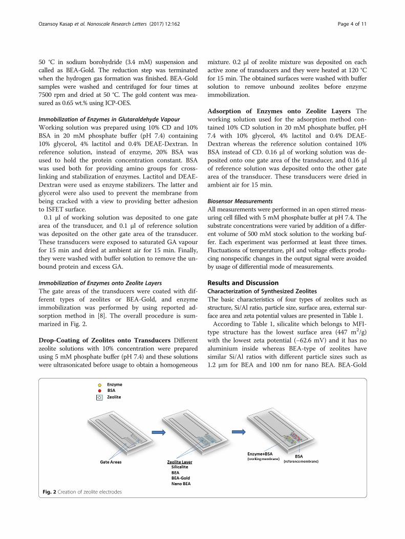
Immobilization of Enzymes onto Zeolite LayersThe gate areas of the transducers were coated with dif-ferent types of zeolites or BEA-Gold, and enzymeimmobilization was performed by using reported ad-sorption method in [8]. The overall procedure is sum-marized in Fig. 2.
Drop-Coating of Zeolites onto Transducers Differentzeolite solutions with 10% concentration were preparedusing 5 mM phosphate buffer (pH 7.4) and these solutionswere ultrasonicated before usage to obtain a homogeneous
Fig. 2 Creation of zeolite electrodes
mixture. 0.2 μl of zeolite mixture was deposited on eachactive zone of transducers and they were heated at 120 °Cfor 15 min. The obtained surfaces were washed with buffersolution to remove unbound zeolites before enzymeimmobilization.
Adsorption of Enzymes onto Zeolite Layers Theworking solution used for the adsorption method con-tained 10% CD solution in 20 mM phosphate buffer, pH7.4 with 10% glycerol, 4% lactitol and 0.4% DEAE-Dextran whereas the reference solution contained 10%BSA instead of CD. 0.16 μl of working solution was de-posited onto one gate area of the transducer, and 0.16 μlof reference solution was deposited onto the other gatearea of the transducer. These transducers were dried inambient air for 15 min.
Biosensor MeasurementsAll measurements were performed in an open stirred meas-uring cell filled with 5 mM phosphate buffer at pH 7.4. Thesubstrate concentrations were varied by addition of a differ-ent volume of 500 mM stock solution to the working buf-fer. Each experiment was performed at least three times.Fluctuations of temperature, pH and voltage effects produ-cing nonspecific changes in the output signal were avoidedby usage of differential mode of measurements.
Results and DiscussionCharacterization of Synthesized ZeolitesThe basic characteristics of four types of zeolites such asstructure, Si/Al ratio, particle size, surface area, external sur-face area and zeta potential values are presented in Table 1.According to Table 1, silicalite which belongs to MFI-
type structure has the lowest surface area (447 m2/g)with the lowest zeta potential (−62.6 mV) and it has noaluminium inside whereas BEA-type of zeolites havesimilar Si/Al ratios with different particle sizes such as1.2 μm for BEA and 100 nm for nano BEA. BEA-Gold
Table 1 Physicochemical characteristics of used zeolites
Sample name Structure Si/Ala Particle sizeb SBET, (m2/g)c Sext, (m
2/g)d Zeta potential, (mV)e
Silicalite MFI No Al. ~470 nm 447 96 −62.6
BEA BEA 21.5 ± 0.7 ~1.2 μm 743 128 −47.9
BEA-Gold BEA 21.0 ± 0.9 ~1.2 μm 776 160 −47.2
Nano BEA BEA 20.8 ± 0.8 ~100 nm 696 183 −36.6aMeasured by EDXbMeasured by SEM and Master sizercMeasured by Multipoint BETdMeasured by t-plot methodeMeasured by zeta potential at pH 7
Ozansoy Kasap et al. Nanoscale Research Letters (2017) 12:162 Page 5 of 11
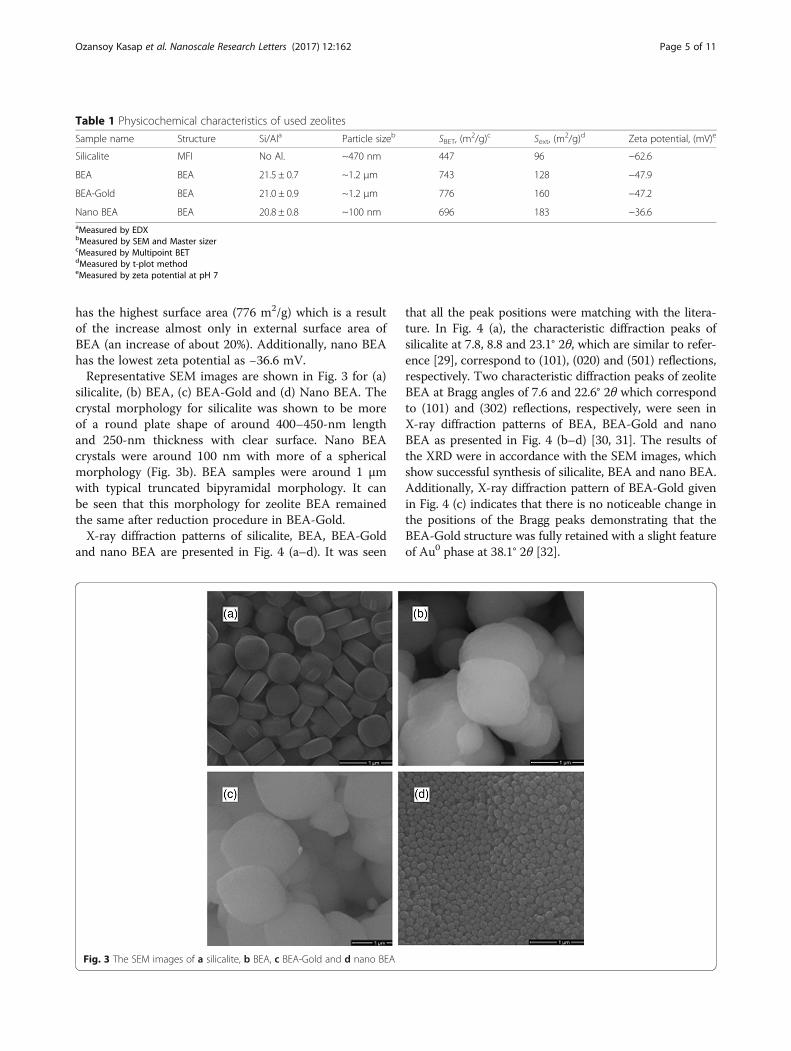
has the highest surface area (776 m2/g) which is a resultof the increase almost only in external surface area ofBEA (an increase of about 20%). Additionally, nano BEAhas the lowest zeta potential as −36.6 mV.Representative SEM images are shown in Fig. 3 for (a)
silicalite, (b) BEA, (c) BEA-Gold and (d) Nano BEA. Thecrystal morphology for silicalite was shown to be moreof a round plate shape of around 400–450-nm lengthand 250-nm thickness with clear surface. Nano BEAcrystals were around 100 nm with more of a sphericalmorphology (Fig. 3b). BEA samples were around 1 μmwith typical truncated bipyramidal morphology. It canbe seen that this morphology for zeolite BEA remainedthe same after reduction procedure in BEA-Gold.X-ray diffraction patterns of silicalite, BEA, BEA-Gold
and nano BEA are presented in Fig. 4 (a–d). It was seen
Fig. 3 The SEM images of a silicalite, b BEA, c BEA-Gold and d nano BEA
that all the peak positions were matching with the litera-ture. In Fig. 4 (a), the characteristic diffraction peaks ofsilicalite at 7.8, 8.8 and 23.1° 2θ, which are similar to refer-ence [29], correspond to (101), (020) and (501) reflections,respectively. Two characteristic diffraction peaks of zeoliteBEA at Bragg angles of 7.6 and 22.6° 2θ which correspondto (101) and (302) reflections, respectively, were seen inX-ray diffraction patterns of BEA, BEA-Gold and nanoBEA as presented in Fig. 4 (b–d) [30, 31]. The results ofthe XRD were in accordance with the SEM images, whichshow successful synthesis of silicalite, BEA and nano BEA.Additionally, X-ray diffraction pattern of BEA-Gold givenin Fig. 4 (c) indicates that there is no noticeable change inthe positions of the Bragg peaks demonstrating that theBEA-Gold structure was fully retained with a slight featureof Au0 phase at 38.1° 2θ [32].
Fig. 4 The X-ray diffraction patterns of a silicalite, b BEA and cBEA-Gold and d nano BEA
Ozansoy Kasap et al. Nanoscale Research Letters (2017) 12:162 Page 6 of 11
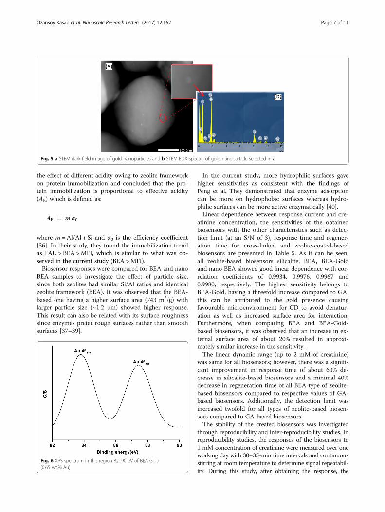
Additional insight for gold characterization in zeoliteBEA was investigated by ICP-OES, STEM, STEM-EDXand XPS. According to ICP-OES, BEA-Gold contains0.65% gold, whereas zeolite BEA has no gold inside. InTable 2, it can be noticed that Si/Al ratio of the zeoliteremains approximately the same after ion exchange andreduction procedure, which suggests that the local struc-ture of zeolite BEA did not collapse.The presence of gold nanoparticles can be seen in
STEM images presented in Fig. 5. The heavy Au atomsclearly stand out on the light background of zeolite BEA.Additionally, STEM-EDX at the shown point of Fig. 5acan be shown as additional evidence for the existing goldnanoparticles. The average size of gold nanoparticlesproduced was 10 nm according to the taken STEM im-ages. This result showed that gold nanoparticles were onthe zeolite surface rather than in the pores of zeolitesince the pore diameters are 5.6 × 5.6 Å and 7.7 × 6.6 Åfor zeolite BEA.The XPS spectrum of BEA-Gold given in Fig. 6 shows
two bands at 83.8 and 87.4 eV, corresponding toAu(0)4f7/2 and Au4f5/2, respectively. The band positionsshow good correspondence with literature [28, 33].Cross-sectional SEM images of (a) silicalite, (b) BEA,
(c) BEA-Gold and (d) nano BEA on silicon wafers arepresented in Table 3 with corresponding AFM imagesand surface roughness values. As seen from the cross-
Table 2 ICP results of BEA and BEA-Gold
ICP BEA BEA-Gold
Si(%) 35.80 ± 1.6 34.70 ± 0.7
Al(%) 1.52 ± 0.03 1.52 ± 0.02
Na(%) 0.45 ± 0.01 0.03 ± 0.01
Au(%) – 0.65 ± 0.02
Si/Al (mole) 23.55 22.83
sectional SEM images, the thickness of the coated zeolitelayer was in the range of 10–20 μm. According to theobtained AFM images, electrode surfaces modified withnano BEA has the smoothest surface with a surfaceroughness value of 0.08 ± 0.01 nm. On the other hand,BEA-Gold, BEA and silicalite have similar surface rough-ness values of 48.85 ± 8.3, 42.00 ± 4.9, 44.45 ± 4.03,respectively.According to the contact angle measurements pre-
sented in Table 4, zeolite-coated surfaces were becomingmore hydrophilic when compared to GA cross-linkedsurfaces. The contact angles were in the following order:BEA = BEA-Gold < Nano BEA < Silicalite < GA. This isan expected result, since it was known that a decrease inAl+3 content, which is related to ion exchange capacityof a zeolite, produces more hydrophobic nature [34].
Characteristics of Zeolite-Modified ISFET BiosensorsThe creatinine-sensitive biosensor based on ion sensitivefield-effect transistor (ISFET) operates according to thehydrolysis reaction of creatinine catalysed by creatininedeiminase:
The enzymatic reaction occurring at the dielectric gateof the transducer produces a pH increase that is propor-tional to creatinine concentration, which makes it de-tectable by the currently developed ISFET device [2].In this study, GA cross-linking, the traditional method
of enzyme immobilization technique, was used as a con-trol group to compare the effect of zeolites on character-istics of zeolite-modified potentiometric biosensors suchas sensitivity, linear range, response time, detection limitand regeneration time. Therefore, firstly, the calibrationcurves for all biosensors up to 10 mM of creatinine con-centration were obtained and presented in Fig. 7. Asseen in Fig. 7, all zeolite-based biosensors regardless oftype had higher sensitivities than GA-based biosensor.The sensitivities of the biosensors decreased in the orderof BEA-Gold > BEA > nano BEA > Silicalite > GA. BEAzeolites, having aluminium inside, regardless of its par-ticle size, gave higher response than silicalite which hasno aluminium in its structure. This result is consistentwith our previous results showing that the sensitivity ofurease biosensor based on ISFET developed by nanoBEA coating is higher compared to silicalite-coated one[20] and the same trend was observed for amperometricglucose biosensor [22]. This result can be attributed tothe presence of aluminium in zeolite BEA and nanoBEA, providing both hydrophilicity and Brønsted acidsites (Si–OH–Al) which are strongly effective in proteinadsorption [35, 36]. Additionally, Tavolaro et al. studied
Fig. 5 a STEM dark-field image of gold nanoparticles and b STEM-EDX spectra of gold nanoparticle selected in a
Ozansoy Kasap et al. Nanoscale Research Letters (2017) 12:162 Page 7 of 11
the effect of different acidity owing to zeolite frameworkon protein immobilization and concluded that the pro-tein immobilization is proportional to effective acidity(AE) which is defined as:
AE ¼ m α0
where m = Al/Al + Si and α0 is the efficiency coefficient[36]. In their study, they found the immobilization trendas FAU > BEA >MFI, which is similar to what was ob-served in the current study (BEA >MFI).Biosensor responses were compared for BEA and nano
BEA samples to investigate the effect of particle size,since both zeolites had similar Si/Al ratios and identicalzeolite framework (BEA). It was observed that the BEA-based one having a higher surface area (743 m2/g) withlarger particle size (~1.2 μm) showed higher response.This result can also be related with its surface roughnesssince enzymes prefer rough surfaces rather than smoothsurfaces [37–39].
Fig. 6 XPS spectrum in the region 82–90 eV of BEA-Gold(0.65 wt.% Au)
In the current study, more hydrophilic surfaces gavehigher sensitivities as consistent with the findings ofPeng et al. They demonstrated that enzyme adsorptioncan be more on hydrophobic surfaces whereas hydro-philic surfaces can be more active enzymatically [40].Linear dependence between response current and cre-
atinine concentration, the sensitivities of the obtainedbiosensors with the other characteristics such as detec-tion limit (at an S/N of 3), response time and regener-ation time for cross-linked and zeolite-coated-basedbiosensors are presented in Table 5. As it can be seen,all zeolite-based biosensors silicalite, BEA, BEA-Goldand nano BEA showed good linear dependence with cor-relation coefficients of 0.9934, 0.9976, 0.9967 and0.9980, respectively. The highest sensitivity belongs toBEA-Gold, having a threefold increase compared to GA,this can be attributed to the gold presence causingfavourable microenvironment for CD to avoid denatur-ation as well as increased surface area for interaction.Furthermore, when comparing BEA and BEA-Gold-based biosensors, it was observed that an increase in ex-ternal surface area of about 20% resulted in approxi-mately similar increase in the sensitivity.The linear dynamic range (up to 2 mM of creatinine)
was same for all biosensors; however, there was a signifi-cant improvement in response time of about 60% de-crease in silicalite-based biosensors and a minimal 40%decrease in regeneration time of all BEA-type of zeolite-based biosensors compared to respective values of GA-based biosensors. Additionally, the detection limit wasincreased twofold for all types of zeolite-based biosen-sors compared to GA-based biosensors.The stability of the created biosensors was investigated
through reproducibility and inter-reproducibility studies. Inreproducibility studies, the responses of the biosensors to1 mM concentration of creatinine were measured over oneworking day with 30–35-min time intervals and continuousstirring at room temperature to determine signal repeatabil-ity. During this study, after obtaining the response, the
Table 3 Cross-sectional SEM and AFM images of silicalite, BEA, BEA-Gold and nano BEA on silicon wafer with their representativesurface roughness values (Ra)
Ozansoy Kasap et al. Nanoscale Research Letters (2017) 12:162 Page 8 of 11
working cell was rinsed with buffer three times to discardthe products of enzymatic reaction on the biosensorsurface. This measurement was repeated for six times forevery type of immobilization. Finally, the relative standard
deviations (RSD) for determining creatinine are presentedin Fig. 8. As seen, the tested biosensors had rather good re-producibility as a sign of stable operation. The RSD valuesfor all biosensors were in the range 1–5% with BEA having
Table 5 Characteristics of creatinine-based biosensors withdifferent types of enzyme immobilizationMaterial Linear equation
between response(y) and creatinineconcentration (x)
Sensitivity,μA/mM
Detectionlimit, μM
Responsetime, s
Regenerationtime, s
GA y = 10.131x + 0.9594,R2 = 0.9807
10 10 240 300
Silicalite y = 20.79x − 0.3002,R2 = 0.9934
21 5 90 80
BEA y = 24.663x + 0.6437,R2 = 0.9976
25 5 150 180
BEA-Gold
y = 29.591x − 0.3703,R2 = 0.9967
30 5 150 120
NanoBEA
y = 22.942x + 0.0394,R2 = 0.9980
23 5 120 120
Table 4 Contact angles of biosensor surfaces
Sample name Contact angle, Ɵ (°)
Plain surface 56
GA 66
Silicalite 29
BEA 6
BEA-Gold 5
Nano BEA 15
Ozansoy Kasap et al. Nanoscale Research Letters (2017) 12:162 Page 9 of 11
the highest RSD value of 4.67%, while the lowest RSD valuebelongs to the silicalite-based biosensors (1.04%).For inter-reproducibility studies, several clean elec-
trodes were drop-coated with zeolites then CD wasadsorbed on these transducers as described in the chap-ter titled ‘Immobilization of enzymes onto zeolite layers’.After obtaining the response to 1 mM of creatinine inthree repetitions, the enzyme and zeolites were removedfrom the transducers with ethanol-wetted cotton. There-after, the whole procedure was repeated three times andRSD values of biosensors were calculated. According tothe results, the highest RSD (36.03%) belonged to thetraditional method prepared with GA and the least RSDbelonged to BEA-Gold (3.19%), while the intermediatevalues belonged to Silicalite (13.27%), BEA (10.7%) andNano BEA (18.5%). The low RSD for zeolite-based bio-sensors compared to GA-based biosensors can be due tothe better controlled procedure of adsorption. Addition-ally, low RSD of BEA-Gold can be due to the presenceof gold nanoparticles, which will bind covalently theamine groups of the enzyme and actively stabilize theenzyme. This result is consistent with the study of Goleet al., where pepsin-gold colloid conjugates were pre-pared by just mixing under protein-friendly conditions
Fig. 7 The calibration curves for determination of creatinineobtained with biosensors created by CD immobilization; a in GAvapour, adsorption on: b silicalite, c BEA, d BEA-Gold and e NanoBEA. The measurements were carried out in 5 mM phosphate buffersolution (pH 7.4)
and then demonstrated that pepsin showed significantcatalytic activity. They explained the binding mechanismof the enzyme to the gold nanoparticle by covalent inter-actions between thiol groups in cysteine residues as wellas amine groups in lysine residues of pepsin with thesurface of gold. This interaction resulted in a more stabi-lized enzyme with its activity [41].
ConclusionsIn this study, creatinine-based ISFET biosensors have beensuccessfully produced by modifying the gate of ISFET withfour different types of zeolites separately. Their responsesand other biosensor characteristics such as sensitivity,detection limit, response time and regeneration time arecompared with covalently cross-linked CD as a controlgroup. All zeolite-based biosensors had higher sensitivities,lower detection limits, response and regeneration time thanGA-based biosensors with good reproducibility showingthat zeolites can be good alternatives for creatinine
Fig. 8 Reproducibility of signals obtained with biosensors created byCD immobilization; a in GA vapour, adsorption on: b silicalite, c BEA,d BEA-Gold and e Nano BEA. The measurements were carried out in5 mM phosphate buffer solution (pH 7.4)
Ozansoy Kasap et al. Nanoscale Research Letters (2017) 12:162 Page 10 of 11
biosensor production. The incorporation of zeolite andgold nanoparticles for adsorption of CD on ISFET-basedbiosensor was studied for the first time. The sensitivitiesof the obtained biosensors were decreasing in followingorder: BEA-Gold > BEA > nano BEA > Silicalite > GA. TheBEA-Gold-based biosensor resulted in threefold increasedsensitivity compared to GA-based biosensor, which couldbe attributed to favourable microenvironment for CD toavoid denaturation as well as increased surface area pro-duced by gold nanoparticles with good stability. This re-sult showed that gold nanoparticles can be used withzeolite to improve the characteristics of ISFET-basedbiosensors.
AcknowledgementsThe authors gratefully acknowledge the financial support of this study by theEuropean IRSES-NANODEV project, Ministry of Education and Science ofUkraine (project DZ/45-2015) and STCU Project 6052 “Enzyme multibiosensorsystem for renal dysfunction diagnosis and hemodialysis control”.
Authors’ ContributionsBOK synthesized and characterized the zeolites with different properties. SVMperformed the enzyme immobilization on the zeolite-modified ISFET andstudied the analytical characteristics of obtained creatinine biosensors withBOK. BOK, SVM and BAK processed the obtained results and wrote and ar-ranged the article. BAK planned and supervised the experiments performedby BOK and SVD. OOS and SVD proposed the idea of the development ofpotentiometric creatinine biosensor based on zeolite-modified electrodes. Allauthors read and approved the final manuscript.
Competing InterestsThe authors declare that they have no competing interests.
Author details1Department of Micro and Nanotechnology, Middle East Technical University,Ankara 0631, Turkey. 2Laboratory of Biomolecular Electronics, Institute ofMolecular Biology and Genetics, National Academy of Sciences of Ukraine,150 Zabolotnogo Str., 03680 Kyiv, Ukraine. 3Institute of High Technologies,Taras Shevchenko National University of Kyiv, 64 Volodymyrska St., Kyiv01003, Ukraine. 4Central Laboratory, Middle East Technical University, Ankara0631, Turkey.
Received: 22 December 2016 Accepted: 22 February 2017
Electrochemical biosensor for creatinine based on the immobilization ofcreatininase, creatinase and sarcosine oxidase onto a ferrocene/horseradishperoxidase/gold nanoparticles/multi-walled carbon nanotubes/Tefloncomposite electrode. Electrochim Acta 97:175–83
2. Soldatkin AP, Montoriol J, Sant W, Martelet C, Jaffrezic-Renault N (2002)Creatinine sensitive biosensor based on ISFETs and creatinine deiminaseimmobilised in BSA membrane. Talanta 58:351–7
3. George SK, Dipu MT, Mehra UR, Singh P, Verma AK, Ramgaokar JS (2006)Improved HPLC method for the simultaneous determination of allantoin,uric acid and creatinine in cattle urine. J Chromatogr B Anal TechnolBiomed Life Sci 832:134–7
4. Costa ACO, da Costa JL, Tonin FG, Tavares MFM, Micke GA (2007)Development of a fast capillary electrophoresis method for determinationof creatinine in urine samples. J Chromatogr A 1171:140–3
7. Walcarius A (2008) Electroanalytical applications of microporous zeolites andmesoporous (organo)silicas: recent trends. Electroanalysis 20:711–38
8. Kirdeciler SK, Soy E, Öztürk S, Kucherenko I, Soldatkin O, Dzyadevych S et al(2011) A novel urea conductometric biosensor based on zeoliteimmobilized urease. Talanta 85:1435–41
9. Saiapina OY, Pyeshkova VM, Soldatkin OO, Melnik VG, Kurç BA, Walcarius Aet al (2011) Conductometric enzyme biosensors based on natural zeoliteclinoptilolite for urea determination. Mater Sci Eng C 31:1490–7
10. Liu B, Hu R, Deng J (1997) Characterization of immobilization of an enzymein a modified Y zeolite matrix and its application to an amperometricglucose biosensor. Anal Chem 69:2343–8
11. Phadtare S, Vinod VP, Mukhopadhyay K, Kumar A, Rao M, Chaudhari RV et al(2004) Immobilization and biocatalytic activity of fungal protease on goldnanoparticle-loaded zeolite microspheres. Biotechnol Bioeng 85:629–37
12. Kumar S, Ahlawat W, Kumar R, Dilbaghi N (2015) Graphene, carbonnanotubes, zinc oxide and gold as elite nanomaterials for fabrication ofbiosensors for healthcare. Biosens Bioelectron 70:498–503
13. Crumbliss, AL, Henkens, RW, Perine, SC, Tubergen, KR, Kitchell, BS,Stonehuerner J (1990). Biosensor Technology: Fundamentals andapplications. Buck, R. P., Hatfield, W. E., Umana, M., Bowden EF, editor.Marcel Dekker: New York
14. Crumbliss AL, Perine SC, Stonehuerner J, Tubergen KR, Zhao J, Henkens RW(1992) Colloidal gold as a biocompatible immobilization matrix suitable forthe fabrication of enzyme electrodes by electrodeposition. BiotechnolBioeng 40:483–90
15. Kharitonov AB, Shipway AN, Willner I (1999) An Au nanoparticle /bisbipyridinium field-effect transistor for the sensing of adrenaline. AnalChem 71:5441–3
16. Gun J, Rizkov D, Lev O, Abouzar MH, Poghossian A, Schöning MJ (2009)Oxygen plasma-treated gold nanoparticle-based field-effect devices astransducer structures for bio-chemical sensing. Microchim Acta 164:395–404
17. Gun J, Schöning MJ, Abouzar MH, Poghossian A, Katzd E (2008) Field-effectnanoparticle-based glucose sensor on a chip: amplification effect ofcoimmobilized redox species. Electroanalysis 20:1748–53
18. Poghossian A, Weil MH, Bäcker M, Mayer D, Schöning MJ (2012) Field-effectdevices functionalised with goldnanoparticle/ macromolecule hybrids: Newopportunities for a label-free biosensing. Procedia Eng 47:273–6
19. Poghossian A, Bäcker M, Mayer D, Schöning MJ (2015) Gating capacitivefield-effect sensors by the charge of nanoparticle/molecule hybrids.Nanoscale 7:1023–31
20. Shelyakina MK, Soldatkin OO, Arkhypova VM, Kasap BO, Akata B, Dzyadevych SV(2014) Study of zeolite influence on analytical characteristics of urea biosensorbased on ion-selective field-effect transistors. Nanoscale Res Lett 9:124
21. Soldatkin OO, Shelyakina MK, Arkhypova VN, Soy E, Kirdeciler SK, OzansoyKasap B et al (2015) Nanosized zeolites as a perspective material forpotentiometric biosensors creation. Nanoscale Res Lett 10:59
22. Soldatkin OO, Ozansoy Kasap B, Akata Kurc B, Soldatkin AP, Dzyadevych SV,El’Skaya AV (2014) Elaboration of new method of enzyme adsorption onsilicalite and nano beta zeolite for amperometric biosensor creation.Biopolym Cell 30:291–8
23. Soldatkin OO, Kucherenko IS, Marchenko SV, Ozansoy Kasap B, Akata B,Soldatkin AP et al (2014) Application of enzyme/zeolite sensor for ureaanalysis in serum. Mater Sci Eng C 42:155–60
24. Marchenko SV, Soldatkin OO, Kasap BO, Kurc BA, Soldatkin AP, DzyadevychSV (2016) Creatinine deiminase adsorption onto silicalite-modified pH-FETfor creation of new creatinine-sensitive biosensor. Nanoscale Res Lett 11:173
26. Pavluchenko AS, Kukla AL, Goltvianskyi YV, Soldatkin OO, Arkhypova VM,Dzyadevych SV, Soldatkin AP (2011) Investigation of stability of the pH-sensitive field-effect transistor characteristics. Sens Lett 9:2392
27. Prokesova P, Mintova S, Cejka JBT (2003) Preparation of nanosized micro/mesoporous composites via simultaneous synthesis of Beta/MCM-48phases. Microporous Mesoporous Mater 64:165–174
28. Zahmakiran M, Özkar S (2010) The preparation and characterization of gold(0)nanoclusters stabilized by zeolite framework: Highly active, selective and reusablecatalyst in aerobic oxidation of benzyl alcohol. Mater Chem Phys 121:359–63
29. Yeong YF, Abdullah AZ, Ahmad AL, Bhatia S (2009) Synthesis, structure andacid characteristics of partially crystalline silicalite-1 based materials.Microporous Mesoporous Mater 123:129–39
30. Prokesova P, Mintova S, Cejka J, Bein T (2003) Preparation of nanosizedmicro/mesoporous composites. Mater Sci Eng C 23:1001–5
Ozansoy Kasap et al. Nanoscale Research Letters (2017) 12:162 Page 11 of 11
31. Narasimharao K, Hartmann M, Thiel HH, Ernst S (2006) Novel solid basiccatalysts by nitridation of zeolite beta at low temperature. MicroporousMesoporous Mater 90:377–83
32. Zeng S, Ding S, Li S, Wang R, Zhang Z (2014) Controlled growth of goldnanoparticles in zeolite L via ion-exchange reactions and thermal reductionprocesses. Inorg Chem Commun 47:63–6
33. Jiang P, Xie S, Yao J, Pang S, Gao H (2001) The stability of self-organized 1-nonanethiol-capped gold nanoparticle monolayer. J Phys D Appl Phys 34:2255–9
34. Matsui M, Kiyozumi Y, Yamamoto T, Mizushina Y, Mizukami F, Sakaguchi K(2001) Selective adsorption of biopolymers on zeolites. Chem - A Eur J 7:1555–60
35. Sakaguchi K, Matsui M, Mizukami F (2005) Applications of zeolite inorganiccomposites in biotechnology: current state and perspectives. Appl MicrobiolBiotechnol 67:306–11
36. Tavolaro P, Tavolaro A, Martino G (2009) Influence of zeolite PZC and pH onthe immobilization of cytochrome c: a preliminary study regarding thepreparation of new biomaterials. Colloids Surfaces B Biointerfaces 70:98–107
37. Rechendorff K, Hovgaard MB, Foss M, Zhdanov VP, Besenbacher F (2006)Enhancement of protein adsorption induced by surface roughness.Langmuir 22:10885–8
38. Jensen UB, Ferapontova EE, Sutherland DS (2012) Quantifying proteinadsorption and function at nanostructured materials: Enzymatic activity ofglucose oxidase at GLAD structured electrodes. Langmuir 28:11106–14
39. Kirdeciler SK, Ozen C, Akata B (2014) Fabrication of nano- to micron-sizedpatterns using zeolites: its application in BSA adsorption. MicroporousMesoporous Mater 191:59–66
40. Peng Y, Zhu-Ping H, Yong-Juan X, Peng-Cheng H, Ji-Jun T (2013) Effect ofsupport surface chemistry on lipase adsorption and activity. J Mol Catal B:Enzym 94:69–76
41. Gole A, Dash C, Ramakrishnan V, Sainkar SR, Mandale AB, Rao M et al (2001)Pepsin - gold colloid conjugates: preparation, characterization, andenzymatic activity. Langmuir 17:1674–9
Submit your manuscript to a journal and benefi t from: