Biosynthesis, Glycosylation and Intracellular Processing of the NeuroglandularAntigen, a Human Melanoma-associated Antigen1
Walter T. Dixon, Douglas J. Demetrick, Kunio Ohyama, Lydia K. J. Sikora, and L. Martin Jerry2
Oncology Research Group [W. T. D., L. K. J. S., K. O., L. M. J.], and Department of Pathology [D. J. DJ, The University of Calgary, and Tom Baker Cancer Centre[L. M. J.J, Calgary, Alberta, Canada T2N 4N2, and Department of Biochemistry [K. O.], Tokyo College of Pharmacy, Tokyo, Japan 192-03
ABSTRACT
Neuroglandular antigen (NGA) was identified as a human melanoma-associated antigen by a panel of murine monoclonal antibodies of bothIgG2a (LS62, LS76, LS159) and IgGl (LS113, LS140, LS152) subclasses, developed in this laboratory (L. Sikora, A. Pinto, D. Demetrick,W. Dixon, S. Urbanski, and L. M. Jerry, Int. J. Cancer, 39: 138-145,1987). Monoclonal antibody LS62 was used to immunoprecipitate NGAfrom radiolabeled cultured melanoma cells, and it behaved as a heterogeneous glycoprotein "smear" on sodium dodecyl sulfate-polyacrylamidegel electrophoresis analysis (M, 29,000-70,000). Radioactive pulse-chasetime course experiments using human melanoma cells cultured in thepresence or absence of inhibitors of protein glycosylation showed thatthe antigen consisted of a core protein with a molecular weight of 22,000on sodium dodecyl sulfate-polyacrylamide gel electrophoresis. This molecule was modified by the addition of at least three TV-linkedoligosac-charide side chains (as revealed by limited W-glycanase digestion) to givea precursor form with a molecular weight of approximately 34,000.Subsequent processing steps yielded a heterogeneous family of glycopro-teins with varying amounts of covalently attached carbohydrate. Muchof this heterogeneity in both molecular weight and pi (as revealed bytwo-dimensional electrophoresis) could be removed by treatment of theantigen with neuraminidase, suggesting heavy sialylation of the glycoprotein.
NGA could be detected on the surface of melanoma cells by fluorescence-activated cell sorter analysis, surface radioiodination, and, as previously shown, immunoperoxidase staining. However, there was a largerintracellular pool of the molecule and the antigen was rapidly releasedinto the culture supernatant. The function of NGA remains unknown butits elevated expression in transformed melanocytes have prompted thischaracterization to understand its biochemical nature and relation toother melanoma-associated antigens.
INTRODUCTION
Monoclonal antibodies have been used extensively in characterizing the antigenic repertoire of human melanocytes, especially malignant melanoma cells (for reviews, see Refs. 1-6).The underlying hope of most investigators has been to identifyunique melanoma-specific tumor antigens which could be exploited for immunodiagnostic or immunotherapeutic strategies(6). However, this goal remains largely unfulfilled (for exceptions, see Refs. 5 and 7) with the realization that most of theantigens described were melanocyte differentiation antigens orwere found on other normal or malignant cell types. Suchantigens could only be classified as melanoma associated ratherthan melanoma specific (2, 3). Despite this, the antigens detected by these MAbs3 have proved to be extremely useful in
Received 2/27/89; revised 2/8/90.The costs of publication of this article were defrayed in part by the payment
of page charges. This article must therefore be hereby marked advertisement inaccordance with 18 U.S.C. Section 1734 solely to indicate this fact.
1W. D., L. S., and D. D. acknowledge the generous support of the Alberta
Heritage Fund for Medical Research, Medical Research Council of Canada, andthe National Cancer Institute of Canada.
2To whom requests for reprints should be addressed, at Tom Baker CancerCentre, 1331-29 Street N.W., Calgary, Alberta, Canada T2N 4N2
delineating pathways of melanocyte differentiation (8-10). It isnow possible to combine a knowledge of antigenic phenotypewith other markers of differentiation such as pigmentation andmorphology to consider a cell as representing early, intermediate, or late stages of melanocyte differentiation (11, 12). Inaddition, the use of MAbs directed against melanoma tumor-associated antigens has enabled the construction of hypotheticalpathways for malignant transformation of melanocytes andsubsequent stages of tumor progression (13, 14).
We have previously described the identification, using murineMAbs, of an antigen, termed NGA which is expressed inabnormal human melanocytes (15). A panel of antibodies werefound to recognize this antigen in n-octyl-a-o-glucopyranosideextracts (by enzyme-linked immunosorbent assay) or by histo-chemical staining of all melanoma cell lines and the majorityof metastatic melanoma tumors. Minimal reactivity was seenagainst comparable preparations of normal adult necropsy tissue but some reactivity was seen against nonmelanoma tumors(15).
Preliminary identification of the glycoprotein nature of thismelanoma-associated antigen was made by immunoprecipita-tion of NGA from [35S]methionine-labeled cell lysates of G361human melanoma cells and subsequent SDS-PAGE analysis. Aheavily glycosylated glycoprotein smear was observed whichcould be reduced to a single band of approximately MT 23,500when cell cultures were treated with an inhibitor of protein 7V-glycosylation (15). Heavily glycosylated melanoma-associatedantigens with various characteristics similar to those of NGAhave been described in the literature previously (16-26). Inparticular, the independently developed MAbs ME491 (20-23)and NKI-C3 (17-19), which have been shown by competitiveinhibition studies to recognize the same antigen (19), immunoprecipitate a glycoprotein with electrophoretic mobilities,after the appropriate treatments, similar to, if not identical withglycosylated or nonglycosylated NGA. In this paper we presentfurther data, using our prototype antibody LS62, concerningthe biosynthesis of the mature NGA molecule, the nature of itscarbohydrate modifications, and indications of its cellular distribution.
MATERIALS AND METHODS
Cells
Human melanoma cell line G361 was obtained from the AmericanType Culture Collection (Rockville, MD) (CRL 1424). Human B-lymphoblastoid cell line T-5.1 was obtained from Dr. D. Pious, University of Washington, Seattle, WA. Both cell lines were grown inRPMI 1640 (Gibco, Burlington, Ontario, Canada) supplemented with10% (v/v) heat-inactivated fetal bovine serum (Flow) and grown in a5% (v/v) CG-2incubator at 37°C.
Monoclonal Antibodies
LS62 (IgG2A) and LSI09 (IgGl) were prepared as described previously (15). LS62 and LS109 immunoadsorbents were prepared bycoupling purified ascites fluid (LSI09) or protein A-Sepharose-purifiedantibody (LS62) to CNBr-activated Sepharose (Pharmacia, Baie
D'Urfe, Quebec, Canada). MAb L243 (16) was a gift from Dr. Ted
Zipf, M. D. Anderson Hospital and Tumor Institute, Houston, TX,and was also coupled to CNBr-activated Sepharose. Normal mouseserum-agarose was obtained from Sigma (St. Louis, MO). Goat anti-mouse IgG-FITC was from Cappell (Organon-Teknika, Scarborough,Ontario, Canada).
Enzymes
Neuraminidase (Clostridium perfringens) Type X (EC 3.2.1.18) wasobtained from Sigma. A'-Glycanase (peptide /V-glycosidase F, EC3.2.2.18), Endo H (endo-/3-A'-acetylglucosaminidase H, EC 3.2.1.96),and 0-glycanase (O-glycopeptide endo-a-A'-acetylgalactosaminyl hy-
drolase, EC 3.2.1.97) were obtained from Genzyme (Boston, MA).
Chemicals and Radiochemicals
Tunicamycin and monensin were obtained from Sigma and weredissolved in dimethyl sulfoxide (10 mg/ml) and 95% (v/v) ethanol (10mM), respectively. Sodium [125l)iodide (506.9 MBq/ÃÃg)was obtainedfrom Amersham (Oakville, Ontario, Canada) and L-[35S]methionine(29.6 TBq/mmol) was from NEN/DuPont (Mississauga, Ontario, Can-ada).
Radiolabeling
In Vivo Labeling
Exponentially growing G361 cells (10") were collected by scraping
and centrifugation, washed with cold, sterile PBS, recentrifuged, finallyresuspended in 10 ml of methionine-depleted media (Select-amine kit,Gibco), and transferred to 80-cm2 culture flasks. After a 2-h preincu-bation, 0.5 mCi of [35S]methionine was added and the incubation was
continued. For experiments involving the inhibitors tunicamycin andmonensin, cells were pretreated for 16 h with media supplemented withinhibitor at the concentrations given in the figure legends, prior tolabeling. In the case of pulse-chase experiments, after the initial labeling"pulse" period (usually 30 min or 2 h), the medium containing the [35S]
methionine was removed. The adherent cells were rinsed once withnonradioactive media and 10 ml fresh prewarmed media were addedand the "chase" continued for varying periods of time. The experiment
was terminated by removal of the media and one rinse of the adherentcells with ice-cold PBS. The cells were rapidly harvested by scrapinginto cold PBS and centrifuging to yield a cell pellet. All liquid wasremoved from the pellet and it was frozen at -20°C until used. For
immunoprecipitation experiments, the cell pellet was thawed in thepresence of 500 n\ lysis buffer (10 mM Tris-Cl, pH 7.6, 1 mM MgCl2,1% (v/v) NP-40, 0.5% (w/v) sodium deoxycholate, 0.25% (v/v) aproti-nin (Sigma), 0.1% sodium azide, (10 mM phenylmethylsulfonyl fluoride) and kept on ice for 30 mins with occasional vortexing. Theresulting lysate was centrifuged in an Eppendorf microfuge for 30 minand the supernatant from this step was used for immunoprecipitationexperiments.
In Vitro Labeling
Surface Labeling of Cells. G361 or T-5.1 cells (5 x IO6) were
harvested and washed as described above, resuspended in 1.0 ml ofPBS, and placed on ice. Sodium [125I]iodide(0.5 mCi) was added andthe suspension was transferred to an lodo-Gen (Pierce, Terochem,Edmonton, Alberta, Canada)-coated tube and incubated for 5 min onice (27). The reaction was terminated by transferring the labeled cellsuspension to 12 ml cold media (containing 1 mM KI) in a conicalcentrifuge tube and centrifuging to pellet the cells. The resulting pelletwas resuspended in 12ml fresh cold media and this washing step wasrepeated twice. After thorough draining of the final washed pellet, thecells were lysed in 500 ^1 of lysis buffer and processed for subsequentimmunoprecipitation as described previously. For experiments involving the glycosylation inhibitor tunicamycin, cells were preincubated for16 h prior to labeling in media containing 10 ng/m\ of the drug.
Labeling of Cell Lysates. Cells (5 x 10') were harvested and washed
as described for surface labeling and the cell pellet was lysed on ice for
30 min in 500 ^1 of lysis buffer. The lysate was centrifuged as describedand the supernatant was transferred to a fresh tube. Sodium [12!I]iodide(0.5 mCi) was added to the lysate and this was transferred to an lodo-Gen-coated tube as described previously. After a 5-min incubation onice, the labeling mixture was applied to a PD-10 column (Pharmacia)preequilibrated with wash buffer 1 [0.5 M NaCI, 5 mM EDTA, 50 mMTris-Cl, pH 7.5, 1% (v/v) NP-40] containing 1% FCS. Radiolabeledprotein was eluted from the column with the same buffer (minus PCS)and 1.0-ml fractions were collected. Ten-¿ilaliquots of each fractionwere counted and the radioactive protein peak fractions were pooledand processed for immunoprecipitation as described below. For experiments involving the glycosylation inhibitor tunicamycin, cells werepreincubated for 16 h prior to labeling in media containing 10 ^g/mlof the drug.
Immunoprecipitations
Ten-^1 aliquots of lysate supernatants from in vivo labeled G361 cellswere precipitated with trichloroacetic acid, collected on glass fiber discsand counted by liquid scintillation. For time course studies, all lysateswere balanced for total radioactivity (cpm) prior to immunoprecipitation.
The lysate supernatants from in vivo labeling or in vitro surfaceiodination, or the pooled labeled peak from total cell lysate iodinationwere diluted with 10-20 volumes of heat-inactivated FCS containing1% (v/v) NP-40, 0.1% NaNj, 0.25% aprotinin, 10 mM iodacetamide, 1mM phenylmethylsulfonyl fluoride. These diluted mixtures were pre-cleared 3 times over 18 h with 50 ¿tl(packed volume) of normal mouseserum agarose beads and end-over-end rotation at 4°C.After centrifug
ing to remove the preclearing beads, the supernatant was transferred toclean tubes and 50 ^1 (packed volume) of LS62, LS109, LS19, or MAb243 immunoadsorbents were added. The specific immunoprecipitationwas incubated with end-over-end mixing at 4'C for 18 h. The immu-
noadsorbent beads were collected by centrifugation and washed with 5changes of wash buffer 1 and two changes of wash buffer 2 (same aswash buffer 1 except no detergent). The washed beads were transferredto 1.5-ml Eppendorf tubes and prepared for enzyme treatment or SDS-PAGE under reducing or nonreducing conditions.
For immunoprecipitation of NGA from culture media, the 10-mlsupernatants from the labeling experiments were decanted into conical15-ml tubes and centrifuged to remove cell debris. The clarified supernatants were transferred to fresh tubes and protease inhibitors wereadded, as described above. NP-40 detergent was added to 1% (v/v) andthe supernatants were precleared and treated with specific antibody asdescribed for cell lysates.
Enzyme Treatments
The immunoadsorbent beads from the previous step were washedtwice in either PBS (pH 7.2) or 200 mM sodium phosphate (pH 8.6),1.25% NP-40, 0.17% (w/v) SDS for neuraminidase or W-glycanase,respectively. The beads were boiled for 3 min in 100 ^1 of one of theincubation buffers, and cooled to room temperature. Ten milliunits ofneuraminidase or 10 units of jV-glycanase were added to the beads andthe suspension was incubated at 37°Cfor varying lengths of time.
Control digestions were carried out on identical ¡mmunoprecipitatesfor the maximum time of experimental digestions under identicalconditions (minus enzyme). For Endo H digestion the washed immunoadsorbent beads were resuspended in 0.5% (w/v) SDS and boiled for3 min. The sample was diluted to 0.2% (w/v) SDS with 50 mM sodiumphosphate buffer (pH 6.0) and 20 mIU/ml (final concentration) ofEndo H enzyme were added to the appropriate samples. Controlsamples were incubated (minus enzyme) for the duration of the 16-hdigestion period. For sequential neuraminidase/O-glycanase digestion,the washed immunoadsorbent beads were boiled for 3 min in 0.1 % (w/v) SDS and diluted with the appropriate reagents to give final concentrations of 10 mM calcium acetate, 20 mM sodium cacodylate, pH 6.5,1.5% (v/v) NP-40. Neuraminidase (1 unit/ml) was added to the sampleand incubated at 37"C for 2 h, followed by the addition of 4 milliunitsof O-glycanase and further incubation at 37°Cfor 20 h.
All of the above enzyme or control incubations were terminated by
the addition of 2 volumes of twice concentrated SDS-PAGE samplebuffer and processing for SDS-PAGE analysis.
Electrophoresis
Immunoadsorbent beads were resuspended in 100 M' of Laemmlisample buffer (28) with or without dithiothreitol for reducing or non-reducing conditions. The samples were boiled for 5 min, cooled, andloaded on SDS-PAGE gels consisting of a 5% (w/v) acrylamide stackinggel and a 15% (w/v) separating gel in either a mini, medium, or largeformat gel apparatus. 14C-labeled molecular weight standards (Amer-
shani) were used to calibrate all gels. The markers were; myosin (M,200,000), phosphorylase b (M, 100,000 and 92,500) bovine serumalbumin (M, 69,000), ovalbumin (M, 46,000), carbonic anhydrase (M,30,000), soybean trypsin inhibitor (M, 21,500), and lysozyme (M,14,300).
Two-dimensional (isoelectric focusing/SDS-PAGE) electrophoresiswas carried out on an ISODALT (Electronucleonics, Fairfield, NJ)apparatus and ampholines (LKB 4-6 Pharmacia/LKB) were used in thefirst dimension (29).
Gels containing '"I-labeled samples were fixed and processed forautoradiography, whereas [35S]methionine-containing gels were processed for fluorography (30). Exposed X-ray films from fluorographywere scanned with a Bio-Rad 620 scanning densitometer (Bio-Rad,Richmond, CA).
Flow Cytometry Analysis
FITC-Labeling. Indirect immunofluorescence was used to investigatethe reactivity of LS62 monoclonal antibody with G361 melanoma cells,which were harvested by scraping. Goat anti-mouse IgG-FITC was usedas the second stage antibody and the staining protocol of Loken andHerzenberg as modified by Zipf et al. (31) was followed. An OrthoSystem 50H Cytofluorograf was used to analyze the populations ofstained cells and the resulting data were recorded on an Ortho 2150Computer System.
RESULTS
Antigen Characterization and Use of Inhibitors of ProteinGlycosylation. The antigen immunoprecipitated by MAb LS62from lysates of metabolically labeled G361 melanoma cellsappeared as a heterogeneous smear with a molecular weight ofapproximately 29,000-70,000 on SDS-PAGE (Fig. 1, Lane 1).Smearing to higher molecular weight ranges was sometimesseen with longer fluorographic exposures (e.g., Fig. 6). Incontrast, when G361 cells were first preincubated with tunica-mycin, an inhibitor of /V-linked glycosylation (32), the heterogeneity was removed and a discrete band with a molecularweight of 23,500 was seen (Fig. 1, Lane 3), as described previously (15). In addition, when cells were pretreated with theionophore monensin, which inhibits terminal glycosylation byblocking vesicle transport in the Golgi apparatus (33), an intermediate molecular weight form of NGA could be seen (Fig. 1,Lane 2). This consisted of a doublet with molecular weights of28,000 and 32,000 with very little of the heterogeneity seen inthe immunoprecipitates from untreated cells. Control immu-noprecipitations using preimmune serum as a control antibodydid not show these specific bands (e.g., Fig. 2, Track C). Takentogether these results suggested that the heterogeneity of thisantigen resided in its covalenti} attached V-linkt'd carbohydrateand not in its protein "backbone." It also revealed that the
protein moiety contained the antigenic determinant and thatthis epitope was unaffected by subsequent glycosylation.
Pulse-Chase Experiments and Release of NGA into CultureMedia. In an attempt to study these covalent modifications,pulse-chase experiments were carried out to follow the biosynthesis of NGA. A pulse-chase time course experiment over 6 h
200 -
100 -92.5~
69 -
kDa46
30 -
14.3-
Fig. 1. Fluorograph of SDS-PAGE profiles of LS62 immunoprecipitates fromcell lysates of G361 melanoma cells. Lysates were prepared from untreated cells(Lane 1) or cells treated with the inhibitors; monensin (Lane 2) at 20 UM, ortunicamycin (Lane 3) at 10 ng/ml as described in "Materials and Methods."
Ordinale, molecular weight in thousands.
is shown in Fig. 2. Immunoprecipitable NGA precursors appeared very rapidly (within 5 min) after the start of labelingand consisted of a minor band at M, 29,000 as well as a majorbroad band at M, 34,000. The upper component of this bandwhich on some gels appeared as a closely spaced doublet (datanot shown) seemed to disappear during the chase period andwas increasingly replaced by a smear of radioactivity upwardfrom a constant M, 34,000 component. It is not known whetherthis represents some proteolytic or carbohydrate processing ofa precursor or simply the gradual replacement of a discreteprotein band with a less discernible, more diffuse posttransla-tionally modified species.
Similar behavior of NGA was observed in an extended pulse-chase time course (Fig. 3), which, in addition, showed NGAeither turning over with an apparent half-life of between 22 and46 h, or else leaving the cellular pool. A higher-molecular-weight band (M, 83,000) which had been indicated in Fig. 2(upper arrow) and was not apparent in control immunoprecip-itations was more evident when samples for SDS-PAGE wereprepared under nonreducing conditions. However, we have noevidence that this band is related to NGA. Apart from thisobservation, NGA behaved similarly under reducing or nonreducing SDS-PAGE conditions (data not shown).
When an extended pulse-chase time course was conductedwith cells which had been pretreated with tunicamycin, therewas a very slight increase in the molecular weight of theimmunoprecipitated antigen from M, 22,000 to 23,500 withtime (Fig. 4). This increase might be due to direct O-glycosy-lation of the protein on serine or threonine residues, or other
MINUTESFig. 2. Fluorograph of SDS-PAGE profiles of LS62 immunoprecipitates
during a pulse-chase time course of NGA synthesis in G361 melanoma cells.After a 30-min pulse, the cells were chased from time 0 for up to 360 min. LaneC shows the results of an immunoprecipitation using preimmune mouse serum.Ordinate, molecular weight in thousands.
posttranslational modification which might occur regardless ofthe inhibition of jV-glycosylation. It also appeared that the NGAwhich lacked TV-linkedcarbohydrate was subject to slower turnover in the G361 cells. Perhaps this reflects a lack of appropriatecarbohydrate structure (or resultant changes in protein conformation) necessary for targeting the NGA for degradation orsecretion.
In this latter regard, culture supernatants from G361 cellsundergoing metabolic labeling were also examined for the appearance of NGA. Fig. 5 shows a comparison of SDS-PAGEprofiles of NGA immunoprecipitated from either cell lysates orspent media after a 16-h labeling period. Although the absoluteamounts of antigen differ, the overall profiles were very similarand both glycosylated and nonglycosylated species of NGAwere present in the culture supernatants. It was interesting toobserve that, even in cells which had not been treated withglycosylation inhibitors, there was evidence of nonglycosylatedforms of NGA in the culture media. Furthermore, cells whichhad been treated with monensin to block transport into thetrans-Golgi cisternae (33, 34), were still able to release partiallyglycosylated forms of NGA into the media. Perhaps this provides evidence for a default pathway for the release of non- orpartially glycosylated species of the molecule, which mightbypass certain compartments of the Golgi during transport tothe plasma membrane. Since the vast majority of both untreatedand treated cells were viable at the end of the experiment(>90%), it seemed that NGA must be rapidly shed, if notactively secreted by the G361 melanoma cells (35).
Mr(kDo)
/ \
15/ '-v..
22
46
68
96
Fig. 3. Densitometer scanning of a fluorograph of an SDS-PAGE gel showinga pulse-chase time course of NGA synthesis over a more extended time coursethan in Fig. 2. The scan plots show the absorbance of the fluorographic image ofthe immunoprecipitates subtracted for the background absorbance of the X-rayfilm. Abscissa, molecular weight in thousands.
200 -
k Da
46
30 -
21.5-
14.3-
.25 1522
HOURS
4668
9616O
Fig. 4. Fluorograph of SDS-PAGE profiles of LS62 immunoprecipitatesduring a pulse-chase time course of NGA synthesis in G361 melanoma cellstreated with tunicamycin (10 jig/ml). Ordinale, molecular weight in thousands.
MEDIAFig. 5. Fluorograph of SDS-PAGE profiles of LS62 immunoprecipitates from
cells and media. The cells were either untreated (C) or treated with monensin(M) or tunicamycin (T) prior to radiolabeling. Ordinate, molecular weight inthousands.
In order to test this latter possibility, a time course of therelease of NGA into the culture supernatant of G361 cellsduring metabolic labeling was carried out (Fig. 6). Immunoprecipitates at each time point were divided into 2 aliquots, one ofwhich was subjected to Endo H digestion while the other servedas a control. In early time points (<2 h) the types of NGAwhich were released into the culture media were the M, 29,000and Mr 34,000 cotranslationally glycosylated forms (Fig. 6).These early forms of NGA were susceptible to Endo H digestion, and removal of their saccharide moieties generated the M,23,500 deglycosylated form of the molecule. The posttransla-tionally modified forms of NGA which appear as heterogeneoussmeared bands on SDS-PAGE gels were not apparent in themedia until later in the time course, consistent with knowntransit times of glycoproteins through Golgi-mediated secretorypathways (36). It appears that the NGA species which weremore slowly released into the media during the time coursewere increasingly resistant to complete Endo H digestion, consistent with their acquisition of complex saccharide moieties(37).
Cell Surface versusIntracellular Distribution of NGA. In order
to examine the cell surface versus intracellular distribution ofNGA, a vectorial iodination protocol was used. M Ab LSI 09which recognizes an abundant heterodimeric melanoma-associated cell surface molecule on G361 cells and MAb L243which recognizes HLA-DR molecules on the surface of T-5.1lymphoblastoid cells (38) were used as controls for the method.When G361 or T-5.1 cells were surface labeled and immuno-
precipitation was carried out on the subsequent lysates, strongantigen bands were detected in short (3-day) autoradiographicexposures of SDS-PAGE gels, using the LSI 09 or L243 MAbs,respectively (Fig. 1A). The appearance of both antigens on thecell surface was apparently unaffected by pretreatment of thecells with tunicamycin. When the same protocol was followedusing the LS62 MAb, a very weak antigen signal (3-weekexposure) was obtained (Fig. IB, Lanes 1 and 2). Again tunicamycin had no obvious influence on the appearance of theantigen on the cell surface. When total cell lysates of G361 andT-5.1 cells were iodinated and immunoprecipitations were carried out using the same antibodies, a dramatically differentobservation was made (Fig. 8, Lanes 1 and 5) with the LS62MAb. This time there was no difficulty in detecting NGA witha short (3-day) autoradiographic exposure. It appeared to bepresent in G361 cells at levels comparable to the antigendetected by LSI09 and suggested that although some NGA ispresent on the cell surface, the bulk of it must be intracellular.
Since it was possible that the observed weak antigen expression (as judged by surface iodination) on the surface of G361cells was due to leakage of the cells and subsequent labeling ofinternal pools, fluorescence-activiated cell sorter analysis ofLS62 binding to cells was examined. It was observed that therewas a low but significant level of fluorescent antibody bindingof LS62 to the surface of G361 cells, compared to an isotype-matched antibody control (Fig. 9). Using a negative controlcutoff point of 50 fluorescence intensity units, approximately35% of the G361 cells exhibited positivity with LS62 staining.
Heterogeneity of Covalently Attached Carbohydrate. In orderto examine the heterogeneity of glycosylated NGA, 2-dimen-sional gels were run to separate the various glycosylated speciesof the antigen by both isoelectric point and apparent molecularweight (Fig. 10). At least 10 isoelectric point variants couldbeen seen in the fluorograph with the most acidic forms alsobeing the highest molecular weight forms. It appeared that theleast glycosylated forms lack acidic carbohydrate residues (probably sialic acid), and in fact when NGA from tunicamycin-
Fig. 6. Fluorograph of SDS-PAGE profilesof a time course of LS62 immunoprecipitatesfrom culture media of cells undergoing metabolic labeling. The immunoprecipitate fromeach time point was divided into two aliquotsand was either untreated (—)or treated (+)with Endo H, as described in "Materials andMethods." NMS, normal mouse serum. Ordi
Fig. 7. Autoradiographs of SDS-PAGEprofiles of immunoprecipitates from lysates ofsurface-iodinated G361 cells with (A) antibodies LSI09 (Lanes 1 and 2) and L243 (T-5.1cells) (Lanes 3 and 4) (3-day autoradiographicexposure); and {11}antibodies LS62 (Lanes Iand 2) and LS109 (Lanes 3 and 4) (3-weekautoradiographic exposure). The right-handlane of each pair shows the immunoprecipitates from cells treated with tunicamycin. Ordinate, molecular weight in thousands.
kDa
200 -
92.5-
69 -
46 -
30 -
B
kDa
200 -
92.5-
69 -
46 -
30 -
14.3-
1234
kDa
30 -
69
46
kDa
30 -
14.3-
Fig. 8. Autoradiographs of SDS-PAGE profiles of immunoprecipitates fromiodinated whole cell lysates of G361 using MAbs LS62 (Lanes 1 and 5), LS109(Lanes 2 and 6), and L243 (T-5.1 cells) (Lanes 3 and 7). The left-hand side ofthe gel shows results from untreated cells and the right-hand side shows theresults obtained with tunicamycin. Lane 4 shows a marker with a molecularweight of 21,500 (methylated soybean trypsin inhibitor) from Amersham. Ordinate, molecular weight in thousands.
Fig. 9. Fluorescence-activated cell sorter analysis plot of G361 melanoma cellindirect fluorescence after incubation with LS62 primary antibody (- ) or anisotype-matched mouse myeloma primary antibody ( ..... ). Ordinate, molecularweight in thousands.
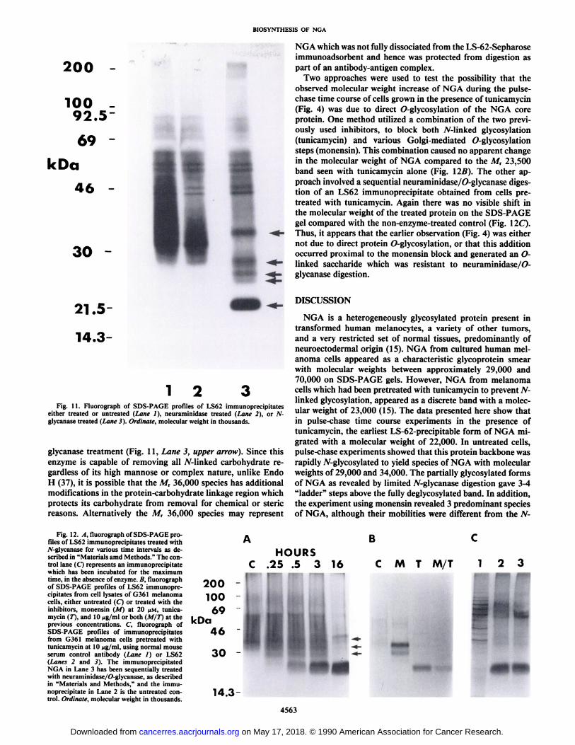
treated cells was run under identical conditions the core proteinmigrated off the basic end of the IEF gradient (data not shown).The heavy sialylation apparent from the 2-dimensional separation was confirmed when NGA immunoprecipitates weretreated with neuraminidase (Fig. 11, Lane 2). Much of theheterogeneity seen in the control track (Fig. 11, Lane 1) was
3.7 5.5
pH
6.7
Fig. 10. Fluorograph of 2-dimensional ISODALT gel of an LS62 immuno-precipitate separated according to pi and molecular weight. Ordinate, molecularweight in thousands.
removed by this enzyme treatment and gave a profile of NGAwhich was reminiscent of the result obtained with monensin(Fig. 1, Lane 2). Additional experiments using sequential lectin-affinity precipitation have shown that NGA exists in formswhich carry high mannose, hybrid type and/or complex N-linked oligosaccharide side chains.4 This heterogeneity mayrepresent biosynthetic intermediate forms of NGA during gly-coprotein processing or functionally different mature forms ofthe molecule.
When NGA immunoprecipitates were treated briefly with theenzyme jV-glycanase which removes AMinked oligosaccharideside chains (39) the predominant species was the core proteinat M, 23,500 (Fig. 11, Lane 3). In addition, the intermediate,partially digested forms of the glycoprotein can be visualized atmolecular weights of 25,000, 26,500 and 29,000 (arrows). It isbelieved that these represent three discrete additions of /V-linkedoligosaccharides to the protein moiety. When the enzyme wasallowed to digest NGA to completion it was seen that theseintermediate glycosylated forms disappeared to yield the M,23,500 core protein (Fig. 12/1). It was also noted that a M,36,000 component of NGA appeared to be resistant to the A7-
4 K. Ohyama, W. T. Dixon, L. K. J. Sikora, H. Muzik, and L. M. Jerry,
characterization of the carbohydrate structure of neuroglandulur antigen (NGA),a human melanoma-associated antigen, manuscript in preparation.
NGA which was not fully dissociated from the LS-62-Sepharoseimmunoadsorbent and hence was protected from digestion aspart of an antibody-antigen complex.
Two approaches were used to test the possibility that theobserved molecular weight increase of NGA during the pulse-chase time course of cells grown in the presence of tunicamycin(Fig. 4) was due to direct 0-glycosylation of the NGA coreprotein. One method utilized a combination of the two previously used inhibitors, to block both TV-linked glycosylation(tunicamycin) and various Golgi-mediated 0-glycosylationsteps (monensin). This combination caused no apparent changein the molecular weight of NGA compared to the M, 23,500band seen with tunicamycin alone (Fig. Ì2B).The other approach involved a sequential neuraminidase/0-glycanase digestion of an LS62 immunoprecipitate obtained from cells pre-treated with tunicamycin. Again there was no visible shift inthe molecular weight of the treated protein on the SDS-PAGEgel compared with the non-enzyme-treated control (Fig. 11C).Thus, it appears that the earlier observation (Fig. 4) was eithernot due to direct protein O-glycosylation, or that this additionoccurred proximal to the monensin block and generated an O-linked saccharide which was resistant to neuraminidase/O-glycanase digestion.
21.5-
14.3-
1 2Fig. 11. Fluorograph of SDS-PAGE profiles of LS62 immunoprecipitates
either treated or untreated (Lane /), neuraminidase treated (Lane 2), or 7V-glycanase treated (Lane 3). Ordinate, molecular weight in thousands.
glycanase treatment (Fig. 11, Lane 3, upper arrow). Since thisenzyme is capable of removing all TV-linked carbohydrate regardless of its high marinóse or complex nature, unlike EndoH (37), it is possible that the M, 36,000 species has additionalmodifications in the protein-carbohydrate linkage region whichprotects its carbohydrate from removal for chemical or stericreasons. Alternatively the M, 36,000 species may represent
Fig. 12. X, fluorograph of SDS-PAGE profiles of LS62 immunoprecipitates treated with/V-glycanase for various time intervals as described in "Materials amd Methods." The con
trol lane (C) represents an immunoprecipitatewhich has been incubated for the maximumtime, in the absence of enzyme. B, fluorographof SDS-PAGE profiles of LS62 immunoprecipitates from cell lysates of G361 melanomacells, either untreated (C) or treated with theinhibitors, monensin (M) at 20 //M. tunicamycin (T), and 10 ng/ml or both (M/T) at theprevious concentrations. C, fluorograph ofSDS-PAGE profiles of immunoprecipitatesfrom G361 melanoma cells pretreated withtunicamycin at 10 ¿ig/ml,using normal mouseserum control antibody (Lane I) or LS62(Lanes 2 and .<). The immunoprecipitatedNGA in Lane 3 has been sequentially treatedwith neuraminidase/O-glycanase, as describedin "Materials and Methods," and the immu
noprecipitate in Lane 2 is the untreated control. Ordinate, molecular weight in thousands.
DISCUSSION
NGA is a heterogeneously glycosylated protein present intransformed human melanocytes, a variety of other tumors,and a very restricted set of normal tissues, predominantly ofneuroectodermal origin (15). NGA from cultured human melanoma cells appeared as a characteristic glycoprotein smearwith molecular weights between approximately 29,000 and70,000 on SDS-PAGE gels. However, NGA from melanomacells which had been pretreated with tunicamycin to prevent N-linked glycosylation, appeared as a discrete band with a molecular weight of 23,000 (15). The data presented here show thatin pulse-chase time course experiments in the presence oftunicamycin, the earliest LS-62-precipitable form of NGA migrated with a molecular weight of 22,000. In untreated cells,pulse-chase experiments showed that this protein backbone wasrapidly 7V-glycosylated to yield species of NGA with molecularweights of 29,000 and 34,000. The partially glycosylated formsof NGA as revealed by limited 7V-glycanase digestion gave 3-4"ladder" steps above the fully deglycosylated band. In addition,
the experiment using monensin revealed 3 predominant speciesof NGA, although their mobilities were different from the N-
glycanase intermediate digestion products due to partial post-translational modification. It has been reported that individual^-linked saccharide moieties contribute M, 2,000-4,000 incremental shifts in molecular weights on SDS-PAGE gels (37),therefore the difference between the "naked" protein (M,
22,000) and the Mr 36,000 form of NGA would suggest 3-7 N-linked chains per polypeptide. Our results with both monensin(3 discrete bands) and N-glycanase (3-4 digestion intermediateladder steps) would support the lower end of this estimate.
Two-dimensional separations of NGA showed the considerable isoelectric point heterogeneity of the molecule, in additionto its wide apparent molecular weight range. The higher molecular weight species of NGA were also the most acidic in nature.This suggested that NGA might be rich in sialic acid residues,a conclusion that received support from the observed decreasein molecular weight heterogeneity of the molecule after neura-minidase treatment.
NGA appeared to be present at relatively low levels on thesurface of the plasma membrane of cultured melanoma cells byseveral experimental criteria, including vectorial iodination andFACS analysis, but was detected at higher levels intracellularly.The fact that NGA appears to be actively secreted into themedia of cultured melanoma cells suggests that the low level ofexpression seen on the cell surface may reflect NGA which isin transit through the plasma membrane. Immunocytochemicalstaining of fixed melanoma cells using MAb LS62 followed bya peroxidase-conjugated second antibody revealed a granularintracellular distribution of the antigen,5 reminiscent of secre
tory granules, or perhaps melanosomes (40). In relation to thislatter possibility, it is interesting that MAb ME491, whichappears to recognize a molecule very similar to NGA, wasobtained by immunizing mice with a crude melanosomal preparation from human melanoma cell lines (20). In contrast, MAbNK1/C3, which has been reported to recognize the same antigen as MAb ME491 (19), was developed by using purifiedplasma membranes from cultured human melanoma cells asthe immunogen (18). Using immunoelectron microscopy, theNKI/C3 reactive antigen was localized to melanosomes, vacuoles, and also the plasma membrane. In addition, experimentsusing a metabolic inhibitor, sodium azide, showed that therelease of the NKJ/C3 antigen into cell culture supernatantswas an active process, rather than a passive shedding mechanism. This suggested that a common membrane-associatedelement, either arising from, or giving rise to these differentorganelle fractions and ultimately released from the cell, perhaps as a melanosome, was the target for both ME491 andNKI/C3 antibodies. The exact relationship of these MAbs toLS62 is at present unknown; however, the molecules theyrecognize appear to share many common features. During thepreparation of this manuscript investigators using MAb NKI/C3 and a polyclonal rabbit antiserum directed against the sameantigen have obtained results with a variety of different techniques, which further support the identity of NGA with theantigen recognized by both NKI/C3 and ME491 (41).
In addition, investigators using the ME491 antibody havesucceeded in cloning and sequencing the gene which encodesthe ME491 antigen by an elegant series of experiments utilizingDNA-mediated gene transfer protocols (42). Their deducedamino acid sequence predicts the existence of four hydrophobictransmembrane domains in the protein, consistent with thenotion of a membrane-inserted location. Their estimate of 3potential sites for asparagine-linked TV-glycosylation in the
5V. Feyles, W. Dixon, R. McGarry, and L. M. Jerry, unpublished observations.
ME491 antigen, when compared to our estimate of 3-4 N-oligosaccharide moieties in NGA, provides additional indirectevidence that these are the same molecule.
ME491 antigen expression is considered a marker for earlystages of melanocytic tumor progression (21, 42). Such stagesof the lesion, including dysplastic nevus and radial growth phaseof melanoma, express the antigen strongly, whereas the laterstages of vertical growth phase and metastatic melanoma showlower or minimal expression (21). We have not seen comparablestage-specific expression of NGA in the striking way describedfor the ME491 antigen and elevated but variable levels of NGAare seen in all stages of progression from nevocellular nevus tometastatic melanoma (15).
We have previously suggested the possibility that NGA mightbe a marker for the proliferative status of cells. This assertionwas based on observations of NGA expression being high inrapidly dividing cultured melanoma patient fibroblasts in vitro,whereas the antigen was undetectable in quiescent counterpartsof the same cell type in vivo (15). Investigators using MAbME491 have also suggested a functional relationship betweenthe expression of the antigen and growth and proliferation.Their interpretation, however, is that the antigen and the genewhich encodes it may be involved in growth inhibition sincethey assert that expression is lower when the tumor progressesto more malignant, and faster growing stages (42). Some evidence in support of their hypothesis comes from another recentstudy in which the authors demonstrate the inhibition of ribo-somal RNA gene transcription in SW948 colorectal carcinomacells by internalized MAb ME491 (43). The suggest that theantibody may be mimicking an as yet unidentified growthfactor-like ligand which is internalized via its putative receptor,the ME491 antigen. We have obtained preliminary data usingsimultaneous indirect immunofluorescence and cell cycle analysis methods (data not shown) which demonstrate cell cyclecorrelated expression or secretion of NGA. If this molecule isindeed a growth factor receptor such temporally controlledexpression could conceivably play a role in regulating proliferation. We are currently isolating and characterizing complementary DNA clones for NGA expression by an alternativestrategy to that used to isolate the gene for ME491. We hopeto use the resulting structural information to address thesequestions concerning the function of NGA, and also to confirmor refute identity of the gene encoding NGA with that forME491/NKI/C3.
ACKNOWLEDGMENTS
We are grateful to H. Muzik, S. Nishikawa, L. Campbell, K. Kirton,and V. Nguyen for technical assistance. The authors thank Drs. R.McGarry, V. Feyles, and K. Maeda for helpful discussions and L.Strong for preparing the manuscript.
REFERENCES
1. Jerry, L. M., Masri, M., Phillips, T. M., Sikora, L., Jones, C, Dixon, W.,and Demetrick, D. Markers of human malignant melanoma: towards aBiology of the host-tumor response. In: A. S. Daar (ed.), Tumor Markers:Concepts and Clinical Applications, pp. 428-496. London: Blackwell, 1985.
2. Hersey, P. Review of melanoma antigens recognized by monoclonal antibodies (MAbs). Their functional significance and applications in diagnosis andtreatment of melanoma. Pathology, 17: 346-354, 1985.
3. Sulitzeanu, D. Human cancer-associated antigens: present status and implications for immunodiagnosis. Adv. Cancer Res., 44: 1-42, 1985.
4. Reisfeld, R. A., Harper, J. R., and Bumol, T. F. Human tumor-associatedantigens defined by monoclonal antibodies. Crit. Rev. Immunol., 55: 27-53,1984.
5. Reisfeld, R. A., and Cheresh, D. A. Human tumor antigens. Ad. Immunol.,40:323-377, 1987.
6. Hellstrom, J., and Hellstrom, K. E. Diagnostic uses of anti-melanomaantibodies: In: H. Z. Kupchik (ed.), Cancer Diagnosis in Vitro Using Monoclonal Antibodies, Immunology Series, Vol. 39, pp. 123-139. New York:Marcel Dekker, 1988.
7. Mattes, M. J., Real, F. X., Furukawa, K., Old, L. J., and Lloyd, K. O. ClassI (unique) tumor antigens of human melanoma: partial purification andcharacterization of the FD antigen and analysis of a mouse polyclonalantiserum. Cancer Res., 47: 6614-6619, 1987.
8. Real, F. X., Houghton, A. N., Albino, A. P., Cordon-Cardo, C, Melamed,M. R., Oettgen, H. F., and Old, L. J. Surface antigens of melanomas andmelanocytes defined by mouse monoclonal antibodies: specificity analysisand comparisons of antigen expression in cultured cells and tissues. CancerRes., 45: 4401-4411, 1985.
9. Mancianti, M. L., Herlyn, M., Neil, D., and Jambrosic, J., Rodeck, U.,Becker, D., Diamond, L., Clark, W. H., and Koprowski, H. Growth andphenotypic characterizations of human nevus cells in culture. J. Invest.Dermatol., 90: 134-141, 1988.
10. Real, F. X., Fliegel, B., and Houghton, A. N. Surface antigens of humanmelanoma cells cultured in serum-free medium: induction of expression ofmajor histocompatibility complex class II antigens. Cancer Res., 48: 686-693, 1988.
11. Houghton, A. N., Real, F. X., Davis, L. J., Cordon-Cardo, C., and Old, L.J. Phenotypic heterogeneity of melanoma: relation to the differentiationprogram of melanoma cells. J. Exp. Med., 164: 812-829, 1987.
12. Herlyn, M., Clark, W. H., Rodeck, U., Mancianti, M. L., Jambrosic, J., andKoprowski, H. Biology of disease: biology of tumor progression in humanmelanocytes. Lab. Invest., 56:461-474, 1987.
13. Lehmann, J. M., Holzmann, B., Brattar!, E. W., Schmiegelow, P., Rieth-muller, G., and Johnson, J. P. Discrimination between benign and malignantcells of melanocytic lineage by two novel antigens, a glycoprotein with amolecular weight of 113,000 and a protein with a molecular weight of 76,000.Cancer Res., 47: 841-845, 1987.
14. Holzmann, B., Brocker, E. B., Lehmann, J. M., Ruiter, D. J., Sorg, C.,Riethmuller, G., and Johnson, J. P. Tumor progression in human malignantmelanoma: five stages defined by their antigenic phenotypes. Int. J. Cancer,59:466-471, 1987.
15. Sikora, L. K. J., Pinto, A., Demetrick, D. J., Dixon, W. T., Urbanski, S. J.,Temple, W., and Jerry, L. M., Characterization of a novel neuroglandularantigen (NGA) expressed on abnormal human melanocytes. Int. J. Cancer,39: 138-145, 1987.
16. Brown, J. P., Wright, P. W., Hart, C. E., Woodbury, R. G., Hellstrom, K.E., and Hellstrom, I. Protein antigens of normal and malignant human cellsidentified by immunoprecipitation with monoclonal antibodies. J. Biol.Chem., 255:4980-4983, 1980.
17. Mackie, R. M., Campbell, I., and Turbiti, M. L. Use of NKI C3 monoclonalantibody in the assessment of benign and malignant melanocytic lesions. J.Clin. Pathol., 37: 367-372, 1984.
18. Vennegoor, C., Calafat, J., Hageman, P. L., van Buitenen, F., Janssen, H.,Kolk, A., and Rumke, P. L. Biochemical characterization and cellular localization of a formalin-resistant melanoma-associated antigen reacting withmonoclonal antibody NKI/C-3. Int. J. Cancer, 35: 287-295, 1985.
19. Vennegoor, C., and Rumke, P. Circulating melanoma-associated antigendetected by monoclonal antibody NKI/C-3. Cancer Immunol. Immunother.,25:93-100, 1986.
20. Atkinson, B., Ernst, C. S., and Ghrist, B. F. D., Heerlyn, M., Blaszczyk, M.,Ross, A. H., and Heerlyn, D., Steplewski, Z., and Koprowski, H. Identification of melanoma-associated antigen using fixed tissue screening of antibodies. Cancer Res., 44: 2577-2581, 1984.
21. Atkinson, B., Ernst, C. S., Ghrist, B. F. D., Ross, A. H., Clark, W. H.,Heerlyn, M., Heerlyn, D., Maul, G., Steplewski, Z., and Koprowski, H.Monoclonal antibody to a highly glycosylated protein reacts in fixed tissuewith melanoma and other tumors. Hybridoma, 4: 243-255, 1985.
22. Ross, A. H., Dietzshold, B., Jackson, D. M., Earley, J. J., Jr., Ghrist, B. D.F., Atkinson, B., and Koprowski, H. Isolation and amino terminal sequencing
of a novel melanoma-associated antigen. Arch. Biochem. Biophys., 242:540-548, 1985.
23. Ernst, C. S., Shen, J-W., Litwin, S., Heerlyn, M., Koprowski, H., and Sears,H. F. Multiparameter evaluation of the expression in situ of normal andtumor-associated antigen in human colorectal carcinoma. J. Nati. CancerInst., 77:387-395, 1986.
24. Ross, A. H., Pleasure, D., Sonnenfeld, K., Atkinson, B., Kreider, B., Jackson,D. M., Taff, I., Scarpini, E., Lisak, R. P., and Koprowski, H. Expression ofmelanoma-associated antigens by normal and neurofibroma schwann cells.Cancer Res., 46: 5887-5892, 1986.
25. Donoso, L. A., Folberg, R., Edelberg, K., Arbizo, V., Atkinson, B., andHerlyn, M. Tissue distribution and biochemical properties of an ocularmelanoma-associated antigen. J. Histochem. Cytochem., 33: 1190-1196,1985.
26. Donoso, L. A., Felberg, N. T., and Edelberg, K. Metastatic uveal melanoma:an ocular melanoma-associated antigen in the serum of patients with met-astatic disease. J. Immunoassay, 7: 273-283, 1986.
27. Salacinski, P. R. P., McLean, C., Sykes, J. E. C., Clement-Jones, V. V., andLowry, P. J. lodination of proteins, glycoproteins and peptides using a solid-phase oxidizing agent, 1,3,4,6-tetrachloro-3,6-diphenyl glycoluril (lodogen).Anal. Biochem., 117: 136-146, 1981.
28. Laemmli, U. K. Cleavage of structural proteins during the assembly of thehead of bacteriophage T4. Nature (Lond.), 227: 665-666, 1970.
29. O'Farrell, P. H. High resolution two-dimensional electrophoresis of proteins.J. Biol. Chem., 250:4007-4021, 1975.
30. Laskey, R. A., and Mills, A. D. Quantitative film detection of 3H and 14, inpolyacrylamide gels by fluorography. Eur. J. Biochem., 56:334-341, 1975.
31. Zipf, T. F., Lauzon, G. J., and Longenecker, B. M. A monoclonal antibodydetecting a 39,000 molecular weight molecule that is present on B lymphocytes and chronic lymphocytic leukemia cells but is rare on acute lymphocyticleukemia blasts. J. Immunol., 131: 3064-3072, 1983.
32. Elbein, A. D. Inhibitors of the biosynthesis and processing of /V-linkedoligosaccharide chains. Annu. Rev. Biochem., 56: 497-534, 1987.
33. Griffiths, G., Quinn, P., and Warren, G. Dissection of the Golgi complex. I.Monensin inhibits the transport of viral membrane proteins from medial totrans Golgi cisternae in baby hamster kidney cells infected with Semlikiforest virus. J. Cell Biol., 96: 835-850, 1983.
34. Dunphy, W. G., and Rothman, J. E. Compartmental organization of theGolgi stack. Cell, 42: 13-21, 1985.
35. Herlyn, M., Rodeck, U., and Koprowski, H. Shedding of human tumor-associated antigens in vitro and in vivo. Adv. Cancer Res., 49:189-221,1987.
36. Rodriguez-Boulan, E., Miske, D. E., Salas, D. V., Salas, P. J. I., ad Bard, E.Protein sorting in the secretory pathway. Curr. Top. Membr. Transp. 24:251-294, 1985.
37. Lewis, V., Green, S. A., Marsh, M., Vihko, P., Helenius, A., and Mellman,I. Glycoproteins of the lysosomal membrane. J. Cell Biol., 700: 1839-1847,1985.
38. Lampson, L., and Levy, R. Two populations of la-like molecules on a humanB cell line. J. Immunol., /25: 293-298, 1980.
39. Maley, F., Trimble, R. B., Tarentino, A. L., and Plummer, T. H. Characterization of glycoproteins and their associated oligosaccharides through theuse of endoglycosidases. Anal. Biochem., 180: 195-204, 1989.
40. Hoyt, R. F., Sorokin, S. P., McDowell, E. M., and Trump, B. F. Periodicacid-Schiff-lead hematoxylin as a marker for the endocrine phenotype inhuman lung tumors. Arch. Pathol. Lab. Med., 110: 943-951, 1986.
41. Gruters, R. A., Calafat, J., Vennegoor, C. J. G. M., Jansen, H., and Ploegh,H. L. Structural heterogeneity of a human melanoma-associated antigen.Cancer Res., 49: 459-465, 1989.
42. Hotta, H., Ross, A. H., Huebner, K., Isobe, M., Wendeborn, S., Chao, M.V., Rkriardi, R. P., Tsujimoto, Y., Croce, C., and Koprowski, H. Molecularcloning and characterization of an antigen associated with early stages ofmelanoma tumor progression. Cancer Res., 48: 2955-2962, 1988.
43. Rackowicz-Szulcznska, E. M., and Koprowski, H. Nuclear uptake of monoclonal antibody to a surface glycoprotein and its effect on transcription. Arch.Biochem. Biophys., 277: 366-379, 1989.
1990;50:4557-4565. Cancer Res Walter T. Dixon, Douglas J. Demetrick, Kunio Ohyama, et al. AntigenNeuroglandular Antigen, a Human Melanoma-associated Biosynthesis, Glycosylation and Intracellular Processing of the