Biphasic mechanosensitivity of TCR mediated adhesion of T lymphocytes A. Wahl 1† , C. Dinet 1‡ , P. Dillard 1,2! , P-H. Puech 2 , L. Limozin 2 , K. Sengupta 1* 1 CINaM AMU-CNRS, UMR 7325, Campus de Luminy, Case 913, Luminy, Marseille, France. 2 Lab. Adhesion & Inflammation INSERM UMR 1067/ CNRS UMR 7333/ AMU UM 61, Case 937, Luminy, Marseille, France. *Correspondence to: [email protected]Current address: † Laboratoire de Chimie Bactérienne, UMR7283, Institut de Microbiologie de la Méditerranée, Centre National de la Recherche Scientifique, Aix-Marseille Université, Marseille, France. ‡ Dept. of Physics, Durham University, South Rd, Durham DH1 3LE, UK. ! Department of Cellular Therapy, Oslo University Hospital – Norwegian Radium Hospital, Norway. Force sensitivity of the T cell receptor (TCR) is now believed to be essential for immune recognition, but cellular mechanosensitivity of T cells is still poorly understood. Here we show that T cells adhering via the TCR-complex respond to environmental stiffness in an unusual biphasic fashion. As the stiffness increases, adhesion and spreading first increase, then decrease, attaining their maximal values on an optimally stiff surface, with stiffness comparable to certain antigen presenting cells. Remarkably, in presence of additional ligands for the integrin LFA-1, spreading increases monotonously with stiffness up to a saturation value. Using a mesoscopic semi-analytical model linking spreading to molecular characteristics of bonds, we identify force sensitivity of the off-rate and the effective bond stiffness as the crucial parameters that determine monotonic or biphasic mechanosensitive behavior. (which was not peer-reviewed) is the author/funder. All rights reserved. No reuse allowed without permission. The copyright holder for this preprint . http://dx.doi.org/10.1101/232041 doi: bioRxiv preprint first posted online Dec. 10, 2017;
Transcript
Biphasic mechanosensitivity of TCR mediated adhesion of T lymphocytes
A. Wahl1†, C. Dinet1‡, P. Dillard1,2!, P-H. Puech2, L. Limozin2, K. Sengupta1*
1 CINaM AMU-CNRS, UMR 7325, Campus de Luminy, Case 913, Luminy, Marseille, France.
2 Lab. Adhesion & Inflammation INSERM UMR 1067/ CNRS UMR 7333/ AMU UM 61, Case
Current address: † Laboratoire de Chimie Bactérienne, UMR7283, Institut de Microbiologie de
la Méditerranée, Centre National de la Recherche Scientifique, Aix-Marseille Université,
Marseille, France. ‡ Dept. of Physics, Durham University, South Rd, Durham DH1 3LE, UK.
! Department of Cellular Therapy, Oslo University Hospital – Norwegian Radium Hospital,
Norway.
Force sensitivity of the T cell receptor (TCR) is now believed to be essential for immune
recognition, but cellular mechanosensitivity of T cells is still poorly understood. Here we show
that T cells adhering via the TCR-complex respond to environmental stiffness in an unusual
biphasic fashion. As the stiffness increases, adhesion and spreading first increase, then decrease,
attaining their maximal values on an optimally stiff surface, with stiffness comparable to certain
antigen presenting cells. Remarkably, in presence of additional ligands for the integrin LFA-1,
spreading increases monotonously with stiffness up to a saturation value. Using a mesoscopic
semi-analytical model linking spreading to molecular characteristics of bonds, we identify force
sensitivity of the off-rate and the effective bond stiffness as the crucial parameters that determine
monotonic or biphasic mechanosensitive behavior.
(which was not peer-reviewed) is the author/funder. All rights reserved. No reuse allowed without permission. The copyright holder for this preprint. http://dx.doi.org/10.1101/232041doi: bioRxiv preprint first posted online Dec. 10, 2017;
Mechanosensitivity has emerged as a hallmark of many biological systems – from molecular to
tissue level, and is implicated in health and disease (1–3). Soft and deformable substrates are
used to study the impact of environmental stiffness on cells and to measure cell-generated forces.
Pioneering single cell studies showed that for fibroblast cells grown on gels of various stiffness,
locomotion and focal adhesions are regulated by substrate flexibility (4). Mechano-sensitivity
has now been demonstrated for almost all cell types. Readouts include extent of spreading, cell
stiffness, motility, differentiation and traction forces (5–7), and all typically increase
monotonically with substrate stiffness, usually reaching a saturation value at a particular value of
the stiffness. From a theoretical point of view, adhesion and mechanosensing can be treated
either within cell-scale macroscopic models (3) that usually predict a monotonous increase in
cell spreading and traction with stiffness, or using microscopic models that account for molecular
mechanosensitivity (8), and may predict biphasic behavior in force or velocity (9). Experimental
reports of such biphasic behavior is very rare for cells adhering to a surface, examples include
speed of migrating neutrophils (10), actin retrograde flow (8) and traction forces in beating
muscle cells (11) or talin-silenced fibroblasts (12), the adhesion area still being monotonous
whenever reported.
A vast majority of experimental studies on mechanotransduction are conducted on focal adhesion
forming cells. Leucocytes in general and lymphocytes in particular have been much less studied,
despite evidence that T cells response is sensitive to substrate stiffness (13, 14), but contradictory
trends were been reported (15, 16), which probably hinted at a non-monotonicity. The ability of
T cells to recognize pathogens and pathological cells is now known to depend on their
(which was not peer-reviewed) is the author/funder. All rights reserved. No reuse allowed without permission. The copyright holder for this preprint. http://dx.doi.org/10.1101/232041doi: bioRxiv preprint first posted online Dec. 10, 2017;
mechanosensitivity (13, 14), which implicates the CD3 domain of the T cell receptor (TCR)
complex (17). Here we explore mechanosensing in T cells via the TCR-CD3 complex, in
presence or absence of the ligand ICAM-1 for the T cell integrin LFA-1. We follow the
spreading of Jurkat T cells, which on this cell type is a marker of activation (18), on
functionalized surfaces of polydimethylsiloxane- based (PDMS silicone) elastomers with
stiffness (quantified in terms of Young’s modulus) ranging from 500 Pa to several MPa. To
cover such a large range while retaining similar surface chemistry, different silicone types with
adapted base/cross-linker ratio were prepared and characterized in terms of their Young’s
modulus from force curves obtained using atomic force microscopy nano indentations. The
PDMS surface was then functionalised with either anti-CD3 alone with estimated molecular
coverage of about ~ 400 molecules/µm2, or, additionally with ICAM-1 at similar coverage
(Table S1).
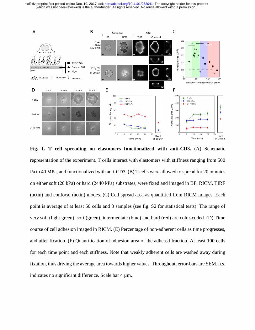
In a first set of experiments, T cells were allowed to interact with anti-CD3 functionalized
surfaces (Fig. 1A) for twenty minutes, allowing them enough time to spread but not retract. They
were then fixed and stained for actin, TCR or the zeta-chain-associated protein kinase 70 (ZAP-
70 – one of the first molecules to be recruited to the TCR complex upon TCR engagement). Cells
were imaged in bright field, reflection interference contrast microscopy (RICM), total internal
reflection fluorescence (TIRF) and confocal microscopy (Fig. 1B and fig. S1). As can be
immediately seen, cells spread more on 20 kPa than on 2440 kPa elastomers but the actin is
peripherally distributed in both cases. RICM images were analyzed to quantify the cell spreading
area (19–21) and the actin TIRF images to quantify the extent of actin peripherality. Fig 1C and
fig. S2 summarize the final cell area measured for different elastomer stiffness. Note that for the
(which was not peer-reviewed) is the author/funder. All rights reserved. No reuse allowed without permission. The copyright holder for this preprint. http://dx.doi.org/10.1101/232041doi: bioRxiv preprint first posted online Dec. 10, 2017;
same stiffness, changing the silicone type does not affect cell area. Cells on the softest elastomer
(QGel 920 1:0.95 at 0.5 kPa) spread moderately to an area of about 200 µm2. The area increases
as the stiffness is increased to 1 kPa (CY52 1:1) and then to 4 and 5 kPa (QGel 920 1:1.1 and
Sylgard 184 1:58, called soft), reaching a maximum of about 300 µm2. Thereafter the cell spread
area decreases with increasing stiffness, falling back to roughly 200 µm2 for 2 MPa (Sylgard 184
1:10, called hard) and to less than 150 µm2 at 7 MPa (Sylgard 184 1:10, rigidified by plasma-
treatment). On equivalently functionalized glass, provided that non-specific interactions are fully
blocked, here using ligands immobilized on supported lipid bilayers (21), the cells spread to a
mere 120 µm2 (without full blocking, area on anti-CD3 coated glass is high at ~300 µm2). This
remarkable spreading behavior is reproduced in human primary CD4+ T cells, which spread
more on soft than on hard PDMS substrates (fig. S3).
T cells tend to spread isotropically on functionalized glass resulting in a roughly circular shape,
whereas on the elastomers studied here, they may exhibit irregular star-like shapes (fig. S4). The
shape of a cell is mainly determined by its actin cytoskeleton. The distribution of actin at the
adhesive interface of a fully spread T cell tends to be peripheral, but the peripherality becomes
less pronounced in weakly spread cells (21, 22). Here, a peripheral ring-like distribution is seen
for all stiffness values (Fig. 1B, fig. S1, quantified in fig. S5), even when the cell area is
relatively small. The recruitment of TCR and ZAP-70 kinase imaged in TIRF do not show
appreciable variations with stiffness (fig S1), hinting at a mechanistic rather than signalling
based mechanosensing mechanism.
(which was not peer-reviewed) is the author/funder. All rights reserved. No reuse allowed without permission. The copyright holder for this preprint. http://dx.doi.org/10.1101/232041doi: bioRxiv preprint first posted online Dec. 10, 2017;
Fig. 1. T cell spreading on elastomers functionalized with anti-CD3. (A) Schematic
representation of the experiment. T cells interact with elastomers with stiffness ranging from 500
Pa to 40 MPa, and functionalized with anti-CD3. (B) T cells were allowed to spread for 20 minutes
on either soft (20 kPa) or hard (2440 kPa) substrates, were fixed and imaged in BF, RICM, TIRF
(actin) and confocal (actin) modes. (C) Cell spread area as quantified from RICM images. Each
point is average of at least 50 cells and 3 samples (see fig. S2 for statistical tests). The range of
very soft (light green), soft (green), intermediate (blue) and hard (red) are color-coded. (D) Time
course of cell adhesion imaged in RICM. (E) Percentage of non-adherent cells as time progresses,
and after fixation. (F) Quantification of adhesion area of the adhered fraction. At least 100 cells
for each time point and each stiffness. Note that weakly adherent cells are washed away during
fixation, thus driving the average area towards higher values. Throughout, error-bars are SEM. n.s.
indicates no significant difference. Scale bar 4 µm.
(which was not peer-reviewed) is the author/funder. All rights reserved. No reuse allowed without permission. The copyright holder for this preprint. http://dx.doi.org/10.1101/232041doi: bioRxiv preprint first posted online Dec. 10, 2017;
We next explored the dynamics of T cells on soft (5 kPa), intermediate (130 kPa) and hard (2.5
MPa) substrates. Fig. 1D shows an example of single cell time-lapse RICM demonstrating that
the cells on hard substrates lag behind in spreading already in the time window 0 to 5 minutes, a
period shown earlier to be critical for antigen recognition (18). Fig. 1E and F quantify this effect
at the scale of the population. It is seen that on hard substrates there is a population of non-
adherent cells that never adhere (Fig. 1E). Furthermore, the cells that do adhere, spread much
less on the hard substrate (Fig. 1F). The time evolution of the actin organization is similar in the
two cases (fig. S6).
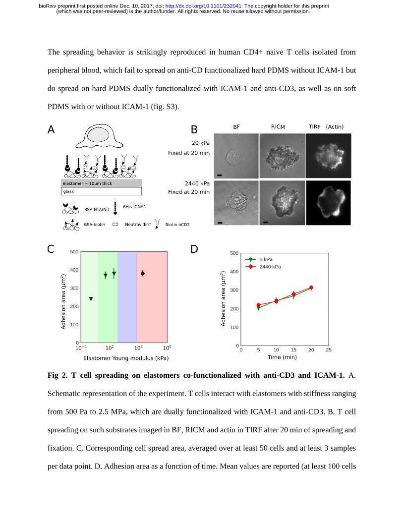
In the next set of experiments, the role of LFA-1 was explored by dual functionalization of the
substrates with anti-CD3 and ICAM-1. As negative control, and consistent with past reports of
cells on glass supports with only ICAM-1 on surface (21), there is no adhesion on soft (5 kPa) or
hard (2MPa) PDMS (fig. S7). Interestingly, this is also the case when cells are simultaneously
stimulated with anti-CD3 in solution. For the case of co-functionalization with both anti-CD3 and
ICAM-1 (Fig. 2A), the cells spread the least on the softest substrates used and they spread more
as the substrate stiffness is increased, already reaching a saturation value of about 400 µm2 in ~kPa
range (earlier designated as soft, Fig. 2B,C). Thus the maximal area reached here is somewhat
larger than that with anti-CD3 alone at 300 µm2. Analysis of time-lapse RICM data shows that the
spreading dynamics does not differ between the 5 kPa and 2.5 MPa cases (Fig. 2D). In terms of
actin organization, no clear difference is detected between soft and hard on one hand and with or
without ICAM-1 on the other hand; the actin is mainly peripheral in all cases (fig. S5).
(which was not peer-reviewed) is the author/funder. All rights reserved. No reuse allowed without permission. The copyright holder for this preprint. http://dx.doi.org/10.1101/232041doi: bioRxiv preprint first posted online Dec. 10, 2017;
The spreading behavior is strikingly reproduced in human CD4+ naive T cells isolated from
peripheral blood, which fail to spread on anti-CD functionalized hard PDMS without ICAM-1 but
do spread on hard PDMS dually functionalized with ICAM-1 and anti-CD3, as well as on soft
PDMS with or without ICAM-1 (fig. S3).
Fig 2. T cell spreading on elastomers co-functionalized with anti-CD3 and ICAM-1. A.
Schematic representation of the experiment. T cells interact with elastomers with stiffness ranging
from 500 Pa to 2.5 MPa, which are dually functionalized with ICAM-1 and anti-CD3. B. T cell
spreading on such substrates imaged in BF, RICM and actin in TIRF after 20 min of spreading and
fixation. C. Corresponding cell spread area, averaged over at least 50 cells and at least 3 samples
per data point. D. Adhesion area as a function of time. Mean values are reported (at least 100 cells
(which was not peer-reviewed) is the author/funder. All rights reserved. No reuse allowed without permission. The copyright holder for this preprint. http://dx.doi.org/10.1101/232041doi: bioRxiv preprint first posted online Dec. 10, 2017;
for each time point and each stiffness, average area is slightly over-estimated due to 30 to 40 non-
adherent cells ignored for the analysis). Error bars are SEM. Scale bar 4 µm.
We next prepared soft, intermediate and hard substrates, dually functionalized with anti-CD28
and anti-CD3 in equi-molar ratio, to assess the impact of engaging the co-stimulatory molecule
CD-28. Interestingly, the presence of anti-CD28 has no impact on the cell area, which remains
unchanged with respect to the case without anti-CD28 (fig. S8). This is consistent with earlier
work suggesting T cell mechanosensing is actuated through CD3 rather than CD28 (17). Since
acto-myosin generated forces are thought to be at the heart of mechanosensing, we tested the
effect of inhibiting myosin IIA activity through the drug blebbistatin. It was shown earlier that in
case of cells that spread weakly on mobile ligands, myosin treatment rescues spreading (21).
However, surprisingly, no effect of myosin IIA inhibition was observed here (fig. S9).
The lack of a role for myosin, a major player in traditional clutch based models (8), led us to
identify actin polymerization generated forces as the basis for theoretical description of
mechanosensitive spreading. In the past, mesoscopic models have linked actin polymerization
driven cellular scale spreading (21) or traction force generation by a filopodium pulled by optical
tweezers (23), to force dependence of bond kinetics. Here we couple the two approaches to
model the edge of the cell where lamellipodia-like protrusions get pushed forward by actin
polymerization just behind the membrane (Fig 3A). Within a quasi-one-dimensional model of
such a cell edge, the actin is modelled as a strip which polymerizes at a given velocity, and
pushes the frontier of the cell forward, at the same time generating a retrograde flow of the actin
(which was not peer-reviewed) is the author/funder. All rights reserved. No reuse allowed without permission. The copyright holder for this preprint. http://dx.doi.org/10.1101/232041doi: bioRxiv preprint first posted online Dec. 10, 2017;
away from the edge with a velocity vp (Fig 3B). The final cell spreading area (A) is then set by a
balance of the actin generated force (F, expressed as force density) and a tensile force that limits
spreading, such that F is proportional to A (see Supplementary Materials).
The ligands are tightly bound to the elastic substrate and form bonds with cellular
transmembrane receptors, whose intracellular moieties interact with actin via adapter proteins
that are mostly known for integrins but are still a matter of debate for TCR. For simplicity, the
entire complex is modelled here as a single spring-like bond (henceforth called a linker, Fig. 3C).
F is transmitted to these bonds such that the force f on a single bond is F/(nN) where n is the
bound fraction, and N the ligand density. The bond kinetics is defined by a constant on-rate (kon)
and a force dependent off-rate given by B
f/f0
offoffekk where 0
offk is the off-rate at zero force and
f B is the characteristic Bell force at which a bond becomes force sensitive. The linker has an
intrinsic bond elasticity kb which is renormalized by the substrate elasticity (E) in such a way that
it increases with E, reaching a saturation when the value of E exceeds kb/a, a being a molecular
length scale (23). Note that for anti-CD3 alone (henceforth called TCR case), the linker
characteristics may be measurable from single-molecule experiments. In presence of ICAM-1
(henceforth called TCR+LFA-1) however, a single effective linker represents both TCR and
LFA-1 mediated bonds – the attributed molecular parameters are effective quantities that
embody the real parameters corresponding to each linker type as well as possible cooperative
effects.
The model self-consistently determines F and n, for given N and vp values which were measured
independently. A fit of the model to area data is able to capture the experimentally observed
stiffness dependence (Fig. 3D). For a given vp, both F and f initially increase with E via the
renormalization of kb (Fig. 3E,F). However, when f becomes appreciably larger than f B, bonds
(which was not peer-reviewed) is the author/funder. All rights reserved. No reuse allowed without permission. The copyright holder for this preprint. http://dx.doi.org/10.1101/232041doi: bioRxiv preprint first posted online Dec. 10, 2017;
start breaking faster than they form and n drops, leading to a drop in F at E>E*, even though f
continues to increase with E. In the TCR case, the bonds are predicted to be stiff and very
sensitive to force (high kb and low f B), ensuring that koff overtakes kon before E reaches kb/a. In
the case of TCR+LFA1 however, kb saturates before E* can be attained. The existence of E* is
an indicator of whether or not the force, and therefore the area, exhibits biphasic behavior. Fig.
S10 show plots of E* as a function of the parameters kb, f B, and 0
offk (keeping kon constant). It is
again seen that independent of 0
offk , high kb and low f B are essential for biphasic behavior. From
the fits, the value of f B is expected to be about 0.2 pN for TCR and about the double for
TCR+LFA1.
Fig 3. The model and fit to data. A. Schematic representation of the model of T cell spreading
at the cell scale. B. Mesoscopic view where cells interact with elastomers through bonds of one
or two types, that link the actin to the substrate. C. The entire actin-receptor-ligand-substrate
(which was not peer-reviewed) is the author/funder. All rights reserved. No reuse allowed without permission. The copyright holder for this preprint. http://dx.doi.org/10.1101/232041doi: bioRxiv preprint first posted online Dec. 10, 2017;
connection is represented as a single effective linker. D. Fit of model to normalized area data. E.
Force density in Pascal. F. Force on a single bond and the fraction of receptors that are bound.
Previous single-molecule probing of TCRs on T cells (24) yielded different force response
compared to cell-free experiments with purified TCRs (25). Furthermore, the characteristic force
f B for a typical ligand-receptor bond is expected to be in the pN range. This leads us to speculate
that the highly force-sensitive bond described above for the TCR case is in fact the previously
mooted CD3 to actin link (21, 26), rather than the ligand-receptor (anti-CD3/TCR) bond. On
integrin engagement, this putative link is probably reinforced and the effective link is less force
sensitive. Interestingly, measurements in the range of elasticity spanning up to few kPa do report
higher traction forces and activation on stiffer substrates (13, 15, 27). This range, roughly
coinciding with the first rising phase of our biphasic curve, is also the physiological range of
elasticity for professional antigen presenting cells, which may be modified under pathological
conditions (28). Whether T cells exploit the potentially biphasic response of the TCR-CD3
complex for their physiological function remains to be explored, but the putative CD3-actin link
proposed here as highly force sensitive should inform future studies on T cell mechanoresponse.
References and Notes
1. D. E. Discher, P. Janmey, Y.L. Wang, Tissue Cells Feel and Respond to the Stiffness of
Their Substrate. Science 310, 1139–1143 (2005).
2. V. Vogel, M. Sheetz, Local force and geometry sensing regulate cell functions. Nat Rev
Mol cell Biol. 7, 265–275 (2006).
3. U. S. Schwarz, S. A. Safran, Physics of adherent cells. Rev Mod Phys. 85, 1327–1381
(2013).
4. R. J. Pelham, Y. l Wang, Cell locomotion and focal adhesions are regulated by substrate
flexibility. Proc Natl Acad Sci U S A. 94, 13661–-13665 (1997).
5. A. J. Engler, S. Sen, H. L. Sweeney, D. E. Discher, Matrix elasticity directs stem cell
(which was not peer-reviewed) is the author/funder. All rights reserved. No reuse allowed without permission. The copyright holder for this preprint. http://dx.doi.org/10.1101/232041doi: bioRxiv preprint first posted online Dec. 10, 2017;
6. J. Solon, I. Levental, K. Sengupta, P. C. Georges, P. A. Janmey, Fibroblast Adaptation and
Stiffness Matching to Soft Elastic Substrates. Biophys J. 93, 4453–4461 (2007).
7. A. Zemel, F. Rehfeldt, A. E. X. Brown, D. E. Discher, S. A. Safran, Optimal matrix
rigidity for stress-fibre polarization in stem cells. Nat Phys. 6, 468–473 (2010).
8. C. E. Chan, D. J. Odde, Traction Dynamics of Filopodia on Compliant Substrates. Science
322, 1687–1691 (2008).
9. B. L. Bangasser, S. S. Rosenfeld, D. J. Odde, Determinants of maximal force transmission
in a motor-clutch model of cell traction in a compliant microenvironment. Biophys J. 105,
581–592 (2013).
10. K. M. Stroka, H. Aranda-Espinoza, Neutrophils display biphasic relationship between
migration and substrate stiffness. Cell Motil Cytoskeleton. 66, 328–341 (2009).
11. S. R. Peyton, A. J. Putnam, Extracellular matrix rigidity governs smooth muscle cell
motility in a biphasic fashion. J Cell Physiol. 204, 198–209 (2005).
12. A. Elosegui-Artola et al., Mechanical regulation of a molecular clutch defines force
transmission and transduction in response to matrix rigidity. Nat Cell Biol. 18, 540–548
(2016).
13. M. Saitakis et al., Different TCR-induced T lymphocyte responses are potentiated by
stiffness with variable sensitivity. Elife. 6, e23190 (2017).
14. K. H. Hu, M. J. Butte, T cell activation requires force generation. J Cell Biol,
jcb.201511053 (2016).
15. E. Judokusumo, E. Tabdanov, S. Kumari, M. L. Dustin, L. C. Kam, Mechanosensing in T
lymphocyte activation. Biophys J. 102, L5--L7 (2012).
16. R. S. O’Connor et al., Substrate Rigidity Regulates Human T Cell Activation and
Proliferation. J Immunol. 189, 1330–1339 (2012).
17. K. T. Bashour et al., CD28 and CD3 have complementary roles in T-cell traction forces.
Proc Natl Acad Sci U S A. 111, 2241–2246 (2014).
18. A. Brodovitch et al., T lymphocytes need less than 3 min to discriminate between peptide
MHCs with similar TCR-binding parameters. Eur J Immunol. 45, 1635–1642 (2015).
19. K. Sengupta, H. Aranda-Espinoza, L. Smith, P. Janmey, D. Hammer, Spreading of
neutrophils: from activation to migration. Biophys J. 91, 4638–4648 (2006).
20. L. Limozin, K. Sengupta, Quantitative reflection interference contrast microscopy (RICM)
in soft matter and cell adhesion. ChemPhysChem. 10, 2752–2768 (2009).
21. P. Dillard, R. Varma, K. Sengupta, L. Limozin, Ligand-mediated friction determines
morphodynamics of spreading T cells. Biophys J. 107, 2629–2638 (2015).
22. J. Hong, S. Murugesan, E. Betzig, J. A. Hammer, Contractile actomyosin arcs promote the
activation of primary mouse T cells in a ligand-dependent manner. PLoS One. 12, 1–22
(2017).
23. P. Sens, Rigidity sensing by stochastic sliding friction. Europhysics Lett. 104, 38003
(which was not peer-reviewed) is the author/funder. All rights reserved. No reuse allowed without permission. The copyright holder for this preprint. http://dx.doi.org/10.1101/232041doi: bioRxiv preprint first posted online Dec. 10, 2017;
28. N. Bufi et al., Human primary immune cells exhibit distinct mechanical properties that are
modified by inflammation. Biophys J. 108, 2181–2190 (2015).
Acknowledgments: We thank Anne Charrier for help and guidance for elastomer stiffness
measurements and critical reading of the manuscript, M. Biarnes-Pelicot for cell culture and
isolation, CINaM-PLANET for profilometer measurements, the PCC culture facility and L.
Borge for cells, Anais Sadoum for complementary experiments, Emmanuelle Bouveret for
generous gift of YFP-his and Pierre Sens for fruitful discussions. We also thank Pierre Bongrand
for stimulating discussions and critical reading of the manuscript. This work was partially funded
by European Research Council via grant no. 307104 FP/2007-2013/ERC-Stg SYNINTER.
Additional data and text can be found in the Supplementary Materials.
(which was not peer-reviewed) is the author/funder. All rights reserved. No reuse allowed without permission. The copyright holder for this preprint. http://dx.doi.org/10.1101/232041doi: bioRxiv preprint first posted online Dec. 10, 2017;