257 Neurology India | May-Jun 2009 | Vol 57 | Issue 3 Address for correspondence: Dr. Basant K Misra, Department of Neurosurgery and Gamma Knife Surgery, P D Hinduja National Hospital and Medical Research Centre, VS Marg, Mahim, Mumbai-400 016, India. E-mail: [email protected]DOI: 10.4103/0028-3886.53263 Introduction Since its rst successful removal, the history and progress of vestibular schwannoma (VS) management parallels that of neurosurgery. [1] Thus it is but natural that the present day trend of minimal invasiveness should also have a major inuence in the management of a patient with VS. In fact, Cushing’s recommendation of subtotal excision in VS is as old as neurosurgery itself. [2] While the debate regarding the best microsurgical approach to a given VS continues, it has been surpassed by the debate about the role of observation, gamma knife radiosurgery (GKR) and microsurgery in the mana gement of VS. [3] The changes that have taken place in the management of VS in our practice over the last two decades as well as the current protocol and results are presented here. Materials and Methods An analysis of 909 cases of cerebellopontine angle lesions operated by the rst author since 1987 yielded 559 cases of VS ope rated at two institutes til l 2008. Microsurgery was the primary option in 438 and GKR was done in 139, of which 18 were previously operated by the authors. A prescription dose of 13 Gray (Gy) to 50% isodose Curr ent treatment strategy in the management of vestibular schwannoma Basant K Misra, Harshad R Purandare, Rahul S Ved, Anshul A Bagdia, Pandurang B Mare Department of Neurosurgery and Gamma Knife Surgery, P D Hinduja National Hospital and Medical Research Centre, VS Marg, Mahim, Mumbai-400 016, India Abstract Background: The changing trends in the management of vestibular schwannoma (VS) in our practice over the last two decades as well as the current status are presented here. Materials and Methods: The observat ions are based on the experience of 559 consecutive cases of VS operated by the first author between 1987 and 2008, 438 of which were operated by microsurgery and 139 by gamma knife radiosurgery (GKR) (18 ofwhich were previously operated by the authors). A detailed analysis of microsurgicallymanaged patients in two different periods (100 consecutive patients each before 1993 and 2008) were compared to see the changing trend and document current results. Results and Discussion: In the initial experience (1990s), the emphasis in microsurgery was preser ving life, total excision of tumor and preser vation of function in t hat order. In the 21 st century , the emphasis in microsurgery has been all about functional preser vation. In 100 consecutive cases of VS (excluding neurofibr omatosis-2) that were treated microsurgicallybetween 2005-08, there were four small tumors (<2 cm), 14 medium-sized tumors (2-3 cm) and 82 large tumors (≥3 cm). The total excision rate was 83%. The facial nerve anatomical preservation rate was 96% and function was Grade III House-Brackmann (HB) or better in 87%. Both the total excision rate and facial function of Grade II HB or better were 100% in cases with tumor size less than three cm. Functional hearing preservation was achieved in ten cases. There was no operative mortality . Conclusion: Total excis ion of VS, though aimed at, is no more pursued at the cost of facial function. Moreover, microsurgery, radiosurgery and observation are all valid options in the management ofVS and choosing the correct modality helps in achieving optimal outcome. Key words: Acoustic neuroma, microsurgery, observation, radiosurgery, treatment Part of the material was presented as Presidential Oration by Dr. BK Misra in NSI Annual Conference 2008 Original Article
Address for correspondence: Dr. Basant K Misra,Department of Neurosurgery andGamma Knife Surgery, P D Hinduja
National Hospital and MedicalResearch Centre, VS Marg, Mahim,Mumbai-400 016, India.E-mail: [email protected]
DOI: 10.4103/0028-3886.53263
Introduction
Since its rst successful removal, the history and progressof vestibular schwannoma (VS) management parallelsthat of neurosurgery. [1] Thus it is but natural that thepresent day trend of minimal invasiveness should alsohave a major in uence in the management of a patientwith VS. In fact, Cushing’s recommendation of subtotalexcision in VS is as old as neurosurgery itself. [2]While thedebate regarding the best microsurgical approach to agiven VS continues, it has been surpassed by the debateabout the role of observation, gamma knife radiosurgery(GKR) and microsurgery in the management of VS. [3] Thechanges that have taken place in the management of VS
in our practice over the last two decades as well as thecurrent protocol and results are presented here.
Materials and Methods
An analysis of 909 cases of cerebellopontine angle lesionsoperated by the rst author since 1987 yielded 559 casesof VS operated at two institutes till 2008. Microsurgerywas the primary option in 438 and GKR was done in 139,of which 18 were previously operated by the authors.A prescription dose of 13 Gray (Gy) to 50% isodose
Current treatment strategy in themanagement of vestibular schwannoma
Basant K Misra, Harshad R Purandare, Rahul S Ved, Anshul A Bagdia, Pandurang B Mare
Department of Neurosurgery and Gamma Knife Surgery, P D Hinduja National Hospital and Medical Research Centre, VS Marg, Mahim, Mumbai-400 016, India
Abstract
Background: The changing trends in the management of vestibular schwannoma (VSour practice over the last two decades as well as the current status are presented hereMaterials and Methods: The observations are based on the experience of 559 consecutivcases of VS operated by the first author between 1987 and 2008, 438 of whichwere operated by microsurgery and 139 by gamma knife radiosurgery (GKR) (18 which were previously operated by the authors). A detailed analysis of microsurgicamanaged patients in two different periods (100 consecutive patients each before 1993and 2008) were compared to see the changing trend and document current results.Results and Discussion: In the initial experience (1990s), the emphasis in microsurgery wpreserving life, total excision of tumor and preservation of function in that order. In the 2st century, the emphasis in microsurgery has been all about functional preservation. In 10consecutive cases of VS (excluding neurofibromatosis-2) that were treated microsurgicbetween 2005-08, there were four small tumors (<2 cm), 14 medium-sized tumors (2-3cm) and 82 large tumors (≥3 cm). The total excision rate was 83%. The facial nervanatomical preservation rate was 96% and function was Grade III House-Brackmann (Hor better in 87%. Both the total excision rate and facial function of Grade II HB or bett
were 100% in cases with tumor size less than three cm. Functional hearing preservatiwas achieved in ten cases. There was no operative mortality. Conclusion: Total excisof VS, though aimed at, is no more pursued at the cost of facial function. Moreovemicrosurgery, radiosurgery and observation are all valid options in the management
VS and choosing the correct modality helps in achieving optimal outcome.
was employed in the majority of the cases. A detailedanalysis of microsurgically managed patients in twodifferent periods (100 consecutive patients each before1993 and 2008) were compared to see the changing trendand document current results. Though not includedin these gures, there were a number of cases of VSwhich were only observed. Retromastoid retrosigmoid
route was the microsurgical approach in all the cases.Till the early 90s, microsurgery was performed in thepark bench position. In the last 15 years the patientshave been operated in the supine position (barringa few) with a sandbag under the shoulder and headturned contralaterally with the sagittal plane parallel tothe oor and neck exed. Some of the key steps in theretrosigmoid approach employed by the authors wereopening of the foramen magnum in large tumors, initialcerebrospinal uid (CSF) release, minimal/no retractionof cerebellum, de ning and remaining in the tumor-arachnoid plane, signi cant decompression of tumorbefore dissection of the tumor capsule, six to eight mmdrilling of internal auditory meatus (IAM) to preservethe labyrinth, excision of the lateral extent of tumor inthe IAM with endoscopic assistance, sealing of the IAMby fat and brin glue, reinforcing by fat the primarilyclosed dura and replacement of the bone ap beforeclosure of the wound in layers.
While intraoperative facial nerve monitoring wascarried out in the majority of the cases, intraoperativebrainstem auditory evoked response (BAER) wasavailable only in the second phase. While a large numberof patients were deaf preoperatively, there were somepatients (even with large tumours) who had preservedhearing. All patients who had a preserved hearing hada preoperative BAER. Intraoperative BAER monitoringwas carried out only when wave one to wave ve couldbe detected preoperatively. It is also important to notethat BAER could not be elicited (or only wave one waselicited) preoperatively in a number of patients in spiteof the presence of functional hearing in audiogram.Constant irrigation while drilling IAM, no coagulationnear the nerves, and side to side dissection and not inthe medial to lateral or lateral to medial direction werepracticed to preserve functional integrity of the cranialnerves. The patients with an uneventful postoperative
period were usually given the option to be dischargedon the fourth or fth postoperative day (POD) after acontrast enhanced computed tomography (CECT) of thebrain and to come back for suture removal on the eighthPOD. The rst follow-up was after six to eight weekswith a contrast magnetic resonance imaging (MRI) of thebrain, and subsequently at six-monthly intervals (withrepeat imaging at six-monthly intervals for patients withsubtotal excision). The facial nerve function at the rstfollow-up at six to eight weeks (at discharge in patientslost to follow-up) was recorded for this tabulation.
Results
A consecutive series of 100 cases of VS treatedmicrosurgically by the senior author was published in1993.[4] The changes in the treatment strategy and theresults achieved were analyzed by comparing the seriesof 1993 to 100 consecutive microsurgically treated VS
from 2005-08. The tumor size was smaller in the secondgroup indicating earlier detection [Table 1]. In spite ofa trend towards earlier detection, there were still 39%giant tumors (four cm or more) in the 2008 group. Thetotal excision rate was a high, 94% in the rst phasewhile it was 83% in the second phase. It is important tomention that while the total excision was classi ed on apostoperative contrast MRI in the second phase, in the‘93 series, CECT scan was more often the postoperativeimaging modality. In the second phase, total excision ratewas 100% in tumors less than three cm, 86% in tumorsthat were three to four cm and 72% in giant tumors (fourcm or bigger). However, the facial function was betterin the second phase with 96% facial nerve continuity inthe later phase as opposed to 77% in the earlier phase.
In the last 100 cases, overall 87% patients had facial nervefunction of Grade III House and Brackmann [5] (HB) orbetter [Table 2].
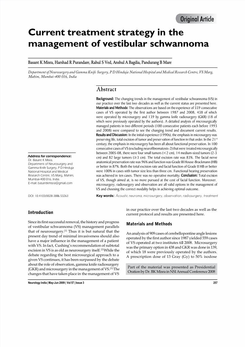
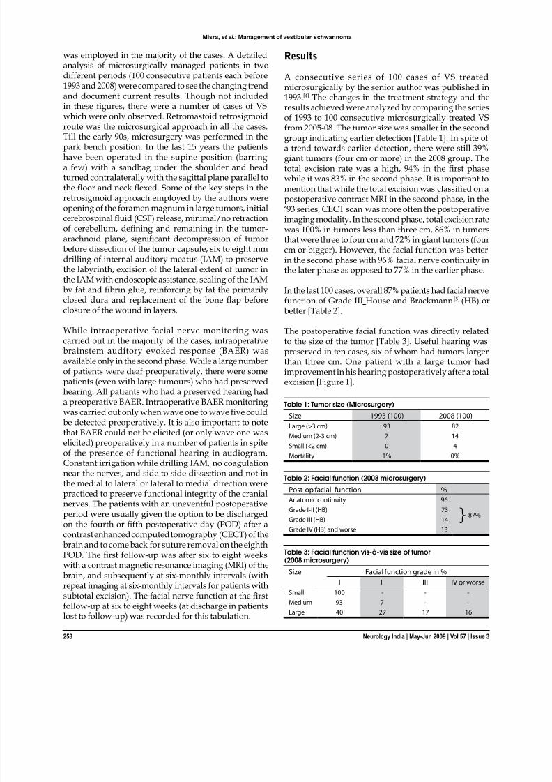
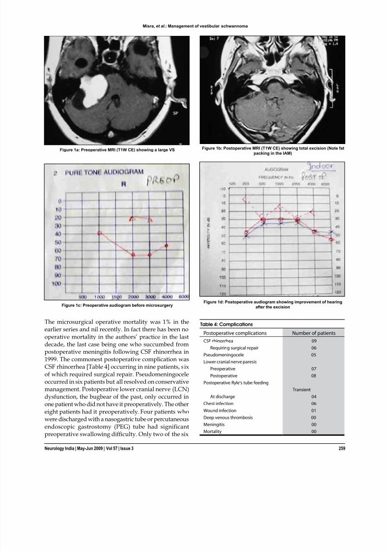
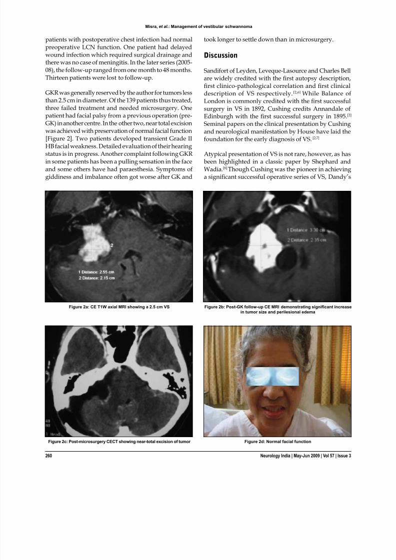
The postoperative facial function was directly relatedto the size of the tumor [Table 3]. Useful hearing waspreserved in ten cases, six of whom had tumors largerthan three cm. One patient with a large tumor hadimprovement in his hearing postoperatively after a totalexcision [Figure 1].
The microsurgical operative mortality was 1% in theearlier series and nil recently. In fact there has been nooperative mortality in the authors’ practice in the lastdecade, the last case being one who succumbed frompostoperative meningitis following CSF rhinorrhea in1999. The commonest postoperative complication wasCSF rhinorrhea [Table 4] occurring in nine patients, sixof which required surgical repair. Pseudomeningoceleoccurred in six patients but all resolved on conservativemanagement. Postoperative lower cranial nerve (LCN)dysfunction, the bugbear of the past, only occurred inone patient who did not have it preoperatively. The othereight patients had it preoperatively. Four patients whowere discharged with a nasogastric tube or percutaneousendoscopic gastrostomy (PEG) tube had significantpreoperative swallowing dif culty. Only two of the six
Figure 1a: Preoperative MRI (T1W CE) showing a large VS
Figure 1c: Preoperative audiogram before microsurgery
Figure 1b: Postoperative MRI (T1W CE) showing total excision (Note fatpacking in the IAM)
Figure 1d: Postoperative audiogram showing improvement of hearingafter the excision
Table 4: Complications
Postoperative complications Number of patientsCSF rhinorrhea 09
outstanding results after total excision of VS were equallyimportant in paving the way for many neurosurgeonsto conquer what Cushing named “the bloody angle, thegloomy corner of neurological surgery”. [2,9] Noteworthycontributions in re ning microsurgical excision of VShave been made by House, Malis, Yasargil and Samiiover the years. [6,10-12] Lars Leksell, the founder of GKR and
Ladislav Steiner from Sweden have established and thePittsburgh group popularized the role of radiosurgeryas an established management protocol in VS. [13,14]
MicrosurgeryIn the microsurgical era, three different surgicalapproaches, retrosigmoid (RS), translabyrinthine (TL)and the middle cranial fossa (MCF) are commonlyutilized. All the patients in the authors series have,however, been operated through the retrosigmoid routeas this is the only route that is not only adequate for allsizes of tumor but also suitable for preservation of facialfunction and hearing. No doubt enough literature isavailable now to document TL and MCF approaches asexcellent means of tackling selected tumors. [15] Thoughthe authors have used both these approaches extensivelyfor other skull base lesions, it has not been necessary tochange the strategy of retrosigmoid route for VS overthe years. [16-21] The authors’ indication for microsurgeryas the rst choice today are• All tumors > 2.5 cm in the largest diameter.• Any patient with brainstem dysfunction, signi cant
mass effect, raised intracranial pressure (ICP) orrapidly progressing symptomatology.
• Young patient (< 50 years) with any tumor size.
Though there have been some changes in the techniqueemployed, the basic retromastoid retrosigmoid approachhas not changed over the years. [4,16-21] Presently, theauthor employs a supine position with head turned to thecontralateral side (with a sandbag under the ipsilateralshoulder) as opposed to the lateral position, and acraniotomy as opposed to craniectomy earlier.
The facial nerve preservation rate was a high 96% in 2008as compared to 77% in 1993. A tradeoff has been a lowertotal excision rate (83% compared to 94% in 1993) because
of the recent emphasis on good facial function. Hencea thin sliver of tumor left over the facial nerve, whichshowed up as enhancing tumor on postoperative MRI,was accepted as better for the patient though classi edas subtotal excision. The authors are well aware of theproblem of poor follow-up in our country and the riskof subtotal excision. Another problem is the inability ofthe patient to go for radiosurgery for a subtotally excisedtumor because of the low socioeconomic status. Henceproper preoperative counseling and individualizingthe goal of surgery in a given patient will go a long
way to protect the patient’s interest. However, it is alsoimportant to remember that a thin sliver of tumor leftattached to the nerves infrequently grows to producesymptoms and even if it grows, it can be operatedagain with excellent results and hence the emphasis onfunctional preservation. Though hearing preservationafter microsurgery is unpredictable, especially in large
tumors, six patients with large tumors had useful hearingpostoperatively, one of whom in fact had improvementin his hearing. Very few authors have been able toachieve that. [12,22]
RadiosurgeryMore and more cases of VS are being treated byradiosurgery the world over, especially as patientsare presenting earlier with smaller tumors. Minimalhospitalization, no general anesthesia and avoiding anopen surgery are particularly appealing to the patient.Reports of long-term control with good cranial nervefunction has led to increased enthusiasm for its use by thephysician. [23-26] Earlier reports of increased cranial nervedysfunction have reduced signi cantly with reductionof tumor dose. The concern, however, is long-termcontrol with the lower dosage. The other major concernhas been the risk of malignant degeneration followingradiosurgery. However, to date, there is no proof tosuggest any realistic risk of malignancy following GKR. [27] Presently 12-14 Gray (Gy) is prescribed to 50% isodosein GKR. The reported long-term control rate is between93% and 97%. Though, there have been recent reportsof even improved hearing after GKR, preservation isunpredictable. [28-30] Today our indications for GKR are
patients with tumors of 2.5 cm or less in maximumdiameter and• high medical risk• elderly patients or• recurrent or residual tumor showing growth.
The best modality of treatment for VS less than three cm,especially in young patients, is still controversial. [3,19,29,31] There is no Class one evidence till date to recommend onemodality over the other. While post-procedure morbidityis generally less and cranial nerve function is better withradiosurgery, the control/eradication rate is better with
microsurgery.[3,29]
Long-term data of tumor control withradiosurgery is still not available. In a recent study ofsmall VS comparing functional outcomes followingradiosurgery and microsurgery, Coelho et al. , concludedthat microsurgery was better than radiosurgery inimbalance control and resulted in minimal morbiditycomparable to radiosurgery. [31]
Even today, the authors advocate microsurgery as therst choice in young patients with tumors less than
2.5 cm as our results in this group are excellent with
Misra, et al .: Management of vestibular schwannoma
100% preservation of facial function and eradication oftumor for good. Our present management protocol forextracanalicular VS is presented in Table 5.
ObservationStudies on th e natural history of VS have shown that thereare some tumors which do not grow and remain static
over a long period of time.[32,33]
Intracanalicular acousticneuroma (ICAN), small tumors and tumors picked upincidentally, thus could merit a period of follow-up. Dueto the low socioeconomic status of many patients andpoor follow-up in our country, one is weary of advisingobservation except in ICAN. Though the numbers arenot included in this series, the authors have advisedobservation in some patients mainly elderly, those withminimal or only auditory/vestibular dysfunction andthose with incidentally picked up tumors; provided thereis no doubt about follow-up of such patients.
Special situations (SS): Some special clinical settings need
particular mention. Our protocols in such situations areas follows:-
SS 1: VS in a pregnant lady (i) Patients with small tumors are allowed to carry on
through pregnancy and normal delivery.(ii) Patients with tumors less than three cm but
minimally symptomatic are advised to continuepregnancy but advised elective Caesarean sectiondelivery.
(iii) Symptomatic patients with large tumors are advisedmicrosurgery, preferably after the rst trimester.Quick subtotal excision is performed to providemaximum safety to both mother and child. The
tumor is usually much more vascular in pregnancy.The patient can then go for normal pregnancy anddelivery. The residual tumor, if signi cant, can thenbe excised at a later date.
SS 2: Cystic/Hemorrhagic tumor Patients harboring cystic tumors and those presenting
with macro hemorrhages should be treated bymicrosurgery. Large-size, Antoni B type tumors andtelangiectasia pattern in the tumor make it more proneto both cystic and hemorrhagic transformation. [16] These tumors have a friable capsule leading to greaterdissection dif culty. However, no effort should be sparedfor total excision as these have a higher risk of recurrenceand postoperative hemorrhage.
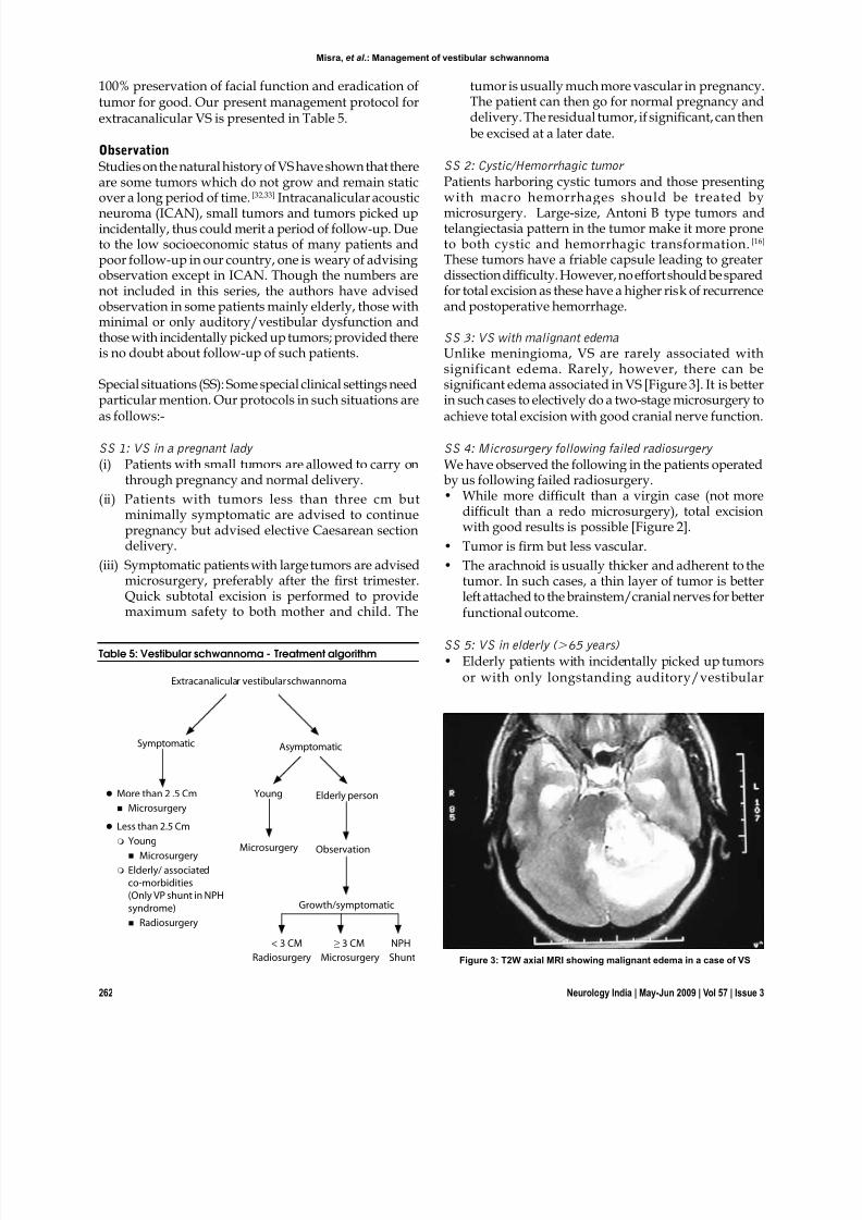
SS 3: VS with malignant edemaUnlike meningioma, VS are rarely associated withsignificant edema. Rarely, however, there can besigni cant edema associated in VS [Figure 3]. It is better
in such cases to electively do a two-stage microsurgery toachieve total excision with good cranial nerve function.
SS 4: Microsurgery following failed radiosurgery We have observed the following in the patients operatedby us following failed radiosurgery.• While more dif cult than a virgin case (not more
dif cult than a redo microsurgery), total excisionwith good results is possible [Figure 2].
• Tumor is rm but less vascular.• The arachnoid is usually thicker and adherent to the
tumor. In such cases, a thin layer of tumor is betterleft attached to the brainstem/cranial nerves for betterfunctional outcome.
SS 5: VS in elderly (>65 years)• Elderly patients with incidentally picked up tumors
or with only longstanding auditory/vestibular
Figure 3: T2W axial MRI showing malignant edema in a case of VS
Misra, et al .: Management of vestibular schwannoma
dysfunction should be observed for a period of timebefore considering any treatment.
• Patients with small tumors will generally not needany treatment in their lifetime.
• Patients presenting with normal pressurehydrocephalus (NPH) syndrome are better treatedby a ventriculo-peritoneal shunt and generally willnot need direct surgery for the tumor.
• While radiosurgery is the preferred modality forelderly patients with tumors less than three cm,patients with larger symptomatic tumors should beoperated and age alone should not be a criterion towithhold microsurgery. Dramatic improvement inthe quality of life is possible in such patients withmicrosurgery.
Conclusion
Vestibula r schwannomas of all sizes can be operatedmicrosurgically by the retrosigmoid approach withgratifying results. Both facial function and hearing can bepreserved after microsurgery even in large tumors; henceno effort should be spared to save these nerves. There isno doubt, however, that some patients are better servedwith GKR and yet some others by no intervention at all.Equally important to remember is that microsurgery forVS is a major procedure, there is a steep learning curveand one can lose a patient from a seemingly innocuousfew drops of postoperative CSF rhinorrhea.
In summary, the approach for a given patient should bepatient-oriented and not surgeon-oriented. Meticulousattention to minute details of each aspect of microsurgeryis essential to avoid catastrophe. The indications for GKRand microsurgery are still evolving. Selection of theappropriate option by a surgeon with ample experience,though not easy, is critical to an optimal outcome.
Acknowledgments
Authors acknowledgment the help of Sarthak B Misra, MBBS,for English language editing and Sudha Nair for secretarialhelp.
References
1. Moskowitz N, Long DM. Acoustic neuromas: Historical review of acentury of operative series. Neurosurg Quart 1991;1:2-18.2. Cushing H. Tumours of the nervus acusticus and the syndrome of the
cerebellopontine angle. W.B. Saunders Co; 1917. p. 1-295.3. Pollock BE. Vestibular Schwannoma management: An evidence based
comparison of stereotactic radiosurgery and microsurgical resection.Progr Neurosurg 2008;21:222-7.
4. Misra BK, Rout D, Bhiladvala DB. Current status of acoustic neuromasurgery. In: Book of Abstracts . Madras: Neuroconf; 1993.
5. House JW, Brackmann DE. Facial nerve grading system. OtolaryngolHead Neck Surg 1985;93:146-7.
6. Rand RW, Dirks DD, Morgan DE, Bentson JR. Acoustic neuromas.In: Youmans JR, editor. Neurological surgery. 2nd ed. W.B. SaundersCo; 1982. p. 2967-3003.
7. House WF. Monograph 2- acoustic neuroma. Arch Otolaryngol1968;88:575-715.
8. Shephard RH, Wadia NH. Some observations on atypical features in Acoustic Neuroma. Brain 1956;79:282-318.
9. Dandy WE. Results of removal of acoustic tumours by the unilateralapproach. AMA Arch Surg 1941;42:1026-33.
10. Malis LI. Microsurgical treatment of acoustic neuromas. In: Honda H,editor. Microneurosurgery. Baltimore: Univ Park Press; 1975. p. 105-20.
11. Yasargil MG, Fox JL. The microsurgical approach to Acoustic Neuroma.Surg Neurol 1974;2:393-8.
12. Mathies C, Samii M. Vestibular and auditory function: Options in largeT3 and T4 tumour. Neurochirurgiere 2002;48:461-70.
13. Prasad D, Steiner M, Steiner L. Gamma Surgery for vestibularschwannoma. J Neurosurg 2000;92:745-59.
14. Flickinger JC, Kondziolka D, Niranjan A, Lunsford LD. Results of acoustic neuroma radiosurgery: An analysis of 5 years experience using current methods. J Neurosurg 2001;94:1-6.
15. Haddad GF, Al-Mefty O. The road less travelled: Trans-temporal accessto CPA. Clin Neurosurg 1994;41:150-67.
16. Misra BK, Rout D, Bhiladvala DB, Radhakrishnan V. Spontaneoushaemorrhage in Acoustic Neuromas. Br J Neurosurg 1995;9:219.
17. Misra BK. Microsurgical Approach to Cerebello pontine angle tumour.In: 11th ICNS, Monduzzi Editors 1997. p. 363-9.
18. Misra BK. Management of Acoustic Neuroma: An overview in Braintumour surgery. In: Kanno T, editor. Japan: Brain Tumour Society;2000. p. 133-8.
19. Misra BK. Changing Trends in the management of acoustic neuroma.Progr Clin Neurosci 2003;18:34-40.
20. Misra BK, Recent trends in the management of acoustic neuroma. In:Khamlichi AE, editor. Medimond: 13th World Congress of NeurologicalSurgery; 2005. p. 213-20.
21. Misra BK. Modern management protocol and outcome in AcousticNeuroma. Asian J Neurosurg 2007;1:30-6.
22. Nair S, Rao RM, Kachhara R. Current perspectives in hearing preservation in large acoustic schwannomas. Progr Clin Neurosci2004;19:216-25.
23. Gabert K, Regis J, Delsanti C, Roche PH, Facon F, Tamura M,et al . Preserving hearing function after gamma knife radiosurgery for
unilateral vestibular schwannoma. Neurochirugiere 2004;50:350-7.24. Lunsford LD, Niranjan A, Flickinger JC, Maitz A, Kondziolka D.
Radiosurgery of vestibular schwannoma: Summary of experience in829 cases. J Neurosurg 2005;102:195-9.
25. Kano H, Kondziolka D, Khan A, Flickinger JC, Lunsford LD.Predictors of hearing preservation after stereotactic radiosurgery foracoustic neuroma. J Neurosurg 2009:Mar 13. [Epub ahead of print].
26. Thomas C, Di Maio S, Ma R, Vollans E, Chu C, Clark B, et al . Hearing Preservation following fractionated stereotactic radiotherapy for
vestibular schwannomas: Prognostic implications of cochlear dose. JNeurosurg 2007;107:917-26.
27. Rowe J, Grainger A, Walton L, Silcocks P, Radatz M, Kemeny A. Risk of malignancy after gamma knife s tereotactic radiosurgery. Neurosurgery 2007;60:60-6.
28. Niranjan A, Lunsford D, Flickinger JC, Maitz A, Kondziolka D. Canhearing improve after acoustic tumour radiosurgery? Neurosurg ClinN Am 1999;10:305-16.
29. Regis J, Pellet W, Delsanti C, Dufour H, Roche PH, Thomassin JM,et al . Functional outcome after gamma knife surgery or microsurgery for vestibular schwannomas. J. Neurosurg 2002;97:1091-100.
30. Niranjan A, Mathieu D, Flickinger JC, Kondziolka D, Lunsford LD.Hearing preservation after intracanalicular vestibular schwannomaradiosurgery. Neurosurgery 2008;63:1054-62.
31. Coelho DH, Roland JT Jr, Rush SA, Narayana A, St Clair E,Chung W, et al Small vestibular schwannomas with no hearing:comparison of functional outcomes in stereotactic radiosurgery andmicrosurgery. Laryngoscope 2008;118:1909-16.
32. Raut VV, Walsh RM, Bat h AP, Bance ML, Guha A, Tator CH,et al . Conservative management of Vestibular schwannomas - Secondreview of a prospective longitudinal study. Clin Otolaryngol Allied Sci2004;29:505-14.
33. Ojemann RG. Management of acoustic neuroma. Clin Neurosurg 1993;40:498-535.
Accepted on 19-06-09Source of Support: Nil, Con ict of Interest: None declared.
Misra, et al .: Management of vestibular schwannoma


![Application Note: QP/C++ MISRA-C++:2008 Compliance Matrix · 2019-07-11 · Association (MISRA) Guidelines for the use of the C++ Language in Critical Systems [MISRA-C++:2008]. This](https://static.documents.pub/doc/80x56/5e719371323a91004e4f3d79/application-note-qpc-misra-c2008-compliance-2019-07-11-association-misra.jpg)


