Blast and Ballistic Injury: Principles of Management Jon Clasper MBA DPhil DM FRCSEd(Orth) FRCS Col L/RAMC Defence Professor Trauma & Orthopaedics Visiting Professor in Bioengineering, Imperial College London
Transcript
Blast and Ballistic Injury: Principles of Management
Jon Clasper MBA DPhil DM FRCSEd(Orth) FRCS
Col L/RAMC
Defence Professor Trauma & Orthopaedics
Visiting Professor in Bioengineering, Imperial College London
Foreword Prof Tim Briggs, BOA President, and Kate Brown
The 14th century heralded the advent of gunpowder and a change from purely penetrating wounds to
ballistic injuries. Since then, military surgery has taught us much about the management of blast and
ballistic wounding patterns. Early examples include Ambroise Pare’s treatment of open fractures with
reduction and stabilisation at the Battle of Turin, the formalisation of debridement by Desault and the
introduction of true Damage Control Surgery by Baron Jean Larrey during the Napoleonic Wars. More
recently, the development of antisepsis with Lister’s carbolic acid and Sir Alexander Fleming’s discovery of
penicillin came from the First and Second World Wars respectively. We continue to revisit the surgical
lessons learnt from our military history and never has that been so pertinent than during the recent
conflicts in the Middle East. The tempo of war has now changed from the traditional format of enemy
lines to a more blurred, almost guerilla-style fighting. Military personnel undergo attacks not only from
traditional ballistic weapons but also from Improvised Explosive Devices (IEDs). The injuries that result
from these weapons are devastating, causing significant polytrauma in multiple casualties. Research from
the operational theatre has served to assess and understand how the surgical management principles
taught are borne out in practice and what is their clinical effectiveness.
Professor Jon Clasper’s expert opinion on the management of blast and ballistic injuries outlines the most
current guidelines, drawing together contemporaneous evidence coupled with personal clinical and
academic experience in the field. He describes the principles of wound management, in particular how to
assess for viability. There is continuing argument as to how aggressive this should be which can be
confusing for the more inexperienced surgeon. He outlines which antimicrobials should be used, and
describes why gram-negative therapy is often not indicated. How to assess, bind and debride pelvic
injuries is described and what the common pitfalls are including mal -placement of the binder and where
to inspect for contaminating debris. How fasciotomies should be performed is delineated and when they
should be performed. Finally, the topic of amputations is reviewed. This is a highly sensitive issue with
significant implications. Whether or not they can be objectively scored is examined and the method by
which the amputations should be performed.
4
These principles are just as relevant in civilian practice as in the theatre of war. Although more prevalent
in the United States, gun crime is still a significant contributor to all -cause trauma in the United Kingdom.
With the advent of Major Trauma Centres, the majority of these patients will be managed in hospitals
with experience in such injuries. However, this will not always be the case and smaller more peripheral
hospitals will need guidance. Blast weapons have been used on numerous occasions, most notably
recently in the tube bombings July 2005 and the Boston marathon bomb April 2013. Even large MTCs are
less experienced in such wounding mechanisms.
About the Author Prof Jon Clasper MBA DPhil DM FRCSEd(Orth) FRCS FHEA
Professor Clasper qualified from Glasgow University in 1986 and completed his basic surgical training in
the Armed Forces. His higher surgical training included a year at the Major Trauma Centre in Baltimore,
USA, as well as 3 years at Oxford. His clinical post is as Consultant Trauma and Orthopaedic Surgeon at
Frimley Park NHS Foundation Trust.
As Defence Professor in Trauma and Orthopaedics, he is responsible for the orthopaedic research focus of
the British military. This is a joint post between the military and the Royal College of Surgeons of
Edinburgh, and was established in June 2009 to complement the Defence Professor of Surgery within the
Academic Department of Military Surgery and Trauma.
Jon was one of the founding members of Imperial Blast, a multidisciplinary group of clinicians, scientists
and engineers collaborating to study the effects of high strain rates on biological structures. Following
significant funding, this initiative has now developed into the Royal British Legion Centre for Blast Injury
Studies at Imperial College.
Jon is the author of a number of book chapters, and of specific papers on trauma and upper limb surgery.
He has gained doctorates in both Medicine and Philosophy.
In 2010 Jon was appointed as a Visiting Professor in Bioengineering at Imperial College London.
Jon has deployed on many operational tours including Northern Ireland, Sierra Leone, Bosnia and Kosovo.
He served in both Gulf Wars, and has deployed to Afghanistan 3 times.
His clinical and academic experience in both military and civilian settings pertain him to be an authority
on the management of blast and ballistic injuries.
Introduction
The aim of this short guide is to outline the principles in the management of blast and ballistic injuries.
Whilst once the domain of the military, with the global rise in terrorism, particularly suicide bombers, it is
likely that more civilian surgeons will be exposed to these wounds. Although the principles in the
management of these injuries are similar to the management of civilian open fractures, some of the
injuries, particularly the blast pelvis or traumatic amputations will be alien to most people. In addition the
technique of amputations for trauma will not be familiar to most orthopaedic surgeons.
5
Management of the Ballistic Wound
The principles of wound care are similar to civilian trauma, although some techniques do differ.
Antibiotics, although an important factor, are secondary to early surgery and should not be considered as
an alternative form of treatment. Neurovascular and radiological assessment should be undertaken prior
to debridement when possible.
Debridement
General Principles
Clothing and gross debris should be removed.
Pre-operative washing of the limb with copious volumes of warmed soap/detergent solution is essential
to remove skin contamination and fully identify all injuries.
Where practical all wounds should be photographed prior to debridement. Photographs allow further
wound management to be planned. Pre-operative photographs are also an invaluable tool for
demonstrating the rationale for amputation to casualties post-operatively.
The wound should be debrided by each layer around the full circumference of the wound in a methodical
manner. Macroscopically non-viable tissue should be excised.
Initial wound debridement should be achieved through sharp dissection using scissors or scalpel . While
the use of diathermy to control bleeding is acceptable its use to excise muscle and to assess contractility
is not recommended as it leaves points of dead burnt muscle which may predispose to infection.
The use of a pneumatic tourniquet will limit blood loss during debridement and improve visualisation of
the operative field, however, it may compromise the assessment of muscle viability and add further
ischaemic stress to already damaged but viable tissue.
In the case of amputations formal flaps should not be fashioned at the initial debridement as this may
involve the sacrifice of viable muscle, which may be required for reconstruction but all viable skin in
continuity should be preserved. (1) (See also Amputations Section).
Technique
Skin
At the initial debridement it is likely that the wound edges will be traumatised. However, skin is resilient
and debridement should be restricted to obviously non-viable tissue. When debridement of skin is
performed there is no requirement to excise to bleeding dermis as skin perfusion may be altered by
hypotension, and by transient vasoconstriction which can be associated with ballistic injury. Similarly
small flecks of embedded contamination are not an indication for excision.
Extension of traumatic wounds may be necessary to allow access to the underlying zone of injury. Wound
extensions should facilitate fasciotomy and any subsequent procedures. Skin incisions should be in the
6
long axis of limbs except where they cross the flexor surfaces of joints where they should be placed
obliquely to reduce the risk of scar contractures.
Fat/fascia
Debridement of fat and fascia should be limited to macroscopically damaged tissue to avoid undermining
skin, which is reliant on its attachment to fat and fascia for its blood supply. Injudicious debridement of
this layer may compromise the use of fascio-cutaneous flaps during reconstruction.
Fasciotomies of muscle compartments are required to expose the underlying zone of injury, to treat
actual or impending compartment syndrome and to reduce the risk of occult compartment syndrome
occurring during evacuation or where prolonged sedation is anticipated. Where performed, fasciotomies
should be along the full length of the muscle compartment (see fasciotomy section).
Muscle
The 4 C’s of colour, contractility, consistency and capillary bleeding may not be reliable indicators of
microscopic muscle damage. Altered consistency correlates moderately well with microscopic muscle
damage but colour variation, contractility and capillary bleeding correlate more weakly (2). In the absence
of other criteria they should be used together to judge how much macroscopically damaged muscle
should be excised. In situations where serial marginal debridement is to be carried out it is suggested that
muscle which is traumatised but not clearly non-viable is left for subsequent reassessment.
Blood vessels/Nerves and Tendons
Traumatised nerves, tendons and blood vessels should be clearly documented in the operation note.
Primary vascular reconstruction may be necessary in some proximal limb wounds with associated
vascular trauma. However, reconstruction of divided nerves and tendons should not be undertaken at the
initial debridement.
Divided blood vessels should be ligated unless reconstruction is required. Divided nerves and tendons can
be tagged/marked with sutures, however this should not add to the existing damage to the structure.
In the absence of clinical signs of vascular injury it is not necessary to identify major blood vessels beyond
the zone of injury at the time of initial debridement.
Bone
Exposed cancellous bone should be curetted and contaminated cortical bone trimmed with bone
nibblers. Periosteum should be preserved where possible. Bone in continuity should be preserved even
where stripped of all periosteum and soft tissue.
Bone fragments with soft tissue attachments which are absent or very tenuous should usually be
discarded.
Prior to irrigation of the wound a final circumferential check of the debrided wound to confirm adequacy
and check for haemostasis should be performed.
7
Irrigation
Fluid constituents
Wounds should be irrigated with warmed sterile saline where available. Where resources are limited
warmed drinking water is an adequate alternative (3,4). Antiseptic, antibiotic and soap solutions are
associated with higher rebound bacterial counts at 48 hours (5) and should be avoided. In addition
antiseptic solutions are associated with cytotoxicity (6,7) and reduced microvascular blood flow (8), and
antibiotic solutions are associated with wound healing problems without a reduction in the rate of
positive wound cultures (9,10).
Fluid Delivery
Irrigation should be via low-pressure systems such as syringe, giving-set or cutting the corner off a bag of
fluid. High-pressure pulsatile lavage systems are associated with damage to cortical bone (11), periosteal
stripping (12), muscle damage and propagation of bacteria into tissues (13). Even low-pressure pulsatile
lavage strips periosteum (12) and demonstrates a higher rebound bacterial count at 48 hours (5).
Fluid Volumes
Suitable volumes for irrigation of wounds are 9L for massive energy wounds such as those caused by close
proximity blast/explosions, 6L for high-energy ballistic wounds and 3L for lower energy narrow channel
ballistic wounds (14) or superficial soft tissue wounds.
Dressings
At present there is no evidence that any specific dressings are associated with any reduction in extremity
war wound infection rates.
Negative pressure wound therapy (NPWT) is being commonly used on military casualties in Birmingham.
This technique appears effective, but evidence is limited at present.
Interval to reassessment/closure
Ballistic wounds caused by penetrating projectiles should be reassessed, usually with a plan to achieve
wound closure at around 5 days. There is no requirement for an earlier reassessment or dressing change
if the debridement is considered complete and the casualty remains well and apyrexial.
If tissue of doubtful viability was left at the initial debridement the casualty should be returned to the
operating theatre at around 48 hours for assessment of viability, further debridement if necessary and
application of new dressings.
Close proximity blast/explosion wounds
These devastating injuries represent a new challenge; in previous conflicts many casualt ies did not survive
long enough to reach hospital.
The debridement of these wounds is extremely challenging. It is unlikely to be achieved in a single surgical
episode due to the physiological frailty of the casualty as well as the complex and contaminated nature of
the wound. Debridement may initially be limited to removal of grossly dead tissue rather than the layer-
8
by-layer procedure described above but each opportunity to reduce the burden of wound contamination
should be taken. This can be a concurrent activity during life-saving damage control surgery.
A philosophy of serial marginal debridement should be adopted (15). This describes the process by which,
at each visit to the operating theatre, the traumatised tissue within the extensive zone of injury is
reassessed and debrided or left as appropriate. At each procedure it may be possible to carry the
debridement deeper into the track of the most complex wounds and remove devitalised tissue where
appropriate.
Low energy soft tissue wounds
While every wound must be explored, not all need the full surgical debridement described above.
Narrow channel soft tissue wounds
A low energy narrow channel wound is characterised by entry and exit wounds of less than 1cm, no
wound cavity, no swelling of intervening tissue and no other signs of injury to important structures. In this
situation it is sufficient to excise the entrance and exit wounds and irrigate the connecting channel.
Superficial soft tissue wounds
Small superficial wounds that do not penetrate the deep fascia do not require any formal surgical
intervention. The wounds should be scrubbed, irrigated with normal saline and left to heal by secondary
intention. The casualty should be given oral antibiotics and dressings should be changed every 48 hours.
Antimicrobials Although responsible for decreasing the infection burden of combat injury, the widespread use of
antimicrobial agents has also created the latest challenge in wound care: multi -drug resistant bacteria.
Antimicrobial agents are merely an adjunct to wound care, to be chosen and administered only after due
consideration. This notion of selecting therapy to target agents posing the greatest threat to those
recently injured has been discussed by Hutley and Green (16).
Intravenous antibiotics should be commenced within three hours of injury with as narrow a spectrum as wounding pattern allows.
Routine Gram–negative cover for all wounds is discouraged. Recommendations for agent of choice and duration of cover differ, however regardless of choice,
duration should be short (48-72 hrs) following initial procedure with a subsequent short course to cover wound closure at which point antibiotics are stopped.
Co-amoxiclav is appropriate for extremity wounding, and visceral injury
Any further antibiotic therapy should be guided by clinical and microbiology assessment. Tetanus immunisation status should be addressed.
References for this section 1. Clasper J, Lower Limb Trauma Working Group. Amputations of the lower l imb: a multidisciplinary
consensus. J R Army Med Corps 2007; 153: 172-4. 2. Sculley RE, Artz CP, Sako Y. Evaluation of the surgeon’s criteria for determining the viability of muscle during debridement. Arch Surg 1956; 73: 1031-5.
3. Moscati RM, Mayrose J, Fincher L, Jehle D. A multicenter comparison of tap water versus sterile saline for wound irrigation. Acad Emerg Med 2007; 14: 404-9.
9
4. Svodoba SJ, Owens BD, Gooden HA, Melvin ML, Baer DG, Wenke JC. Irrigation with potable water versus normal saline in a contaminated musculoskeletal wound model. J Trauma 2008; 64: 1357-9. 5. Owens BD, White DW, Wenke JC. Comparison of irrigation solutions and devices in a contaminated musculoskeletal wound survival model. J Bone Joint Surg [Am] 2009; 91: 92-8.
6. Lineaweaver W, McMorris S, Soucy D, Howard R. Cellular and bacterial toxicities of topical antimicrobials. Plast Reconstr Surg 1985; 75: 394-6. 7. Bhandari M, Adili A, Schemitsch EH. The efficacy of low pressure lavage with different irrigating solutions to
remove adherent bacteria from bone. J Bone Joint Surg [Am] 2001; 83: 412-9. 8. Crowley DJ, Kanakaris NK, Giannoudis PV. Irrigation of the wounds in open fractures. J Bone Joint Surg [Br] 2007; 89: 580-5. 9. Anglen JO. Comparison of soap and antibiotic solutions for irrigation of lower l imb open fracture wounds. A
prospective, randomised study. J Bone Joint Surg [Am] 2005; 87: 1415-22. 10. Conroy BP, Anglen JO, Simpson WA, Christensen G, Phaup G, Yeager R, Gainor BJ. Comparison of castile soap, benzalkonium chloride and bacitracin as irrigation solutions for complex contaminated orthopaedic wounds. J
Orthop Trauma 1999; 13: 332-7. 11. Bhandari M, Schemitsch EH, Adili A, Lachowski RJ, Shaughnessy SG. High and low pressure pulsatile lavage of contaminated tibial fractures: An in vitro study of bacterial adherence and bone damage. J Orthop Trauma 1999; 13: 526-33.
14. Giannou C, Baldan M. Red Cross Wound Score and Classification System. In: Gi annou C, Baldan M (eds) War Surgery Volume 1; Working with limited resources in armed conflict and other situations of violence. Geneva: International Committee of the Red Cross, 2010; Ch 4
15. Taylor CJ, Hettiaratchy S, Jeffrey SL, Evriviades D, Kay AR. Contemporary Approaches To Definitive Extremity Reconstruction Of Military Wounds. J R Army Med Corps 2009; 155: 302-7. 16. Hutley EJ, Green AD. Infection in wounds of conflict--old lessons and new challenges. J.R.Army Med.Corps 2009; 155: 315-9.
10
Pelvic Injuries Following Explosions
Improvised explosive devices (IEDs) have become the weapon of choice in Afghanistan, and were
responsible for 60% of coalition forces’ fatalities in 2009 and 2010. This period has also seen the
emergence of one of the most challenging injury patterns seen in trauma (1), the unstable pelvic fracture,
with severe perineal trauma, and associated severe limb injuries often with traumatic amputations.
The increased survival in the current conflict in Afghanistan has been well documented, and this is related
to an integrated systematic approach to military trauma, from improved training and pre -hospital care to
after-event clinical governance review (2). This, together with improved personal protection (body
armour) has resulted in early survivors with injuries that were universally fatal in previous conflicts. These
casualties bleed to death, rapidly, and the initial treatment is therefore focused on controlling
catastrophic haemorrhage (3). This applies particularly to the management of traumatic amputations, but
is challenging when the injuries are very proximal.
Pre-hospital Care If available, and if enough limb remains, a tourniquet should be immediately applied proximal to the
injury.
If this is not effective or possible, direct pressure at the site of bleeding is effective, but can be difficult to
maintain, particularly when casualties have to be moved. The military and some ambulance services carry
topical haemostatic agents, which should be considered.
All casualties with severe limb injuries, particularly traumatic amputations, are assumed to have pelvic
fractures, and a pelvic binder should be applied. Care must be taken to position the binder correctly, even
if wounds are present. Binders that are too high do not effectively close the pelvis (4).
Resuscitation requires vascular access. Peripheral venous access is frequently impossible particularly if all
4 limbs have been injured. Intraosseus devices such as the EZIO® (Vidacare®, Shavano Park, Texas, USA)
and FAST-1TM (PyngMedical, Richmond, BC, Canada) are used by the UK military. The EZIO® is usually
inserted into the proximal tibia or iliac crest, but when the patient has sustained traumatic lower
extremity amputations, or iliac placement is contraindicated by the presence of pelvic injuries, the
humeral head is an alternative site.
Hospital Phase The hospital reception and initial assessment of casualties with proximal traumatic extremity
amputations requires communication, preparation, organisation and leadership.
Key preparations include the immediate availability of an operating theatre and operating room staff, and
notification of the blood transfusion laboratory. Delay will kill these casualties; it must be minimised.
The anteroposterior pelvic radiograph is the key investigation in identifying the unstable pelvic ring, even
with the binder in place. If there is radiological evidence of a pelvic fracture, and the patient is responding
to resuscitation, CT scanning should be performed prior to removal or loosening of the binder.
11
Non-responders, or transient responders require proximal surgical control, and should be taken
immediately to the operating theatre. If delays are likely operating in the Emergency Department should
be considered.
If a clear need for operative proximal vascular control is identified prior to the arrival of the patient, the
trauma team is relocated to the operating theatre, and the casualty taken straight there, bypassing the
ED.
Whilst some of this may seem aggressive, this approach has been adopted by the military, with
demonstrable success. Patients with severe pelvic injuries who have arrived at hospital undergoing
cardio-pulmonary resuscitation, have left hospital alive.
Resuscitation Techniques are discussed in detail elsewhere (5).
Surgical Strategy The initial surgical management is focused on gaining control of haemorrhage. The proximal extension of
the zone of injury is usually worse than external appearances suggest.
Amputation below mid-thigh level can usually be managed with a combination of tourniquet and, if
necessary, surgical control at common femoral artery level.
If the injury is more proximal but unilateral, an extraperitoneal approach with control at external iliac
level should be considered.
With bilateral and more proximal injuries, immediate laparotomy and control at the level of the distal
aorta or iliac system can be achieved more rapidly, and limits blood loss from both the amputation sites
and pelvic injuries.
Laparotomy can be performed with a pelvic binder still applied, and following proximal control of
bleeding, conversion to an external fixator, if indicated, should be performed at this stage.
Debridement of bilateral lower extremity wounds should ideally be performed by two teams working
concurrently, to minimise ischaemic injury and on-table time. Contaminating debris is frequently found
forced into intermuscular planes and around neurovascular bundles.
Proximal vascular control is maintained until debridement is complete rather than released immediately
after femoral vessel ligation, as additional blood loss may worsen any coagulopathy.
Many patients with high traumatic amputations caused by IEDs also sustain extensive perineal injuries,
which can present complex management dilemmas. Faecal diversion should be considered, but is the
subject of some debate. Vascularised testicular remnants should be preserved. Scrotal and penile injuries
are debrided and, where possible, closed. Urethral injuries are treated with urethral and/or suprapubic
catheterisation.
12
Contamination often extends through the sacro-iliac joint into the paraspinal muscles, it is usually
impossible to debride this at the first operation due to the severity of the injuries in an uns table casualty.
Whilst not ideal this has to be accepted as part of the damage control nature of the surgery.
Wounds should be inspected every 48 hours and debrided as necessary.
For patients with associated upper limb injuries, the policy in military casualties evacuated to Birmingham
is to try and optimise the upper limbs by early internal fixation as appropriate before redebriding the
pelvis. Functional recovery should be optimised, rather than delaying upper limb reconstruction by
constantly destabilising the patient attempting to clear the pelvic contamination first.
References for this section 1. Bailey JR, Stinner DJ, Blackbourne LH, Hsu JR, Mazurek MT. Combat related pelvis fractures in nonsurvivors. J Trauma 2011;71:S58-61. 2. Russell RJ, Hodgetts TJ, McLeod J, Starkey K, Mahoney P, Harrison K, Bell E. The role of trauma scoring in developing trauma clinical governance in the defence medical services. Philos Trans R Soc Lond B Biol Sci 2011, Jan
27;366:171-91. 3. Hodgetts TJ, Mahoney PF, Russell MQ, Byers M. ABC to <C>ABC: redefining the military trauma paradigm. Emerg Med J. 2006 Oct;23:745-6.
4. Bonner TJ, Eardley WG, Newell N, Masouros S, Matthews JJ, Gibb I, Clasper JC. Accurate placement of a pelvic binder improves reduction of unstable fractures of the pelvic ring. J Bone Joint Surg Br. 2011;93:1524-8. 5. Early management of proximal traumatic lower extremity amputation and pelvic injury caused by improvised
explosive devices (IEDs). Jansen JO, Thomas GO, Adams SA, Tai NR, Russell R, Morrison J, Clasper J, Midwinter M. Injury. 2011 Sep 8. [Epub ahead of print]
Introduction Acute Compartment Syndrome (ACS) is more common with ballistic and blast injury.
The diagnosis is clinical, although pressure monitoring can be used if the condition is anticipated or
suspected and accurate clinical examination is not possible, such as with a head injured patient (1). As
pressure monitoring has not been shown to be more accurate than clinical monitoring (2), it is not
indicated in a conscious patient if regular assessment is possible.
Regular re-assessment is required as there may be a delay in presentation, with a recent paper reporting
a mean delay between injury and fasciotomy of 22 hours even in monitored patients (3). Even though the
development of ACS may be delayed, once the diagnosis has been made, fasciotomy must not be
delayed. It has been reported that a delay in treatment is associated with an adverse outcome and an
increase in complications such as amputation and even death (4).
General Principles Fasciotomy can be associated with a high rate of complications (5), and in many cases this is due to
technique rather than the underlying injury.
Unfortunately most of the following guidelines below have had to be relearned in recent conflicts. They
represent what we believe to be current best practice. They are meant to guides, particularly for the
relatively inexperienced, and should not be regarded as superior to any other published guides:
Following diagnosis, fasciotomy should be carried out as soon as practical due to the increase in
complications with delay (4).
All compartments should be decompressed. In the lower leg failure to decompress the deep posterior is relatively common and missed compartment syndrome is associated with a high complication rate (5).
Full length incisions must be performed. A common error is the incision is too short; usually the decompression appears adequate but post-operative muscle swelling leads to ACS recurrence due to the tourniquet effect of tight skin and fascia at the extremes of the incision.
Incisions should be placed with regard to later reconstructive options. If the medial incision to decompress the lower leg (see below) is placed too posteriorly the perforating vessels may be damaged, limiting the options for local flaps.
All planned incisions must be pre-marked, including possible surgical extensions prior to any incision being made. It should be noted that significant swelling may obscure the normal anatomical features.
In most circumstances any associated fracture should be reduced and stabilised. Most fasciotomies are carried out through a longitudinal skin incision. If the incision is performed in the presence of an unstable displaced fracture, particularly with rotational abnormalities, the final incision, after reduction of the fracture may expose bone, or again compromise reconstructive options.
Un-necessary exposure of the bone by the fasciotomy incision must be avoided as this will also compromise reconstructive options, possibly necessitating flap rather than split skin graft coverage.
14
If the medial incision to decompress the lower leg (see below) is placed too anteriorly, the subcutaneous surface of the tibia may be exposed. The upper tibia may also be exposed due to failure to appreciate the flare of the proximal tibia.
Anatomical Locations
Upper arm
This has 2 compartments; the flexor containing the biceps and related muscles, and extensor containing
the triceps. Both can be decompressed by a single longitudinal incision, which may be possible through an
open wound.
If exposure of the vessels is also required, the incision can be placed medially.
A lateral longitudinal incision can be used to avoid exposing the artery, but if external fixation is also
required, closure of a lateral wound may be compromised.
Forearm
The forearm has superficial and deep flexor compartments and an extensor compartment, and release
can be performed through a single curved volar incision, and several incisions have been recommended
(6,7).
However, for the inexperienced, it is advisable to carry this out through 2 separate incisions to avoid
inadequate release and compromising potential wound closure options.
The following method is suggested:
Identify both epicondyles and identify the midpoint of a line joining the 2. Mark a 2nd point midway between the 1st point and the medial epicondyle. In other words this is a point ¼ of the way along a line from the medial to lateral epicondyles.
At the proximal wrist crease mark a point in line with the ulnar border of the ring finger. A straight incision should be made joining these points.
This incision will not allow adequate access to the lateral muscles, and to decompress these, a separate incision is made. For this a straight, longitudinal incision is made along a line joining the lateral epicondyle of the humerus with the radial styloid. An incision along the proximal 1/3 to ½ of this line is made.
If extension into the palm is required to release the carpal tunnel, a short transverse incision is made in the proximal wrist crease to a point marked in line with the radial side of the ring finger.
The incision is then extended longitudinally into the palm. To avoid contractures transverse or oblique incisions should always be used when the flexor surface of a joint is crossed.
Hand
The compartments of the hand contain the small muscles that provide fine motor control of the digits.
Missed or untreated compartment syndrome of the hand can be devastating for hand function, with only
very limited reconstruction options being possible. Unlike with the foot, there is no debate amongst hand
surgeons of the need to decompress the hand.
The compartments that require release are: 1. The thenar eminence 2. The hypothenar eminence 3. The interossei and lumbricals 4. The adductor policis
15
The thenar and hypothenar eminences are released via longitudinal incisions along their radial and ulnar
borders, respectively. Up to 50% of patients may have separate compartments within the thenar
eminence and this should be reviewed by direct inspection.
The interossei are approached via 2 longitudinal incisions on the dorsum of the hand, the first over the 2nd
metacarpal and the second over the 4th metacarpal. The incisions are deepened either side of the
metacarpals and care should be taken to leave the paratenon of the extensor tendons undisturbed. This
allows release of not only the interossei but also, via the radial extension of the first incision, the
adductor policis muscle that is deep to the 1st dorsal interosseus.
Thigh
The thigh contains 3 compartments, flexor, extensor and a medial adductor compartment. In most
situations release of the anterior and posterior compartments through a single lateral incision may be
sufficient, but penetrating injuries to the thigh may require release of the medial compartment
particularly when associated with a vascular injury. This will require a separate (medial) incision.
Lower leg
There are 4 compartments; anterior and lateral, and superficial and deep posterior compartments.
Failure to release the deep posterior compartment is the most common error in lower limb fasciotomy,
and occurs when releasing the soleus muscle from the posterior aspect of the tibia is mistaken for
releasing the compartment. The posterior tibial artery is located between the 2 posterior compartments,
and this can be used as a landmark during surgery. A 2 incision technique should be used in the lower
limb, decompressing the posterior compartments through a single medial skin incision approximately 1-
1.5 cm (1 finger breadth) posterior to the medial border of the tibia. Incisions too anterior will leave
exposed bone, and incisions too posterior may damage the perforators and compromise subsequent
plastic surgical options.
The anterior and lateral compartments can be decompressed through a single lateral incision. There
should be a 5-7cm skin bridge between these two incisions, which should be drawn with an indelible
marker prior to incision as the surface anatomy may be altered by the first incision. Traumatic wounds
should be incorporated into the skin incisions where practical to avoid leaving two inadequate skin
bridges each side of the wound. Again these should be marked on the skin prior to any incisions being
made.
The lateral compartment can be decompressed through the intermuscular septum between the anterior
and lateral compartments and indeed this method is the recommended standard of care in civilian open
fracture management in the United Kingdom (8). However, the lateral compartment may also be
decompressed through its overlying deep fascia (9). Both techniques require only a single skin incision.
The advantage of the former is that it avoids undermining skin and fat and so preserves its blood supply
and theoretically the use of a lateral fasciocutaneous flap. The latter technique perhaps provides a more
independent decompression of the lateral compartment.
16
Foot
The main indication for fasciotomy is to reduce the morbidity associated with muscle necrosis. With the
foot, there is some controversy regarding the significance of the complications, when compared to the
morbidity associated with the procedure.
The diagnosis of compartment syndrome of the foot remains challenging and the sequelae of missing or
not adequately treating can be disabling and painful (10). However the operative management of
compartment syndrome can be complicated by developing infection of the wounds and fracture sites
especially if there is a need operative fixation and the wounds are likely to require skin grafting for
closure (11). The complications associated with missing or not decompressing a foot with compartment
syndrome includes contracture and toe deformities (12). It is the level of disability from these deformities
that is the focus of the debate as to whether to decompress or not. 2 dorsal incisions are made centred
over the 2nd and 4th metatarsals.
The adductor compartment is released through the 1st webspace. The posterior extent of the incision is
approximately 3cm anterior to the back of the heel, and 3cm above the sole of the foot.
Dressings and Re-inspection Wounds should be re-inspected if any increase in pain is noted and routinely at 3-5 days as early cover /
closure leads to better mobilisation.
References for this section 1. McQueen MM, Court-Brown CM. Compartment monitoring in tibial fractures. The pressure threshold for decompression. J Bone Joint Surg [Br].1996; 78-B: 99-104. 2. Janzing HM, Broos PL. Routine monitoring of compartment pressure in patients with tibial fractures: Beware of overtreatment. Injury 2001; 32: 415-21.
3. Al-Dadah OQ, Darrah C, Cooper A, Donell ST, Patel AD. Continuous compartment pressure monitoring vs clinical monitoring in tibial diaphyseal fractures. Injury 2008; 39: 1204-9. 4. Hayakawa H, Aldington D, Moore RA. Acute traumatic compartment syndrome: a systematic review of
results of fasciotomy. Trauma 2009; 11: 5-15. 5. Ritenour AE, Dorlac WC, Fang R, Woods T, Jenkins DH, Flaherty SF, et al. Complications after fasciotomy revision and delayed compartment release in combat patients. J Trauma 2008: 64; S161-2. 6. Gelberman RH, Zakaib GS, Mubarak SJ, Hargens AR, Akeson WH. Decompression of forearm compartment
syndromes. Clin Orthop 1978; 134: 225-9. 7. Ronel DN, Mtui E, Nolan WB. Forearm compartment syndrome: Anatomical analysis of surgical approaches to the deep space. Plast Recon Surg 2004; 114: 699-705. 8. Nanchahal J, Nayagam S, Khan U, Moran C, Barrett S, Sanderson F, Pall ister I. Standards for the
management of open fractures of the lower limb. London: Royal Society of Medicine Press Ltd, 2009. 9. Clasper JC, Standley D, Hepell S, Jeffrey S, Parker PJ. Limb Compartment Syndrome and Fasciotomy. J R Army Med Corps 2009; 155: 298-301.
10. Myerson M, Manoli A. Compartment Syndromes of the Foot After Calcaneal Fractures. Clin Orthop. 1993; 290: 142-50. 11. Velmahos
GC, Theodorou
D, Demetriades
D et al. Complications and Nonclosure Rates of Fasciotomy for
Trauma and Related Risk Factors. World J Surg 1997; 21: 247-53.
12. Rammelt S, Zwipp H. Reconstructive surgery after compartment syndrome of the lower leg and foot. Eur J Trauma Emerg surg 2008; 34: 237-48.
17
Amputations
Indications The presence of a vascular injury that required repair in a physiologically unstable patient was the main
factor in patients who required an amputation in one study (1). This is unsurprising when considering the
prolonged time needed for revascularization will not be tolerated in a patient who is critically unwell.
Given the advances that have been made in limb salvage techniques, then anatomical indications for
amputation, such as ‘unreconstructable’ should not be used, unless the case has been discussed with a
specialist centre.
The presence of a numb sole of foot has, traditionally, been quoted as an indication for amputation.
However, recovery of protective sensation has been reported in 50% of civilian trauma patients (2), and
has also been noted in 90% of UK military patients.
Scoring systems Several scoring systems have been developed to help the decision to amputate after severe lower limb
trauma. However, none of these scores have been fully validated in subsequent studies, and they must
only be considered as guides; none relate to specific to blast and ballistic injury mechanisms.
Brown et al reported the largest study investigating the use of the Mangled Extremity Severity Score
(MESS) specifically in ballistic lower extremity injuries (1). In their study, 9 patients (10 limbs) with a MESS
7 had limbs that were successfully salvaged, resulting in a positive predictive value of 64.3% if the MESS
criteria of 7 had been used for amputation. This equates to one in three legs being unnecessarily
amputated. The authors concluded that the MESS could not be used to determine the need for primary
amputation in individual patients.
General Principles A trauma amputation should be considered an extension of debridement, rather than a definitive
procedure in its own right.
When the foot remains viable, debridement of the wound, rather than amputation should be performed.
This approach retains the possibility of reconstruction, even when significant foot injuries have occurred
(3).
In 2007 the Limb Trauma Working Group of The Academic Department of Military Surgery and Trauma
held a consensus meeting to discuss amputations from military trauma (4). The output of this meeting
was a series of recommendations, which are noted in Table 1, and expanded below.
Whenever it is practical to do so, the decision by the admitting consultant to perform an amputation
should be discussed with a colleague who should, if possible, perform his or her own clinical assessment
of the patient, and this discussion should be clearly documented.
18
Pre-operative radiographs
These should be obtained unless the resulting delay would compromise the care of the casualty.
Radiographs may help to identify significant proximal contamination as well as the extent of bony injury
that may influence limb salvage (5).
Technique
A guillotine amputation, defined as skin, soft tissue and bone all cut at the same anatomical level utilising
a circumferential skin cut, should be avoided. A guillotine amputation is not a significantly quicker
amputation and will sacrifice viable tissue and subsequent stump fashioning can be technically
challenging. An exception to this guidance may be considered if the amputation is required for extraction
of a trapped casualty when access to the patient and limb is limited.
Medial and lateral longitudinal incisions should be used to extend the wounds and allow adequate
exposure, but this is dependent on individual circumstances, particularly pre-existing wounds.
At initial debridement all viable tissue should be preserved even if bone length appears excessive, or
excess soft tissue is present. This is to avoid compromising definitive closure, particularly if further skin or
soft tissue necrosis occurs.
Definitive flaps should not be performed at the initial debridement. This may result in the excision of
viable tissue, which could be required for definitive wound closure, particularly if f urther excision is
required or non-standard flaps are necessary. Definitive flaps are created at the time of wound closure,
usually 2-5 days later.
Management of the bone
Bone should be cut at the most distal soft tissue levels as excising bone more proximal than soft tissue
may compromise the vascularity of the distal soft tissue and subsequent wound closure. Any bone
protruding beyond soft tissue will require excision and the absence of soft tissue attachments may
compromise its blood supply and predispose to infection.
Definitive bone levels should be determined at the time of wound closure not the initial debridement.
Amputation should not to be carried out at the level of any fracture unless this is the appropriate
skin/soft tissue level. Viable bone distal to a fracture site may be reduced and fixed at definitive closure to
allow for a better functional outcome.
Articular fragments should not be excised if soft tissue attachments remain.
Through Knee Amputations
One area of controversy is the place of through knee amputations, which may be indicated following
trauma. Although traditionally it was felt that these had little place, modern prosthetics result in a better
functional result than a trans-femoral amputation, and the cosmetic result is acceptable. Although it has
been reported that through-knee amputation result in a worse outcome when compared to above-knee
(6), others have stated that the more distal the level, the better the walking independence and functional
outcome, and have noted that disarticulations result in end-bearing stumps (7). This opinion has been
19
confirmed by the Defence Rehabilitation Centre at Headley Court, who probably have more experience
with through knee amputations than any other centre in Europe.
The amputation is performed through the joint and the distal femoral condyles should not be excised.
The Gritti-Stokes technique of attaching the decorticated patella to the distal condyles should not be
performed.
Wound Closure
No part of the wound to be closed at initial surgery; even if closed without tension, post-operative/injury
oedema will occur and this may result in soft tissue swelling, ischaemia and necrosis.
Bone does not have to be covered by soft tissue at the time of the initial debridement, instead soft tissue
can be folded across and a moist dressing applied.
No attempt should be made to prevent skin retraction as this may result in ischaemia of deep tissues and
further swelling resulting in further skin loss.
20
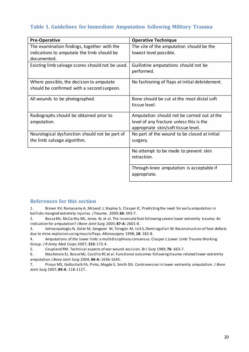
Table 1. Guidelines for Immediate Amputation following Military Trauma
Pre-Operative Operative Technique The examination findings, together with the
indications to amputate the limb should be documented.
The site of the amputation should be the
lowest level possible.
Existing limb salvage scores should not be used. Guillotine amputations should not be performed.
Where possible, the decision to amputate
should be confirmed with a second surgeon.
No fashioning of flaps at initial debridement.
All wounds to be photographed. Bone should be cut at the most distal soft tissue level.
Radiographs should be obtained prior to
amputation.
Amputation should not be carried out at the
level of any fracture unless this is the appropriate skin/soft tissue level.
Neurological dysfunction should not be part of the limb salvage algorithm.
No part of the wound to be closed at initial surgery.
No attempt to be made to prevent skin retraction.
Through-knee amputation is acceptable if
appropriate.
References for this section 1. Brown KV, Ramasamy A, McLeod J, Stapley S, Clasper JC. Predicting the need for early amputation in ballistic mangled extremity injuries. J Trauma. 2009; 66: S93-7.
2. Bosse MJ, McCarthy ML, Jones AL et al. The insensate foot following severe lower extremity trauma: An indication for amputation? J Bone Joint Surg 2005; 87-A: 2601-8. 3. Selmanpakoglu N, Güler M, Sengezer M, Türegün M, Isik S, Demirogullari M. Reconstructi on of foot defects due to mine explosion using muscle flaps. Microsurgery 1998; 18: 182-8.
4. Amputations of the lower l imb: a multidisciplinary consensus. Clasper J; Lower Limb Trauma Working Group. J R Army Med Corps 2007; 153: 172-4. 5. Coupland RM. Technical aspects of war wound excision. Br J Surg 1989; 76: 663-7. 6. MacKenzie EJ, Bosse MJ, Castil lo RC et al. Functional outcomes following trauma-related lower-extremity
amputation J Bone Joint Surg 2004; 86-A: 1636-1645. 7. Pinzur MS, Gottschalk FA, Pinto, Magde S, Smith DG. Controversies in lower -extremity amputation. J Bone Joint Surg 2007; 89-A: 118-1127.