Blast-induced Mild Traumatic Brain Injury Gregory A. Elder, MDa,b,c,*, Effie M. Mitsis, Phob·d, Stephen T. Ahlers, Phoe, Adrian Cristian, MDd.t Traumatic brain injury (TBI) has been a major cause of mortality and morbidity in the wars in Iraq and Afghanistan, known as Operation Iraqi Freedom (OIF) and Operation Enduring Freedom (OEF). In the popular press, TBI has sometimes been referred to as the signature injury of the Iraq and Afghanistan wars, 1 with estimates that 10% to 20% of returning OIF/OEF veterans have suffered a Most attention focused initially on moderate to severe TBis recognized in theater, 7 and OIF has resulted in the highest number of service-related severe TBis since the Vietnam era. 8 However, it soon became apparent that many OIF/OEF veterans were presenting to Veteran's Affairs 0/A) hospitals and other facilities with symptoms suggestive of the residual effects of mild TBis that were never recognized before discharge. Mild TBis greatly outnumber moderate to severe TBis in this population. 2 • 3 Although diverse mechanisms have resulted in injury, because of the prominent use of improvised Work in the author's labs is supported by grants from the Department of Veterans Affairs (1101RX000179-01 and 101CX000190-01). The views expressed in this article are those of the authors and do not necessarily reflect the official policy or position of the Department of the Navy, Department of Defense, or the United States Government. • Neurology Service, James J. Peters Department of Veterans Affairs Medical Center , 130 West Kingsbridge Road, Bronx, NY 10468, USA b Department of Psychiatry, Mount Sinai School of Medicine, One Gustave l. levy Place, New York, NY 10029, USA < Department of Neurology, Mount Sinai School of Medicine, One Gustave l. levy Place, New York, NY 10029, USA d Rehabilitation Medicine Service, James J. Peters Veterans Affairs Medical Center, 130 West Kingsbridge Road, Bronx, NY 10468, USA • Operational and Undersea Medicine Directorate, Naval Medical Research Center , 503 Robert Grant Avenue, Silver Spring, MD 20910, USA t Department of Rehabilitation Medicine, Mount Sinai School of Medicine, One Gustave L. levy Place, New York, NY 10029, USA * Corresponding author. Neurology Service, James J. Peters Veterans Affairs Medical Center, 130 West Kingsbridge Road, Bronx, NY 10468. E-mail address: gregory.elder@va.gov Psychiatr Clin N Am 33 (2010) 757-781 doi:10.1016/j.psc.2010.08.001 0193-953XI10/$ - see front matter. Published by Elsevier Inc. psych.theclinics.com
Transcript
Blast-induced Mild Traumatic Brain Injury
Gregory A. Elder, MDa,b,c,*, Effie M. Mitsis, Phob·d,
Stephen T. Ahlers, Phoe, Adrian Cristian, MDd.t
Traumatic brain injury (TBI) has been a major cause of mortality and morbidity in the wars in Iraq and Afghanistan, known as Operation Iraqi Freedom (OIF) and Operation Enduring Freedom (OEF). In the popular press, TBI has sometimes been referred to as the signature injury of the Iraq and Afghanistan wars, 1 with estimates that 10% to 20% of returning OIF/OEF veterans have suffered a TBI.2~ Most attention focused initially on moderate to severe TBis recognized in theater, 7 and OIF has resulted in the highest number of service-related severe TBis since the Vietnam era. 8
However, it soon became apparent that many OIF/OEF veterans were presenting to Veteran's Affairs 0/A) hospitals and other facilities with symptoms suggestive of the residual effects of mild TBis that were never recognized before discharge. Mild TBis greatly outnumber moderate to severe TBis in this population.2•3 Although diverse mechanisms have resulted in injury, because of the prominent use of improvised
Work in the author's labs is supported by grants from the Department of Veterans Affairs (1101RX000179-01 and 101CX000190-01). The views expressed in this article are those of the authors and do not necessarily reflect the official policy or position of the Department of the Navy, Department of Defense, or the United States Government. • Neurology Service, James J. Peters Department of Veterans Affairs Medical Center, 130 West Kingsbridge Road, Bronx, NY 10468, USA b Department of Psychiatry, Mount Sinai School of Medicine, One Gustave l. levy Place, New York, NY 10029, USA < Department of Neurology, Mount Sinai School of Medicine, One Gustave l. levy Place, New York, NY 10029, USA d Rehabilitation Medicine Service, James J. Peters Veterans Affairs Medical Center, 130 West Kingsbridge Road, Bronx, NY 10468, USA • Operational and Undersea Medicine Directorate, Naval Medical Research Center, 503 Robert Grant Avenue, Silver Spring, MD 20910, USA t Department of Rehabilitation Medicine, Mount Sinai School of Medicine, One Gustave L. levy Place, New York, NY 10029, USA * Corresponding author. Neurology Service, James J. Peters Veterans Affairs Medical Center, 130 West Kingsbridge Road, Bronx, NY 10468. E-mail address: [email protected]
Psychiatr Clin N Am 33 (2010) 757-781 doi:10.1016/j.psc.2010.08.001 0193-953XI10/$ - see front matter. Published by Elsevier Inc.
psych.theclinics.com
Report Documentation Page Form ApprovedOMB No. 0704-0188
Public reporting burden for the collection of information is estimated to average 1 hour per response, including the time for reviewing instructions, searching existing data sources, gathering andmaintaining the data needed, and completing and reviewing the collection of information. Send comments regarding this burden estimate or any other aspect of this collection of information,including suggestions for reducing this burden, to Washington Headquarters Services, Directorate for Information Operations and Reports, 1215 Jefferson Davis Highway, Suite 1204, ArlingtonVA 22202-4302. Respondents should be aware that notwithstanding any other provision of law, no person shall be subject to a penalty for failing to comply with a collection of information if itdoes not display a currently valid OMB control number.
1. REPORT DATE 2010 2. REPORT TYPE
3. DATES COVERED 00-00-2010 to 00-00-2010
4. TITLE AND SUBTITLE Blast-induced Mild Traumatic Brain Injury
5a. CONTRACT NUMBER
5b. GRANT NUMBER
5c. PROGRAM ELEMENT NUMBER
6. AUTHOR(S) 5d. PROJECT NUMBER
5e. TASK NUMBER
5f. WORK UNIT NUMBER
7. PERFORMING ORGANIZATION NAME(S) AND ADDRESS(ES) Naval Medical Research Center,Operational and Undersea MedicineDirectorate,Silver Spring,MD,20910
8. PERFORMING ORGANIZATIONREPORT NUMBER
9. SPONSORING/MONITORING AGENCY NAME(S) AND ADDRESS(ES) 10. SPONSOR/MONITOR’S ACRONYM(S)
11. SPONSOR/MONITOR’S REPORT NUMBER(S)
12. DISTRIBUTION/AVAILABILITY STATEMENT Approved for public release; distribution unlimited
13. SUPPLEMENTARY NOTES
14. ABSTRACT
15. SUBJECT TERMS
16. SECURITY CLASSIFICATION OF: 17. LIMITATION OF ABSTRACT Same as
Report (SAR)
18. NUMBEROF PAGES
25
19a. NAME OFRESPONSIBLE PERSON
a. REPORT unclassified
b. ABSTRACT unclassified
c. THIS PAGE unclassified
Standard Form 298 (Rev. 8-98) Prescribed by ANSI Std Z39-18
758 Elder et al
explosive devices (lEOs) in both theaters of operation, blast exposure has been the most common cause of TBI.2- 5 More broadly, according to Department of Defense (DoD) statistics of February 6 2010, of the more than 41 ,000 US military casualties in Iraq and Afghanistan, more than 26,000 were caused by explosive devices.9 There are concerns that blast-related TBis may produce both long-term health effects in veterans as well as affecting the in-theater performance of active-duty troops.
This review discusses some of the current controversies related to mild TBI, in particular the distinction between mild TBI and posttraumatic stress disorder (PTSD). The problem of distinguishing between the 2 disorders is not new and has roots dating back to the historical entity known as shell shock. 10 During World War I (WWI), while British troops were engaged in the static trench warfare that was characteristic of fighting on the frontlines in Europe, they were exposed to a variety of blasts at close range, including artillery barrages and mortar attacks. Jn this era before the advent of steel helmets, symptoms developed that were reminiscent of both the postconcussion syndrome and what would now be called PTSD. A variety of names for this entity were used but the most enduring label was shell shock. The disorder became so common during WWI that 10% of British battle casualties were diagnosed with shell shock, accounting for one-seventh of all discharges from the British Army, one-third of cases when physical wounds were excluded. A vigorous debate took place concerning whether shell shock represented a physical injury or was the result of psychic trauma. The debate ended without any clear resolution, but with most clinicians probably favoring a psychological explanation. As World War II (WWII) began, hoping to avoid another epidemic, including associated pension claims, the British government went so far as to ban the use of the term shell shock. 10 Despite this, soldiers continued to be exposed to blasts and to present with a similar range of symptoms. The controversy regarding physical versus psychological injury continued without any clear resolution.
MECHANISMS OF BLAST-RELATED INJURY
A variety of explosives including mortar shells, rocket propelled grenades, and lEOs cause blast injuries. In Iraq and Afghanistan, lEOs have been the most common cause of blast injuries and are estimated to be responsible for about 40% of coalition deaths in Iraq, and a roughly similar percentage ofTBis.11 Although diverse in design, lEOs typically consist of an explosive charge coupled to a detonator. 12 The explosive charge may be a conventional artillery shell or be made from commercially manufactured or homemade explosives. lEOs often incorporate shrapnel-generating materials including nails, ball bearings, scrap metal, or other particulate material. Devices used to trigger lEOs may be sophisticated or simple, ranging from electronic transmitters to trip wires, tilt switches, motion detectors, or thermal or pressure-sensitive switches. lEOs are often placed along transport routes and triggered to detonate beneath vehicles. Vehicle-borne devices may contain large quantities of explosives and are placed in strategic locations or driven into their targets.
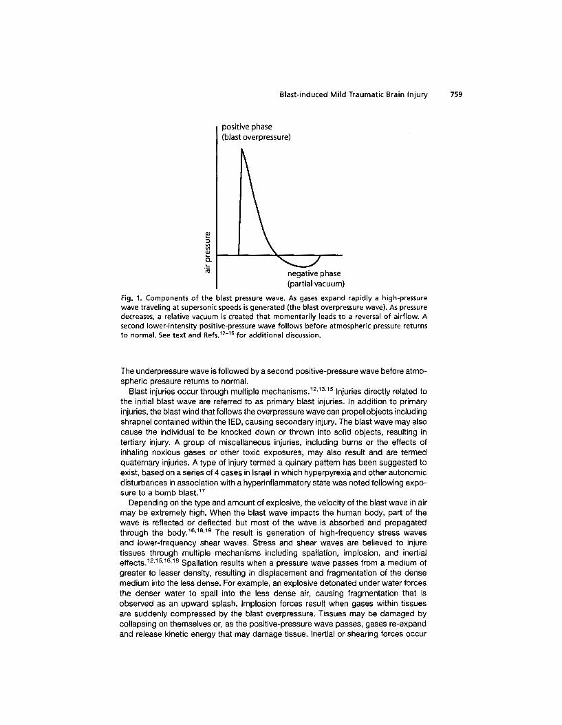
During a detonation, a solid or liquid explosive is converted nearly instantaneously into a gas, creating a pulse of increased air pressure lasting only milliseconds. 12- 16 The gases rapidly expand as a sphere from the point source, forming a high-pressure wave, known as the blast overpressure wave (Fig. 1). The overpressure wave travels faster than the speed of sound and is followed closely by a blast wind generated by the mass displacement of air caused by the expanding gases. Following the overpressure wave, pressure decreases creating a relative vacuum or blast underpressure wave during which a momentary reversal of airflow or reversed blast wind occurs.
Blast-induced Mild Traumatic Brain Injury 759
positive phase (blast overpressure)
negative phase (partial vacuum)
Fig. 1. Components of the blast pressure wave. As gases expand rapidly a high-pressure wave traveling at supersonic speeds is generated (the blast overpressure wave). As pressure decreases, a relative vacuum is created that momentarily leads to a reversal of airflow. A second lower-intensity positive-pressure wave follows before atmospheric pressure returns to normal. See text and Refs. 12- 16 for additional discussion.
The underpressure wave is followed by a second positive-pressure wave before atmospheric pressure returns to normal.
Blast injuries occur through multiple mechanisms.12•13•
15 1njuries directly related to the initial blast wave are referred to as primary blast injuries. In addition to primary injuries, the blast wind that follows the overpressure wave can propel objects including shrapnel contained within the lED, causing secondary injury. The blast wave may also cause the individual to be knocked down or thrown into solid objects, resulting in tertiary injury. A group of miscellaneous injuries, including burns or the effects of inhaling noxious gases or other toxic exposures, may also result and are termed quaternary injuries. A type of injury termed a quinary pattern has been suggested to exist, based on a series of 4 cases in Israel in which hyperpyrexia and other autonomic disturbances in association with a hyperinflammatory state was noted following exposure to a bomb blast.17
Depending on the type and amount of explosive, the velocity of the blast wave in air may be extremely high. When the blast wave impacts the human body, part of the wave is reflected or deflected but most of the wave is absorbed and propagated through the body. 16•18•19 The result is generation of high-frequency stress waves and lower-frequency shear waves. Stress and shear waves are believed to injure tissues through multiple mechanisms including spallation, implosion, and inertial effects.12
•15
•16•18 Spallation results when a pressure wave passes from a medium of
greater to lesser density, resulting in displacement and fragmentation of the dense medium into the less dense. For example, an explosive detonated under water forces the denser water to spall into the less dense air, causing fragmentation that is observed as an upward splash. Implosion forces result when gases within tissues are suddenly compressed by the blast overpressure. Tissues may be damaged by collapsing on themselves or, as the positive-pressure wave passes, gases re-expand and release kinetic energy that may damage tissue. Inertial or shearing forces occur
760 Elder et al
when tissues of different densities are propelled at different speeds as the overpressure wave passes through organs or tissues.12·16•18 These forces are similar in their pathophysiological effects to the acceleration/deceleration forces that occur in closed-impact TBis, such as those associated with motor vehicle accidents, when tissues of different densities may be damaged by their collision with one another, or the cytoarchitecture of a tissue may be disrupted.
Factors that affect the degree of primary blast injury include distance from the detonation, the blast wave's peak overpressure, its duration, and other characteristics of the overpressure wave form. 15
•16
•18 The orientation of the body to the blast wave,
as well as environmental factors, may also influence primary blast effects. For example, explosions within enclosed structures or adjacent to walls become amplified by shockwave reflection and cause greater injury than if exposure occurs in an open space. 12
Organs regarded as most vulnerable to blast effects are those having air-fluid interfaces.12·13 The tympanic membrane is regarded as the most susceptible structure in the body, and ruptured tympanic membranes have been commonly observed in relation to blast injuries in Iraq and Afghanistan.13·2o-22 The lungs and abdominal viscera are also highly sensitive to primary blast injury, with pulmonary injury being one of the most common life-threatening injuries in those close to detonation.12
•13
THE PRIMARY BLAST WAVE AND THE BRAIN
How the primary blast wave affects the brain is at present incompletely understood. Computer simulations23-29 predict various potential mechanisms of injury, including induction of high strain effects in traditional coup and contrecoup regions29 and high shear stresses in white matter regions that could be associated with diffuse axonal injury (DAI).23 Some models also predict preferential damage to the brainstem.29 Others suggest that, as the blast wave passes through the head because of the mechanical properties of the skull, there is significant blast pressure magnification caused by reflection of the blast wave off the skull, with the highest mechanical damage predicted in focal areas on the opposite side of the head.24 Blast waves have also been predicted to generate sufficient force to produce potentially damaging skull flexures. 25
Besides direct effects of the primary blast wave on brain, a thoracic mechanism has also been proposed to contribute to brain injury.30·31 Specifically, this theory proposes that a high-pressure blast wave hitting the body compresses the abdomen and chest, inducing oscillating high-pressure waves that can be transmitted through the systemic circulation to the brain, leading to preferential damage to cellular elements close to cerebral vessels. It has also been suggested that blast overpressure may cause sudden hyperinflation of the lungs, inducing a vasovagal response that could lead to apnea, bradycardia, and hypotension, causing cerebral hypoxemia.15
Understanding the mechanisms that underlie primary blast injury has practical implications for protection of troops in the field. In particular, the thoracic mechanism implies that even blast-resistant helmets would not protect against brain injury. In addition, although current body armor protects the trunk from projectile injuries, there are suggestions that it may intensify the blast wave by serving as an improved contact surface for shock propagation31 or serve as a reflecting surface that concentrates the energy of the blast wave by causing it to resonate internally.15 If the thoracic hypothesis is correct, new types of body armor would need to be designed that could absorb or deflect blast wave energies to prevent central nervous system (CNS) injury.
Blast-induced Mild Traumatic Brain Injury 761
Other mechanisms of CNS injury that have been suggested include that the primary blast wave may cause formation of air emboli leading to cerebral infarction.32 It has been also suggested that the blast wave may be focused through the orbital sockets and nasal sinuses, causing preferential injury to the orbitofrontal cortex.33
EXPERIMENTAL STUDIES IN ANIMALS
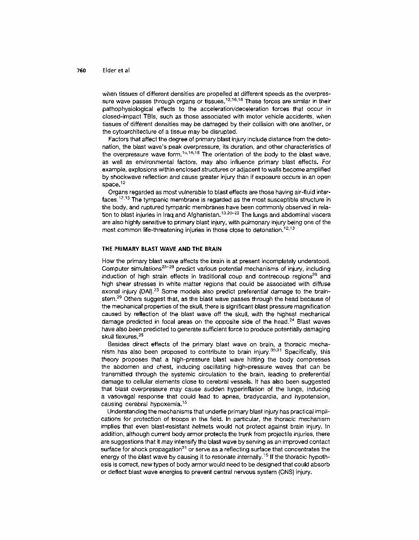
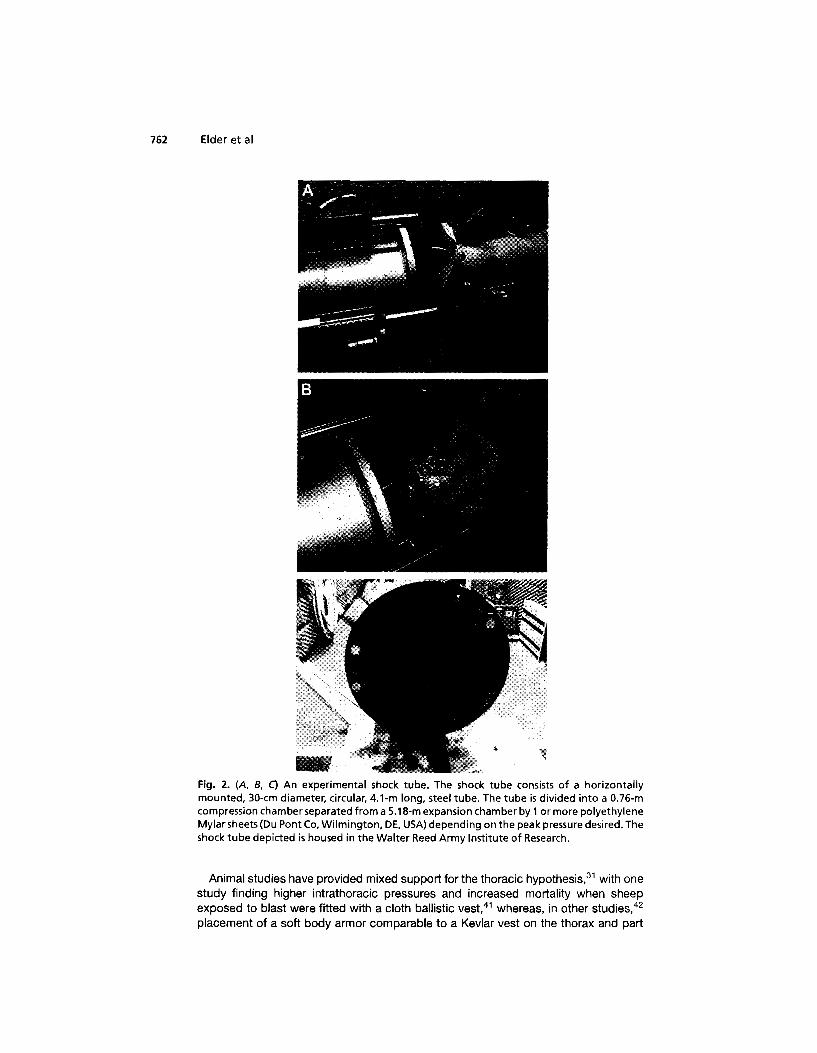
Animals have been exposed to various forms of blast ranging from direct exposure to live explosives to controlled blast waves produced by compressed-air generators. In most studies, to concentrate the blast wave, anesthetized animals have been placed in special holders designed to limit body movement. The animals are secured in the end of a metal tube termed a shock tube if live explosives are used or a blast tube if compressed air is used. Effects of body alignment can be determined by altering the animal's orientation within the tube and, by applying appropriate shielding, it is possible to isolate the effects of body versus head exposure. An example of a blast tube apparatus is shown in Fig. 2.
Although live explosives may best model exposure in the field, this approach affords less experimental control over the physical characteristics of the blast wave as well as difficulty in separating primary from secondary injury. Pressure generators allow blast overpressure effects to be studied in isolation, offering more experimental control. However, a limitation of both shock and blast tubes is that, although they replicate the ideal blast wave, they lack the capability to model the nonideal blast wave, with its multiple shock and expansion fronts, that occurs in real-life settings. Some studies have used open-field exposure or exposure in simulated bunkers or other types of enclosures. For example, Bauman and colleagues34 recently described a swine model of blast injury in which, in addition to exposure in a blast tube, pigs were exposed to an explosive charge detonation while secured in a simulated Humvee or in a 4-sided building.
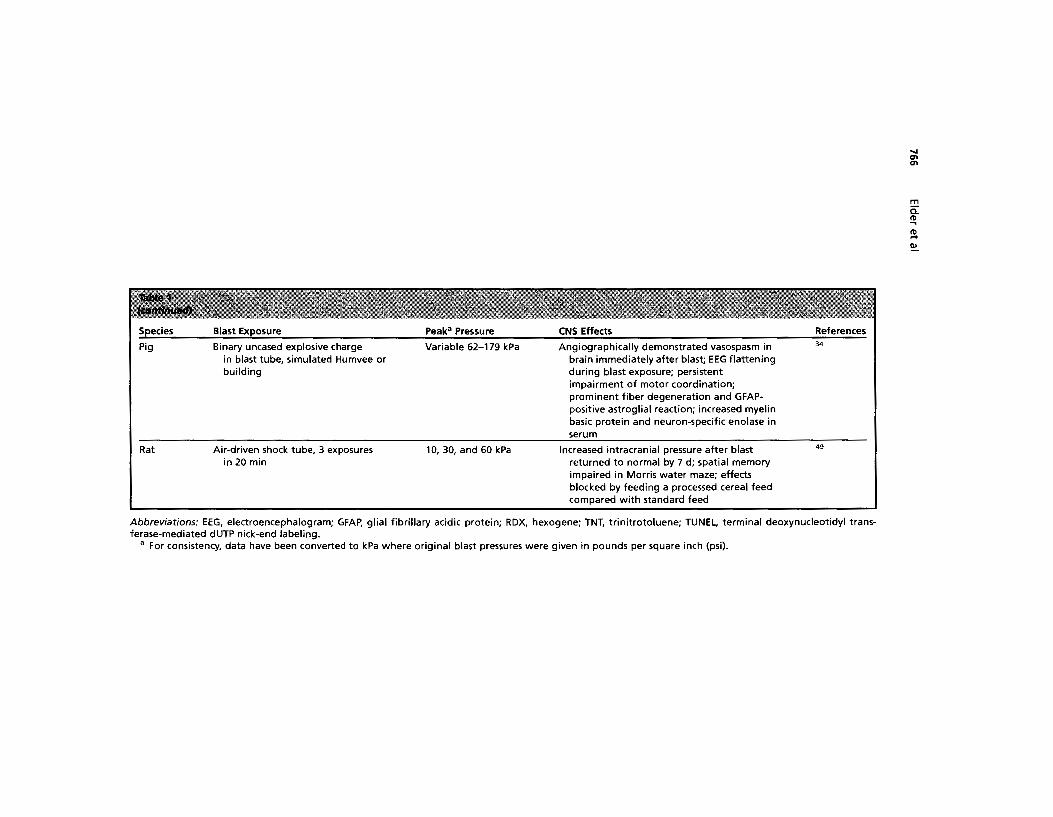
Several studies have looked at the effect of blast exposure on the CNS (Table 1). Most have used rats but some have studied rabbits,35 pigs,34
•36•37 or nonhuman primates.38 One study examined the effects of blast injury to the brain in whales at sea.39 The choice of species has both practical and theoretical implications. Practically, rodents are less expensive to study than larger animals. However, rodents suffer the disadvantage of having smooth brains lacking the gyri, sulci, and proportion of white matter found in human brain, anatomical factors that may affect the mechanical properties of the brain's reaction to blast exposure. Species such as pigs and nonhuman primates offer the advantage of having a brain more similar to humans, but a significant disadvantage lies in their cost and availability.
Studies in rats and pigs have established that the primary blast wave is transmitted through the skull to the brain. 34.4° In pigs, a transient flattening of the electroencephalogram (EEG) with apnea has been observed immediately after blast exposure36;
effects that might, in part, be explained by the vasovagal mechanism alluded to earlier. However, disruption of the EEG in pigs has been seen when the head, but not the body, was exposed to blast injury,34 arguing that a cerebral mechanism may be involved. Prominent vasospasm has also been shown angiographically immediately following blast injury in pigs.34
Pathologically, high-level blast exposure seems particularly prone to inducing hemorrhagic lesions including intraparenchymal, subdural, and subarachnoid bleeding. Blast injury also induces a variety of histological effects including neuroaxonal, glial, microglial, and myelin abnormalities, sometimes with apoptotic neurons (see Table 1). Increased energy consumption and evidence of oxidative stress have been observed, as well as persistent cognitive and motor deficits.
762 Elder et al
Fig. 2. (A B, Q An experimental shock tube. The shock tube consists of a horizontally mounted, 30-cm diameter, circular, 4.1-m long. steel tube. The tube is divided into a 0. 76-m compression chamber separated from a 5.18-m expansion chamber by 1 or more polyethylene Mylar sheets (DuPont Co, Wilmington, DE, USA) depending on the peak pressure desired. The shock tube depicted is housed in the Walter Reed Army Institute of Research.
Animal studies have provided mixed support for the thoracic hypothesis,31 with one study finding higher intrathoracic pressures and increased mortality when sheep exposed to blast were fitted with a cloth ballistic vest,41 whereas, in other studies,42
placement of a soft body armor comparable to a Kevlar vest on the thorax and part
Blast-induced Mild Traumatic Brain Injury 763
of the abdomen reduced mortality and ameliorated the widespread fiber degeneration that was prominent in brains of unprotected rats during exposure to a 126-kPa blast. The importance of vagally mediated effects has been supported by experiments in rabbits35 and rats43 that found that animals with bilateral vagotomies had less bradycardia, hypotension, and apnea; effects that could contribute to cerebral damage. Axonal degeneration in the optic nerve and central visual pathways has also been observed,44 an effect consistent with suggestions that blast forces may be focused by the orbital sockets. 33
One limitation of most animal studies to date is that they have used relatively powerful blast exposures delivered to anesthetized animals at levels high enough to induce significant pulmonary pathology.45-47 Only a few studies have examined effects of lower-level blasts that are probably more comparable with the mild TBI exposures that are the most common exposure in the current war zones. Moochhala and co1Jeagues48 studied the effects of 2.8- to 20-kPa exposures in rats. Although blast-exposed animals showed no deficits in a passive avoidance-learning task, the 20 kPa-exposed animals showed impaired motor function, and histologically scattered hyperchromatic and apoptotic neurons were found in the cerebral cortex 24 hours after exposure. Saljo and colleagues49 also found that exposure of rats to 1 0- to 60-kPa blasts increased intracranial pressure in a dose-dependent manner and cognitive function, as judged by a Morris water maze, was impaired at 2 days after exposure to 10- or 30-kPa blasts. Collectively, these studies clearly indicate that the primary blast wave has CNS effects and that these effects may be apparent even at modest blast pressures.
BLAST-RELATED TBI IN HUMANS
Blast injury is infrequent in civilian life. A survey of 57,392 trauma cases seen in a large urban trauma center found only 89 cases of blast injury (0.2%),50 with private dwelling explosions and industrial accidents being the most common causes. The best understood pathophysiological mechanisms associated with the type of blunt impact TBI seen most commonly in civilian settings are bleeding, direct tissue damage, and DAI.51 DAI results when angular forces cause shearing or stretching of axons that can lead to impaired axonal transport that is pathologically associated with focal axonal swellings. DAI is common following closed head injuries and most commonly affects tracts at gray/white matter junctions, particularly in frontal and temporal regions. Contusions occur as the result of coup/contrecoup injuries, most affecting the frontotemporal regions and occipital lobes.
It is unclear whether blast-related brain injury is similar to the blunt impact injury seen in civilian life or whether blast injury may produce pathophysiologically distinct changes. Few human cases of blast exposure have come to autopsy and all of them sustained such severe injuries that they died within a few days of injury.52 The most prominent features in 2 cases from WWI studied by Mott53 were multiple punctate hemorrhages in subcortical gray and white matter regions. Cohen and Biskind54
identified 9 cases from WWII in the archives of the Armed Forces Institute of Pathology, all whom died within 5 days of injury. These cases also exhibited a prominent hemorrhagic component with diffuse leptomeningeal bleeding, intracerebral clots, and multifocal hemorrhages in white matter.
Close exposure to a high-pressure blast wave can clearly cause moderate to severe TBI, likely activating many of the same pathophysiological cascades seen in closed impact injury. However, these injuries are a mix of secondary and tertiary injuries, making the contribution of primary blast difficult to ascertain. Belanger and
Species Blast Exposure
Rhesus monkey
Rabbit
Rat
Pig
Rat
Rat
Air pressure-driven shock tube
Air pressure-driven shock tube
Air pressure-driven shock tube
Open exposure to RDXfTNT mixture
Air pressure-driven shock tube
Nitrate-based conventional explosive (TNT compound B); exposed in a simulated bunker
Peaka Pressure
207, 276, and 345 kPa
304 kPa
104-110 and 129-173 kPa
200-300 kPa
""338.9 kPa
Equivalent to 110 kg TNT
CNS Effects
Transient impairment in performance on auditory and visual discrimination avoidance task
Acutely in medulla oblongata, increased lipid peroxidation products, glucose and lactate concentrations, lactate/pyruvate ratio and increased phosphocreatine/adenosine triphosphate ratio, all indicative of increased energy consumption
Axonal degeneration in the optic nerve and central visual pathways
Reduction of amplitude of EEG immediately after blast accompanied by apnea
Swollen neurons; glial reaction; myelin debris; increased pinocytotic activity; increased total nitrite/nitrate; increased superoxide dismutase activity, superoxide anion and malondialdehyde generation; decreased glutathione peroxidase activity; impaired performance active avoidance task
Transient widespread microglial response in various brain regions and presence of cells expressing major histocompatibility complex class I and II (Ia) antigen and neurons in the cerebral and cerebellar cortex with darkened dendrites; transient disruption of the ultrastructure of the choroid plexus with widened perivascular spaces and the presence of cells expressing immune-related markers among the epiplexus cells
References 38
35
44
36
45,46
102-104
'.I
t
m c:: II> ..., II> .... ~
Rat
Minke whale
Rat
Rat
Rat
Pig
Rat
Pentaerythrittetranitrate plastic explosive in a metal tube
Harpoon tipped with a grenade containing 30 g penthrite
Pentaerythrittetranitrate plastic explosive in a metal tube detonated in concrete bunker
Air pressure-driven shock tube
Single shock produced by silver azide explosion applied directly to the brain after craniotomy
154 or 240 kPa
Unknown
2.8 or 20 kPa
40 kPa
1 or 10 MPa
Redistribution of phosphorylated neurofilament H protein from axon to perikarya; induction of c-jun, c-fos, c-myc, and ~amyloid precursor protein in neurons in multiple brain regions including hippocampus and cortex; induction of scattered neuronal apoptosis; microglial reaction and induction of GFAP-positive astrocytes
Extensive macroscopic and microscopic intracerebral, subarachnoid and subdural hemorrhage, severity related to proximity of explosion to head
Decreased rotarod and grip strength in 20 kPaexposed animals; scattered hyperchromatic and TUNEL-positive apoptotic cells in cerebral cortex after 20 kPa exposure; effects of blast reversed by aminoguanidine
Transmission of blast pressure waves to brain
At 10 MPa, gross and microscopic hemorrhage in cortical and subcortical regions; induction of apoptotic neurons with evidence of caspase 3 activation; at 1 MPa, spindle-shaped changes in neurons and elongation of neuronal nuclei without other evidence of damage
Firing of a howitzer, bazooka, or automatic rifle 9-30 kPa Blast wave transmitted to brain; at 30-kPa in open air; automatic rifle fire in concrete exposure grossly visible subdural, enclosure; local body exposure from air subarachnoid, and intraparenchymal pressure-driven shock tube; underwater hemorrhages; microscopic intraparenchymal exposure hemorrhages
Air pressure-driven shock tube 126 and 147 kPa Hemorrhage, necrosis and widespread fiber degeneration in brain; impaired beam walking and impaired spatial learning in Morris water maze; less fiber degeneration in 126 kPa-exposed animals wearing a Kevlar vest
105-108
39
48
40
109
37
42
(continued on next page)
!!: Qj
7 :;· a. c n (1) a. s: 0:: ::;l Qj
c 3 Qj .... ;:;· tll iil :;· 5" c:· -<
~ Cl'l Ul
Species
Pig
Rat
Blast Exposure
Binary uncased explosive charge in blast tube, simulated Humvee or building
Air-driven shock tube, 3 exposures in 20 min
Peak" Pressure
Variable 62-179 kPa
10, 30, and 60 kPa
CNS Effects
Angiographically demonstrated vasospasm in brain immediately after blast; EEG flattening during blast exposure; persistent impairment of motor coordination; prominent fiber degeneration and GFAPpositive astroglial reaction; increased myelin basic protein and neuron-specific enolase in serum
Increased intracranial pressure after blast returned to normal by 7 d; spatial memory impaired in Morris water maze; effects blocked by feeding a processed cereal feed compared with standard feed
• For consistency, data have been converted to kPa where original blast pressures were given in pounds per square inch (psi).
-..j 0'1 0'1
m a: 1'1) .... !l ~
Blast-induced Mild Traumatic Brain Injury 767
colleagues55 compared neuropsychological test results in a group of primarily OIF/ OEF veterans who sustained TBis as a result of blast versus those who sustained TBis from blunt force trauma and found no major differences in patterns between blast and non-blast-injured subjects, thus providing no support at the neuropsychological level that blast is different. However, distinct pathophysiological mechanisms might produce similar functional consequences. It is also unclear whether long-term effects may be caused by a primary blast wave sufficient only to produce mild TBI and whether multiple exposures to this type of blast, which is common among troops in Iraq, 2 can lead to significant long-term effects. There are few neuropathological data on mild TBI in general, and none related to blast-induced mild TBI.
NEUROIMAGING IN MILD TBI
Use of in vivo measures to understand the mechanisms of brain damage, particularly mild TBI, as a result of acute and repeated exposure to blast is in its infancy. Conventional structural imaging techniques such as computed tomography (CT) and magnetic resonance imaging (MRI) have been used historically in both civilian and military patients with TBI, and are capable of rapid identification of contusions or hemorrhages in the dural and parenchymal spaces as well as cerebral edema. However, these techniques often result in negative findings in nonblast, civilian mild TBI (data on the use of CT and MAt in military patients following blast exposure is limited at this juncture), because they are considered to be tess sensitive to the small size of mild TBI lesions and to DAI,56•57 which is widely hypothesized to be a principle mechanism of damage in mild TBI. Hence, an absence of visible damage on structural scans does not necessarily mean an absence of abnormality, as the lesions associated with mild TBI may be too small, thus failing to achieve the current threshold of detection for these techniques.58•59 In addition, conventional MRI is sensitive to blood from torn vessels but is not sensitive to axonal damage itself, thus likely underestimating the presence of DAI, especially in milder cases of injury in which hemorrhagic lesions are uncommon.
Functional imaging, such as positron emission tomography (PET), is also used in the evaluation of TBI but is not typically the standard of practice in clinical or military settings during the acute stage of assessment in mild TBI. PET studies have been used extensively in research studies evaluating brain glucose metabolic activity and cerebral blood flow in civilian patients with TBI and have contributed considerably to understanding of abnormalities in subtle brain injury. Recent studies suggest that PET imaging may be more sensitive to mild trauma than is conventional structural imaging such as CT or MRI.60•61 For example, PET imaging has often shown abnormalities in mild TBI that show some correlation with prognosis, Glasgow Coma Scale (GCS), and neuropsychological deficits in the absence of findings on structural imaging. In what may be an important direction in the understanding of chronic symptoms of mild TBI in the absence offindings on CT or MRI, and thus for clinical practice, recent review articles60
•61 have generally concluded that functional brain imaging is
more sensitive than anatomical imaging in the period of months and even years after the injury.62
Diffusion tensor imaging (DTI) is a new imaging technique that provides a more direct measure of the integrity of white matter fibers via the DTI metrics of fractional anisotropy (FA) and mean diffusivity (MD). White matter tracts normally constrain the isotropic diffusion of water. An FA value approaching 1.0 reflects maximal anisotropic diffusion, and values approaching zero indicate compromised white matter integrity. The MD values are typically increased in brain trauma, but may be
768 Elder et al
decreased, depending on TBI severity or the time frame in which DTI is conducted after injury (eg, see Refs.63-66). Because DTI is considered to be sensitive to subtle forms of damage and to DAI, it holds promise as a sensitive tool in identifying impair· ment in blast-related mild TBI.
An increasing number of DTI studies, conducted primarily in civilian mild TBI, have emerged. Most,56·65•67...o9 but not all,63•64•66 of these studies indicate reductions in FA at sites of axonal shearing injury, indicating reduced directionality in the diffusion of water molecules. The discrepancies found in some of these studies in outcomes (ie, decreased or increased FA) may be related to scanning in the acute versus chronic phase of injury, age of the patient population (adolescent vs adults), methodology used, and selection of regions of interest. Newer studies exploring the relationship between DTI and cognitive outcomes have shown significant correlations between DTI findings and neuropsychological function, particularly in the cognitive domains of learning, memory, and processing speed.68•
7o-73
In response to funding opportunities initiated primarily by the DoD and the Veterans Affairs Administration to address the effects of blast in military personnel, neuroimaging in OIF/OEF veterans with mild TBI is now being conducted by investigators at several VA medical centers across the country. Using more sensitive imaging methods to show brain abnormalities in veterans with chronic postconcussive symptoms years after their last exposure to blast will undoubtedly inform the debate as to whether blast mild TBI is a distinct pathophysiological entity. Identifying biomarkers specific to blast would improve diagnostic accuracy, treatment interventions, and overall patient management.
To our knowledge, only 2 studies have been published with findings in postdeployed veterans and service members with both mild and moderate blast TBI, 74.75 1 using [18F]fluoro-2-deoxyglucose PET (FDG PET)75 and the other DTI.74 Peskind and colleagues75 compared 12 Iraq war veterans with a mean age of 32 years who had at least 1 blast exposure that resulted in acute mild TBI as defined by the American Congress of Rehabilitation Medicine criteria and had persistent postconcussive symptoms with a group of civilian controls who were, on average, 20 years older than the veteran group. At the time of PET scanning, veterans were on average 3.5 (± 1.2) years after their last exposure to blast. Compared with controls, veterans with blast exposure mild TBI with or without PTSD showed consistent regional hypometabolism in infratentorial (cerebellum, vermis, and pons) and medial temporal regions. Veterans also exhibited subtle impairments in complex information processing, some reductions in verbal fluency, processing speed, and aspects of attention and working memory. These findings are the first to show with FDG PET that blast exposure resulting in mild TBI may have a neurobiological substrate that is measurable and that the persistent postconcussive symptoms with which so many OIF/OEF veterans present at VA hospitals should not be solely attributed to psychiatric disorders until the contribution of structural brain injury has been fully evaluated and established.
In the 1 published study using DTI, Levin and colleagues74 examined the effects of mild to moderate blast-related TBI in 37 OIF/OEF veterans and service members (mean age 31.5 ± 7.2 years} who were on average 871.5 (± 343.1) days after injury compared with a group of OIF/OEF veterans without blast exposure (N = 15) who sustained injury to other body regions or had no injury. Using manual measurement of single-slice regions of interest, quantitative tractography, and voxel-based methods of DTI analysis, this group found no between-group differences in white matter integrity. The distributions of FA and the apparent diffusion coefficient were similar across neuroanatomic regions of white matter known to be vulnerable to axonal injury. The correlations between DTI data and postconcussion symptoms were weak. Definitive
Blast-induced Mild Traumatic Brain Injury 769
conclusions regarding blast exposure mild TBI cannot be determined from the findings of 1 study. The Levin and colleagues74 study may be especially limited by the sample size of the comparison group, which was less than half of the blast-exposed group. Furthermore, there is a lack of information regarding specifics of the comparison group (ie, type of extracranial injuries that were present in 8 of the 15 subjects).
Systematic and comprehensive studies using functional imaging methods in veterans and service members with blast exposure mild TBI with acute and chronic postconcussive symptoms are beginning to emerge. These comprehensive studies are timely because of the significant number of veterans returning from Iraq and Afghanistan with chronic and disabling sequelae of brain injury. Advanced neuroimaging techniques hold promise for the detection and characterization in vivo of subthreshold, but clinically significant, abnormalities in blast mild TBI.
DISTINGUISHING BLAST-RELATED MILD TBI FROM PTSD
One of the striking features of the mild TBI cases being seen in the current OIF/OEF veterans is the high prevalence of PTSD. PTSD or depression is present in more than one-third of OIF/OEF veterans with suspected postconcussion syndromes secondary to mild TBI. 2 This coincidence could reflect dual exposure to blast as well as stressors that can independently cause PTSD. However, the clinical distinction between a postconcussion syndrome and PTSD is often difficult, with the 2 disorders having many overlapping symptoms. In both disorders, complaints of fatigue, irritability, and poor sleep are frequent. Impaired concentration, attention, and memory are also common symptoms and neuropsychological test profiles may look similar with deficits in attention, working memory, executive functioning, and episodic memory prominent in both disorders.76 In practice, when a documented episode of TBI is present, the clinical distinction between the 2 disorders is usually based on the predominant symptoms, a postconcussion syndrome being suspected when patients have more organic symptoms such as headache, dizziness, visual complaints, hearing loss, balance problems, and cognitive disturbance, whereas PTSD is more likely to be diagnosed when the predominant symptoms include nightmares, hyperarousal, avoidance, and re-experiencing phenomena. The more severe cognitive impairments and associated neurological deficits make cases of moderate to severe TBI easy to recognize. What is more difficult is separating the postconcussion syndrome of mild TBI from PTSD, and sometimes even deciding whether a TBI has occurred may be difficult in cases of mild TBI in which distinguishing transient neurological dysfunction from a psychologically based stress reaction is not always easy.
An increasing number of reports are beginning to address the issue of overlap. The first such study was that of Hoge and colleagues2 who surveyed more than 2700 US Army infantry soldiers from 2 brigades, 3 to 4 months after returning from a 1-year deployment in Iraq. Questionnaires were used to elicit information regarding the occurrence of a TBI and other injuries during deployment as well as current general health status and the presence of symptoms suggestive of a postconcussion syndrome, PTSD, or depression. The most frequent TBI exposure was blast and, among the sample, 5% reported a TBI with loss of consciousness (LOC) that was most typically on the order of a few seconds to 3 minutes. Ten percent reported a TBI without LOC (ie, reported being dazed/confused), giving a total of 15% reporting a TBI. All but 4 of the 384 TBis reported were mild TBis. In soldiers who reported a mild TBI, complaints of headache and poor memory and concentration were frequent, suggesting that a persistent postconcussion syndrome was present. Of those reporting
770 Elder et al
TBI with LOC, 44% met criteria for PTSD, whereas PTSD was present in 27% of those reporting altered mental status without LOC. In addition, major depression was present in 23% and 8% respectively. This high coincidence of PTSD and depression led the investigators to perform a covariate analysis for the 2 disorders and, after adjusting for the coexistence of PTSD and depression, a mild TBI history was no longer significantly associated with adverse physical health outcomes or symptoms, except for headache.
In a subsequent study, this same group determined whether a blast mechanism identifies individuals at higher risk of persistent postconcussive symptoms following TBI.5 Anonymous surveys were administered to 3952 US Army infantry soldiers 3 to 6 months after returning from a year-long deployment to Iraq; 14.9% of the total sample met criteria for having suffered a concussion with most (72.2%) reporting a blast mechanism of injury. Of those who suffered a concussion, 34.2% reported LOC and 63.5% only an alteration of consciousness. Among those with LOC, a blast mechanism was associated with headaches and tinnitus. However, among soldiers reporting only transient alterations of consciousness without LOC, blast was not associated with adverse health outcomes, arguing that a history of blast exposure was associated with persistent postconcussive symptoms only in those whose TBI involved LOC and not most mild TBI cases as currently defined.
In a large randomly selected cohort of more than 5800 UK military personnel deployed to Iraq, Fear and colleagues 77 also found that postconcussive symptoms were associated with exposure to blast during deployment. However, similar symptoms were as likely to occur with other in-theater experiences not associated with blast exposure, such as aiding the wounded or potential exposure to depleted uranium. They concluded that postconcussive symptoms are common in returning troops and, although some may be related to blast exposure, the association is not specific.
One interpretation of these findings is that the current screening procedures and definitions of mild TBI are flawed78 and that much of what is presently being called blast-related mild TBI is really PTSD. Similar suggestions have been made concerning the postconcussion syndrome following closed head injuries in civilian cases, with one prospective study reporting that postconcussive symptoms are as common following trauma without TBI as with mild TBI. 79 Clearly, many of the symptoms of postconcussion syndrome overlap with PTSD and are not specific to either disorder.
However, other studies have suggested that the link may be more than coincidental. Mora and colleagues80 reviewed the records of 333 patients admitted consecutively to the United States Army Institute of Surgical Research burn center for explosionrelated injuries between March 2003 and March 2006 and examined the prevalence of PTSD in patients with burns with and without primary blast injury or mild TBI as defined by LOC. They found a greater prevalence of PTSD in patients with burns with primary blast injury and mild TBI than in patients with burns injured by other mechanisms. Walilko and colleagues,81 in a study of 124 survivors of the Oklahoma City bombing, explored the relationships between PTSD and physical injuries. They found a significant association between PTSD and head/brain injuries, whereas PTSD was not highly correlated with other injuries. Collectively, these studies could be seen as arguing that TBI may predispose to the development of PTSD.
TBI and PTSD can be considered as different ends of a spectrum, with TBI being the classic example of an organic brain disease and PTSD a psychologically based reaction to a stressor that was not associated with physical injury. Indeed, it has been suggested that the posttraumatic amnesia associated with TBI may protect against PTSD, based on the notion that amnesia for the event precludes formation of the core
Blast-induced Mild Traumatic Brain Injury 771
affective responses associated with the development of PTSD. 82 There are likely mitigating factors that allow the development of PTSD even in the context of amnesia for the event, including that, in the context of mild TBI, amnesia may be only partial, allowing for some aspects of the experience to be encoded. 83 In addition, secondary sources such as family or friends may provide enough information to allow the victim to reconstruct the incident, and experiences in the posttrauma period, such as events witnessed after regaining consciousness or subsequent medical procedures, may be psychologically traumatic in and of themselves. PTSD has been documented in moderate and severe TBI.83 However, one prospective study found that PTSD rates were higher in subjects who remembered the TBI incident compared with those with no memory for the event.84 Studies on whether PTSD rates are higher in mild compared with moderate or severe TBI have given more mixed results.83•85 Stress experiences are also typically not limited to single, isolated events, and service personnel in a war zone inevitably have exposure to PTSD stressors independent of TBI events, making coexistence of the 2 disorders easy to imagine.
A second possibility to explain the PTSD!TBI interface is that the 2 conditions are not coincidental but rather that TBI may increase the risk of developing PTSD following a psychological trauma. Physical injury of any type, even if not involving the brain, has been reported to increase the risk of developing PTSD.86 Studies of Vietnam veterans have also suggested that TBI is associated with more severe PTSD87 and, in OEF/OIF veterans, PTSD is more prevalent in veterans reporting mild TBI, compared both with veterans who suffered no injury2
•6 or with those who suffered injuries not involving the
head. 2 In the study by Hoge and colleagues, 2 mild TBI was associated with PTSD even after controlling for the intensity of combat experience.
These observations thus raise the question of whether a neural insult might alter reactions to psychological stressors and increase the likelihood that PTSD will develop. One mechanism whereby TBI might predispose to PTSD would be if blastrelated injury damaged brain structures that are involved in the development of PTSD.33
•76
•88•89 Current biological models of PTSD postulate that key frontal and limbic structures, including the prefrontal cortex, amygdala, and hippocampus, are involved in the development of PTSD. 76
·90
•91 These models suggest that a key compo
nent of the disorder is inadequate frontal inhibition of the amygdala, a limbic structure believed to be central to the fear response and the formation of fear associations. Exaggerated amygdala responses are believed to heighten responses to psychological threats. A substantial body of functional neuroimaging data is consistent with such models, suggesting that there is heightened amygdala activity with decreased hippocampal and orbital frontal activity in PTSD,90•
91 and, in a study of penetrating brain injuries in Vietnam veterans, damage to the amygdala was associated with less PTSD compared with other lesion locations.92 Damage to the prefrontal cortex by TBI could therefore predispose individuals to abnormally sustained responses to psychological stressors. Damage to regions such as the hippocampus might also impair the cognitive reserve needed to deal with psychological stressors afterward.
At present, the regional specificity of blast-related brain injury is not known. What is also not known is the degree to which subclinical blast exposure (ie, not sufficient to produce a transient neurological alteration qualifying as a TBI) may affect the brain. Multiple subclinical exposures are common in the war zones in Iraq and Afghanistan and could affect the brain even in the absence of a diagnosable TBI event. Largely because of research in the sport's medicine literature, the definition of concussion, which used to require LOC, has been expanded to include even the most transient alterations of consciousness. If subclinical blast exposure affects the brain, this definition would need to be expanded even further to capture the full spectrum of blast
772 Elder et al
effects on brain, and would suggest that the TBI model may be misleading with regard to blast and fail to capture what is really a spectrum of blast-related brain injury.
The distinction between blast-related brain injury and PTSD has more than academic significance because it affects treatment strategies as well as patient education. The pathophysiology of PTSD is most commonly conceptualized as an abnormally sustained stress response. Treatment of PTSD is focused on normalization of stress reactions through psychologically based as well as pharmacologically based treatments, the latter often involving the use of selective serotonin reuptake inhibitors. By contrast, TBI treatments are based on an organic model that presumes that structural brain alterations have occurred and that recovery depends on neurological factors. Treatments focus on improving attention and concentration with agents such as psychostimulants, or improving compensatory strategies through cognitivebehavioral therapies. Pharmacological interventions that improve one condition may worsen the other.93 For example, a-adrenergic blockers such as prazocin, which are given to improve sleep in PTSD, may worsen TBI-related cognitive complaints, and agents that improve cognitive function, such as methylphenidate, may exacerbate PTSD symptoms. It is also possible that persistent cognitive deficits associated with TBI may complicate cognitive-behavioral approaches to PTSD because both exposure-based and cognitive-behavioral interventions depend on some measure of cognitive reserve, making it plausible that cognitive deficits associated with TBI may reduce responsiveness to PTSD therapies.
The diagnosis of TBI may also be viewed as carrying the implication of permanent brain injury, and labels can unintentionally affect prognosis. By contrast, although the diagnosis of PTSD may be regarded as more benign in one sense, it still carries a stigma in the minds of some. This attitude may be particularly prevalent in the culture of the military, in which service personnel may be more comfortable and receptive of treatment depending on the diagnosis.
DIAGNOSIS AND SCREENING FOR BLAST-RELATED TBI
The diagnosis of moderate to severe TBI is straightforward even in theater because the traumatic incident is generally apparent along with prolonged alterations of consciousness; other clinical signs and symptoms, and often neuroimaging abnormalities, are discovered later. By contrast, accurate identification of mild TBI can be challenging because of the more subtle signs of injury, the paucity of objective physical findings, and the overlap of postconcussion symptoms with those of other disorders including depression, PTSD, and the effects of chronic pain. In theater, it can even be difficult to establish whether a TBI event has occurred because of the transient nature of the neurological dysfunction and the difficulty of distinguishing TBI from an acute stress reaction. The problem is compounded by the diagnosis frequently not being made until much later when it is difficult for the veteran to recall the events surrounding a blast that occurred several months or even years earlier. It is often difficult to reconstruct the time course of the veteran's present symptoms in relation to the TBI event, information that is critical to establishing the existence of a postconcussion syndrome. Soldiers in these wars are also in constant exposure to blasts, some close by and some at a distance, and the effects of repeated subclinical exposure to blasts (ie, less than the threshold of producing a TBI event) are unknown.
It is important to obtain as many details about specific events as possible. Good detective work is often necessary. Obtaining medical records or talking to key people familiar with the event can be challenging, but it can yield useful information. As in civilian head trauma, the diagnosis of blast-related mild TBI is largely
Blast-induced Mild Traumatic Brain Injury 773
based on establishing an accurate history of the specific events that led to a brain injury. It is important to determine whether there was direct trauma to the brain (ie, falls, motor vehicle accidents, bullets) or indirect trauma (ie, close exposure to blasts). It is clearly important to establish the presence of symptoms such as headaches, dizziness, impaired balance, memory loss, inability to focus, difficulty making decisions, mental fatigue, irritability, and problems with sleep soon after the event and their persistence to the present day. In addition, the effects of these symptoms on current functioning and interpersonal relationships should be ascertained: For example, are they having difficulties at work keeping up with job requirements or getting along with supervisors and coworkers? If in school, are they receiving low grades in course work? To what extent have the often-reported behavioral changes ~rritability, low frustration tolerance, anger outbursts) affected their lives? The effect of these symptoms on key social relationships with spouses, children, and friends should also be determined.
The physical examination is often limited in aiding the diagnosis of mild TBI in blastinjured veterans, especially if they are far removed from the immediate event. Nevertheless, a thorough neurological and cognitive examination should be performed that assesses orientation, concentration, immediate and delayed recall, as well as motor strength, balance, and gait. Assessment for tympanic membrane rupture is also important because this has been described as a marker of concussive blast injury in soldiers. 13
•2o-22 Standard neuroimaging, such as CT scan and MRI, are often not help
ful, but should be considered on a case-by-case basis. Current research is exploring the usefulness of functional imaging as an adjuvant to the diagnostic process. One new technique being used in blast-related mild TBI is DTI, which holds promise as a sensitive tool in identifying impairment in mild injury (as discussed earlier). Although there is much interest in establishing biomarkers for TBI,94
•95 none currently exist. To deal with the problem of missed cases, both the DoD and VA have implemented
population-based screening procedures for mild TBI. 3•96•97 1n recognition of the importance of early diagnosis, the DoD in 2007 implemented screening processes throughout the course of combat operations, beginning with troops in the field who may have experienced a blast-related TBL As soon as practical, soldiers are evaluated by medics using a structured assessment tool, the Military Acute Concussion Evaluation (MACE).98 The MACE consists of 13 items (8 history and 5 examination). In the history assessment, the clinician records details of the incident. Items such as a description of the event, cause of the injury (ie, explosion, blast, fall, motor vehicle accident, gunshot wound), wearing of a helmet, amnesia surrounding the event, LOC, and symptoms are recorded. In the examination, the clinician evaluates 5 domains: (1) orientation; (2) immediate memory; (3) neurological screening of vision, speech, and motor function; (4) concentration; and (5) delayed recall. Total maximum score for the examination is 30. Scores less than 25 may indicate cognitive impairment. 96 The usefulness of this instrument for predicting subsequent functional impairment has not been established, but it holds the potential for providing the most objective history regarding the initial injury.
More recently, for screening in the field, the DoD has moved away from what has been called a symptom-based approach to an incident-based approach to facilitate early detection.99 This new protocol, which was being implemented in spring 2010, makes medical evaluations mandatory for those involved in incidents such as being close to explosions or blasts, whereas, in the past, evaluations were performed after such incidents only if soldiers reported symptoms. As part of this initiative, the DoD is also focusing on educating commanders as well as troops in the field on the symptoms of TBI and the importance of early diagnosis and treatment.
774 Elder et al
Service members returning from OIF/OEF undergo a range of mandatory health assessments as part of their Postdeployment Health Assessment, which includes assessment for self-reported TBis. One of these assessments, the Brief Traumatic Brain Injury Screen (BTBIS) comprises 3 questions designed to determine whether an injury occurred, whether that injury was associated with a TBI, and whether there are current symptoms that are consistent with an ongoing postconcussion syndrome.98
The BTBIS is used to identify those who may have sustained an injury that was not identified acutely. Since May 2008, baseline neurocognitive testing before deployment has been performed using the Automated Neuropsychological Assessment Metrics (ANAM)97• The ANAM TBI military battery is completed during a 15-minute computerized assessment and includes tests of code substitution, simple reaction time, matching to sample, reaction time, and mathematical processing. The ANAM thus provides a baseline against which cognitive functioning after injury can be compared.
The VA has mandated TBI screening for all OIF/OEF veterans who present to VA hospitals for any reason, using a 4-question screening procedure, 98 the basis of which is, like the BTBIS, to establish whether a TBI event is likely to have occurred and whether the veteran is currently symptomatic. Although such screens lack specificity and are subject to both false-positive and false-negative results, they are suitable for large-scale postdeployment screening.98The VA has also established a polytrauma/ TBI system of care nationwide to deal with identified cases.100 A positive screen triggers referral to a clinic within this network where a secondary TBI evaluation is performed. If a TBI is confirmed, the veteran is referred for appropriate clinical services.
Research efforts are also underway, both within the VA and DoD, to increase understanding of the pathophysiology and natural history of blast-related TBI. 97 Among these efforts are the establishment of a DoD TBI registry and studies to validate the MACE as well as other screening tools. Active efforts are also underway to apply the most modern neuroimaging techniques to blast-related TBI and to use advanced computer modeling methods to simulate the effects of blast.
TREATMENT PRINCIPLES FOR THE VETERAN WITH A BLAST-RELATED MILD TBI
The cornerstones in the treatment of veterans with mild TBI are education, symptom management, and care coordination. Veterans and their families are educated on the causes, symptoms, treatments, and prognosis of mild TBI. The educational interventions take into account the veteran's cognitive and emotional impairments as well as their cultural and religious beliefs and preferred method of learning. Educational materials must be written at an appropriate reading level and in a language that the veteran can readily understand. The information provided often needs to be repeated at several visits and by different providers, therefore consistency in the content and method of education by all providers is important.
In educating the veteran with a mild TBI, it is important that the instruction occurs in a quiet environment that is conducive to learning and that an adequate amount of time is allowed for the visits. Material can be reinforced through alternate means such as telephone calls whenever necessary. Family education is critical to the successful reintegration of the veteran back to the community and home life. Families are encouraged to participate in educational activities and support groups. Some examples of general educational content include (1) compensatory strategies for impaired memory and concentration; (2) relaxation techniques; (3) anger management techniques; (4) diet and exercise; (5) strategies for successful reintegration in work, school, and social activities; (6) limiting alcohol and caffeine intake; and (7) avoidance of high-risk behavior that could increase the risk of additional head injuries.
Blast-induced Mild Traumatic Brain Injury 775
The symptoms associated with mild TBI can be broadly divided into 3 groups: physical, cognitive, and behavioral. It is common for veterans to complain of concurrent symptoms in each of these groups. It is therefore important to prioritize and treat the symptoms that cause the veteran the most distress first. Although the treatment of specific symptoms associated with mild TBI is beyond the scope of this article, some general principles are briefly discussed. The reader is directed to the VNDoD Clinical Practice Guideline for more specific treatment recommendations in this population. 1 01
Injured brains are sensitive to the side effects of medications, therefore close monitoring during treatment is suggested to evaluate for potential toxicities and drug-drug interactions. Clinicians should avoid medications that can lower the seizure threshold, cause drowsiness or slow thinking, as well as those medications associated with increasing risk of suicidal ideation, because suicide risk is higher in this population.
Some commonly used interventions in the symptom management of veterans with mild TBI include (1) physical therapy for the treatment of musculoskeletal pain syndromes and balance disorders; (2) voice recorders, global positioning systems (GPS) and personal digital assistants as aids to veterans with memory impairments; (3) cognitive remediation training; (4) sunglasses for veterans whose eyes are sensitive to sunlight; (5) speech pathology referrals for teaching of communication skills; (6) mental health referrals for management of depression, PTSD, and anxiety; (7) referrals to substance abuse treatment specialists as needed; (8) teaching of sleep management techniques such as avoidance of alcohol, avoidance of caffeine products, stimulants, and nicotine before a sleep period, waking up at regular times, avoidance of naps during the daytime, and stimulating activities immediately before sleep. Vigilance for conditions such as sleep apnea is recommended, with referrals for sleep studies or to sleep specialists as needed.
Case management in this population is important to ensure that care is coordinated and that social service needs are adequately addressed. The veteran should also be educated on return to activities such as work, school, and leisure. The decision on when and how to return is made based on the severity of the cognitive, physical, and emotional impairments and the type of work previously engaged in. Safety is a primary consideration if the veteran previously engaged in high-risk work. Another consideration is the ability to perform specific tasks with competence. Successful reintegration may involve a period of work restriction or accommodation such as provision of additional time to complete tasks and working in a quiet environment with additional supervision.
CONCLUDING REMARKS
TBI has been a major cause of mortality and morbidity in the wars in Iraq and Afghanistan. In both theaters of operation, blast exposure has been the most common cause of TBI. Blast injuries occur through multiple mechanisms that likely activate many of the same pathophysiological cascades seen in closed impact injuries in civilian life. What is less clear is whether the primary blast wave causes brain damage through mechanisms that are pathophysiologically distinct from those common in civilian TBI and, in particular, whether multiple exposures to low-level blast can lead to long-term sequelae. One of the other striking features of the mild TBI cases being encountered in OIF/OEF veterans is the high prevalence of PTSD. At present, it is unclear whether this association reflects coincident exposure or whether the relationship is more complex with, for example, TBI increasing the risk of developing PTSD by damaging brain structures that are involved in mediating PTSD. Resolution of these issues affects treatment strategies as well as strategies for protection of troops in
776 Elder et al
the field and is an area of high priority for research both within the DoD and Department of Veteran's Affairs.
REFERENCES
1. Alvarez L. War veterans' concussions are often overlooked. New York Times. August 25, 2008: A 1.
2. Hoge CW, McGurk D, Thomas JL, et al. Mild traumatic brain injury in U.S. soldiers returning from Iraq. N Engl J Med 2008;358:453-63.
3. Tanielian T, Jaycox LH, editors. Invisible wounds of war: psychological and cognitive injuries, their consequences, and services to assist recovery. Santa Monica (CA): Rand Corporation; 2008.
4. Terrio H, Brenner LA, Ivins BJ, et al. Traumatic brain injury screening: preliminary findings in a US Army Brigade Combat Team. J Head Trauma Rehabil2009;24:14-23.
5. Wilk JE, Thomas JL, McGurk DM, et al. Mild traumatic brain injury (concussion) during combat: lack of association of blast mechanism with persistent postconcussive symptoms. J Head Trauma Rehabil 2010;25:9-14.
6. Schneiderman AI, Braver ER, Kang HK. Understanding sequelae of injury mechanisms and mild traumatic brain injury incurred during the conflicts in Iraq and Afghanistan: persistent postconcussive symptoms and posttraumatic stress disorder. Am J Epidemic! 2008;167:1446-52.
7. Warden DL, Ryan L, Helmick K, et al. War neurotrauma: the defense and veterans brain injury center (DVBIC) experience at the Walter Reed Army Medical Center. J Neurotrauma 2005;22: 1178.
8. Bell RS, Vo AH, Neal CJ, et al. Military traumatic brain and spinal column injury: a 5-year study of the impact blast and other military grade weaponry on the central nervous system. J Trauma 2009;66:S 104-11.
9. Available at: http://siadapp.dmdc.osd.mil/personnei/CASUALTY/castop.htm. Accessed February 6, 2010.
10. Jones E, Fear NT, Wessely S. Shell shock and mild traumatic brain injury: a historical review. Am J Psychiatry 2007;164:1641-5.
11. Brookings Institution, Saban Center for Middle East Policy. Iraq index: tracking variables of reconstruction and security in post-Saddam Iraq. April 27, 2010. Available at: www.brookings.edu/iraqindex. Accessed May 2, 2010.
12. Wolf SJ, Bebarta VS, Bonnett CJ, et al. Blast injuries. Lancet 2009;37 4:405-15. 13. DePalma RG, Burris DG, Champion HR. et al. Blast injuries. N Engl J Med 2005;
352: 1335-42. 14. Taber KH, Warden OL, Hurley RA. Blast-related traumatic brain injury: what is
known? J Neuropsychiatry Clin Neurosci 2006; 18:141-5. 15. Cernak I, Noble-Haeusslein LJ. Traumatic brain injury: an overview of pathobi
ology with emphasis on military populations. J Cereb Blood Flow Metab 2010; 30:255-66.
16. Leung LY, VandeVord PJ, Dal Cengio AL, et al. Blast related neurotrauma: a review of cellular injury. Mol Cell Biomech 2008;5:155-68.
17. Kluger Y, Nimrod A, Biderman P, et al. The quinary pattern of blast injury. Am J Disaster Med 2007;2:21-5.
blast-related potential mild traumatic brain injury and comorbidities. Clin Neuropsychol2009;23:1315-37.
20. Okie S. Traumatic brain injury in the war zone. N Eng! J Med 2005;352:2043-7.
Blast-induced Mild Traumatic Brain Injury 777
21. Xydakis MS. Bebarta VS, Harrison CD, et al. Tympanic-membrane perforation as a marker of concussive brain injury in Iraq. N Engl J Med 2007;357:830-1.
22. Ritenour AE, Wickley A, Ritenour JS, et al. Tympanic membrane perforation and hearing loss from blast overpressure in Operation Enduring Freedom and Operation Iraqi Freedom wounded. J Trauma 2008;64:S174-8.
23. Chafi MS, Karami G. Ziejewski M. Biomechanical assessment of brain dynamic responses due to blast pressure waves. Ann Biomed Eng 2009;38:490-504.
24. Moore DF, Jerusalem A, Nyein M, et al. Computational biology - modeling of primary blast effects on the central nervous system. Neuroimage 2009;47 (Suppl 2):T10-20.
25. Moss WC, King MJ, Blackman EG. Skull flexure from blast waves: a mechanism for brain injury with implications for helmet design. Phys Rev Lett 2009;103 108702.
26. Taylor PA, Ford CC. Simulation of blast-induced early-time intracranial wave physics leading to traumatic brain injury. J Biomech Eng 2009; 131. 061007.
27. Zhang J, Pintar FA, Yoganandan N, et al. Experimental study of blast-induced traumatic brain injury using a physical head model. Stapp Car Crash J 2009; 53:215--27.
28. Zhang J, Song B. Pintar FA, et al. How to test brain and brain simulant at ballistic and blast strain rates. Biomed Sci lnstrum 2008;44: 129-34.
29. Zhang J, Yoganandan N, Pintar FA, et al. A finite element study of blast traumatic brain injury- biomed 2009. Biomed Sci lnstrum 2009;45:119-24.
30. Bhattacharjee Y. Neuroscience. Shell shock revisited: solving the puzzle of blast trauma. Science 2008;31 9:406-8.
31. Courtney AC. Courtney MW. A thoracic mechanism of mild traumatic brain injury due to blast pressure waves. Med Hypotheses 2009;72:76-83.
32. Phillips YY, Richardson D. Primary blast injury: a brief history. In: Zatchuck R, Jenkins D, Bellamy R, et al, editors. Textbook of military medicine. Part I. Warfare, weapons and the casualty, Conventional warfare. Ballistic, blast and burn injuries, vol. 5. Washington DC: TMM Publications; 1990. p. 221-40.
33. Hoffman SW, Harrison C. The interaction between psychological health and traumatic brain injury: a neuroscience perspective. Clin Neuropsychol 2009;23 1400-15.
34. Bauman RA, LingG, Tong L, et al. An introductory characterization of a combatcasualty-care relevant swine model of closed head injury resulting from exposure to explosive blast. J Neurotrauma 2009;26:841-60.
35. Cernak I, Savic J, Malicevic Z, et al. Involvement of the central nervous system in the general response to pulmonary blast injury. J Trauma 1996;40:S100-4.
36. Axelsson H, Hjelmqvist H, Medin A, et al. Physiological changes in pigs exposed to a blast wave from a detonating high-explosive charge. Mil Med 2000;165:119-26.
37. Saljo A, Arrhen F, Bolouri H, et al. Neuropathology and pressure in the pig brain resulting from low-impulse noise exposure. J Neurotrauma 2008;25: 1397-406.
38. Bogo V, Hutton R, Bruner A. The effects of airblast on discriminated avoidance behavior in rhesus monkeys. In: Technical progress report on contract no. DA-49-146-XZ-372, vol. DASA 2659. Washington, DC: Defense Nuclear Agency; 1971. p. 1-32.
39. Knudsen SK, Oen EO. Blast-induced neurotrauma in whales. Neurosci Res 2003;46:377 -86.
778 Elder et al
40. Chavko M, Koller WA, Prusaczyk WK. et al. Measurement of blast wave by a miniature fiber optic pressure transducer in the rat brain. J Neurosci Methods 2007;159:277-81.
41. Phillips YY, Mundie TG, Yelverton JT, et al. Cloth ballistic vest alters response to blast. J Trauma 1988;28:S 149-52.
42. Long JB, Bentley TL, Wessner KA, et al. Blast overpressure in rats: recreating a battlefield injury in the laboratory. J Neurotrauma 2009;26:827-40.
43. Irwin RJ, Lerner MR. Bealer JF, et al. Shock after blast wave injury is caused by a vagally mediated reflex. J Trauma 1999;47:105-10.
44. Petras JM, Bauman RA, Elsayed NM. Visual system degeneration induced by blast overpressure. Toxicology 1997;121:41-9.
45. Cernak I, Wang Z, Jiang J, et al. Cognitive deficits following blast injury-induced neurotrauma: possible involvement of nitric oxide. Brain lnj 2001; 15:593-612.
46. Cernak I, Wang Z, Jiang J, et al. Ultrastructural and functional characteristics of blast injury-induced neurotrauma. J Trauma 2001 ;50:695-706.
47. Chavko M, Prusaczyk WK, McCarron RM. Lung injury and recovery after exposure to blast overpressure. J Trauma 2006;61 :933-42.
48. Moochhala SM, Md S, Lu J, et al. Neuroprotective role of aminoguanidine in behavioral changes after blast injury. J Trauma 2004;56:393-403.
49. Saljo A, Bolouri H, Mayorga M, et al. Low-level blast raises intracranial pressure and impairs cognitive function in rats: prophylaxis with processed cereal feed. J Neurotrauma 2010;27:383-9.
50. Bochicchio GV, Lumpkins K, O'Connor J, et al. Blast injury in a civilian trauma setting is associated with a delay in diagnosis of traumatic brain injury. Am Surg 2008;74:267-70.
51. Gennarelli TA, Grahm Dl. Neuropathology. In: Silver JM, McAllister TW, Yudofsky SC, editors. Textbook of traumatic brain injury. Arlington (VA): American Psychiatric Publishing; 2005. p. 27-50.
52. Kocsis JD, Tessler A. Pathology of blast-related brain injury. J Rehabil Res Dev 2009;46:667-72.
53. Mott F. The effects of high explosives upon the central nervous system. Lancet 1916;1:441-9.
54. Cohen H, Biskind G. Pathologic aspects of atmospheric blast injuries in man. Arch Pathol 1946;42:12-34.
55. Belanger HG, Kretzmer T, Yoash-Gantz R, et al. Cognitive sequelae of blastrelated versus other mechanisms of brain trauma. J lnt Neuropsychol Soc 2009;15:1-8.
56. Arfanakis K, Haughton VM, Carew JD, et al. Diffusion tensor MR imaging in diffuse axonal injury. AJNR Am J Neuroradiol 2002;23:794-802.
57. Huisman TA, Schwamm LH, Schaefer PW, et al. Diffusion tensor imaging as potential biomarker of white matter injury in diffuse axonal injury. AJNR Am J Neuroradiol 2004;25:370-6.
58. Bigler ED. Quantitative magnetic resonance imaging in traumatic brain injury. J Head Trauma Rehabil 2001 ;16:117-34.
59. Bigler ED, Snyder JL. Neuropsychological outcome and quantitative neuroimaging in mild head injury. Arch Clin Neuropsychol 1995;10: 159-74.
60. Newberg AB, Alavi A. Neuroimaging in patients with head injury. Semin Nucl Med 2003;33:136-47.
61. Van Heertum RL, Greenstein EA. Tikofsky RS. 2-Deoxy-fluorglucose-positron emission tomography imaging of the brain: current clinical applications with emphasis on the dementias. Semin Nucl Med 2004;34:3(){}-12.
Blast-induced Mild Traumatic Brain Injury 779
62. Kato T, Nakayama N, Yasokawa Y, et al. Statistical image analysis of cerebral glucose metabolism in patients with cognitive impairment following diffuse traumatic brain injury. J Neurotrauma 2007;24:919-26.
63. Bazarian JJ, Zhong J, Blyth B. et al. Diffusion tensor imaging detects clinically important axonal damage after mild traumatic brain injury: a pilot study. J Neurotrauma 2007;24:1447-59.
64. Chu Z, Wilde EA, Hunter JV, et al. Voxel-based analysis of diffusion tensor imaging in mild traumatic brain injury in adolescents. AJNR Am J Neuroradiol 2010;31 :340-6.
65. Inglese M, Makani S, Johnson G, et al. Diffuse axonal injury in mild traumatic brain injury: a diffusion tensor imaging study. J Neurosurg 2005;103:298-303.
66. Wilde EA, McCauley SR, Hunter JV, et al. Diffusion tensor imaging of acute mild traumatic brain injury in adolescents. Neurology 2008;70:948-55.
67. Kraus MF, Susmaras T. Caughlin BP, et al. White matter integrity and cognition in chronic traumatic brain injury: a diffusion tensor imaging study. Brain 2007;130: 2508-19.
68. Lipton ML, Gellella E, Lo C, et al. Multifocal white matter ultrastructural abnormalities in mild traumatic brain injury with cognitive disability: a voxel-wise analysis of diffusion tensor imaging. J Neurotrauma 2008;25:1335-42.
69. Miles L, Grossman Rl, Johnson G, et al. Short-term DTI predictors of cognitive dysfunction in mild traumatic brain injury. Brain lnj 2008;22:115-22.
70. Levin HS, Wilde EA, Chu Z, et al. Diffusion tensor imaging in relation to cognitive and functional outcome of traumatic brain injury in children. J Head Trauma Rehabil 2008;23: 197-208.
71. Mathias JL, Bigler ED, Jones NR, et al. Neuropsychological and information processing performance and its relationship to white matter changes following moderate and severe traumatic brain injury: a preliminary study. Appl Neuropsychol 2004; 11: 134-52.
72. Niogi SN, Mukherjee P, Ghajar J, et al. Extent of microstructural white matter injury in postconcussive syndrome correlates with impaired cognitive reaction time: a 3T diffusion tensor imaging study of mild traumatic brain injury. AJNR Am J Neuroradiol 2008;29:967-73.
73. Salmond CH, Menon OK, Chatfield DA, et al. Diffusion tensor imaging in chronic head injury survivors: correlations with learning and memory indices. Neuroimage 2006;29:117-24.
7 4. Levin HS, Wilde E, Troyanskaya M, et al. Diffusion tensor imaging of mild to moderate blast-related traumatic brain injury and its sequelae. J Neurotrauma 201 0;27:683-94.
75. Peskind ER, Petrie EC, Cross DJ, et al. Cerebrocerebellar hypometabolism associated with repetitive blast exposure mild traumatic brain injury in 12 Iraq war Veterans with persistent post-concussive symptoms. Neuroimage [Epub ahead of print]. PMID: 20385245.
76. Vasterling JJ, Verfaellie M, Sullivan KD. Mild traumatic brain injury and posttraumatic stress disorder in returning veterans: perspectives from cognitive neuroscience. Clin Psycho! Rev 2009;29:67 4-84.
77. Fear NT, Jones E, Groom M, et al. Symptoms of post-concussional syndrome are non-specifically related to mild traumatic brain injury in UK Armed Forces personnel on return from deployment in Iraq: an analysis of self-reported data. Psycho! Med 2009;39:1379-87.
78. Hoge CW, Goldberg HM, Castro CA. Care of war veterans with mild traumatic brain injury-flawed perspectives. N Engl J Med 2009;360: 1588-91.
780 Elder et al
79. Meares S, Shores EA, Taylor AJ, et al. Mild traumatic brain injury does not predict acute postconcussion syndrome. J Neurol Neurosurg Psychiatry 2008;79:300-6.
80. Mora AG, Ritenour AE, Wade CE, et al. Posttraumatic stress disorder in combat casualties with burns sustaining primary blast and concussive injuries. J Trauma 2009;66:S 178-85.
81. Walilko T, North C, Young LA, et al. Head injury as a PTSD predictor among Oklahoma City bombing survivors. J Trauma 2009;67:1311-9.
82. JosephS, Masterson J. Posttraumatic stress disorder and traumatic brain injury: are they mutually exclusive? J Trauma Stress 1999;12:437-53.
83. Harvey AG, Brewin CR, Jones C, et al. Coexistence of posttraumatic stress disorder and traumatic brain injury: towards a resolution of the paradox. J lnt Neuropsychol Soc 2003;9:663-76.
84. Gil S, Caspi Y, Ben-Ari IZ, et al. Does memory of a traumatic event increase the risk for posttraumatic stress disorder in patients with traumatic brain injury? A prospective study. Am J Psychiatry 2005;162:963-9.
85. Glaesser J, Neuner F, Lutgehetmann R, et al. Posttraumatic stress disorder in patients with traumatic brain injury. BMC Psychiatry 2004;4:5.
86. Koren D, Norman D, Cohen A, et al. Increased PTSD risk with combat-related injury: a matched comparison study of injured and uninjured soldiers experiencing the same combat events. Am J Psychiatry 2005;162:276-82.
87. Vanderploeg RD. Belanger HG, Curtiss G. Mild traumatic brain injury and posttraumatic stress disorder and their associations with health symptoms. Arch Phys Med Rehabil 2009;90:1084-93.
88. Bryant RA. Disentangling mild traumatic brain injury and stress reactions. N Engl J Med 2008;358:525-7.
89. Elder GA, Cristian A. Blast-related mild traumatic brain injury: mechanisms of injury and impact on clinical care. Mt Sinai J Med 2009;76:111-8.
90. Liberzon I, Sripada CS. The functional neuroanatomy of PTSD: a critical review. Prog Brain Res 2008;167:151-69.
91. Rauch SL, Shin LM. Phelps EA. Neurocircuitry models of posttraumatic stress disorder and extinction: human neuroimaging research-past, present, and future. Bioi Psychiatry 2006;60:376-82.
92. Koenigs M, Huey ED, Raymont V, et al. Focal brain damage protects against post-traumatic stress disorder in combat veterans. Nat Neurosci 2008; 11:232-7.
93. McAllister TW. Psychopharmacological issues in the treatment of TBI and PTSD. Clin Neuropsychol 2009;23:1338-67.
94. Agoston DV, Gyorgy A, Eidelman 0, et al. Proteomic biomarkers for blast neurotrauma: targeting cerebral edema, inflammation, and neuronal death cascades. J Neurotrauma 2009;26:901-11.
95. Svetlov Sl, Larner SF, Kirk DR, et al. Biomarkers of blast-induced neurotrauma: profiling molecular and cellular mechanisms of blast brain injury. J Neurotrauma 2009;26:913-21.
96. Iverson GL, Langlois JA, McCrea MA, et al. Challenges associated with postdeployment screening for mild traumatic brain injury in military personnel. Clin Neuropsychol 2009;23: 1299-314.
97. Jaffee MS, Meyer KS. A brief overview of traumatic brain injury (TBI) and posttraumatic stress disorder (PTSD) within the Department of Defense. Clin Neuropsychol 2009;23:1291-8.
98. Available at: www.dvbic.org. Accessed September 23, 2010. 99. Available at: http://www.defense.gov/news/newsarticle.aspx?id=58342.
Accessed September 23, 2010.
Blast-induced Mild Traumatic Brain Injury 781
100. Available at: http //www.polytrauma.va.gov/. Accessed September 23, 2010. 101. VA/DoD Clinical Practice Guideline for Management of Concussion/Mild Trau
matic Brain Injury (mTBI) 2009. Available at: www.healthquality.va.gov/mtbi/ concussion_mtbi_full_1_0.pdf. Accessed September 23, 2010.
102. Kaur C, Singh J, Lim MK, et al. Macrophages/microglia as 'sensors' of injury in the pineal gland of rats following a non-penetrative blast. Neurosci Res 1997;27:317-22.
103. Kaur C, Singh J, Lim MK, et al. The response of neurons and microglia to blast injury in the rat brain. Neuropathol Appl Neurobiol 1995;21 :369-77.
104. Kaur C. Singh J, Lim MK, et aL Studies of the choroid plexus and its associated epiplexus cells in the lateral ventricles of rats following an exposure to a single non-penetrative blast. Arch Histol Cytol 1996;59:239-48.
105. Saljo A, Bao F, Haglid KG, et al. Blast exposure causes redistribution of phosphorylated neurofilament subunits in neurons of the adult rat brain. J Neurotrauma 2000;17:719-26.
106. Saljo A, Bao F, Hamberger A, et al. Exposure to short-lasting impulse noise causes microglial and astroglial cell activation in the adult rat brain. Pathophysiology 2001 ;8: 105-11.
107. Saljo A, Bao F, Jingshan S, et al. Exposure to short-lasting impulse noise causes neuronal c-Jun expression and induction of apoptosis in the adult rat brain. J Neurotrauma 2002; 19:985-91.
108. Saljo A, Bao F, Shi J, et al. Expression of c-Fos and c-Myc and deposition of beta-APP in neurons in the adult rat brain as a result of exposure to short-lasting impulse noise. J Neurotrauma 2002;19:379-85.
109. Kato K, Fujimura M, Nakagawa A, et al. Pressure-dependent effect of shock waves on rat brain: induction of neuronal apoptosis mediated by a caspasedependent pathway. J Neurosurg 2007;106:667-76.