Bleeding and Bleeding and Thrombosis Disorders Thrombosis Disorders in the ICU in the ICU Division of Critical Care Division of Critical Care Medicine University of Medicine University of Alberta Alberta
Transcript
Bleeding and Bleeding and Thrombosis Disorders Thrombosis Disorders
in the ICUin the ICU
Division of Critical Care Medicine Division of Critical Care Medicine University of AlbertaUniversity of Alberta
OutlineOutline
Normal hemostasisNormal hemostasis Coagulation pathwaysCoagulation pathways Initial investigationsInitial investigations
CoagulopathiesCoagulopathies Pathogenesis of coagulopathiesPathogenesis of coagulopathies Specific causesSpecific causes
Normal hemostatic Normal hemostatic mechanismmechanism
There are four There are four phases to the phases to the normal coagulation normal coagulation cascade:cascade: Blood vessel Blood vessel
constrictionconstriction Platelet aggregationPlatelet aggregation Fibrin generationFibrin generation Vessel repair and Vessel repair and
fibrin degradationfibrin degradation
Constriction of vesselsConstriction of vessels
There are 2 mechanisms for vessel There are 2 mechanisms for vessel constriction:constriction: Local smooth muscle contractile responseLocal smooth muscle contractile response Thromboxane AThromboxane A22 release from endothelium release from endothelium
Formation of the unstable Formation of the unstable platelet plugplatelet plug
Exposure of the subendothelial layers Exposure of the subendothelial layers cause platelets to adhere.cause platelets to adhere.
They release ADP and TxAThey release ADP and TxA22, inducing , inducing further platelet aggregation and activationfurther platelet aggregation and activation
Binding and activation of Binding and activation of plateletsplatelets
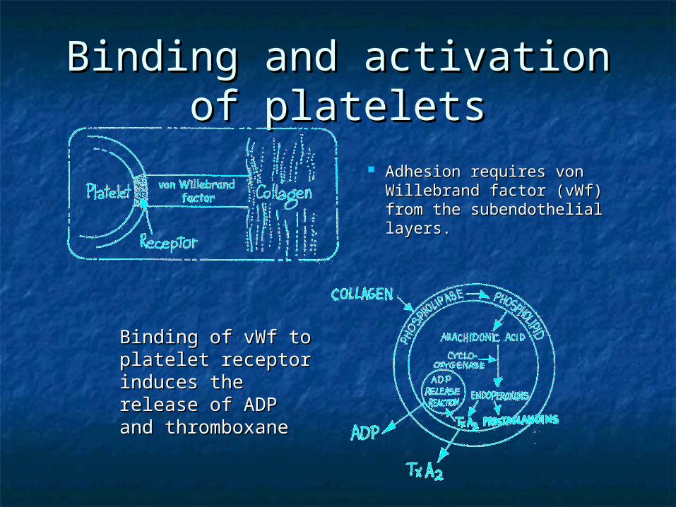
Adhesion requires von Adhesion requires von Willebrand factor (vWf) from Willebrand factor (vWf) from the subendothelial layers.the subendothelial layers.
Binding of vWf to Binding of vWf to platelet receptor platelet receptor induces the release induces the release of ADP and of ADP and thromboxanethromboxane
Generation of fibrinGeneration of fibrin
Aggregated platelets provide a surface on which Aggregated platelets provide a surface on which blood coagulation occurs through the generation blood coagulation occurs through the generation of thrombin and cleavage of fibrinogen.of thrombin and cleavage of fibrinogen.
Stabilization of platelet plug Stabilization of platelet plug with fibrinwith fibrin
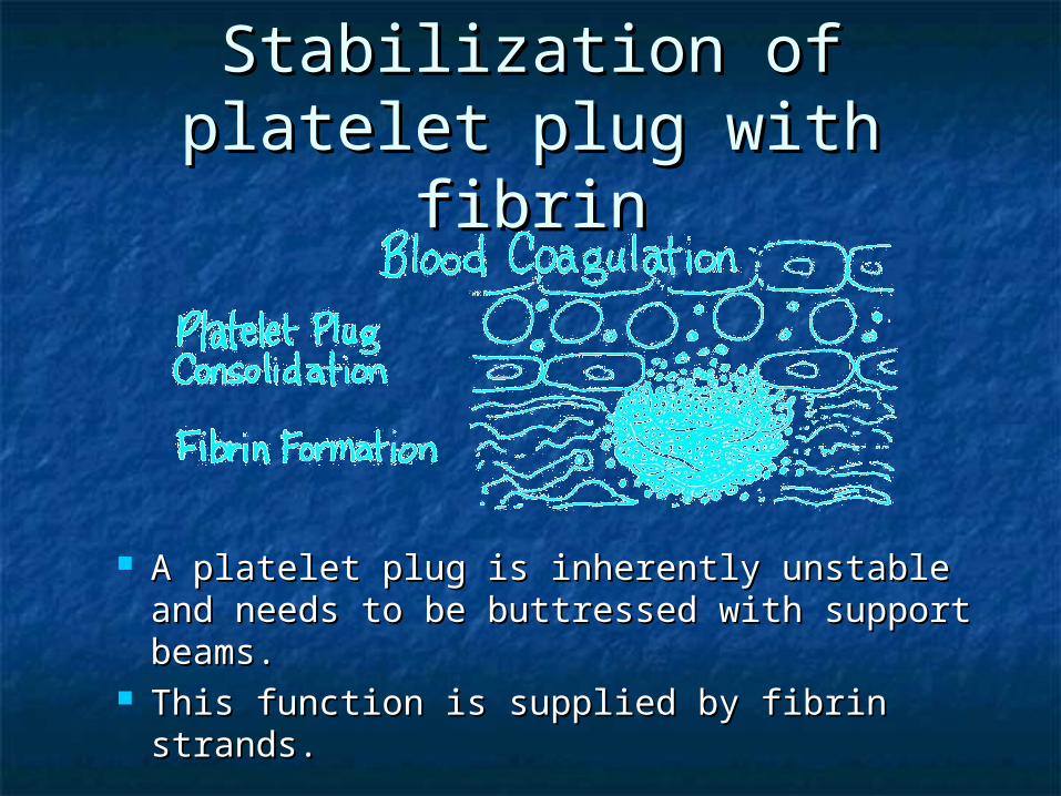
A platelet plug is inherently unstable and A platelet plug is inherently unstable and needs to be buttressed with support beams.needs to be buttressed with support beams.
This function is supplied by fibrin strands.This function is supplied by fibrin strands.
Blood coagulationBlood coagulation
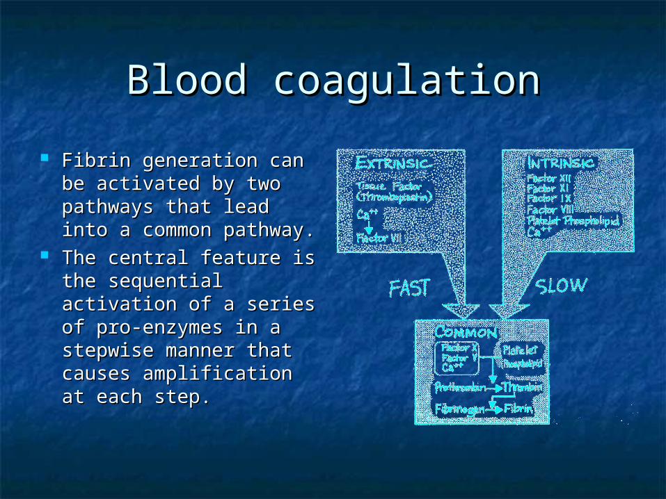
Fibrin generation can Fibrin generation can be activated by two be activated by two pathways that lead into pathways that lead into a common pathway.a common pathway.
The central feature is The central feature is the sequential the sequential activation of a series of activation of a series of pro-enzymes in a pro-enzymes in a stepwise manner that stepwise manner that causes causes amplification at amplification at each step.each step.
Intrinsic clotting systemIntrinsic clotting system
The intrinsic The intrinsic clotting pathway clotting pathway requires at least requires at least four coagulation four coagulation proteins and two proteins and two co-factors.co-factors.
Tested using the Tested using the aPTT.aPTT.
Activation of intrinsic Activation of intrinsic pathwaypathway
The intrinsic The intrinsic pathway is initiated pathway is initiated by the exposure of by the exposure of blood to a blood to a negatively charged negatively charged surface.surface.
Extrinsic clotting systemExtrinsic clotting system The extrinsic The extrinsic
system, in system, in contrast, requires contrast, requires only one only one coagulation protein coagulation protein and two co-factors and two co-factors which allows for which allows for rapid activation. rapid activation.
Tested using the Tested using the INR.INR.
Activation of extrinsic Activation of extrinsic pathwaypathway
Tissue thromboplastin Tissue thromboplastin (also known as tissue (also known as tissue factor) is present in factor) is present in the endothelial but is the endothelial but is only exposed to blood only exposed to blood flow during injury.flow during injury.
Thromboplastin, in the Thromboplastin, in the presence of calcium, presence of calcium, binds to Factor VII to binds to Factor VII to cause the activation of cause the activation of Factor X.Factor X.
The two pathways feed into The two pathways feed into ……
… … the common pathwaythe common pathway
The Common PathwayThe Common Pathway
Both the extrinsic and intrinsic pathway Both the extrinsic and intrinsic pathway converge on the activation of Factor X (this converge on the activation of Factor X (this also facilitates amplification).also facilitates amplification).
Factor Xa converts prothrombin to thrombin, Factor Xa converts prothrombin to thrombin, the final enzyme in the clotting cascade.the final enzyme in the clotting cascade.
Thrombin converts fibrinogen from a soluble Thrombin converts fibrinogen from a soluble plasma protein into an insoluble fibrin clot.plasma protein into an insoluble fibrin clot.
Thrombin also is an important feedback Thrombin also is an important feedback protein on the rest of the clotting cascade protein on the rest of the clotting cascade and platelets.and platelets.
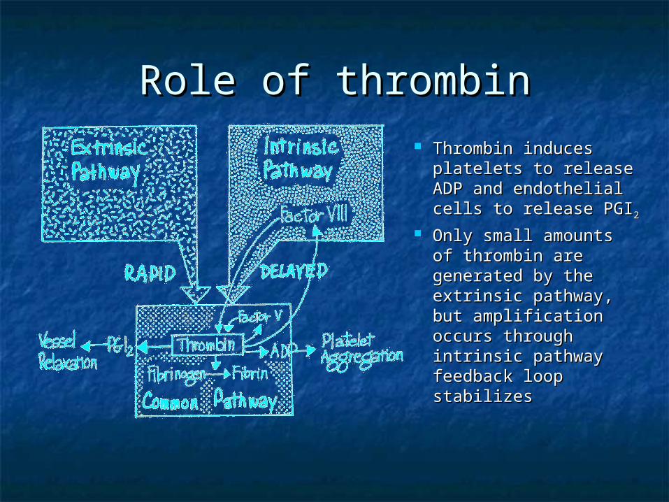
Role of thrombinRole of thrombin Thrombin induces Thrombin induces
platelets to release platelets to release ADP and endothelial ADP and endothelial cells to release PGIcells to release PGI22
Only small amounts Only small amounts of thrombin are of thrombin are generated by the generated by the extrinsic pathway, extrinsic pathway, but amplification but amplification occurs through occurs through intrinsic pathway intrinsic pathway feedback loop feedback loop stabilizesstabilizes
FibrinolysisFibrinolysis
The restore vessel patency, the clot must The restore vessel patency, the clot must be organized and removed by plasmin be organized and removed by plasmin while wound healing and tissue while wound healing and tissue remodeling occur.remodeling occur.
Fibrinolysis and repairFibrinolysis and repair Plasminogen binds fibrin and tissue plasminogen Plasminogen binds fibrin and tissue plasminogen
activator (tPA).activator (tPA). This complex then converts the plasminogen to plasmin.This complex then converts the plasminogen to plasmin. Plasmin cleaves fibrin in addition to fibrinogen and a Plasmin cleaves fibrin in addition to fibrinogen and a
variety of plasma proteins and clotting factors.variety of plasma proteins and clotting factors. tPA is an endothelial cell enzyme that is released in tPA is an endothelial cell enzyme that is released in
response to thrombin, serotonin, bradykinin, cytokines response to thrombin, serotonin, bradykinin, cytokines and epinephrine.and epinephrine.
When fibrin is degraded by plasmin it exposes new lysine When fibrin is degraded by plasmin it exposes new lysine terminals on the clot that act as further binding sites for terminals on the clot that act as further binding sites for plasminogen creating a positive feedback loop.plasminogen creating a positive feedback loop.
InvestigationsInvestigations
The basic investigations into any The basic investigations into any coagulation problem are:coagulation problem are:
1.1. INRINR
2.2. aPTTaPTT
3.3. Platelet countPlatelet count
4.4. FibrinogenFibrinogen Three patterns of defects can be Three patterns of defects can be
seen.seen.
Elevated INR, Normal aPTTElevated INR, Normal aPTT
Causes include:Causes include:1.1. Congenital factor VII deficiencyCongenital factor VII deficiency2.2. Vitamin K deficiencyVitamin K deficiency3.3. WarfarinWarfarin4.4. SepsisSepsis5.5. DIC (occasionally)DIC (occasionally)
Normal INR, Elevated aPTTNormal INR, Elevated aPTT
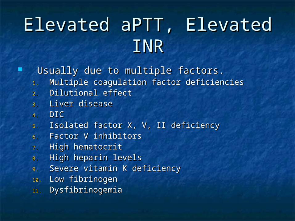
Usually due to multiple factors.Usually due to multiple factors.1.1. Multiple coagulation factor deficienciesMultiple coagulation factor deficiencies2.2. Dilutional effectDilutional effect3.3. Liver diseaseLiver disease4.4. DICDIC5.5. Isolated factor X, V, II deficiencyIsolated factor X, V, II deficiency6.6. Factor V inhibitorsFactor V inhibitors7.7. High hematocritHigh hematocrit8.8. High heparin levelsHigh heparin levels9.9. Severe vitamin K deficiencySevere vitamin K deficiency10.10. Low fibrinogenLow fibrinogen11.11. DysfibrinogemiaDysfibrinogemia
InvestigationsInvestigations
When a low platelet count, a blood smear When a low platelet count, a blood smear should be sent to rule out clumping and should be sent to rule out clumping and microangiopathic process.microangiopathic process.
Low fibrinogen levels reflect either severe Low fibrinogen levels reflect either severe liver disease, consumptive coagulopathy, or liver disease, consumptive coagulopathy, or massive transfusion (dilution effect).massive transfusion (dilution effect).
Other bleeding defects are harder to Other bleeding defects are harder to diagnosis on routine testing and are usually diagnosis on routine testing and are usually platelet function defects or increases in platelet function defects or increases in fibrinolysis.fibrinolysis.
Transfusion TherapyTransfusion Therapy
Replacement therapy should be based on the Replacement therapy should be based on the lab results and the patient’s clinical situation.lab results and the patient’s clinical situation.
The transfusion trigger for platelets can be The transfusion trigger for platelets can be less than 10,000/uL if the patient is not less than 10,000/uL if the patient is not bleeding, is not on platelet inhibitors, has bleeding, is not on platelet inhibitors, has preserved renal function, and does not have preserved renal function, and does not have DIC.DIC.
The usual dose of platelets is one The usual dose of platelets is one plateletpheresis unit.plateletpheresis unit.
For a fibrinogen level < 1 g/L, transfusion of For a fibrinogen level < 1 g/L, transfusion of 10 units of cryoprecipitate should increase the 10 units of cryoprecipitate should increase the fibrinogen level by 1.0 g/L.fibrinogen level by 1.0 g/L.
Transfusion TherapyTransfusion Therapy
In patients with an INR of >1.6 and an In patients with an INR of >1.6 and an abnormal aPTT, FFP is given abnormal aPTT, FFP is given dependent on the aPTT.dependent on the aPTT.
aPTT >1.5 times normal – 2-4 units of aPTT >1.5 times normal – 2-4 units of FFPFFP
aPTT >2 times normal – 15-30 ml/kg aPTT >2 times normal – 15-30 ml/kg of FFPof FFP
Repeat laboratory tests should be Repeat laboratory tests should be sent frequently to reassess adequacy sent frequently to reassess adequacy of treatment.of treatment.
Three general mechanisms:Three general mechanisms: Decreased productionDecreased production
Causes include thrombopoietin underproduction in Causes include thrombopoietin underproduction in liver disease, bone marrow suppression by viral, liver disease, bone marrow suppression by viral, drugs, toxins, nutritional deficiency, congenital or drugs, toxins, nutritional deficiency, congenital or acquired disorders of hematopoiesis.acquired disorders of hematopoiesis.
Increased destructionIncreased destruction Caused by immune or nonimmune processes.Caused by immune or nonimmune processes.
Dilution or distributionDilution or distribution Caused by massive transfusion or splenic Caused by massive transfusion or splenic
sequestration in splenomegaly.sequestration in splenomegaly.
Causes of Increased Platelet Causes of Increased Platelet DestructionDestruction
Specific Causes of Specific Causes of Thrombocytopenia - HITThrombocytopenia - HIT
Antibodies form against the Antibodies form against the heparin/platelet factor IV (PF4) complex.heparin/platelet factor IV (PF4) complex.
Despite the low platelets, thrombosis is Despite the low platelets, thrombosis is the major clinical problem.the major clinical problem.
Occurs 1-5% when unfractionated heparin Occurs 1-5% when unfractionated heparin is used, <1% if LMWH is used.is used, <1% if LMWH is used.
Suspect if platelet count drops 50% from Suspect if platelet count drops 50% from previous level or if count < 100,000/uL.previous level or if count < 100,000/uL.
Usually occurs in 4 days from heparin Usually occurs in 4 days from heparin start.start.
Specific Causes of Specific Causes of Thrombocytopenia - HITThrombocytopenia - HIT
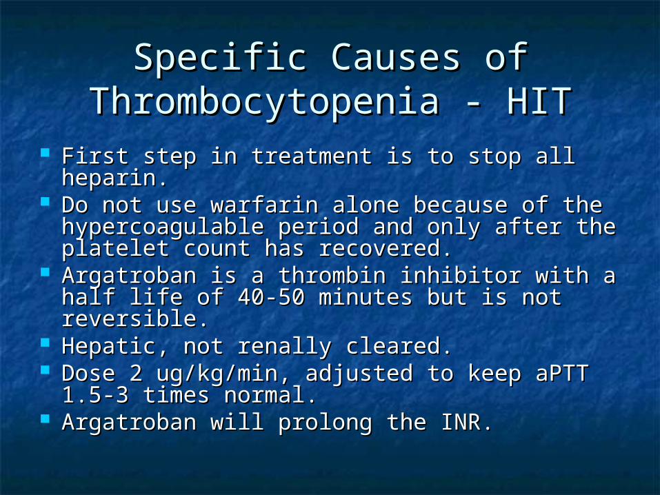
First step in treatment is to stop all heparin.First step in treatment is to stop all heparin. Do not use warfarin alone because of the Do not use warfarin alone because of the
hypercoagulable period and only after the hypercoagulable period and only after the platelet count has recovered.platelet count has recovered.
Argatroban is a thrombin inhibitor with a half Argatroban is a thrombin inhibitor with a half life of 40-50 minutes but is not reversible.life of 40-50 minutes but is not reversible.
Hepatic, not renally cleared.Hepatic, not renally cleared. Dose 2 ug/kg/min, adjusted to keep aPTT 1.5-Dose 2 ug/kg/min, adjusted to keep aPTT 1.5-
3 times normal.3 times normal. Argatroban will prolong the INR.Argatroban will prolong the INR.
Specific Causes of Specific Causes of Thrombocytopenia - HITThrombocytopenia - HIT
Danaparoid is a heparinoid which can Danaparoid is a heparinoid which can be used as an alternative to heparin in be used as an alternative to heparin in HIT.HIT.
There is a 10% cross reactivity with There is a 10% cross reactivity with the antibody responsible for HIT but the antibody responsible for HIT but clinical significance is unknown.clinical significance is unknown.
It has a long half life of 25 hours and is It has a long half life of 25 hours and is not reversible.not reversible.
Monitor using anti-factor Xa levels.Monitor using anti-factor Xa levels.
Specific Causes of Specific Causes of Thrombocytopenia - TTPThrombocytopenia - TTP
Caused by an inhibitor against an enzyme Caused by an inhibitor against an enzyme responsible for cleaving vWF causing spontaneous responsible for cleaving vWF causing spontaneous platelet aggregation.platelet aggregation.
Often spontaneous but can be induced by drugs Often spontaneous but can be induced by drugs such as cyclosporin, tacrolimus, and ticlopidine.such as cyclosporin, tacrolimus, and ticlopidine.
Suspect in patients presenting with fever, Suspect in patients presenting with fever, thrombocytopenia, hemolysis, neurological thrombocytopenia, hemolysis, neurological symptoms, and renal dysfunction.symptoms, and renal dysfunction.
Plasma exchange is gold standard followed by Plasma exchange is gold standard followed by steroids.steroids.
Exchanges should be 1.5 plasma volume/day until Exchanges should be 1.5 plasma volume/day until LDH normalizes and platelets rebound.LDH normalizes and platelets rebound.
Specific Causes of Specific Causes of Thrombocytopenia - HELLPThrombocytopenia - HELLP
Presents as part of the spectrum of pre-Presents as part of the spectrum of pre-eclampsia, generally > 28 weeks.eclampsia, generally > 28 weeks.
Pre-eclampsia need not be severe.Pre-eclampsia need not be severe. First sign is a fall in platelets, then First sign is a fall in platelets, then
abnormal liver function tests, and abnormal liver function tests, and hemolysis.hemolysis.
Can progress to liver failure and death from Can progress to liver failure and death from hepatic rupture.hepatic rupture.
Delivery usually resolves the condition.Delivery usually resolves the condition. May require steroids or plasma exchange if May require steroids or plasma exchange if
condition worsens.condition worsens.
Specific Causes of Specific Causes of Thrombocytopenia – CAPSThrombocytopenia – CAPS
Catastrophic antiphospholipid Catastrophic antiphospholipid antibody syndrome (CAPS) is caused antibody syndrome (CAPS) is caused by widespread microthrombi in by widespread microthrombi in multiple vascular fields.multiple vascular fields.
Patients generally have a known Patients generally have a known autoimmune condition with autoimmune condition with anticardiolipin antibodies.anticardiolipin antibodies.
Treatment is with plasmaphreresis Treatment is with plasmaphreresis and immunosuppression.and immunosuppression.
Renal failure can cause bleeding from Renal failure can cause bleeding from platelet dysfunction.platelet dysfunction.
Best treatment is aggressive dialysis to Best treatment is aggressive dialysis to control the uremia and DDAVP (20 ug IV) for control the uremia and DDAVP (20 ug IV) for acute bleeding or pre-procedure.acute bleeding or pre-procedure.
ASA irreversibly inhibits platelet function ASA irreversibly inhibits platelet function and so platelets must be replaced if and so platelets must be replaced if bleeding occurs.bleeding occurs.
Other drugs such as ketorolac and Other drugs such as ketorolac and hydroxyethyl starch reversible inhibit and hydroxyethyl starch reversible inhibit and platelet function will recover as the drug platelet function will recover as the drug clears.clears.
Patients can present in one of four ways:Patients can present in one of four ways:1.1. AsymptomaticAsymptomatic - Laboratory evidence only but - Laboratory evidence only but
no bleeding or thrombosis.no bleeding or thrombosis.2.2. BleedingBleeding – Caused by factor and platelet – Caused by factor and platelet
depletion, platelet dysfunction and fibrinolysis. depletion, platelet dysfunction and fibrinolysis. Present with diffuse bleeding from multiple Present with diffuse bleeding from multiple sites.sites.
3.3. ThrombosisThrombosis – Despite the microthrombi, – Despite the microthrombi, macrothrombi are unusual but can occur in macrothrombi are unusual but can occur in cancer patients.cancer patients.
4.4. Purpura fulminansPurpura fulminans – Described in more detail – Described in more detail later.later.
Diagnosis established by the history and Diagnosis established by the history and presence of thrombocytopenia, presence of thrombocytopenia, microangiopathic changes on the smear.microangiopathic changes on the smear.
FDP or D-dimers are always elevated due FDP or D-dimers are always elevated due to the marked fibrinolysis.to the marked fibrinolysis.
Both the INR and aPTT are elevated.Both the INR and aPTT are elevated. Fibrinogen is low.Fibrinogen is low. Liver failure can mimic DIC but factor VIII Liver failure can mimic DIC but factor VIII
levels are low in DIC (consumed) and levels are low in DIC (consumed) and normal in liver failure (produced by the normal in liver failure (produced by the endothelium not the liver).endothelium not the liver).
First, treat the underlying condition.First, treat the underlying condition. Initiate other supportive measures as Initiate other supportive measures as
necessary (i.e. intubation, vasopressors).necessary (i.e. intubation, vasopressors). Low levels of evidence suggest that low doses Low levels of evidence suggest that low doses
of heparin in non-bleeding patients may be of heparin in non-bleeding patients may be helpful.helpful.
Both factor and platelet replacement in Both factor and platelet replacement in bleeding patients and those at risk for major bleeding patients and those at risk for major bleeding can be life-saving but only supportive bleeding can be life-saving but only supportive until the underlying condition can be treated.until the underlying condition can be treated.
DIC in association with limb ecchymosis DIC in association with limb ecchymosis and skin necrosis.and skin necrosis.
Often associated with meningococcemia Often associated with meningococcemia and post-splenectomy sepsis.and post-splenectomy sepsis.
Optimum therapy has not been Optimum therapy has not been established but includes:established but includes: rhAPC infusionrhAPC infusion Blood products to keep INR < 2, aPTT < 1.8 Blood products to keep INR < 2, aPTT < 1.8
normal, and platelets >50000/uLnormal, and platelets >50000/uL
Body stores are normally low and Body stores are normally low and require 40-80 ug/day.require 40-80 ug/day.
Once depleted, production of factors Once depleted, production of factors II, VII, IX, and X fall and INR rapidly II, VII, IX, and X fall and INR rapidly rises.rises.
The diagnosis should be suspected The diagnosis should be suspected when there is a history of prolonged when there is a history of prolonged antibiotic use, biliary obstruction, antibiotic use, biliary obstruction, and pre-existing malnutrition.and pre-existing malnutrition.
Defined as requiring more than one blood Defined as requiring more than one blood volume in 24 hours or less.volume in 24 hours or less.
Coagulation defects occur from dilution of Coagulation defects occur from dilution of the plasma volume by fluid resuscitation the plasma volume by fluid resuscitation or red cell transfusions and consumption or red cell transfusions and consumption from the underlying disorder.from the underlying disorder.
It is difficult to predict the degree of It is difficult to predict the degree of coagulopathy from the amount of blood coagulopathy from the amount of blood transfused. Therefore, monitoring the transfused. Therefore, monitoring the patient’s coagulation status during patient’s coagulation status during massive transfusion is critical. massive transfusion is critical.
Platelets < 50,000/uLPlatelets < 50,000/uL give 6-8 units of random donor plateletsgive 6-8 units of random donor platelets
Fibrinogen < 1 g/LFibrinogen < 1 g/L give 10 units of cryoprecipitategive 10 units of cryoprecipitate
Hematocrit < 30%Hematocrit < 30% give red cellsgive red cells
INR > 1.6 and aPTT abnormalINR > 1.6 and aPTT abnormal give 2-4 units of FFPgive 2-4 units of FFP
Coagulation Defects – Use of Coagulation Defects – Use of Factor VIIaFactor VIIa
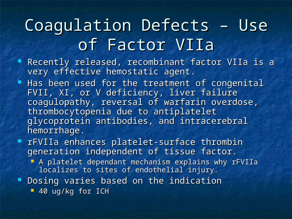
Recently released, recombinant factor VIIa is a very Recently released, recombinant factor VIIa is a very effective hemostatic agent.effective hemostatic agent.
Has been used for the treatment of congenital FVII, Has been used for the treatment of congenital FVII, XI, or V deficiency, liver failure coagulopathy, XI, or V deficiency, liver failure coagulopathy, reversal of warfarin overdose, thrombocytopenia reversal of warfarin overdose, thrombocytopenia due to antiplatelet glycoprotein antibodies, and due to antiplatelet glycoprotein antibodies, and intracerebral hemorrhage.intracerebral hemorrhage.
rFVIIa enhances platelet-surface thrombin rFVIIa enhances platelet-surface thrombin generation independent of tissue factor.generation independent of tissue factor. A platelet dependant mechanism explains why rFVIIa A platelet dependant mechanism explains why rFVIIa
localizes to sites of endothelial injury.localizes to sites of endothelial injury. Dosing varies based on the indication Dosing varies based on the indication
40 ug/kg for ICH40 ug/kg for ICH
ThrombosisThrombosis
Thrombosis - IncidenceThrombosis - Incidence
Most ICU patients have one or more Most ICU patients have one or more risk factors for thrombosis.risk factors for thrombosis.
The true incidence is unknown but The true incidence is unknown but estimates run up to 33%.estimates run up to 33%.
15% of DVT are in the upper limbs 15% of DVT are in the upper limbs and are associated with central lines.and are associated with central lines.
Trauma and surgical patients inevidently have Trauma and surgical patients inevidently have endothelial damage triggering coagulation.endothelial damage triggering coagulation.
Inflammation and stress response causes Inflammation and stress response causes thrombocytosis, hyperfibrinogenemia, altered thrombocytosis, hyperfibrinogenemia, altered coagulation factors, and elevated levels of PAI-1 coagulation factors, and elevated levels of PAI-1 which tips the balance toward thrombosis.which tips the balance toward thrombosis.
Patients are immobilized, ventilated and Patients are immobilized, ventilated and sedated which alters blood flow causing stasis.sedated which alters blood flow causing stasis.
There are many inherited There are many inherited hypercoagable states.hypercoagable states.
These increase the relative risk by 10 These increase the relative risk by 10 fold on top of the acquired risks.fold on top of the acquired risks.
The most important risk factors for The most important risk factors for DVT are a previous episode of DVT are a previous episode of thrombosis and a family history of thrombosis and a family history of DVT.DVT.
Genetic Risk Factors for Genetic Risk Factors for ThrombosisThrombosis
Risk FactorRisk Factor Prevalence Prevalence in the in the General General PopulationPopulation
Prevalence in Prevalence in Patients with Patients with DVTDVT
Elevated factor VIII levelElevated factor VIII level 11%11% 25%25%
Thrombosis – PE Thrombosis – PE PathophysiologyPathophysiology
Pulmonary artery obstruction causes a rise in Pulmonary artery obstruction causes a rise in pulmonary artery pressure that leads to right pulmonary artery pressure that leads to right ventricular failure.ventricular failure.
RV failure leads to a rise in central venous pressures RV failure leads to a rise in central venous pressures and fall in cardiac output.and fall in cardiac output.
RV oxygen demands is higher in the presence of RV oxygen demands is higher in the presence of hypoxia, hypotension, and reduced coronary perfusion hypoxia, hypotension, and reduced coronary perfusion causing infarction.causing infarction.
Infarction leads to worsening RV function and output Infarction leads to worsening RV function and output perpetuating the vicious cycle.perpetuating the vicious cycle.
Patients with a PFO can also develop paradoxical Patients with a PFO can also develop paradoxical embolism.embolism.
A PE with PFO can lead to greater hypoxia due to an A PE with PFO can lead to greater hypoxia due to an intracardiac right-to-left shunt.intracardiac right-to-left shunt.
Thrombosis - DiagnosisThrombosis - Diagnosis
Clinical symptoms and signs are fraught with poor Clinical symptoms and signs are fraught with poor sensitivity and specificity.sensitivity and specificity. Acute onset dyspneaAcute onset dyspnea Pleuritic chest painPleuritic chest pain CoughCough HemoptysisHemoptysis TachypneaTachypnea Sinus tachycardiaSinus tachycardia SyncopeSyncope
Although the gold standard test is pulmonary Although the gold standard test is pulmonary angiogram, the most practical in the critically ill angiogram, the most practical in the critically ill patient is CT chest angiogram.patient is CT chest angiogram.
V/Q scan is likewise impractical because of the V/Q scan is likewise impractical because of the long image acquisition time.long image acquisition time.
Thrombosis – Use of echo in Thrombosis – Use of echo in PEPE
Sensitivity and specificity of TEE in the Sensitivity and specificity of TEE in the diagnosis of proximal PE is 84%.diagnosis of proximal PE is 84%.
RV volume and pressure overload following RV volume and pressure overload following massive PE is readily identified.massive PE is readily identified.
Echo findings includes dilated right atrium Echo findings includes dilated right atrium and ventricle, increased ratio of RV to LV and ventricle, increased ratio of RV to LV chamber size, paradoxical septal bulging chamber size, paradoxical septal bulging and a reduction in RV function.and a reduction in RV function.
In severe cases of RV volume and pressure In severe cases of RV volume and pressure overload, an under filled and overload, an under filled and hyperdynamic left ventricle will be evident.hyperdynamic left ventricle will be evident.
Once detected, both DVT and PE require Once detected, both DVT and PE require full anticoagulation with heparin.full anticoagulation with heparin.
LMWH is a therapeutic option but can be LMWH is a therapeutic option but can be contraindication by it long half life and contraindication by it long half life and renal clearance.renal clearance.
Coumadin is best avoided in the critical Coumadin is best avoided in the critical care phase because of the number of care phase because of the number of interactions with other medications and interactions with other medications and difficult reversibility.difficult reversibility.
Thrombosis – PE and tPAThrombosis – PE and tPA
While most of the treatment for PE is While most of the treatment for PE is supportive (oxygen, vasopressors etc.), supportive (oxygen, vasopressors etc.), intravenous tPA for clot lysis is indicated for intravenous tPA for clot lysis is indicated for the following conditions:the following conditions: Persistent hypotensionPersistent hypotension Severe hypoxemiaSevere hypoxemia Large perfusion defectLarge perfusion defect RV dysfunctionRV dysfunction Free floating RV thrombusFree floating RV thrombus PFOPFO Maybe – RV dilation and/or hypokinesis without Maybe – RV dilation and/or hypokinesis without
Preventative strategies fall into two groups Preventative strategies fall into two groups – pharmacological (UFH and LMWH) and – pharmacological (UFH and LMWH) and mechanical (stockings and compression mechanical (stockings and compression devices).devices).
Heparin – either UFH or LMWH – in the Heparin – either UFH or LMWH – in the mainstay of prophylaxis. Reduces mainstay of prophylaxis. Reduces incidence by over 50%.incidence by over 50%.
LMWH is as effective as UFH but is LMWH is as effective as UFH but is superior in trauma and spinal cord injury.superior in trauma and spinal cord injury.
The incidence of hemorrhagic complications is The incidence of hemorrhagic complications is no different in treated and control patients.no different in treated and control patients.
Only contraindications to prophylaxis heparin Only contraindications to prophylaxis heparin is:is: Intracranial bleedingIntracranial bleeding Spinal cord injury associated with hematomaSpinal cord injury associated with hematoma Uncontrolled ongoing bleedingUncontrolled ongoing bleeding Uncorrected coagulopathyUncorrected coagulopathy
Warning: Liver failure patients may be thrombotic despite Warning: Liver failure patients may be thrombotic despite an elevated INR.an elevated INR.
Those at high risk for bleeding should receive Those at high risk for bleeding should receive a pneumatic compression devices until able a pneumatic compression devices until able to be converted to heparin.to be converted to heparin.
SummarySummary
Normal hemostasisNormal hemostasis Coagulation pathwaysCoagulation pathways Initial investigationsInitial investigations
CoagulopathiesCoagulopathies Pathogenesis of coagulopathiesPathogenesis of coagulopathies Specific causesSpecific causes