78
Blood Biomarkers for Traumatic Brain Injury Pashtun Shahim Department of Neurochemistry Institute of Neuroscience and Physiology Sahlgrenska Academy University of Gothenburg Gothenburg 2015

Blood Biomarkers for Traumatic Brain Injury
Pashtun Shahim
Department of Neurochemistry Institute of Neuroscience and Physiology
Sahlgrenska Academy University of Gothenburg
Gothenburg 2015

Cover illustration: Katarina Gren. All previously published papers were reproduced with permission from the publisher. Blood Biomarkers for Traumatic Brain Injury © Pashtun Shahim 2015 [email protected] ISBN: 978-91-628-9670-6 (Print) ISBN: 978-91-628-9671-3 (E-pub) http://hdl.handle.net/2077/39572 Printed by Ineko AB, Gothenburg, Sweden, 2015

To my parents and siblings

ABSTRACT
Traumatic brain injury (TBI) is the major cause of death and disability following blunt head trauma. The term mild TBI (mTBI) or concussion are used interchangeably in the literature. Concussion or mTBI is recognized as a clinical syndrome caused by biochemically induced alterations of brain function secondary to head trauma, typically affecting memory and orientation and may involve loss of consciousness. The diagnosis of mTBI is mainly based on clinical symptoms. Objective tools such as computed tomography or magnetic resonance imaging are mainly used to exclude structural brain damage, e.g., intracranial bleeding. Biomarkers are molecules that can be measured in accessible biological fluids, that reflect physiological, pharmacological, or disease processes and can suggest the etiology of, susceptibility to, activity level of, or progress of a disease. The overall aim of this dissertation was to assess both novel and previously studied blood biomarkers reflecting neuronal injury and dysfunction in individuals with mild to severe traumatic brain injury (sTBI). The main research question we sought to answer was whether mTBI was associated with biomarker evidence of axonal injury. For this purpose we used sera from ice hockey players competing in the Swedish Hockey League. In addition, we developed a novel ultrasensitive digital ELISA based on Single molecule array (Simoa) platform for detection of neurofilament light protein (NFL) in serum. NFL is a neuron-specific protein, which is difficult to measure in blood using standard immunochemical techniques due to suboptimal analytical sensitivity. We assessed the diagnostic and prognostic utility of this assay in patients with sTBI, who were treated at the Neurointensive Care Unit at the Sahlgrenska University Hospital. The main findings of these studies were that mTBI in professional ice hockey players is associated with altered serum levels of biomarkers associated with neuronal injury. The levels of these biomarkers were also related to the number of days it took for the athletes to return to play. In the context of sTBI, NFL levels assessed in serum showed

high diagnostic accuracy, and the levels in serum were also related to overall clinical outcome at follow-up 12 months after the injury. Our overall conclusion is that the novel blood biomarkers presented in this thesis are promising diagnostic and prognostic tools for mild to severe TBI. Furthermore, the findings of this thesis may be extended to other neurological disorders associated with axonal injury, where tracking disease progression and evaluating the efficacy of novel therapy were previously limited to analyses of cerebrospinal fluid. Keywords: traumatic brain injury, blood biomarker, ice hockey players, neuronal injury; tau, NFL, SNTF, VILIP-1, S100B, NSE ISBN: 978-91-628-9670-6 (Print) ISBN: 978-91-628-9671-3 (E-pub) http://hdl.handle.net/2077/39572
POPULÄRVETENSKAPLIG SAMMANFATTNING De vanligaste orsakerna till traumatiska hjärnskador (traumatic brain injury; TBI) är fallolyckor och trafikolyckor, men TBI har också blivit vanligare i kontaktidrotter som ishockey, boxning, amerikansk fotboll och rugby. I dagsläget är det svårt att säkert säga om hjärnan har påverkats eller ej vid en lättare huvudskada. En biomarkör som avspeglar en patofysiologisk process ger ett objektivt biologiskt mått som kan användas för att diagnostisera en sjukdom, följa ett sjukdomsförlopp, utvärdera behandlingseffekten och förutspå sjukdomsprognosen. Eftersom likvor står i direkt kontakt med hjärnan, anses denna vara en optimal källa för att mäta biomarkörer som avspeglar patologiska processer i hjärnan. Idag finns väletablerade markörer i likvor som avspeglar nervcellskada. Vissa av dessa markörer, t.ex. total tau (T-tau), är i hög grad specifika för centrala nervsystemet, men det har fram tills nu varit svårt att mäta dem i blod p.g.a. deras låga koncentrationer. Det övergripande syftet med denna avhandling var att ta fram nya biomarkörer i blod samt jämföra dessa markörer, med befintliga väletablerade markörer för neuronal skada hos individer med båda lätta och svåra skallskador. I den första studien utvärderades ett mycket specifikt hjärnprotein som utrycks i kortikala axoner, T-tau, hos professionella ishockeyspelare i den Svenska Hockey Ligan (SHL, tidigare Elitserien). Vi analyserade T-tau i blod med hjälp av en ny högkänslig metod kallad Single molecule array (Simoa) som är ca tusen gånger känsligare än motsvarande standardmetod för att mäta detta protein. Tidigare har T-tau endast kunnat mätas i likvor. Vi fann att T-tau i plasma ökade hos ishockeyspelare efter hjärnskakning jämfört med försäsongsvärden och nivån av T-tau en timme efter hjärnskakning korrelerade med antalet dagar spelarna var borta ifrån spel, vilket kan tolkas som ett mått på graden av hjärnpåverkan. I den andra studien mättes, hos samma studiekohort, tau-A och tau-C som är nedbrytningsprodukter av T-tau. I tidigare studier har man sett att tau-fragment ökar i blodet hos patienter med Alzheimers sjukdom. Vår hypotes var att tau-fragment

passerar blodhjärnbarriären lättare än T-tau vid en skada på kortikala axoner, och följaktligen borde möjligheten att detektera hjärnskadan i blod öka. I denna studie fann vi att tau-A korrelerade med graden av hjärnpåverkan, ungefär på samma sätt som T-tau. I den tredje studien analyserades en annan ny axonal markör, αII-spectrin N-terminal fragment (SNTF), hos hockeyspelarna. Vi fann att SNTF ökade hos hockeyspelare efter en hjärnskakning och nivåerna 12-36 timmar efter hjärnskakning korrelerade med graden av hjärnpåverkan. I den fjärde studien utvärderade vi visinin-like protein-1 (VILIP-1), en neuronal skademarkör som tidigare visats öka i djurmodeller för TBI. I blod hos hockeyspelare ökade VILIP-1 inte efter en hjärnskakning. Däremot fann vi att VILIP-1 ökade signifikant efter en träningsmatch utan hjärnskakning, vilket talar emot användbarheten av VILIP-1 som en markör för hjärnskada. I den femte och sista studien i denna avhandling, utvecklade vi en ny immunkemisk metod baserat på Simoa-plattformen för detektion av neurofilament light (NFL) protein i blod. NFL har tidigare visats öka i likvor hos boxare som utsätts för upprepat trauma mot huvudet. Vi fann att nivåerna av NFL var både diagnostiska och prognostiska hos patienter med svår TBI.


Pashtun Shahim
1
LIST OF PAPERS This thesis is based on the following studies, referred to in the text by their Roman numerals. Paper I Shahim P, Tegner Y, Wilson DH, Randall J, Skillbäck T, Pazooki D, Kallberg B, Blennow K, Zetterberg H. Blood biomarkers for brain injury in concussed professional ice hockey players. JAMA Neurology 2014; 71(6): 684-92. Paper II Shahim P, Linemann T, Inekci D, Karsdal MA, Blennow K, Tegner Y, Zetterberg H, Henriksen K. Serum tau fragments predict return to play in concussed professional ice hockey players. Journal of Neurotrauma 2015; doi: 10.1089/neu.2014.3741. Paper III Siman R, Shahim P, Tegner Y, Blennow K, Zetterberg H, Smith DH. Serum SNTF increases in concussed professional ice hockey players and relates to the severity of postconcussion symptoms. Journal of Neurotrauma 2015; 32(17): 1294-300. Paper IV Shahim P, Mattsson N, Macy EM, Crimmins DL, Ladenson JH, Zetterberg H, Blennow K, Tegner Y. Serum visinin-like protein-1 in concussed professional ice hockey players. Brain Injury 2015; 29(7-8): 872-6. Paper V Shahim P, Gren M, Liman V, Andreasson U, Norgren N, Tegner Y, Mattsson N, Andreasen N, Öst M, Zetterberg H, Nellgård B, Blennow K. An ultrasensitive method for quantitating serum neurofilament light protein after traumatic brain injury. Submitted.

Blood Biomarkers for Traumatic Brain Injury
2
CONTENT
ABBREVIATIONS .............................................................................................. 5 INTRODUCTION ................................................................................................ 7
Acute TBI ...................................................................................................... 8 Catastrophic brain injury ......................................................................... 8 Sports-related TBI .................................................................................... 8
Diagnosis of mTBI ........................................................................................ 9 Management of mTBI ................................................................................. 10 Second impact syndrome ............................................................................ 10 Postconcussive syndrome ........................................................................... 11 Pathophysiology of TBI .............................................................................. 11
Neurometabolic cascades ....................................................................... 12 Repetitive brain trauma .......................................................................... 14
Neuropathology of TBI ............................................................................... 15 Tau pathology ........................................................................................ 15 Plaque pathology .................................................................................... 16 Microglia ................................................................................................ 18 Genetic susceptibility ............................................................................. 19 Outcome assessment after TBI .............................................................. 19
Treatments for TBI ...................................................................................... 20 Biomarkers for TBI ..................................................................................... 21
AIM ................................................................................................................ 23 Paper I ......................................................................................................... 23 Paper II ........................................................................................................ 23 Paper III ....................................................................................................... 23 Paper IV ...................................................................................................... 23 Paper V ........................................................................................................ 23
PATIENTS AND METHODS .............................................................................. 24 Subjects and study settings ......................................................................... 24

Pashtun Shahim
3
Blood and CSF sampling ............................................................................ 25 Analytical methods ...................................................................................... 25
ELISA .................................................................................................... 26 Electrochemiluminescence assays ......................................................... 27 Single molecule counting ....................................................................... 27 Single Molecule Arrays ......................................................................... 27 Tau fragment .......................................................................................... 30
Statistical analyses ...................................................................................... 30 Ethics ........................................................................................................... 31
RESULTS ......................................................................................................... 32 Paper I ......................................................................................................... 32
Background ............................................................................................ 32 Main results ............................................................................................ 32
Paper II ........................................................................................................ 33 Background ............................................................................................ 33 Main results ............................................................................................ 33
Paper III ....................................................................................................... 34 Background ............................................................................................ 34 Main results ............................................................................................ 34
Paper IV ...................................................................................................... 34 Background ............................................................................................ 34 Main results ............................................................................................ 35
Paper V ........................................................................................................ 35 Background ............................................................................................ 35 Main results ............................................................................................ 36
DISCUSSION .................................................................................................... 37 Serum total tau in mild traumatic brain injury ............................................ 37 Tau fragments .............................................................................................. 39 Spectrin N-terminal fragment following mild traumatic brain injury ......... 39 Visinin-like protein-1 is not sensitive for mild brain injury ....................... 40

Blood Biomarkers for Traumatic Brain Injury
4
An ultrasensitive method for quantitating serum neurofilament light protein after traumatic brain injury ......................................................................... 41
CONCLUSION .................................................................................................. 44 The role of blood biomarkers in TBI research ............................................ 44 The future of TBI therapy: a role for blood biomarkers? ........................... 46
FUTURE PERSPECTIVES .................................................................................. 47 ACKNOWLEDGEMENT .................................................................................... 48 REFERENCES .................................................................................................. 51
Pashtun Shahim
5
ABBREVIATIONS AAN American Academy of Neurology AD Alzheimer’s disease ADAM A disintegrin and metalloproteinase APOE apolipoprotein E APP amyloid precursor protein AUROC area under the receiver operating characteristic Aβ amyloid-beta BACE1 β-site amyloid precursor protein-cleaving enzyme 1 BBB blood-brain barrier CNS central nervous system CSF cerebrospinal fluid CT computed tomography CTE chronic traumatic encephalopathy DAI diffuse axonal injury ELISA enzyme-linked immunosorbent assay fMRI functional magnetic resonance imaging GCS Glasgow coma scale GFAP glial fibrillary acid protein GOS Glasgow outcome scale MRI magnetic resonance imaging mTBI mild traumatic brain injury NFL neurofilament light NMDA N-methyl-D-aspartate NSE neuron-specific enolase P-tau phosphorylated tau PCS post-concussive symptom RPCSQ Rivermead Post-Concussion Symptoms Questionnaire RTP return to play S100B S100 calcium-binding protein B SAC Standardized Assessment of Concussion sAPP soluble amyloid precursor protein Scat3 sport concussion assessment tool 3 SHL Swedish Hockey League Simoa single molecule arrays SNTF spectrin-N terminal fragment sTBI severe traumatic brain injury

Blood Biomarkers for Traumatic Brain Injury
6
T-tau total tau TBI traumatic brain injury VILIP-1 visinin-like protein 1

Pashtun Shahim
7
INTRODUCTION Traumatic brain injury (TBI) is a heterogeneous disorder characterized by acute brain injury resulting from external mechanical forces exerted on the head [1]. In clinical settings, the Glasgow Coma Scale (GCS) is commonly used to stratify the severity of TBI [2], where mild TBI (mTBI), moderate TBI and severe (sTBI) constitute a GCS score of 13-15, 9-12 and 3-8 respectively (Table 1) [3]. The incidence of mTBI has been estimated to about 600 per 100 000 each year [4] and of sTBI about 17 per 100 000 each year [5].
Table 1. Glasgow Coma Scale
Behavior Response Score Eye Opening Response Spontaneously 4
To speech 3 To pain 2 No response 1
Best Verbal Response Oriented to time, place, and person 5 Confused 4 Inappropriate words 3 Incomprehensible sounds 2 No response 1
Best Motor Response Obeys commands 6 Moves to localized pain 5 Flexion withdrawal from pain 4 Abnormal flexion (decorticate) 3 Abnormal extension (decerebrate) 2 No response 1
Total Score: 15 Comatose: 8 points or less Totally unresponsive: 3 points Mild brain injury = 13-15 points; Moderate Brain Injury = 9-12 points; Severe Brain Injury = 3-8 points
TBI generally falls into two categories: acute and chronic [6]. Acute brain injury comprises mTBI and catastrophic brain injury that may

Blood Biomarkers for Traumatic Brain Injury
8
lead to death, most commonly due to subdural hematoma. Chronic traumatic brain injury, sometimes called dementia pugilistica or chronic traumatic encephalopathy (CTE), is a neurodegenerative disorder triggered by repeated head trauma. In professional boxers and athletes, CTE may start several years after the end of the sports career.
Acute TBI
Catastrophic brain injury Catastrophic brain injury refers to severe brain trauma associated with intracranial bleeding or cerebral contusions, which may result in death or long-term neurologic deficits. The most common cause of death in sport-related TBI is subdural hematoma, particularly in boxers [7, 8]. In boxing, about ten deaths occur each year, most of which are following knockout or technical knockouts [9]. Surprisingly, most deaths are in lower weight classes. Catastrophic brain injury occurs also in American football. During the second half of the 20th century, more than 400 American football players and approximately 30 ice hockey players died from brain or spinal cord injury in the United States while playing [10].
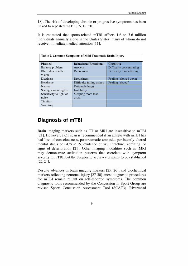
Sports-related TBI In the sports literature, mTBI is traditionally referred to as concussion or sports-related mTBI. According to the American Academy of Neurology sports-related mTBI is defined as a clinical syndrome of biomechanically induced alteration of brain function, typically affecting memory and orientation, which may involve loss of consciousness [11]. Some of the more common symptoms categorized by different domains are presented in Table 2. Symptoms of mTBI usually resolve within days to weeks, but 10-15% of the cases remain symptomatic more than one-year post injury [12-15]. In recent years, many professional athletes who suffered repeated mTBI have retired due to chronic post-concussive symptoms [6, 16-
Pashtun Shahim
9
18]. The risk of developing chronic or progressive symptoms has been linked to repeated mTBI [16, 19, 20]. It is estimated that sports-related mTBI affects 1.6 to 3.6 million individuals annually alone in the Unites States, many of whom do not receive immediate medical attention [11].
Diagnosis of mTBI Brain imaging markers such as CT or MRI are insensitive to mTBI [21]. However, a CT scan is recommended if an athlete with mTBI has had loss of consciousness, posttraumatic amnesia, persistently altered mental status or GCS < 15, evidence of skull fracture, vomiting, or signs of deterioration [21]. Other imaging modalities such as fMRI may demonstrate activation patterns that correlate with symptom severity in mTBI, but the diagnostic accuracy remains to be established [22-24]. Despite advances in brain imaging markers [25, 26], and biochemical markers reflecting neuronal injury [27-30], most diagnostic procedures for mTBI remain reliant on self-reported symptoms. The common diagnostic tools recommended by the Concussion in Sport Group are revised Sports Concussion Assessment Tool (SCAT3), Rivermead
Table 2. Common Symptoms of Mild Traumatic Brain Injury Physical Behavioral/Emotional Cognitive Balance problem Anxiety Difficulty concentrating Blurred or double vision
Depression Difficulty remembering
Dizziness Drowsiness Feeling “slowed down” Headache Difficulty falling asleep Feeling “dazed” Nausea Fatigue/lethargy Seeing stars or lights Irritability Sensitivity to light or noise
Sleeping more than usual
Tinnitus Vomiting

Blood Biomarkers for Traumatic Brain Injury
10
Post-Concussion Symptoms Questionnaire (RPCSQ) [31] or Standardized Assessment of Concussion [32, 33], which can be used during sideline evaluation.
Management of mTBI The latest guidelines on the management of sports-related TBI suggest any player suspected of having experienced mTBI should not be allowed to return to play in the same game or day of play [21, 32]. The guidelines also recommend physical and cognitive rest until the symptoms resolve [33]. Once symptoms diminish, a step-wise return to play, with at least 24 hours between each step, should be followed (Table 3).
Table 3. Graduated Return to Play Protocol
Rehabilitation stage Functional exercise at each stage 1. No activity When asymptomatic, proceed to level 2 2. Light aerobics Walking or stationary cycling, no
resistance training 3. Sport-specific exercise Skating in hockey, running in soccer 4. Non-contact training drill May start progressive resistance training 5. Full-contact practice Participate in normal training activities 6. Return to play Normal game play
Second impact syndrome Second impact syndrome (SIS) is a rare but severe complication of TBI among athletes. SIS results from sustaining a second head injury, most often a minor blow to the head before the symptoms of the first brain injury has resolved [34]. In SIS the injured athlete collapses into a coma within minutes. The mechanism is thought to be severe cerebrovascular engorgement and cerebral edema leading to brain herniation. SIS is common in adolescent males, and has been recorded in American football players at high school levels, boxing, as well as

Pashtun Shahim
11
ice hockey [35]. The terms SIS has, however, been questioned by some critics who have proposed the replacement of SIS with diffuse cerebral swelling [36].
Postconcussive syndrome Postconcussive syndrome is a clinical disorder referred to as the presence of neurological symptoms lasting more than 3 months, and is observed in 40-80 % of individuals who have suffered from mTBI [12]. In about 10-15% of cases, the symptoms may persist for more than one year [37, 38]. Neuropsychological measures reveal cognitive impairment beyond the subjectively symptomatic time in boxers following mTBI [39]. These subtle subjective and objective neuropsychological deficits following mild head injury is sometimes referred to as subconcussion [40]. Appropriate management of individuals with subconcussion is highly important in order to avoid development of chronic postconcussive syndrome (CPCS) [41]. The exact mechanism of how subconcussion causes CPCS and the relationship to chronic traumatic encephalopathy is yet to be established [42].
Pathophysiology of TBI The neurobiological changes following TBI are complex and not fully understood [6]. Some layers of complexity arise from unique anatomical features such as the elongated shape of a neuron, the skull that both protects the brain and constitutes a harmful closed compartment in the case of intracranial volume expansion, the cerebrospinal fluid [43] and blood-brain barrier (BBB) [44], which both maintain and perpetuate the unique intracerebral environment. The BBB is highly selective and plays an important role in allowing proteins or biomarkers entering the blood stream. In the acute phase of TBI, the disruption of the BBB may cause vasogenic or cytotoxic brain edema [45-47], leading to the expansion

Blood Biomarkers for Traumatic Brain Injury
12
of brain volume, which in turn increases intracranial pressure (ICP), impairs cerebral perfusion and subsequently causes ischemic injury [48]. Structural injuries to the brain can be categorized into focal and diffuse. Focal injuries are usually associated with higher energy trauma and include cortical or subcortical contusions and lacerations as well as intracerebral hemorrhage. Diffuse injury can be seen in mTBI and results from shearing and tearing of brain tissue during acceleration and deceleration. The most common type of diffuse injury is called diffuse axonal injury (DAI), which is caused by shearing or tearing of axons [49, 50]. The majority of individuals with sports-related brain trauma have DAI [33, 51].
Neurometabolic cascades Clinically relevant animal models of mTBI for investigation of the neurometabolic cascades that are set into motion following the biomechanical injury to the brain have been challenging to develop, mainly due to the lower mass of the animal brain and other differences in head and neck anatomy compared to humans [50]. In most animal TBI models, the brain or cortex is subjected to injury by direct crush or compression. These direct crush animal models of TBI, however, have been found to have a high variability in outcome, which further limit their utility as models of human mTBI [52, 53]. Studies using the mild fluid percussion injury model have provided some insight into the dynamic changes of mTBI [54]. Immediately after a closed head injury with acceleration and deceleration forces to the brain, there are stretching and disruption of neuronal and axonal cell membranes [52]. Resulting membrane defects cause a deregulated flux of ions, including an efflux of potassium and influx of calcium, which leads to increased release of excitatory neurotransmitters, particularly glutamate. The binding of glutamate to N-methyl-D-aspartate (NMDA) receptors leads to further depolarization, influx of calcium ions and widespread suppression of neurons with glucose hypometabolism [55, 56]. Increased activity in membrane pumps (to restore ionic balance) raises glucose consumption, depletes energy

Pashtun Shahim
13
stores, causes calcium influx into mitochondria, and impairs oxidative metabolism, and consequently induces anaerobic glycolysis with lactate production, which might cause acidosis and edema [55, 56] (Figure 1).
Figure 1. Schematic flow-chart of molecular changes following acceleration and deceleration forces on the brain.
Blood Biomarkers for Traumatic Brain Injury
14
Repetitive brain trauma Extensive experimental data from animal models indicate that repeated mild head injury with axonal damage increases the brain’s vulnerability for additional severe concussive impacts [56, 57]. In line with these findings are reports of increased risk of memory problems and cognitive impairment in American football players with a history of repeated concussions [58]. In the long term, repetitive brain trauma may cause chronic neurological impairment. The early cases of chronic brain damage were reported by Martland in 1928 [20] in retired boxers, and the condition was termed punch-drunk syndrome. A few years later, in 1937, Millspaugh [59] introduced the term dementia pugilistica, describing the aftermath of repetitive TBI in professional and amateur boxing. In 1949, Critchley [60] coined the term CTE, which has become the prevailing term in modern literature to recognize potential long-term consequences of repetitive TBI, which also occurs in American football players [17, 61] and ice hockey players [62], as well as in war veterans [63]. In 1969, Roberts reported a prevalence of CTE of 17% among retired boxers in the UK [64]. Risk factors for TBI include high number of bouts (n > 20 bouts), older age at retirement from boxing and long length of boxing career (n >10 years) [65]. A positive carrier status of the Alzheimer-associated apolipoprotein E (APOE) ε4 allele has been suggested to be associated with CTE [66, 67]. Epidemiological data in non-boxing sports are scarce [68, 69]. In an autopsy study of 321 American football players Gavett et al. estimated a life-time CTE prevalence of 3.7%, but this figure needs to be validated in a larger scale study [70]. The cellular mechanisms underlying increased susceptibility of the brain to repeated trauma are not fully clear. Experimental data indicate that metabolic dysfunction, including reduced mitochondrial energy in the brain due to increased metabolic demands but decreased energy stores with low ATP/DTP ratio and increased lactate/pyruvate ratio, may play a role [71, 72]. It is also suggested that mild trauma may

Pashtun Shahim
15
induce a type of sodium channelopathy on axons, which in turn may intensify pathophysiological responses to minor injuries [73, 74].
Neuropathology of TBI
Tau pathology Neuropathological findings in the literature on the individuals with mTBI who died of other causes are rare and have been compiled as isolated cases or small case series [75, 76]. The pathological findings observed in these few cases within 24 hours of injury were petechial hemorrhage, myelin destruction and retracted axonal bulbs. In cases with survival times of six weeks or more after TBI, the appearance of glial scars was observed which is now known as astrocytic tangles, which is a characteristic histological finding in CTE along with neurofibrillary tangles (NFT). Neurofibrillary tangle pathology is a prominent finding in the cortex of American football players, boxers, military personnel and others who have suffered repetitive concussive traumatic brain injuries [16, 19, 77-79]. Cortical tangles are also found in Alzheimer’s disease (AD) and other chronic neurological diseases [80]. Tangles are found intracellularly in the cytoplasm of neurons and are composed of thread-like aggregates of hyperphosphorylated tau protein [81]. There are six different isoforms of tau, each containing several serine or threonine residues that can be phosphorylated. Tau is a normal axonal protein that is responsible for microtubule assembly and stability [82]. In AD, tau is frequently found in a hyperphosphorylated form [82]. Hyperphosphorylation of tau causes disassembly of microtubules and thus impaired axonal transport, leading to neuronal and synaptic dysfunction. Hyperphosphorylation also increases tau aggregation and subsequent formation of insoluble fibrils and tangles [83]. Hyperphosphorylated tau found in CTE has chemical and structural resemblances with those found in AD [6]. However, tangles in CTE are found in superficial cortical axons, while tangles in AD are found in both superficial and deep axons [19, 78]. Furthermore, tau pathology in CTE is irregularly distributed, possibly due to many different directions of shearing forces induced by physical trauma [78]. However, animal models of severe contusional TBI

Blood Biomarkers for Traumatic Brain Injury
16
indicate that tau pathology induced by TBI may be mechanistically different from tau pathology due to CTE [84-86]. The underlying molecular mechanisms inducing tau pathology in CTE and how acute TBI leads to CTE are not known in detail. However, experimental animal studies suggest that accumulation of tau pathology is a consequence of repeated trauma (Figure 2) [84]. Experimental animal models exposed to rotational acceleration show both increased tau accumulation and neurofilament in damaged axons [87, 88]. Furthermore, brain trauma in animal models has been shown to increase tau phosphorylation; an abnormality that correlates with the severity of the injury [84].
Figure 2. Schematic flow-chart of the tau pathology in repetitive TBI.
Plaque pathology Besides tangle formation, extensive Aβ pathology has also been reported in TBI patients [19, 89], where the extent of plaque deposition in these patients was comparable with AD cases [90]. Aggregation of Aβ into plaques is one of the key histopathological hallmarks of AD [91]. Aβ is generated from amyloid precursor protein (APP) by cleavage of β-secretase and γ-secretase [82]. β-secretase was identified as β-site APP-cleaving enzyme 1 (BACE1) [82], while γ-secretase is a complex of protein consisting of presenilin, nicastrin, Pen-2 and Aph-1 [92]. APP is mainly expressed in neurons, and under normal conditions β-secretase and γ-secretase (presenilin) are translocated by axonal transport to the synapses, where APP can be cleaved by the secretases to generate Aβ [6]. APP has neurotrophic functions of importance for

Pashtun Shahim
17
neuronal survival after axonal damage [93-95]. Studies of human brain tissue samples have shown accumulation of APP in neurons and axons following brain trauma [89, 96, 97]. The accumulation of APP is even visible after mTBI, and often occurs within hours [50, 76]. In addition to APP, Aβ accumulation has also been reported in TBI [98-100]. The accumulation of Aβ forming plaque around the terminal bulb of disconnected axons is preceded by APP accumulation [90, 98, 101]. Further, repetitive mTBI increases Aβ deposition [102]. Intra-axonal APP accumulation is an established marker of DAI, and is regarded as the gold standard for identifying DAI [97, 103]. The increased APP in DAI is probably due to the role of APP promoting axonal outgrowth after injury [104]. Data from both brain trauma in humans and experimental studies indicate that DAI is a long-term process in which axons continue to degenerate and swell over several weeks to months following the injury (Wallerian degeneration)[105, 106]. In the disconnected axons, both the substrate (APP) and the key enzymes (BACE1 and presenilin) for Aβ production accumulate in the bulb of swollen axons, which may induce abnormal APP metabolism [101]. When this abundance of APP is processed into fragments of Aβ, some will form diffuse plaques (Figure 3) [101]. These plaques, however, are not a consistent finding in TBI [78], and their implication remains speculative. In addition, multiple studies using various techniques demonstrate the absence of Aβ pathology in most cases of acute TBI and CTE, particularly in young individuals and in early stages [107, 108]. It is possible that those with positive Aβ pathology may represent co-incidental age-related changes [109], but in the absence of more data it is hard to draw any definite conclusions on the role of Aβ accumulation in TBI.

Blood Biomarkers for Traumatic Brain Injury
18
Figure 3. Schematic flow-chart of the relationship between amyloid plaque formation and CTE.
Microglia Microglial activation has been reported following human TBI including concussive injury [75, 110]. Microglia is the prime mediators of inflammation in the CNS, and appears in areas of damaged axons in many animal TBI models [111-113]. The specific role of microglia in TBI is currently unknown [114, 115]. Processes related to neuronal plasticity [116] and axonal regeneration [117] are likely also of importance in the aftermath of TBI. Other proteins that may also be involved in the pathogenesis of CTE is TAR DNA binding protein-43 (TDP-43) [118, 119], which is likely part of the normal response to injury [120], and whose potential role in the pathological process of CTE remains to be elucidated.

Pashtun Shahim
19
Genetic susceptibility Evidence suggests that apolipoprotein Ε (apoE) plays a crucial role in lipid delivery for growth and regeneration of axons following TBI [121]. ApoE is the main component of lipoproteins in plasma and plays an important role in the transport of cholesterol and phospholipids. In the CNS, apoE is expressed predominantly by astrocytes, but also microglia have been suggested to play a role in the production of apoE [122]. Following brain injury, large amount of membrane lipids are released from damaged axons, and in response astrocytes increase apoE expression, with release of apoE to the extracellular space to scavenge cholesterol and other lipids for reuse during axonal or synaptic repair and regeneration [123]. The APOE gene has three alleles (APOE ε2, APOE ε3, and APOE ε4). APOE ε3 is the most common in the population. Numerous studies have shown that the APOE ε4 is a strong risk factor for AD [124]. In the context of TBI, inheritance of APOE ε4 may be related to unfavorable outcome after TBI [125] or development of CTE [126]. The mechanism for the association of APOE ε4 allele with unfavorable outcome after TBI remains controversial [125]. Evidence also links the APOE ε4 allele with Aβ generation and plaque formation. Studies show that the APOE ε4 allele is overrepresented in TBI patients who display Aβ deposition [127-129]. In a study on AD transgenic mice exposed to TBI, mice co-expressing apoE4 showed greater Aβ deposition than apoE3 mice [130]. These data suggest that apoE4 may trigger Aβ deposition and plaque formation as part of an acute phase response to brain injury. Furthermore, carriers of APOE ε4 allele have poor neurological long-term outcome, and boxers who possess APOE ε4 allele suffer from more severe CTE [67, 131]. Despite the aforementioned studies linking APOE ε4 to unfavorable outcome after TBI, the role APOE ε4 remains controversial [125, 131, 132].
Outcome assessment after TBI In sports-related mTBI, the severity of the injury can be measured as the time it takes until a player is declared fit to return to unrestricted

Blood Biomarkers for Traumatic Brain Injury
20
competition or reports minimal symptoms through neuropsychological measures such as RPCSQ [31]. Death or severe disability is common after sTBI [133]. Outcome or recovery in these patients can be assessed using the Glasgow Outcome Scale (GOS) [134] (Table 3). GOS is a broad functional outcome scale, taking into account whether the patient depends upon others for daily support due to mental or physical disability and whether major or minor neurological or psychological disabilities beyond that are present or not. Patients are categorized into one of five recovery categories: death, vegetative state, severe disability, moderate and good recovery [135, 136]. Analysis of data on a large amount of patients with moderate to sTBI have revealed that age, pupillary diameter and light reflex, GCS motor score and CT findings are good predictors of GOS score after 6 months [137-139].
Table 4. Glasgow Outcome Scale (GOS)
Score Term Definition
1 Dead No life 2 Vegetative state Unaware of self and environment 3 Severe disability Unable to live independently 4 Moderate disability Able to live independently 5 Mild disability Able to return to work/school
Treatments for TBI For mTBI or concussion, few evidence-based studies exist regarding optimal treatment. Information and physical and cognitive rest continue to be the gold standard, though largely unsubstantiated by clinical data [140-142]. In both athletes and patients gradual return to play or pre-injury activities are recommended [21, 143]. No effective pharmacological treatment is present as of today [15]. Treatment of patients with sTBI by surgical intervention [144] and resuscitative measures [145] are long established, and still evolving

Pashtun Shahim
21
topics within the neurosurgical discipline. Pharmacological treatments of sTBI targeting bradykinin inhibition, glutamate excitotoxicity, calcium-mediated damage, free-radical damage or immunomodulation by glucocorticoids or modulation of brain edema have been largely unsuccessful in human subjects [3, 146].
Biomarkers for TBI Biomarkers are molecules that can be measured in accessible biological fluids, that reflect physiological, pharmacological, or disease processes and can suggest the etiology of, susceptibility to, activity levels of, or progress of a disease. Biomarkers may be classified into at least four categories: diagnostic, prognostic, predictive, and pharmacodynamic. Diagnostic biomarkers are markers that reliably identify a certain disease with clinically meaningful sensitivity and specificity. Prognostic biomarkers are baseline measurements that categorize patients by degree of risk for disease progression and inform about the natural history of the disorder. Predictive biomarkers are baseline characteristics that categorize patients by their likelihood of response to a particular treatment. Finally, pharmacodynamic (or theragnostic) biomarkers are dynamic measurements that show that a biological response has occurred in a patient after a therapeutic intervention. The search for biomarkers in CSF for the management of neurological disorders has been an intense area of research for decades. Owing to the close proximity of CSF to the brain parenchyma, CSF is considered as a suitable biofluid to monitor pathophysiological changes affecting the brain. CSF biomarkers such as white blood cell counts and CSF/blood glucose ratio as well as intrathecal IgG production have primarily been used to identify or rule out CNS infection and inflammation for decades [147, 148]. In recent decades, advances in assay development have enabled researchers to measure biomarkers believed to reflect many aspects of brain pathology, such as axonal (T-tau, NFL, SNTF), amyloid (Aβ) and astroglial pathology (S100B, GFAP) [82, 149]. For example T-tau,

Blood Biomarkers for Traumatic Brain Injury
22
and Aβ measured in CSF have been successful in the diagnosis and prognosis of patients with AD [82, 149]. Aβ (mainly Aβ1-42) concentration in lumbar CSF is inversely related to the amyloid burden the brain [82, 149]. In the context of TBI, increased levels of T-tau and NFL measured in CSF have also been found in athletes and patients with traumatic brain injury [28, 82, 149-152]. Increased levels of GFAP and S100B measured in CSF have also been reported in individuals with traumatic brain injury [150]. CSF is often obtained through lumbar puncture, which may not always be practical to perform on routine clinical basis. Blood-based biomarkers for neuronal injury have been evaluated in few studies of patients with AD and TBI [153, 154]. However, development of blood-based biomarkers has faced several obstacles; one of the major challenges has been the lack of highly sensitive immunochemical methods for detection of CNS-specific markers in peripheral blood. A recently developed ultra-sensitive assay based on single molecule arrays (Simoa) technology has solved part of this problem [155]. Simoa is a technique in which antibody-coated beads capture the target analyte, which is detected using an enzyme-labeled detection antibody; the detection reaction is compartmentalized in femtoliter microwells, which may allow for single analyte readout. Our laboratory has developed a Simoa assay for T-tau, which detects T-tau in plasma and serum with a 1000-fold improvement in analytical sensitivity as compared to regular ELISA [155, 156]. Elevated levels of T-tau in serum were found in patients with cardiac arrest who were resuscitated and serum T-tau concentrations correlated with outcome [156]. This novel method forms a new platform for detection of other injury biomarkers in blood, and may further be extended to other neurological disorders associated with neuronal injury.

Pashtun Shahim
23
AIM The overall aim of this project was to identify and develop blood biomarkers for brain injury and to assess the clinical utility of these biomarkers in professional athletes with mTBI, as well as in patients with sTBI. The specific aims of each paper were:
Paper I To study the diagnostic and prognostic performance of T-tau, S100B and NSE in professional ice hockey players who suffered mTBI.
Paper II To evaluate the diagnostic and prognostic performance of a novel proteolytic cleavage product of tau in the context of sports-related mTBI.
Paper III To study the diagnostic and prognostic performance of SNTF, which is a newly developed biomarker for brain injury, in professional ice hockey players who suffered sports-related mTBI?
Paper IV To evaluate VILIP-1 as biomarker for neuronal injury in sports-related mTBI.
Paper V To develop a novel ultrasensitive digital ELISA based on Simoa technology for assessment of neurofilament light protein (NFL) in serum, and to assess the performance of serum NFL on serial serum samples from individuals with sTBI.

Blood Biomarkers for Traumatic Brain Injury
24
PATIENTS AND METHODS
Subjects and study settings Papers I-IV are based on a prospective cohort study of concussion among professional ice hockey players from the Swedish Hockey League (SHL). 288 professional ice hockey players from the 12 contesting teams participated in the study. Thirty-five players had a concussion from September 13, 2012, to January 31, 2013, and of these 28 underwent repeated blood sampling at 1, 12, 36, and 144 hours, and when the player returned to play. Players (n = 47) from two of the contesting teams underwent blood sampling prior to the start of the season. All players were examined physically and with Standardized Assessment of Concussion [32, 33], prior to the start of the season. The teams’ physicians were present at all regular season games, documenting signs and symptoms of concussion and physical examination findings in the event of a concussion. Physicians also recorded the date when a player had completely recovered from his concussion and was able to return to unrestricted competition. The diagnosis of concussion was made according to the latest diagnostic guidelines on sports-related concussion and players with concussion were managed according to these guidelines. During the pre-season, the team physicians were provided with a concussion kit, containing injury protocol, Rivermead Post-Concussion Syndromes Questionnaire [31], instructions for blood tests, blood sampling equipment and tubes and instructions for the local laboratory to handle blood samples. The study setting for paper V was Sahlgrenska University Hospital. The inclusion criteria were: 1) sTBI with a GCS of 8 or less at admission, 2) admittance to the Neurointensive Care Unit within 48 hours from insult, 3) age ≥ 18 years, 4) acceptance from next-of-kin to participate in the study, and 5) residence in Sweden for 12 months follow-up. The control group consisted of 35 neurologically healthy age-matched subjects with normal mini-mental state examination

Pashtun Shahim
25
(MMSE) scores and no history of head trauma or other potential causes of brain injury. Consecutive blood samples were obtained at admission (n=25) and day 1 (n=56), 2 (n=70), 3 (n=62), 4 (n=64), 6 (n=66), 8-9 (n=57), and 10-12 days (n=50) post-trauma and at 12 months (n=32) clinical follow-up. After clinical and radiological examination, the patients underwent neurosurgical intervention for insertion of an indwelling ventricular catheter for ICP monitoring. In a subset of patients with ventriculostomy (n =32), NFL was also analyzed in vCSF.
Blood and CSF sampling Blood samples were collected by venipuncture into gel-separator tubes for serum and centrifuged within 20-60 minutes. Serum was separated, aliquoted and stored at -80°C pending analysis. In paper V, CSF was collected from ventricular catheter, centrifuged and stored at -80°C pending analysis. All samples were analyzed at the same time using the same batch of reagents by laboratory technicians who were unaware of the clinical information.
Analytical methods Biomarkers were analyzed by enzyme-linked immunosorbent assays (ELISAs), electrochemiluminescent sandwich immunoassays on Cobas e601 (Roche Diagnostics, Mannheim, Germany), Meso Scale Discovery (MSD; Gaithersburg, MD, USA), Single molecule array (Quanterix, Lexington, MA, USA) and microparticle-based immunoassay based on single molecule counting (Singulex, CA, USA).

Blood Biomarkers for Traumatic Brain Injury
26
ELISA ELISAs in this dissertation were either sandwich or competitive assays. In summary, the principle for a sandwich ELISA is immobilizing a specific antigen or analyte through binding onto an immobilized capture antibody, and then binding to a biotinylated detection antibody, which binds to the streptavidin-enzyme complex. The enzyme (e.g., horseradish peroxidase) reacts with a chromogen to develop color. The color intensity is a measure of the analyte concentration in the sample (Figure 4). The principle for competitive ELISA is measuring unlabeled analyte in the test sample by its ability to compete with labeled analyte in the immunoassay. The unlabeled analyte blocks the ability of the labeled analyte to bind because that binding site on the antibody is already occupied. Thus, in a competitive assay, less label measured in the assay means more of the undiluted (test sample) analyte is present. The amount of analyte in the test sample is inversely related to the amount of label measured in the competitive format.
Figure 4. Basic principles of ELISA.
Pashtun Shahim
27
Ventricular CSF NFL levels were measured using a commercial ELISA (NF-light® ELISA, Uman Diagnostics, Umeå, Sweden) [157].
Electrochemiluminescence assays NSE and S100B in serum were measured using immunochemical assays with electrochemiluminescence detection (Elecsys S100 test, Roche Diagnostics, Mannheim, Germany). SNTF was analyzed on Meso Scale Discovery platform (MSD, Gaithersburg, MD, USA) using electrochemiluminescence-based sandwich immunoassay. MSD uses electrochemiluminescent labels called SULFO-TAG, which are conjugated to detecting antibodies, and allow for detection, which can be more sensitive than regular ELISA.
Single molecule counting The single molecule counting (SMC, Singulex, USA) has a similar workflow as traditional ELISA. Briefly, the assay uses paramagnetic microparticles as the solid phase support for immune-capture, which increases the surface area several-fold over plate-based ELISA methods. Further, the instrumentation platform incorporates a digital counting system that detects single molecules as they pass through an interrogation space via capillary flow, resulting in a count of discrete detected events above background rather than a sum of total fluorescence including background. VILIP-1 in serum was analyzed by a microparticle-based immunoassay (Erenna, Singulex, USA).
Single Molecule Arrays Single molecule array technology uses the same reagents as conventional ELISA, but is a bead-based technique that can quantify at femtomolar (fg/mL) concentrations, offering the potential of a 100- to 1000-fold improvement in sensitivity [155]. The high sensitivity is achieved by making use of arrays of femtoliter-sized reaction chambers, which are termed single molecule arrays (Simoa) that can

Blood Biomarkers for Traumatic Brain Injury
28
isolate and detect single enzyme-labeled molecules. Because the array volumes are approximately 2 billion times smaller than conventional ELISA, a rapid build-up of fluorescent product is generated if a labeled protein is present. With diffusion defeated, this local concentration of product can be readily observed (Figure 5). T-tau was measured with a novel immunoassay using Simoa technology (Quanterix, Lexington, MA, USA) [155]. The employed assay uses Tau 5 monoclonal antibody for capture (Covance) and HT7 and BT2 monoclonal antibodies for detection (Thermo Scientific Pierce). This combination of antibodies reacts with both normal and phosphorylated tau with epitopes in the mid-region of the molecule, making the assay specific for all tau isoforms. A digital ELISA was developed for quantification of NFL levels in serum using the Simoa platform (Quanterix, Lexington, MA, USA). Magnetic beads (Quanterix, Lexington, MA, USA) were conjugated with a capture antibody (UmanDiagnostics, Umeå, Sweden), and after incubation with 100 μL 10-fold diluted serum (PBS, 0.1 % Tween 20, 2 % BSA, 10 µg/mL TRU Block (Meridian Life Science, Inc., Memphis, TN, USA), a biotin-labeled detection antibody (UmanDiagnostics, Umeå, Sweden) and streptavidin-conjugated β-galactosidase (Quanterix, Lexington, MA, USA) beads were transferred together with resorufin-D-galactopyranoside substrate (Quanterix, Lexington, MA, USA) to an array of wells, each well only big enough to contain one bead. The array was imaged with a charge-coupled device (CCD) camera imaging system and the images were used to differentiate between empty beads and beads with bound analyte, giving a signal expressed as average enzyme per bead (AEB). To translate AEB to concentration, each sample was plotted against known concentrations of an NFL calibrator (UmanDiagnostics, Umeå, Sweden) run in parallel with the samples. The series diluted calibrator points were run in triplicates while samples were diluted 10-fold and run in duplicates. Limit of detection (LOD) for the NFL assay was 0.29 pg/mL and lower limit of quantification (LLOQ) was 2.7 pg/mL when compensated for a four-fold sample dilution. LOD and LLOQ were determined by mean blank signal + 3 SD and + 10 SD respectively. Average intra-assay duplicate coefficient of variation (CV) for the samples was 6.5% (SD 8.6%).

Pashtun Shahim
29
C D
Figure 5. The principle of how analytes are detected on Simoa platform [155]. (A) Capturing and labeling single protein molecules on beads using standard ELISA reagents. (B) Loading of beads into femtoliter well arrays. (C) Scanning electron microscope image of a section of a femtoliter well array after bead loading. (D) Fluorescence image of a small section of the femtoliter well array after signals from single enzymes are generated. While the majority of femtoliter chambers contain a bead from the assay, only a fraction of those beads possess catalytic enzyme activity, indicative of a single, bound protein. The concentration of protein in bulk solution is correlated to the percentage of beads that have bound a protein molecule.

Blood Biomarkers for Traumatic Brain Injury
30
Tau fragment The assays used for detecting the two neo-epitopes of tau, (tau-A, and tau-C) are solid phase ELISAs. Each neo-epitope constitutes a specific amino acid sequence that has been generated by enzymatic cleavage. The assays in paper II are based on mouse monoclonal antibodies, which are specific for caspase-3 and ADAM10-generated neo-epitope fragments of tau protein. The antibodies only react specifically with the cleaved protein and not with intact (full-length) tau protein, and therefore only detect tau protein fragments. The tau-A assay detects a neo-epitope identified by LC-MS/MS as the amino acid sequence TPRGAAPPGQ [158]. The tau-C assay detects the caspase-3 generated cleavage site of tau at Asp 421 [159].
Statistical analyses For paired observations, the Wilcoxon signed rank test was used. For the group comparison of the biomarker levels after the injury versus the control group, the Mann-Whitney U test was used, and for comparison of biomarker levels at measured time points, the Kruskal-Wallis test was used. We used chi-square test to test for differences in demographic characteristics (categorical variables) between TBI and control groups. The area under the receiver operating characteristic curve (AUC) was calculated for determining the diagnostic accuracy of the biomarkers. The Spearman rank correlation coefficient (ρ) was used for analyses of correlation between biomarkers at 1 hour post-injury and duration of post-concussive symptoms. To analyze biomarker trajectories in different concussion categories in paper IV (RTP > 6 days versus RTP ≤ 6 days), we performed linear mixed effects analysis in which VILIP-1 was the dependent variable and time (sampling number, categorical variable), severity of the concussion (RTP ≤ 6 days vs. RTP > 6 days), the interaction between time and severity were the independent variables. The model included random intercepts and slopes. All tests were two-sided and statistical significance was determined at

Pashtun Shahim
31
P < 0.05. Dunn’s correction was performed for all multiple comparisons except for the linear mixed model analyses. Statistical analyses were performed using R (v. 3.0.3, The R Foundation for Statistical Computing) or GraphPad Prism 5.0 (GraphPad Inc., San Diego, CA).
Ethics The Institutional Review Board for medical research at the University of Gothenburg, Sweden approved the use of human subjects for these studies. All subjects gave informed and written consent. In paper V, written informed consent was obtained from all participants or, if incapable, from their proxy, prior to the inclusion in the study. When circumstances made this inappropriate, e.g. rapid death or lack of next of kin to provide consent, a portion of a blood sample taken for routine clinical purposes was retained and consent was obtained afterwards.

Blood Biomarkers for Traumatic Brain Injury
32
RESULTS
Paper I
Background Axonal injury has been hypothesized to be the primary determinant of outcome following both mild and severe TBI [160]. Tau is an intracellular, microtubule-associated protein that is highly enriched in axons. Its release into the CSF has been interpreted as indicative of axonal injury [28, 151, 152, 161]. The release of tau protein into the blood in the initial stages of mild brain injury may not be enough for a conventional ELISA to detect. The current most commonly used blood biomarkers for brain injury, S100B and NSE, have limited diagnostic and prognostic value [162-164]. With this in mind, we approached the Swedish Hockey League to collaborate on developing new biomarkers of brain injury as well as validating the established biomarkers of brain injury. Elevated levels of CSF biomarkers of axonal damage, e.g. T-tau and NFL, are found after acute damage to the brain such as stroke and subarachnoid hemorrhage, and the levels of these biomarkers correlate with the severity of the brain damage [61, 165]. CSF T-tau has been measured in sTBI and its concentrations correlate with one-year outcome [161, 166]. In the context of contact sports, CSF NFL and T-tau have been shown to increase in amateur boxers, even after bouts without any knockouts [28, 51]. Further, CSF levels of NFL and T-tau correlate with the number and severity of received head blows, and return to normal levels after a period of rest from boxing [28, 51]. These findings indicate that CSF biomarkers may be used to identify brain damage in concussed athletes, and as a guide for medical teams in return to play decisions [167].
Main results Using the ultrasensitive Simoa assay, T-tau was measureable in all samples, also in the absence of CNS injury. The main finding of the

Pashtun Shahim
33
study was that T-tau is elevated in plasma already one hour after a concussion, and that the elevation persists for several (up to 6) days. This is an important finding, as tau is a widely studied brain-specific molecule involved in a wide range of neurodegenerative conditions, including chronic traumatic encephalopathy. T-tau may be useful as a prognostic biomarker, as there was a good correlation between T-tau elevations 1 hour after concussion and the number of days it took for symptoms to resolve. Using receiver operating characteristic analysis, T-tau at 1 hour had high diagnostic accuracy (AUC = 0.80) for discriminating players who had a concussion from those who had played in a friendly game and were not concussed, and it had even better prognostic accuracy for identifying players who had post-concussive symptoms lasting longer than 6 days (AUC = 0.91) compared with non-concussed players.
Paper II
Background Previous studies have indicated that tau fragments may play a role in the pathophysiology of both AD [168] and TBI [169]. Recently, the caspase-3 generated fragment of tau called tau-C was shown to be elevated in serum of AD patients [159]. Caspases are a family of proteins, which are highly involved in neuronal apoptosis [170]. Studies indicate that caspases are activated in the AD brain, although the underlying mechanism is not fully understood yet [168]. Further, serum concentrations of another tau fragment (tau-A), generated by ADAM10 cleavage, correlated with dementia symptoms in AD patients [158].
Main results The major findings of this study were that serum tau-A concentrations at 1 hour and 12 hours post-concussion could predict the number of days it took for the players to return to play, and serum tau-A levels at 1 hour and 12 hours after concussion could discriminate between early

Blood Biomarkers for Traumatic Brain Injury
34
and late return to play. Serum tau-A levels at 1h and 12 hours were related to T-tau. However, there was no relationship between tau-C levels and T-tau.
Paper III
Background Recently the calpain-derived α-spectrin N-terminal fragment (SNTF) was identified as a protein accumulating in axons after traumatic injury, and increased in human blood following mTBI [171-173], including CT-negative mTBI [171]. However, SNTF has not been evaluated before as a biomarker for sports-related concussion.
Main results SNTF increased at 1 hour after concussion and remained significantly elevated from 12 hours to 6 days, before declining to preseason baseline when the players returned to play. In some players, post-concussion symptoms resolved within a few days, and in these cases serum SNTF levels were unchanged from baseline. In sharp contrast, in players withheld from play for 6 days or longer, serum SNTF levels at 36 hours differed significantly from those seen in players with less severe concussions (p = 0.004). Also, SNTF exhibited diagnostic accuracy for concussion, especially so for identifying players with delayed return to play (AUC = 0.87) versus preseason concentrations.
Paper IV
Background To assess visinin-like protein-1 (VLP-1 or VILIP-1), which is a neuronal calcium-sensor protein, as biomarker for TBI. VILIP-1, was originally studied as a stroke marker, and identified as a marker of

Pashtun Shahim
35
neuronal injury in brain injury models [174, 175]. Previous studies have shown that CSF levels of VILIP-1 correlate with the levels of CSF T-tau and phosphorylated tau in AD patients [176]. Increased plasma and CSF VILIP-1 has been reported in AD, where CSF VILIP-1 correlates with CSF T-tau and phosphorylated tau, and with brain volume [177, 178]. To our knowledge, no previous study had tested VILIP-1 in the context of sports-related concussion or mTBI. We tested the specific hypothesis that concussion would result in increased serum VILIP-1 levels.
Main results The main findings of this study were that 1) levels of VILIP-1 1 hour after concussion were not significantly different compared with preseason values, 2) levels of VILIP-1 post-concussion were dynamic and lowest at 36 hours after the injury, 3) levels of VILIP-1 did not correlate significantly with duration of post-concussive symptoms, and 4) levels of VILIP-1 increased significantly after a friendly game without concussion.
Paper V
Background Neurofilament light (NFL) is a highly CNS-enriched protein, abundantly expressed in the long myelinated subcortical axons [167] and also in axons, preferentially myelinated, of the peripheral nervous system [179]. In the context of TBI, NFL in CSF has shown high prognostic utility, both for severe and mild TBI [28], and a recent study indicates that this may hold true also for NFL in serum [180]. In paper V, we developed an ultrasensitive digital ELISA based on Simoa technology [155] to assess NFL levels in serum of patients with sTBI.

Blood Biomarkers for Traumatic Brain Injury
36
Main results NFL levels were significantly higher in TBI patients compared with controls. NFL rose continuously until day 12, and was normalized at 12 months after injury. NFL at admission yielded an AUC of 99% to detect TBI versus controls (AUC 96% for S100B), a diagnostic accuracy that increased to 100% at day 12 (65% for S100B). Importantly, NFL levels at 24 hours post-injury predicted 12-month clinical outcome. In contrast, S100B was not related to clinical outcome.

Pashtun Shahim
37
DISCUSSION
Serum total tau in mild traumatic brain injury The most novel feature of paper I was the use of a digital ELISA based on Simoa technology, which allows ultrasensitive detection of total tau in plasma. The digital ELISA for tau is 1000-fold more sensitive than the corresponding standard immunoassay (INNOTEST tau ELISA), allowing the detection of proteins found in circulation at subfemtomolar concentrations. Traditional ELISA readout systems require comparably large volumes that dilute products requiring millions of enzyme labels to generate signals that are detectable utilizing conventional plate readers. Sensitivity is therefore limited to the picomolar range and above. In contrast, single molecule measurements are digital in nature; each molecule generates a signal that can be counted. It is easier to measure the presence or absence of signal than to detect an absolute amount of signal. The principle of digital ELISA to measure much lower concentrations of proteins than conventional ELISA derives from two effects: (1) the high sensitivity of Simoa to enzyme label, and (2) the low level of background signal that can be achieved by digitizing protein detection. Tau is a widely studied brain-specific molecule involved in a wide range of neurodegenerative conditions, including CTE and AD [6, 113]. In AD, tau protein levels measured in CSF correlates with the rate of regional brain atrophy [181]. Previous studies attempting to quantify tau in blood with the use of conventional ELISA have reported variable results [182-184]. The elevated levels of tau in plasma immediately following mild brain injury is an important finding supporting previous studies of tau in CSF of brain injured humans as well as well as histopathological studies of human brain tissue indicating that mTBI is associated with cortical axonal injury. In paper I, tau showed a bimodal release with a second peak at 36 hours after the injury. The bimodal increase in plasma tau after mTBI may reflect an initial release of cytosolic tau followed by a later release of

Blood Biomarkers for Traumatic Brain Injury
38
microtubule-bound tau from injured axons. The highest levels of plasma tau measured immediately after mTBI may also be secondary to the disruption of BBB, which marks the acute phase of TBI [47]. The greater diagnostic performance of T-tau compared to S100B could also have applications in emergency clinical settings, where S100B has been proposed as a diagnostic tool to identify those patients with mTBI who may be in need of neurosurgical intervention and who thus should be further examined using neuroimaging [185]. Tau may also be useful as a prognostic biomarker, as there was a good correlation between total tau elevations 1 hour after concussion and the number of days it took for symptoms to resolve. Further, the levels of tau remained elevated 6 days post-concussion in players with PCS > 6 days versus players with PCS resolving within 6 days (table 6). It is plausible to assume that tau may be persistently elevated in the subset of TBI patients who are at risk of developing chronic postconcussive syndrome. If these findings are validated in larger cohorts, with extended sampling time points, tau may be helpful for identifying individuals who have not recovered from their brain injury and who should not expose themselves to activities with increased risk of a new concussion to prevent future CTE.
The overall findings of paper I, if validated in a larger scale study, may not only be useful and valuable in detecting subtle or mild brain injury,
Table 5. Blood concentrations of total tau (T-tau), neuron-specific enolase (NSE) and S-100B 144 h post-concussion vs. biomarker changes after a friendly game
Biomarker
After Friendly
Game, Median (Range)
PCS < 6 days PCS > 6 days
Median (Range)
P
valuea
Median (Range)
P
valuea T-tau, pg/mL 5.8 (0.06-12.45) 6.5 (0.93-88.0) 0.60 9.5 (4.0-164.8) 0.02
S-100B, µg/L 0.06 (0.0-0.10) 0.05 (0.03-0.07) 0.81 0.06 (0.03-0.23) 0.62 NSE, µg/L 8.3 (3.8-12.7) 6.5 (3.2-14.1) 0.88 5.4 (3.2-13.8) 0.18 aCompared with biomarker values after a friendly game

Pashtun Shahim
39
but could also be used in diagnosis and prognosis of other neurodegenerative diseases where diagnosis and prognosis have previously been limited to analysis of CSF.
Tau fragments In paper I, we quantified T-tau in plasma of athletes with sports-related concussion using the ultrasensitive Simoa technology. However, it is plausible that tau fragments due to their smaller size may more easily cross the BBB and thus be present in blood at higher concentrations detectable using regular ELISA [186]. The increase in tau-A in sera of the concussed hockey players and the findings of increased tau-A in AD suggests that increased tau-A is caused by axonal injury [158]. Further, the initial levels of tau-A correlated with T-tau levels. Similar to T-tau, tau-A levels in serum correlated with the duration of PCS. These findings indicate potential utility of tau-A as diagnostic and prognostic biomarker for mTBI. In contrast, the levels of tau-C in serum were unchanged in concussion. Both tau-A and tau-C are fragments of the same protein, but cleaved by different mechanisms. We do not have a plausible explanation yet as to why tau-A but not tau-C increases in serum following mTBI. It is possible that for unknown reasons after TBI, tau is preferentially processed into tau-A fragments and not tau-C. However, both tau-A and tau-C are elevated in AD. This finding may be of interest for researchers trying to understand the tau pathology of CTE.
Spectrin N-terminal fragment following mild traumatic brain injury The mechanism underlying the rise in serum SNTF levels following mTBI is not fully understood. Several studies suggest that SNTF is normally absent in healthy neurons, but accumulates as a stable N-terminal 1176 residue spectrin fragment [187] in degenerating neurons after activation of calpain-induced proteases [173, 188]. The rise in serum SNTF following mTBI may be due to the stretch injury by intra-

Blood Biomarkers for Traumatic Brain Injury
40
axonal calcium overload and spectrin proteolysis mediated by the calpain family of calcium-activated proteases [189, 190]. In vitro, stretch-induced axonal injury triggers intra-axonal calcium overload, calpain activation, proteolysis of spectrin and other calpain substrates, subsequently leading to the accumulation of SNTF in the damaged axons [189, 190]. Similar results have been indicated in vivo [191, 192], as well as in brain of injured humans [171]. Further, the correlation between T-tau and SNTF further strengthens the notion that SNTF is a potential biomarker for axonal injury. SNTF levels at 36 hours post-injury correlated with the duration of PCS. It is an interesting finding, which raises the possibility that SNTF may be chronically elevated in subset of patients at risk of developing persistent or chronic neurodegeneration [6, 19, 78].
Visinin-like protein-1 is not sensitive for mild brain injury An important aspect of biomarker research in the context of sports-related concussion is that the biomarker is not markedly affected by physical exertion. The levels of VILIP-1 increased significantly (almost 2-fold) in serum samples of ice hockey players after a friendly game or practice game without concussion, and the levels dropped to normal levels after 12 hours of rest. Similar to S-100B, studies have also reported extra-cerebral expression of VILIP-1 although its gene expression in brain is far higher than in any other organ [193-196]. The findings in paper IV argue against VILLIP-1 being a sensitive and specific protein for mild brain injury. The baseline VILIP-1 values in serum were similar to those observed in prior studies using heparinized plasma; mean 48.6 in 211 cognitively normal individuals [177, 178]. In contrast, in severe acute brain injury, such as stroke, there is a clear increase in serum VILIP-1 [197]. These results indicate that unchanged VILIP-1 in serum following mTBI may not be due to suboptimal assay sensitivity, but rather that VILIP-1 is not a sensitive enough biomarker for mild brain injury. Further, experimental studies suggest that VILIP-1 is unevenly distributed in the brain. It is possible that the most trauma-vulnerable axons may have limited VILIP-1 expression [198].

Pashtun Shahim
41
In addition, we also assessed VILIP-1 in serum of ice hockey players who had post-concussive symptoms persisting longer than 6 days versus those whose symptoms resolved within 6 days, as well as those who suffered loss of consciousness (Figure 6). There were no significant differences between these two categories at any sampling time point. Having said that, in order to definitely rule out VILIP-1 as a biomarker for TBI, VILIP-1 may further to be assessed in CSF and serum of patients with other forms of mTBI, as well as in patients with sTBI.
Figure 6. VILIP-1 measured in serum 1 h after mTBI. There were no significant differences between different categories of concussion based on the resolutions of postconcussive symptoms (PCS). Values are presented as means and error bars indicate standard deviation.
An ultrasensitive method for quantitating serum neurofilament light protein after traumatic brain injury Axonal white matter injury has been hypothesized to be a major determinant of adverse outcomes following TBI [160, 199-201]. However, it has been difficult to acutely assess the severity of axonal injury in human TBI. Similar to tau, NFL is an integral part of the axonal cytoskeleton, mainly myelinated subcortical axons [157, 202]. Elevation of NFL in CSF may serve as a direct biomarker of axonal
Preseason PCS < 6 days PCS > 6 days Unconscious0
20
40
60
VIL
IP-1
, pg/
mL

Blood Biomarkers for Traumatic Brain Injury
42
injury [28, 157]. Further, it is thought that long myelinated axons are more vulnerable to rotational acceleration injury. The finding that NFL increases in serum of sTBI patients is in agreement with a previous study of NFL measured in CSF of amateur boxers with concussive and sub-concussive head trauma [28], as well as a recent study using a conventional ELISA for detection of NFL in serum of TBI patients[180]. The assay used by Nimer et al, however, was approximately 100 times less sensitive than the assay presented in paper VI. The lack of analytical sensitivity for ELISA methods precludes identification of a cut-off value for normality and hinders accurate measurement of mild increases in serum NFL levels. In addition, some samples with very high NFL levels reported in the previous study [203] may be due to interference by heterophilic antibodies, which may explain the limited intra-patient differences seen over time and the poor correlation of NFL with vCSF NFL concentrations (R2 = 0.13, compared with R2 = 0.89 in our study) [203]. An intriguing observation arising from our study is that NFL and S100B showed different dynamics with NFL rising in a linear fashion over time, and S100B returning to approximately normal levels after 2-12 days after injury. The dynamics of NFL fits well with the implication that all axonal injury may not occur or be visible on brain CT or MRI in the initial stages of TBI, but evolves over hours or days as the axonal swelling progresses [204]. The linear rise in serum NFL may also be due to the disruption of the BBB. BBB dysfunction triggers many secondary injuries including brain swelling with higher ICP [205]. The underlying mechanism is assumed to be that primary brain injury disrupts the tight junctions of BBB, allowing an influx of peripheral immune cells and circulating factors [206], which in turn affect interaction between BBB endothelial cells and astrocytic glial cells, further contributing to the disruption of the BBB by increasing its permeability [206]. An important finding of paper V was that initial NFL levels were predictive of 12-month clinical outcome. These findings are in line with previous studies of CSF NFL in humans with mTBI, as well as other axonal degenerative diseases [28, 152, 207-209]. In selected patients, the early indication of potentially poor outcome may be critical in order to motivate extended efforts or to identify patients in

Pashtun Shahim
43
whom further interventions are futile, thus strengthening clinicians in prompt and correct decisions. There are several limitations of this study, one being the lack of neuroimaging data. Second, BBB plays an important role in the release of biomarkers to the blood following injury; biomarkers that specifically reflect the integrity or disruption of BBB would be of great value when interpreting blood concentrations of neuronal injury markers. The overall findings of paper V were that S-NFL is highly diagnostic and prognostic for sTBI. Besides TBI, measuring NFL in serum could also be useful for tracking disease progression, and prognosis, as well as evaluation of novel therapies in several other neurological diseases, where diagnosis and prognosis were previously limited to the analysis of NFL in CSF [207-210].

Blood Biomarkers for Traumatic Brain Injury
44
CONCLUSION The findings presented in these studies show that mTBI or concussion is associated with axonal injury that is detectable using blood biomarkers. Plasma T-tau showed great promise in detecting subtle or mild brain injury. Further, the levels of T-tau in plasma were prognostic since they correlated with the duration of post-concussion symptoms. In the context of sTBI, serum NFL showed both greater diagnostic and prognostic utility than the currently established brain injury biomarker S100B and will now be evaluated in the context of mTBI. Also, relatively new biomarkers such as tau fragments (mainly tau-A) and SNTF showed some promise as biomarkers for brain injury. Serum VILIP-1, which was identified as a biomarker of brain injury in animal models, showed poor diagnostic and prognostic performance for human TBI.
The role of blood biomarkers in TBI research One of the major challenges in mTBI research has been the lack of objective methods to quantify seemingly subtle or mild brain injury. Currently, diagnosis of mTBI is often based on subjective symptoms or with the aid of neuropsychological measures, as mild brain injury below the threshold of detection using conventional brain imaging biomarkers [21, 32]. Neuropsychological tests, however, are not optimal and may not always be objective [211]. Despite normalized neuropsychological tests, the athletes may still have ongoing brain injury [212]. An early return to play may also occur within the windows of increased susceptibility to recurrent injury that exists before the complete neurometabolic resolution of the injury [55, 68]. Although rare, a premature return to play may place the athlete at risk for catastrophic brain injury if sustained a second concussive blow before the first concussion has resolved [34]. Objective and quantifiable measure, such as serum biomarkers, are needed to aid in mTBI diagnosis, prognosis, return to play, and may further elucidate mTBI pathophysiology.

Pashtun Shahim
45
One of the challenges of developing new fluid-based biomarkers is that proteins reflecting the molecular mechanisms of TBI, mainly mTBI are found at very low concentrations in blood, and thus are not easily measured. The currently used brain injury biomarkers, such as S100B, NSE and GFAP lack diagnostic and prognostic value to be safely implemented on routine basis [21, 32]. The findings presented in this dissertation, mainly of paper I, and V, may pave the way for implementation of blood-based biomarkers for monitoring of sports-related mTBI, as well as sTBI [213]. Although the data in studies I-IV should be regarded as preliminary and in need of replication, there are several novel and potentially groundbreaking approaches to these studies. One of these aspects is the use of ultrasensitive digital ELISA based on Simoa technology, allowing quantification of brain-specific proteins in peripheral fluids, which was not possible before. Further, we also evaluated other novel brain injury biomarkers, which were previously not evaluated in the context of TBI, and some of these biomarkers show potential for monitoring TBI. A central aspect of mild and severe TBI management is preventing progression of mTBI or concussion to chronic neurodegeneration or secondary damage. It is implicated that acceleration following TBI may stretch but not initially transect axons in mTBI [214]. However, in a clinical setting, it is almost impossible to identify when an initial injury would develop into a chronic neurodegeneration before the manifestation of the symptoms. Data recently published by Conforti et al. suggest that the process of Wallerian degeneration may have an initiation phase, which is then followed by a latent period during which axonal damage may be reversible, and an execution phase during which axons irreversibly degrade into fragments [105]. Biomarkers that can identify the reversible versus irreversible stages of axonal damage and dysfunction may also be useful in the development of novel treatments for TBI.

Blood Biomarkers for Traumatic Brain Injury
46
The future of TBI therapy: a role for blood biomarkers? Despite preclinical data suggesting many promising pharmacological agents, more than 30 phase III prospective clinical trials on TBI have failed to reach their primary endpoints [204, 215, 216]. Most of these trials targeted single factors proposed to mediate secondary injury. The complexity and diversity of secondary injury have also led to the development of drug candidates that target multiple pathogenic processes; several of these have shown promise in preclinical studies [217], but are now failing in clinical trials [218]. There are several reasons to why past studies have failed to show significant results [219]. Biomarkers could provide major opportunities for the conduct of clinical trials. First, biomarkers of TBI could help identifying and monitoring injury mechanisms, which may lead to novel targets for treatment. Second, biomarkers could be used to identify pharmacodynamic effects of the drug candidates, which would be very useful for dose-finding. Third, they could be used to detect adverse events. Fourth, once we have a set of anti-TBI drug candidates directed against different aspects of the pathogenic cascades that may be activated in TBI, biomarkers may be useful to select the appropriate treatment for individual patients.

Pashtun Shahim
47
FUTURE PERSPECTIVES The large effort put into finding biomarkers for AD diagnosis has resulted in a set of core biomarkers that have been verified in numerous studies, and are now being implemented in international research criteria for AD diagnosis [220, 221]. However, in TBI, implementation of blood biomarkers in clinical routine has been hampered by the lack of brain-specific brain injury biomarkers, as well as assays sensitive enough to detect them in peripheral fluids. Ultrasensitive methods show great promise in quantifying brain-specific proteins, which was not possible to detect before. An important aspect of biomarker research and discovery is developing methods that are reliable, accessible, and cost-effective [222]. Currently, ultrasensitive methods such as Simoa are limited to few highly specialized centers, and the technique requires further developing in order to be placed on point-of care spots at sports arenas and emergency settings. Additionally, TBI is a heterogeneous disorder affecting all cell types in the brain. The biomarkers measured in these studies were mainly axonal injury biomarkers. It is likely that other proteins that reflect astrocytic, endothelial, and microglial pathobiology, as well as amyloid pathology will ultimately be required for blood biomarkers to fulfill their promise in the clinical management of TBI. Innovative methods such as proteomics, microRNA arrays, urinary screens, autoantibody identification and phage display would complement more traditional approaches to aid in the discovery of novel TBI biomarkers.

Blood Biomarkers for Traumatic Brain Injury
48
ACKNOWLEDGEMENT This dissertation is the collaborative work and effort of many people. I am especially grateful to: My main supervisor: Professor Henrik Zetterberg for the continuous support, patience, motivation and immense knowledge. Your enthusiasm, generosity, energy and wisdom have been enormously inspiring. I could not have imagined a better advisor and mentor. My co-supervisors: Professor Kaj Blennow for our enlightening discussions and guidance in both research and clinical work. You have been a brilliant supervisor and thank you for the opportunity to work with you and your group. Niklas Mattsson for bidding on me and introducing me to the world of neuroscience research and statistics. Whenever I hear the word genius, your name pops up in my mind. Niklas Darin for your patience, immense knowledge and guidance in research and clinical work as well as life in general. Rafael Holmgren, for your limitless patience and your natural teaching and mentoring abilities as well as your generosity. It was love at first sight!! Thank you also for all the pep talk, guidance and advice on neurosurgery and life in general. I am grateful for the hospitality you and your lovely family (Theresa, Adam, Simon and Ludvig) have extended to me in Linköping. All co-authors and present and previous co-workers at the Clinical neurochemistry Laboratory and the rest of Department of Neurochemistry: Annika Lekman, Charlotta Otter, Jan-Eric Månsson, Ronald Lautner, Bob Olsson, Staffan Persson, Erik Portelius, Ulf Andreasson, Victor Liman, Jonas, Karin, Dzemila, Monica Christiansson, Ann & Gunnar Brinkmalm, Johan Gobom, Josef, Annika Öhrfelt, Gösta Karlsson, Mariann Wall, Rolf Ekman, Bozena Zayer, Mikko Hölttä and Åsa Källen. Celia, thank you for coordinating the studies as well as helping with the dissertation party.
49
Birgitta Kallberg, Gisela Malmgård, Anna Nyman, and the rest of the staff at the Clinical Chemistry Laboratory in Mölndal for all their help and assistance with the studies of concussion among SHL players. I also would like to thank the Swedish Hockey League and the medical staff of the teams; Anders Wiklund, Bengt Gustafsson, Billy Nilsson, Christer Andersson, Göran Thorén, Henrik Wretling, Jonas Kalman, Karin Rundblad, Lars Lundgren, Leif Gyllefjord, Mattias Hell, Patrik Johansson, Stefan Serenius, Stina Hedin, Torsten Johansson, Ulf Nordström and Örjan Fröjd. Without you guys most of the studies in this dissertation would have not been possible. Yelverton Tegner for your patience, enthusiasm, and mentorship. For all your moral support when I was devoted to clinical work. Most of all thank you for your friendship. You are the most prolific retiree professor I have come to know!! I also would like to thank our collaborators professor Robert Siman and Douglas Smith from the department of Neurosurgery at the University of Pennsylvania on collaborating on SNTF paper. I also would like to thank Professor Ladenson and his team on collaborating on VILIP-1 paper. Kim Henriksen and Thomas Lineman for collaborating on tau fragment study. Bengt Nellgård and Martin Öst for collaborating on severe traumatic brain injury study. Kalliopi Sofu, collaborator who has become a friend, for collaborating on mitochondrial study. David Pazooki for mentoring me during my general surgery internship. I am also grateful for all the students that I have met during my teaching at the Sahlgrenska Academy for their enthusiasm and fruitful discussions. I also would like to thank the students I have supervised during the last year, especially Patrik Hallmén. In parallel with conducting research and writing the dissertation, I have been working at the Department of Neurosurgery at University Hospital Linköping. I would like to thank Patrik Sturnegk and Jan Hillman for giving me the opportunity to work at the department of neurosurgery. Per M Karlsson, Oscar Åneman, Patrik Vigren, Michael Persson, Amir Ramezani, Peter Zsigmond, Peter Milos, Martin Nilsson, Sandro Rossitti, Zoltan Szatmary, Miriam

Blood Biomarkers for Traumatic Brain Injury
50
Rodrigues, Anette Theordorsson, Björn Carlsvärd, Johanna Eneling, Lovisa Tobieson, Nathanael Göransson, Markus K Andersson and all the co-workers at avd 71/Postop and NIVA for your patience and clinical guidance. My friends and family outside the world of biomarkers, traumatic brain injury and neurosurgery. I especially would like to thank my parents; Zarghuna, Baqi, my systers Bahira, Rasta and Angiza, and Mattias for all your love and support. My friends: Frida Aronsson, Carl Axelsson, Henrik Brezicka, Pojan, Jessica, Andreas, Paulina, Erik, Björn Redfors and Emma. Magnus Gren, my friend and mentor. Your input and support during our time at the medical school have been essential. The Ansari family (Kaka doctor, Soraya, Yama, Marjan, Atoosa, Ali-jan, Mejgan, Jon, Masood, Mariam) for your hospitality and making my visits in the States memorable. To my Johanna for all the support and love during the writing of this thesis <3 <3<3. Finally, I would like to express my deepest gratitude to all the SHL/Elitserien players and patients. This work is dedicated to you.

51
REFERENCES 1. Carroll, L., et al., Methodological issues and research
recommendations for mild traumatic brain injury: the who collaborating centre task force on mild traumatic brain injury. Journal of Rehabilitation Medicine, 2004. 36(0): p. 113-‐125.
2. Teasdale, G. and B. Jennett, Assessment of Coma and Impaired Consciousness. The Lancet, 1974. 304(7872): p. 81-‐84.
3. Maas, A.I.R., N. Stocchetti, and R. Bullock, Moderate and severe traumatic brain injury in adults. The Lancet Neurology, 2008. 7(8): p. 728-‐741.
4. Cassidy, J.D., et al., Incidence, risk factors and prevention of mild traumatic brain injury: results of the who collaborating centre task force on mild traumatic brain injury. Journal of Rehabilitation Medicine, 2004. 36(0): p. 28-‐60.
5. Masson, F., et al., Epidemiology of severe brain injuries: a prospective population-‐based study. J Trauma, 2001. 51(3): p. 481-‐9.
6. Blennow, K., J. Hardy, and H. Zetterberg, The neuropathology and neurobiology of traumatic brain injury. Neuron, 2012. 76(5): p. 886-‐99.
7. Guterman, A. and R.W. Smith, Neurological sequelae of boxing. Sports Med, 1987. 4(3): p. 194-‐210.
8. Unterharnscheidt, F., A neurologist's reflections on boxing. I: Impact mechanics in boxing and injuries other than central nervous system damage. Rev Neurol, 1995. 23(121): p. 661-‐74.
9. Baird, L.C., et al., Mortality resulting from head injury in professional boxing. Neurosurgery, 2010. 67(5): p. 1444-‐50; discussion 1450.
10. Maron, B.J., et al., Sudden deaths in young competitive athletes: analysis of 1866 deaths in the United States, 1980-‐2006. Circulation, 2009. 119(8): p. 1085-‐92.

Blood Biomarkers for Traumatic Brain Injury
52
11. Langlois, J.A., W. Rutland-‐Brown, and M.M. Wald, The epidemiology and impact of traumatic brain injury: a brief overview. J Head Trauma Rehabil, 2006. 21(5): p. 375-‐8.
12. Hall, R.C., R.C. Hall, and M.J. Chapman, Definition, diagnosis, and forensic implications of postconcussional syndrome. Psychosomatics, 2005. 46(3): p. 195-‐202.
13. Sterr, A., et al., Are mild head injuries as mild as we think? Neurobehavioral concomitants of chronic post-‐concussion syndrome. BMC Neurol, 2006. 6: p. 7.
14. Schretlen, D.J. and A.M. Shapiro, A quantitative review of the effects of traumatic brain injury on cognitive functioning. Int Rev Psychiatry, 2003. 15(4): p. 341-‐9.
15. Levin, H.S. and R.R. Diaz-‐Arrastia, Diagnosis, prognosis, and clinical management of mild traumatic brain injury. The Lancet Neurology, 2015. 14(5): p. 506-‐517.
16. Baugh, C.M., et al., Chronic traumatic encephalopathy: neurodegeneration following repetitive concussive and subconcussive brain trauma. Brain Imaging Behav, 2012. 6(2): p. 244-‐54.
17. Omalu, B.I., et al., Chronic traumatic encephalopathy in a National Football League player. Neurosurgery, 2005. 57(1): p. 128-‐34; discussion 128-‐34.
18. Omalu, B.I., et al., Chronic traumatic encephalopathy in a national football league player: part II. Neurosurgery, 2006. 59(5): p. 1086-‐92; discussion 1092-‐3.
19. Corsellis, J.A., C.J. Bruton, and D. Freeman-‐Browne, The aftermath of boxing. Psychol Med, 1973. 3(3): p. 270-‐303.
20. Martland, H.S., Punch Drunk. Journal of the American Medical Association, 1928. 91(15): p. 1103.
21. McCrory, P., et al., Consensus statement on concussion in sport: the 4th International Conference on Concussion in Sport held in Zurich, November 2012. Br J Sports Med, 2013. 47(5): p. 250-‐8.
22. Chen, J.K., et al., Functional abnormalities in symptomatic concussed athletes: an fMRI study. Neuroimage, 2004. 22(1): p. 68-‐82.
23. Chen, J.K., et al., A validation of the post concussion symptom scale in the assessment of complex concussion using cognitive

53
testing and functional MRI. J Neurol Neurosurg Psychiatry, 2007. 78(11): p. 1231-‐8.
24. Gosselin, N., et al., Electrophysiology and functional MRI in post-‐acute mild traumatic brain injury. J Neurotrauma, 2011. 28(3): p. 329-‐41.
25. Mac Donald, C.L., et al., Detection of blast-‐related traumatic brain injury in U.S. military personnel. N Engl J Med, 2011. 364(22): p. 2091-‐100.
26. Adam, O., et al., Clinical and imaging assessment of acute combat mild traumatic brain injury in Afghanistan. Neurology, 2015. 85(3): p. 219-‐27.
27. Bigler, E.D., Neuropsychology and clinical neuroscience of persistent post-‐concussive syndrome. J Int Neuropsychol Soc, 2008. 14(1): p. 1-‐22.
28. Zetterberg, H., et al., Neurochemical aftermath of amateur boxing. Arch Neurol, 2006. 63(9): p. 1277-‐80.
29. Neselius, S., et al., Increased CSF levels of phosphorylated neurofilament heavy protein following bout in amateur boxers. PLoS One, 2013. 8(11): p. e81249.
30. Neselius, S., et al., Olympic boxing is associated with elevated levels of the neuronal protein tau in plasma. Brain Inj, 2013. 27(4): p. 425-‐33.
31. King, N.S., et al., The Rivermead Post Concussion Symptoms Questionnaire: a measure of symptoms commonly experienced after head injury and its reliability. J Neurol, 1995. 242(9): p. 587-‐92.
32. Giza, C.C., et al., Summary of evidence-‐based guideline update: evaluation and management of concussion in sports: report of the Guideline Development Subcommittee of the American Academy of Neurology. Neurology, 2013. 80(24): p. 2250-‐7.
33. McCrory, P., et al., Consensus Statement on Concussion in Sport: the 3rd International Conference on Concussion in Sport held in Zurich, November 2008. Br J Sports Med, 2009. 43 Suppl 1: p. i76-‐90.
34. Cantu, R.C., Second-‐impact syndrome. Clin Sports Med, 1998. 17(1): p. 37-‐44.

Blood Biomarkers for Traumatic Brain Injury
54
35. Mori, T., Y. Katayama, and T. Kawamata, Acute hemispheric swelling associated with thin subdural hematomas: pathophysiology of repetitive head injury in sports. Acta Neurochir Suppl, 2006. 96: p. 40-‐3.
36. McCrory, P., Does second impact syndrome exist? Clin J Sport Med, 2001. 11(3): p. 144-‐9.
37. Roe, C., et al., Post-‐concussion symptoms after mild traumatic brain injury: influence of demographic factors and injury severity in a 1-‐year cohort study. Disabil Rehabil, 2009. 31(15): p. 1235-‐43.
38. Williams, W.H., S. Potter, and H. Ryland, Mild traumatic brain injury and Postconcussion Syndrome: a neuropsychological perspective. J Neurol Neurosurg Psychiatry, 2010. 81(10): p. 1116-‐22.
39. Bleiberg, J., et al., Duration of cognitive impairment after sports concussion. Neurosurgery, 2004. 54(5): p. 1073-‐78; discussion 1078-‐80.
40. Guskiewicz, K.M., et al., Measurement of head impacts in collegiate football players: relationship between head impact biomechanics and acute clinical outcome after concussion. Neurosurgery, 2007. 61(6): p. 1244-‐52; discussion 1252-‐3.
41. Bailes, J.E., et al., Role of subconcussion in repetitive mild traumatic brain injury. J Neurosurg, 2013. 119(5): p. 1235-‐45.
42. Harmon, K.G., et al., American Medical Society for Sports Medicine position statement: concussion in sport. Clin J Sport Med, 2013. 23(1): p. 1-‐18.
43. Swedenborg, E., The brain, considered anatomically, physiologically and philosophically. Vol. 1. 1882: James Speirs.
44. Lewandowsky, M., Zur lehre der cerebrospinal flüssigkeit. Z Klinische Medizin, 1900. 40: p. 480-‐494.
45. Unterberg, A.W., et al., Edema and brain trauma. Neuroscience, 2004. 129(4): p. 1021-‐9.
46. Betz, A.L., F. Iannotti, and J.T. Hoff, Brain edema: a classification based on blood-‐brain barrier integrity. Cerebrovasc Brain Metab Rev, 1989. 1(2): p. 133-‐54.
55
47. Simard, J.M., et al., Brain oedema in focal ischaemia: molecular pathophysiology and theoretical implications. Lancet Neurol, 2007. 6(3): p. 258-‐68.
48. Becker, D.P., et al., The outcome from severe head injury with early diagnosis and intensive management. J Neurosurg, 1977. 47(4): p. 491-‐502.
49. Adams, J.H., et al., Diffuse axonal injury in head injury: definition, diagnosis and grading. Histopathology, 1989. 15(1): p. 49-‐59.
50. Johnson, V.E., W. Stewart, and D.H. Smith, Axonal pathology in traumatic brain injury. Exp Neurol, 2013. 246: p. 35-‐43.
51. Neselius, S., et al., CSF-‐biomarkers in Olympic boxing: diagnosis and effects of repetitive head trauma. PLoS One, 2012. 7(4): p. e33606.
52. O'Connor, W.T., A. Smyth, and M.D. Gilchrist, Animal models of traumatic brain injury: a critical evaluation. Pharmacol Ther, 2011. 130(2): p. 106-‐13.
53. Nilsson, P., et al., Changes in cortical extracellular levels of energy-‐related metabolites and amino acids following concussive brain injury in rats. J Cereb Blood Flow Metab, 1990. 10(5): p. 631-‐7.
54. Spain, A., et al., Mild fluid percussion injury in mice produces evolving selective axonal pathology and cognitive deficits relevant to human brain injury. J Neurotrauma, 2010. 27(8): p. 1429-‐38.
55. Giza, C.C. and D.A. Hovda, The Neurometabolic Cascade of Concussion. J Athl Train, 2001. 36(3): p. 228-‐235.
56. Barkhoudarian, G., D.A. Hovda, and C.C. Giza, The molecular pathophysiology of concussive brain injury. Clin Sports Med, 2011. 30(1): p. 33-‐48, vii-‐iii.
57. Laurer, H.L., et al., Mild head injury increasing the brain's vulnerability to a second concussive impact. J Neurosurg, 2001. 95(5): p. 859-‐70.
58. Guskiewicz, K.M., et al., Association between recurrent concussion and late-‐life cognitive impairment in retired professional football players. Neurosurgery, 2005. 57(4): p. 719-‐26; discussion 719-‐26.

Blood Biomarkers for Traumatic Brain Injury
56
59. Millspaugh, J.A., Dementia puglistica. US Nav. Med. Bull., 1937: p. 297-‐303.
60. Critchley, M., Punch-‐drunk syndromes: the chronic traumatic encephalopathy of
boxers. Hommage a Clovis Vincent (Paris), 1949. 61. Hesse, C., et al., Cerebrospinal fluid markers for Alzheimer's
disease evaluated after acute ischemic stroke. J Alzheimers Dis, 2000. 2(3-‐4): p. 199-‐206.
62. McKee, A.C., et al., The neuropathology of sport. Acta Neuropathol, 2014. 127(1): p. 29-‐51.
63. McKee, A.C. and M.E. Robinson, Military-‐related traumatic brain injury and neurodegeneration. Alzheimers Dement, 2014. 10(3 Suppl): p. S242-‐53.
64. Roberts, A., Brain Damage in Boxers. Pitman Medical and Scientific Publishing Co
Ltd, London., 1969. 65. McCrory, P., Sports concussion and the risk of chronic
neurological impairment. Clin J Sport Med, 2011. 21(1): p. 6-‐12.
66. Kutner, K.C., et al., Lower cognitive performance of older football players possessing apolipoprotein E epsilon4. Neurosurgery, 2000. 47(3): p. 651-‐7; discussion 657-‐8.
67. Jordan, B.D., et al., Apolipoprotein E epsilon4 associated with chronic traumatic brain injury in boxing. JAMA, 1997. 278(2): p. 136-‐40.
68. Guskiewicz, K.M., et al., Cumulative effects associated with recurrent concussion in collegiate football players: the NCAA Concussion Study. JAMA, 2003. 290(19): p. 2549-‐55.
69. Pellman, E.J., et al., Concussion in professional football: epidemiological features of game injuries and review of the literature-‐-‐part 3. Neurosurgery, 2004. 54(1): p. 81-‐94; discussion 94-‐6.
70. Gavett, B.E., R.A. Stern, and A.C. McKee, Chronic traumatic encephalopathy: a potential late effect of sport-‐related concussive and subconcussive head trauma. Clin Sports Med, 2011. 30(1): p. 179-‐88, xi.
71. Vespa, P., et al., Metabolic crisis without brain ischemia is common after traumatic brain injury: a combined

57
microdialysis and positron emission tomography study. J Cereb Blood Flow Metab, 2005. 25(6): p. 763-‐74.
72. Vagnozzi, R., et al., Temporal window of metabolic brain vulnerability to concussion: a pilot 1H-‐magnetic resonance spectroscopic study in concussed athletes-‐-‐part III. Neurosurgery, 2008. 62(6): p. 1286-‐95; discussion 1295-‐6.
73. Longhi, L., et al., Temporal window of vulnerability to repetitive experimental concussive brain injury. Neurosurgery, 2005. 56(2): p. 364-‐74; discussion 364-‐74.
74. Yuen, T.J., et al., Sodium channelopathy induced by mild axonal trauma worsens outcome after a repeat injury. J Neurosci Res, 2009. 87(16): p. 3620-‐5.
75. Oppenheimer, D.R., Microscopic lesions in the brain following head injury. J Neurol Neurosurg Psychiatry, 1968. 31(4): p. 299-‐306.
76. Blumbergs, P.C., et al., Staining of amyloid precursor protein to study axonal damage in mild head injury. Lancet, 1994. 344(8929): p. 1055-‐6.
77. Goldstein, L.E., et al., Chronic traumatic encephalopathy in blast-‐exposed military veterans and a blast neurotrauma mouse model. Sci Transl Med, 2012. 4(134): p. 134ra60.
78. McKee, A.C., et al., Chronic traumatic encephalopathy in athletes: progressive tauopathy after repetitive head injury. J Neuropathol Exp Neurol, 2009. 68(7): p. 709-‐35.
79. McKee, A.C., et al., The spectrum of disease in chronic traumatic encephalopathy. Brain, 2013. 136(Pt 1): p. 43-‐64.
80. Wisniewski, K., et al., Alzheimer neurofibrillary tangles in diseases other than senile and presenile dementia. Ann Neurol, 1979. 5(3): p. 288-‐94.
81. Grundke-‐Iqbal, I., et al., Abnormal phosphorylation of the microtubule-‐associated protein tau (tau) in Alzheimer cytoskeletal pathology. Proc Natl Acad Sci U S A, 1986. 83(13): p. 4913-‐7.
82. Blennow, K., M.J. de Leon, and H. Zetterberg, Alzheimer's disease. Lancet, 2006. 368(9533): p. 387-‐403.
83. Mandelkow, E.M. and E. Mandelkow, Biochemistry and cell biology of tau protein in neurofibrillary degeneration. Cold Spring Harb Perspect Med, 2012. 2(7): p. a006247.

Blood Biomarkers for Traumatic Brain Injury
58
84. Tran, H.T., et al., Controlled cortical impact traumatic brain injury in 3xTg-‐AD mice causes acute intra-‐axonal amyloid-‐beta accumulation and independently accelerates the development of tau abnormalities. J Neurosci, 2011. 31(26): p. 9513-‐25.
85. Tran, H.T., et al., Distinct temporal and anatomical distributions of amyloid-‐beta and tau abnormalities following controlled cortical impact in transgenic mice. PLoS One, 2011. 6(9): p. e25475.
86. Tran, H.T., L. Sanchez, and D.L. Brody, Inhibition of JNK by a peptide inhibitor reduces traumatic brain injury-‐induced tauopathy in transgenic mice. J Neuropathol Exp Neurol, 2012. 71(2): p. 116-‐29.
87. Smith, D.H., et al., Accumulation of amyloid beta and tau and the formation of neurofilament inclusions following diffuse brain injury in the pig. J Neuropathol Exp Neurol, 1999. 58(9): p. 982-‐92.
88. Mac Donald, C.L., et al., Detection of traumatic axonal injury with diffusion tensor imaging in a mouse model of traumatic brain injury. Exp Neurol, 2007. 205(1): p. 116-‐31.
89. McKenzie, J.E., et al., Increased numbers of beta APP-‐immunoreactive neurones in the entorhinal cortex after head injury. Neuroreport, 1994. 6(1): p. 161-‐4.
90. Roberts, G.W., D. Allsop, and C. Bruton, The occult aftermath of boxing. J Neurol Neurosurg Psychiatry, 1990. 53(5): p. 373-‐8.
91. Masters, C.L., et al., Amyloid plaque core protein in Alzheimer disease and Down syndrome. Proc Natl Acad Sci U S A, 1985. 82(12): p. 4245-‐9.
92. Bai, X.C., et al., An atomic structure of human gamma-‐secretase. Nature, 2015. 525(7568): p. 212-‐7.
93. Alvarez, J., et al., Axonal sprouting induced in the sciatic nerve by the amyloid precursor protein (APP) and other antiproteases. Neurosci Lett, 1992. 144(1-‐2): p. 130-‐4.
94. Small, D.H., The role of the amyloid protein precursor (APP) in Alzheimer's disease: does the normal function of APP explain the topography of neurodegeneration? Neurochem Res, 1998. 23(5): p. 795-‐806.

59
95. Small, D.H., et al., Neurite-‐outgrowth regulating functions of the amyloid protein precursor of Alzheimer's disease. J Alzheimers Dis, 1999. 1(4-‐5): p. 275-‐85.
96. Gentleman, S.M., et al., Axonal injury: a universal consequence of fatal closed head injury? Acta Neuropathol, 1995. 89(6): p. 537-‐43.
97. Gleckman, A.M., et al., Diffuse axonal injury in infants with nonaccidental craniocerebral trauma: enhanced detection by beta-‐amyloid precursor protein immunohistochemical staining. Arch Pathol Lab Med, 1999. 123(2): p. 146-‐51.
98. Smith, D.H., et al., Amyloid beta accumulation in axons after traumatic brain injury in humans. J Neurosurg, 2003. 98(5): p. 1072-‐7.
99. Chen, X.H., et al., A lack of amyloid beta plaques despite persistent accumulation of amyloid beta in axons of long-‐term survivors of traumatic brain injury. Brain Pathol, 2009. 19(2): p. 214-‐23.
100. Schwetye, K.E., et al., Traumatic brain injury reduces soluble extracellular amyloid-‐beta in mice: a methodologically novel combined microdialysis-‐controlled cortical impact study. Neurobiol Dis, 2010. 40(3): p. 555-‐64.
101. Chen, X.H., et al., Long-‐term accumulation of amyloid-‐beta, beta-‐secretase, presenilin-‐1, and caspase-‐3 in damaged axons following brain trauma. Am J Pathol, 2004. 165(2): p. 357-‐71.
102. Uryu, K., et al., Multiple proteins implicated in neurodegenerative diseases accumulate in axons after brain trauma in humans. Exp Neurol, 2007. 208(2): p. 185-‐92.
103. Gorrie, C., et al., Axonal injury in children after motor vehicle crashes: extent, distribution, and size of axonal swellings using beta-‐APP immunohistochemistry. J Neurotrauma, 2002. 19(10): p. 1171-‐82.
104. Xie, Y., et al., Potential roles of Alzheimer precursor protein A4 and beta-‐amyloid in survival and function of aged spinal motor neurons after axonal injury. J Neurosci Res, 2003. 73(4): p. 557-‐64.
105. Conforti, L., J. Gilley, and M.P. Coleman, Wallerian degeneration: an emerging axon death pathway linking

Blood Biomarkers for Traumatic Brain Injury
60
injury and disease. Nat Rev Neurosci, 2014. 15(6): p. 394-‐409.
106. Kelley, B.J., et al., Traumatic axonal injury in the perisomatic domain triggers ultrarapid secondary axotomy and Wallerian degeneration. Exp Neurol, 2006. 198(2): p. 350-‐60.
107. Geddes, J.F., et al., Neuronal cytoskeletal changes are an early consequence of repetitive head injury. Acta Neuropathol, 1999. 98(2): p. 171-‐8.
108. Hof, P.R., et al., Differential distribution of neurofibrillary tangles in the cerebral cortex of dementia pugilistica and Alzheimer's disease cases. Acta Neuropathol, 1992. 85(1): p. 23-‐30.
109. Tokuda, T., et al., Re-‐examination of ex-‐boxers' brains using immunohistochemistry with antibodies to amyloid beta-‐protein and tau protein. Acta Neuropathol, 1991. 82(4): p. 280-‐5.
110. Smith, C., Review: the long-‐term consequences of microglial activation following acute traumatic brain injury. Neuropathol Appl Neurobiol, 2013. 39(1): p. 35-‐44.
111. Davalos, D., et al., ATP mediates rapid microglial response to local brain injury in vivo. Nat Neurosci, 2005. 8(6): p. 752-‐8.
112. Shitaka, Y., et al., Repetitive closed-‐skull traumatic brain injury in mice causes persistent multifocal axonal injury and microglial reactivity. J Neuropathol Exp Neurol, 2011. 70(7): p. 551-‐67.
113. Brody, D.L., et al., The pathophysiology of repetitive concussive traumatic brain injury in experimental models; new developments and open questions. Mol Cell Neurosci, 2015. 66(Pt B): p. 91-‐8.
114. Loane, D.J. and K.R. Byrnes, Role of microglia in neurotrauma. Neurotherapeutics, 2010. 7(4): p. 366-‐77.
115. Bennett, R.E. and D.L. Brody, Acute reduction of microglia does not alter axonal injury in a mouse model of repetitive concussive traumatic brain injury. J Neurotrauma, 2014. 31(19): p. 1647-‐63.

61
116. Keyvani, K. and T. Schallert, Plasticity-‐associated molecular and structural events in the injured brain. J Neuropathol Exp Neurol, 2002. 61(10): p. 831-‐40.
117. Greer, J.E., M.J. McGinn, and J.T. Povlishock, Diffuse traumatic axonal injury in the mouse induces atrophy, c-‐Jun activation, and axonal outgrowth in the axotomized neuronal population. J Neurosci, 2011. 31(13): p. 5089-‐105.
118. King, A., et al., Abnormal TDP-‐43 expression is identified in the neocortex in cases of dementia pugilistica, but is mainly confined to the limbic system when identified in high and moderate stages of Alzheimer's disease. Neuropathology, 2010. 30(4): p. 408-‐19.
119. McKee, A.C., et al., The neuropathology of chronic traumatic encephalopathy. Brain Pathol, 2015. 25(3): p. 350-‐64.
120. Johnson, V.E., et al., Acute and chronically increased immunoreactivity to phosphorylation-‐independent but not pathological TDP-‐43 after a single traumatic brain injury in humans. Acta Neuropathol, 2011. 122(6): p. 715-‐26.
121. Bennett, R.E., et al., Human apolipoprotein E4 worsens acute axonal pathology but not amyloid-‐beta immunoreactivity after traumatic brain injury in 3xTG-‐AD mice. J Neuropathol Exp Neurol, 2013. 72(5): p. 396-‐403.
122. Huang, Y., et al., Apolipoprotein E: diversity of cellular origins, structural and biophysical properties, and effects in Alzheimer's disease. J Mol Neurosci, 2004. 23(3): p. 189-‐204.
123. Poirier, J., Apolipoprotein E in animal models of CNS injury and in Alzheimer's disease. Trends Neurosci, 1994. 17(12): p. 525-‐30.
124. Bagyinszky, E., et al., The genetics of Alzheimer's disease. Clin Interv Aging, 2014. 9: p. 535-‐51.
125. Teasdale, G.M., G.D. Murray, and J.A. Nicoll, The association between APOE epsilon4, age and outcome after head injury: a prospective cohort study. Brain, 2005. 128(Pt 11): p. 2556-‐61.
126. Jordan, B.D., Chronic traumatic brain injury associated with boxing. Semin Neurol, 2000. 20(2): p. 179-‐85.

Blood Biomarkers for Traumatic Brain Injury
62
127. Roberts, G.W., et al., beta A4 amyloid protein deposition in brain after head trauma. Lancet, 1991. 338(8780): p. 1422-‐3.
128. Nicoll, J.A., G.W. Roberts, and D.I. Graham, Apolipoprotein E epsilon 4 allele is associated with deposition of amyloid beta-‐protein following head injury. Nat Med, 1995. 1(2): p. 135-‐7.
129. Nicoll, J.A., G.W. Roberts, and D.I. Graham, Amyloid beta-‐protein, APOE genotype and head injury. Ann N Y Acad Sci, 1996. 777: p. 271-‐5.
130. Hartman, R.E., et al., Apolipoprotein E4 influences amyloid deposition but not cell loss after traumatic brain injury in a mouse model of Alzheimer's disease. J Neurosci, 2002. 22(23): p. 10083-‐7.
131. Zhou, W., et al., Meta-‐analysis of APOE4 allele and outcome after traumatic brain injury. J Neurotrauma, 2008. 25(4): p. 279-‐90.
132. Lawrence, D.W., et al., The role of apolipoprotein E episilon (epsilon)-‐4 allele on outcome following traumatic brain injury: A systematic review. Brain Inj, 2015. 29(9): p. 1018-‐31.
133. Levati, A., et al., Prognosis of severe head injuries. J Neurosurg, 1982. 57(6): p. 779-‐83.
134. Jennett, B., ASSESSMENT OF OUTCOME AFTER SEVERE BRAIN DAMAGE A Practical Scale. The Lancet, 1975. 305(7905): p. 480-‐484.
135. Wilson, J.T., L.E. Pettigrew, and G.M. Teasdale, Structured interviews for the Glasgow Outcome Scale and the extended Glasgow Outcome Scale: guidelines for their use. J Neurotrauma, 1998. 15(8): p. 573-‐85.
136. Jennett, B. and M. Bond, Assessment of outcome after severe brain damage. Lancet, 1975. 1(7905): p. 480-‐4.
137. Steyerberg, E.W., et al., Predicting outcome after traumatic brain injury: development and international validation of prognostic scores based on admission characteristics. PLoS Med, 2008. 5(8): p. e165; discussion e165.
138. Murray, G.D., et al., Multivariable prognostic analysis in traumatic brain injury: results from the IMPACT study. J Neurotrauma, 2007. 24(2): p. 329-‐37.

63
139. Collaborators, M.C.T., et al., Predicting outcome after traumatic brain injury: practical prognostic models based on large cohort of international patients. BMJ, 2008. 336(7641): p. 425-‐9.
140. Ponsford, J., et al., Impact of early intervention on outcome following mild head injury in adults. J Neurol Neurosurg Psychiatry, 2002. 73(3): p. 330-‐2.
141. Ponsford, J., et al., Long-‐term outcomes after uncomplicated mild traumatic brain injury: a comparison with trauma controls. J Neurotrauma, 2011. 28(6): p. 937-‐46.
142. Bombardier, C.H., et al., Rates of major depressive disorder and clinical outcomes following traumatic brain injury. JAMA, 2010. 303(19): p. 1938-‐45.
143. Wrightson, P. and D. Gronwall, Time off work and symptoms after minor head injury. Injury, 1981. 12(6): p. 445-‐54.
144. Grandhi, R., et al., Surgical management of traumatic brain injury: a review of guidelines, pathophysiology, neurophysiology, outcomes, and controversies. J Neurosurg Sci, 2014. 58(4): p. 249-‐59.
145. Brain Trauma, F., S. American Association of Neurological, and S. Congress of Neurological, Guidelines for the management of severe traumatic brain injury. J Neurotrauma, 2007. 24 Suppl 1: p. S1-‐106.
146. Michinaga, S. and Y. Koyama, Pathogenesis of Brain Edema and Investigation into Anti-‐Edema Drugs. Int J Mol Sci, 2015. 16(5): p. 9949-‐9975.
147. Tamune, H., et al., Cerebrospinal fluid/blood glucose ratio as an indicator for bacterial meningitis. Am J Emerg Med, 2014. 32(3): p. 263-‐6.
148. Axelsson, M., et al., Immunosuppressive therapy reduces axonal damage in progressive multiple sclerosis. Mult Scler, 2014. 20(1): p. 43-‐50.
149. Mattsson, N., et al., CSF biomarkers and incipient Alzheimer disease in patients with mild cognitive impairment. JAMA, 2009. 302(4): p. 385-‐93.
150. Shahim, P., et al., Cerebrospinal fluid biomarkers in neurological diseases in children. Eur J Paediatr Neurol, 2013. 17(1): p. 7-‐13.
Blood Biomarkers for Traumatic Brain Injury
64
151. Magnoni, S., et al., Quantitative assessments of traumatic axonal injury in human brain: concordance of microdialysis and advanced MRI. Brain, 2015. 138(Pt 8): p. 2263-‐77.
152. Magnoni, S., et al., Tau elevations in the brain extracellular space correlate with reduced amyloid-‐beta levels and predict adverse clinical outcomes after severe traumatic brain injury. Brain, 2012. 135(Pt 4): p. 1268-‐80.
153. Blennow, K., et al., Cerebrospinal fluid and plasma biomarkers in Alzheimer disease. Nat Rev Neurol, 2010. 6(3): p. 131-‐44.
154. Skillback, T., et al., Cerebrospinal fluid biomarkers for Alzheimer disease and subcortical axonal damage in 5,542 clinical samples. Alzheimers Res Ther, 2013. 5(5): p. 47.
155. Rissin, D.M., et al., Single-‐molecule enzyme-‐linked immunosorbent assay detects serum proteins at subfemtomolar concentrations. Nat Biotechnol, 2010. 28(6): p. 595-‐9.
156. Randall, J., et al., Tau proteins in serum predict neurological outcome after hypoxic brain injury from cardiac arrest: results of a pilot study. Resuscitation, 2013. 84(3): p. 351-‐6.
157. Norgren, N., L. Rosengren, and T. Stigbrand, Elevated neurofilament levels in neurological diseases. Brain Res, 2003. 987(1): p. 25-‐31.
158. Henriksen, K., et al., An enzyme-‐generated fragment of tau measured in serum shows an inverse correlation to cognitive function. PLoS One, 2013. 8(5): p. e64990.
159. Henriksen, K., et al., Relationship between serum levels of tau fragments and clinical progression of Alzheimer's disease. J Alzheimers Dis, 2015. 43(4): p. 1331-‐41.
160. Smith, D.H., D.F. Meaney, and W.H. Shull, Diffuse axonal injury in head trauma. J Head Trauma Rehabil, 2003. 18(4): p. 307-‐16.
161. Ost, M., et al., Initial CSF total tau correlates with 1-‐year outcome in patients with traumatic brain injury. Neurology, 2006. 67(9): p. 1600-‐4.
162. Routsi, C., et al., Increased levels of serum S100B protein in critically ill patients without brain injury. Shock, 2006. 26(1): p. 20-‐4.

65
163. Stalnacke, B.M., et al., Serum concentrations of two biochemical markers of brain tissue damage S-‐100B and neurone specific enolase are increased in elite female soccer players after a competitive game. Br J Sports Med, 2006. 40(4): p. 313-‐6.
164. Hasselblatt, M., et al., Serum S100beta increases in marathon runners reflect extracranial release rather than glial damage. Neurology, 2004. 62(9): p. 1634-‐6.
165. Nylen, K., et al., CSF -‐neurofilament correlates with outcome after aneurysmal subarachnoid hemorrhage. Neurosci Lett, 2006. 404(1-‐2): p. 132-‐6.
166. Franz, G., et al., Amyloid beta 1-‐42 and tau in cerebrospinal fluid after severe traumatic brain injury. Neurology, 2003. 60(9): p. 1457-‐61.
167. Zetterberg, H., D.H. Smith, and K. Blennow, Biomarkers of mild traumatic brain injury in cerebrospinal fluid and blood. Nat Rev Neurol, 2013. 9(4): p. 201-‐10.
168. Rissman, R.A., et al., Caspase-‐cleavage of tau is an early event in Alzheimer disease tangle pathology. J Clin Invest, 2004. 114(1): p. 121-‐30.
169. Knoblach, S.M., et al., Multiple caspases are activated after traumatic brain injury: evidence for involvement in functional outcome. J Neurotrauma, 2002. 19(10): p. 1155-‐70.
170. Behl, C., Apoptosis and Alzheimer's disease. J Neural Transm, 2000. 107(11): p. 1325-‐44.
171. Siman, R., et al., Evidence That the Blood Biomarker SNTF Predicts Brain Imaging Changes and Persistent Cognitive Dysfunction in Mild TBI Patients. Front Neurol, 2013. 4: p. 190.
172. Siman, R., et al., A panel of neuron-‐enriched proteins as markers for traumatic brain injury in humans. J Neurotrauma, 2009. 26(11): p. 1867-‐77.
173. Roberts-‐Lewis, J.M., et al., Immunolocalization of calpain I-‐mediated spectrin degradation to vulnerable neurons in the ischemic gerbil brain. J Neurosci, 1994. 14(6): p. 3934-‐44.
174. Braunewell, K.H. and A.J. Klein-‐Szanto, Visinin-‐like proteins (VSNLs): interaction partners and emerging functions in

Blood Biomarkers for Traumatic Brain Injury
66
signal transduction of a subfamily of neuronal Ca2+ -‐sensor proteins. Cell Tissue Res, 2009. 335(2): p. 301-‐16.
175. Laterza, O.F., et al., Identification of novel brain biomarkers. Clin Chem, 2006. 52(9): p. 1713-‐21.
176. Lee, J.M., et al., The brain injury biomarker VLP-‐1 is increased in the cerebrospinal fluid of Alzheimer disease patients. Clin Chem, 2008. 54(10): p. 1617-‐23.
177. Tarawneh, R., et al., Visinin-‐like protein-‐1: diagnostic and prognostic biomarker in Alzheimer disease. Ann Neurol, 2011. 70(2): p. 274-‐85.
178. Tarawneh, R., et al., CSF VILIP-‐1 predicts rates of cognitive decline in early Alzheimer disease. Neurology, 2012. 78(10): p. 709-‐19.
179. Schlaepfer, W.W. and R.G. Lynch, Immunofluorescence studies of neurofilaments in the rat and human peripheral and central nervous system. J Cell Biol, 1977. 74(1): p. 241-‐50.
180. Al Nimer, F., et al., Comparative Assessment of the Prognostic Value of Biomarkers in Traumatic Brain Injury Reveals an Independent Role for Serum Levels of Neurofilament Light. PLoS One, 2015. 10(7): p. e0132177.
181. Tarawneh, R., et al., Cerebrospinal Fluid Markers of Neurodegeneration and Rates of Brain Atrophy in Early Alzheimer Disease. JAMA Neurol, 2015. 72(6): p. 656-‐65.
182. Ingelson, M., et al., Tau immunoreactivity detected in human plasma, but no obvious increase in dementia. Dement Geriatr Cogn Disord, 1999. 10(6): p. 442-‐5.
183. Sparks, D.L., et al., Tau is reduced in AD plasma and validation of employed ELISA methods. Am J Neurodegener Dis, 2012. 1(1): p. 99-‐106.
184. Noguchi-‐Shinohara, M., et al., Serum tau protein as a marker for the diagnosis of Creutzfeldt-‐Jakob disease. J Neurol, 2011. 258(8): p. 1464-‐8.
185. Zongo, D., et al., S100-‐B protein as a screening tool for the early assessment of minor head injury. Ann Emerg Med, 2012. 59(3): p. 209-‐18.

67
186. Wang, Y., et al., Will posttranslational modifications of brain proteins provide novel serological markers for dementias? Int J Alzheimers Dis, 2012. 2012: p. 209409.
187. Harris, A.S., D.E. Croall, and J.S. Morrow, The calmodulin-‐binding site in alpha-‐fodrin is near the calcium-‐dependent protease-‐I cleavage site. J Biol Chem, 1988. 263(30): p. 15754-‐61.
188. Siman, R., J.C. Noszek, and C. Kegerise, Calpain I activation is specifically related to excitatory amino acid induction of hippocampal damage. J Neurosci, 1989. 9(5): p. 1579-‐90.
189. Iwata, A., et al., Traumatic axonal injury induces proteolytic cleavage of the voltage-‐gated sodium channels modulated by tetrodotoxin and protease inhibitors. J Neurosci, 2004. 24(19): p. 4605-‐13.
190. von Reyn, C.R., et al., Mechanisms of calpain mediated proteolysis of voltage gated sodium channel alpha-‐subunits following in vitro dynamic stretch injury. J Neurochem, 2012. 121(5): p. 793-‐805.
191. Buki, A., et al., The role of calpain-‐mediated spectrin proteolysis in traumatically induced axonal injury. J Neuropathol Exp Neurol, 1999. 58(4): p. 365-‐75.
192. Saatman, K.E., et al., Traumatic axonal injury results in biphasic calpain activation and retrograde transport impairment in mice. J Cereb Blood Flow Metab, 2003. 23(1): p. 34-‐42.
193. Buttgereit, J., et al., Visinin-‐like protein 1 regulates natriuretic peptide receptor B in the heart. Regul Pept, 2010. 161(1-‐3): p. 51-‐7.
194. Dai, F.F., et al., The neuronal Ca2+ sensor protein visinin-‐like protein-‐1 is expressed in pancreatic islets and regulates insulin secretion. J Biol Chem, 2006. 281(31): p. 21942-‐53.
195. Akagi, T., et al., Visinin-‐like protein-‐1 overexpression is an indicator of lymph node metastasis and poor prognosis in colorectal cancer patients. Int J Cancer, 2012. 131(6): p. 1307-‐17.
196. Mahloogi, H., et al., Overexpression of the calcium sensor visinin-‐like protein-‐1 leads to a cAMP-‐mediated decrease of in

Blood Biomarkers for Traumatic Brain Injury
68
vivo and in vitro growth and invasiveness of squamous cell carcinoma cells. Cancer Res, 2003. 63(16): p. 4997-‐5004.
197. Stejskal, D., et al., Determination of serum visinin like protein-‐1 and its potential for the diagnosis of brain injury due to the stroke: a pilot study. Biomed Pap Med Fac Univ Palacky Olomouc Czech Repub, 2011. 155(3): p. 263-‐8.
198. Bernstein, H.G., et al., Regional and cellular distribution of neural visinin-‐like protein immunoreactivities (VILIP-‐1 and VILIP-‐3) in human brain. J Neurocytol, 1999. 28(8): p. 655-‐62.
199. Perlbarg, V., et al., Relation between brain lesion location and clinical outcome in patients with severe traumatic brain injury: a diffusion tensor imaging study using voxel-‐based approaches. Hum Brain Mapp, 2009. 30(12): p. 3924-‐33.
200. Kinnunen, K.M., et al., White matter damage and cognitive impairment after traumatic brain injury. Brain, 2011. 134(Pt 2): p. 449-‐63.
201. Petzold, A., et al., In vivo monitoring of neuronal loss in traumatic brain injury: a microdialysis study. Brain, 2011. 134(Pt 2): p. 464-‐83.
202. Van Geel, W.J., L.E. Rosengren, and M.M. Verbeek, An enzyme immunoassay to quantify neurofilament light chain in cerebrospinal fluid. J Immunol Methods, 2005. 296(1-‐2): p. 179-‐85.
203. Maas, A.I., N. Stocchetti, and R. Bullock, Moderate and severe traumatic brain injury in adults. Lancet Neurol, 2008. 7(8): p. 728-‐41.
204. Narayan, R.K., et al., Clinical trials in head injury. J Neurotrauma, 2002. 19(5): p. 503-‐57.
205. Pop, V., et al., Early brain injury alters the blood-‐brain barrier phenotype in parallel with beta-‐amyloid and cognitive changes in adulthood. J Cereb Blood Flow Metab, 2013. 33(2): p. 205-‐14.
206. Chodobski, A., B.J. Zink, and J. Szmydynger-‐Chodobska, Blood-‐brain barrier pathophysiology in traumatic brain injury. Transl Stroke Res, 2011. 2(4): p. 492-‐516.

69
207. Skillback, T., et al., CSF neurofilament light differs in neurodegenerative diseases and predicts severity and survival. Neurology, 2014. 83(21): p. 1945-‐53.
208. Tortorella, C., et al., Cerebrospinal fluid neurofilament tracks fMRI correlates of attention at the first attack of multiple sclerosis. Mult Scler, 2015. 21(4): p. 396-‐401.
209. Lu, C.H., et al., Neurofilament light chain: A prognostic biomarker in amyotrophic lateral sclerosis. Neurology, 2015.
210. Hjalmarsson, C., et al., Neuronal and glia-‐related biomarkers in cerebrospinal fluid of patients with acute ischemic stroke. J Cent Nerv Syst Dis, 2014. 6: p. 51-‐8.
211. Erdal, K., Neuropsychological testing for sports-‐related concussion: how athletes can sandbag their baseline testing without detection. Arch Clin Neuropsychol, 2012. 27(5): p. 473-‐9.
212. Broglio, S.P., S.N. Macciocchi, and M.S. Ferrara, Neurocognitive performance of concussed athletes when symptom free. J Athl Train, 2007. 42(4): p. 504-‐8.
213. Shahim, P., K. Blennow, and H. Zetterberg, Tau, s-‐100 calcium-‐binding protein B, and neuron-‐specific enolase as biomarkers of concussion-‐reply. JAMA Neurol, 2014. 71(7): p. 926-‐7.
214. Armstrong, R.C., et al., White matter involvement after TBI: Clues to axon and myelin repair capacity. Exp Neurol, 2015.
215. Maas, A.I., B. Roozenbeek, and G.T. Manley, Clinical trials in traumatic brain injury: past experience and current developments. Neurotherapeutics, 2010. 7(1): p. 115-‐26.
216. Schouten, J.W., Neuroprotection in traumatic brain injury: a complex struggle against the biology of nature. Curr Opin Crit Care, 2007. 13(2): p. 134-‐42.
217. Deutsch, E.R., et al., Progesterone's role in neuroprotection, a review of the evidence. Brain Res, 2013. 1530: p. 82-‐105.
218. Skolnick, B.E., et al., A clinical trial of progesterone for severe traumatic brain injury. N Engl J Med, 2014. 371(26): p. 2467-‐76.
219. Maas, A.I., et al., IMPACT recommendations for improving the design and analysis of clinical trials in moderate to severe

Blood Biomarkers for Traumatic Brain Injury
70
traumatic brain injury. Neurotherapeutics, 2010. 7(1): p. 127-‐34.
220. Dubois, B., et al., Advancing research diagnostic criteria for Alzheimer's disease: the IWG-‐2 criteria. Lancet Neurol, 2014. 13(6): p. 614-‐29.
221. Paterson, R.W., et al., Dissecting IWG-‐2 typical and atypical Alzheimer's disease: insights from cerebrospinal fluid analysis. J Neurol, 2015.
222. Zetterberg, H. and K. Blennow, Fluid markers of traumatic brain injury. Mol Cell Neurosci, 2015.


