54
BLOOD
| Date post: | 28-Dec-2015 |
| Category: |
Documents |
| Upload: | matthew-gilbert |
| View: | 222 times |
| Download: | 1 times |

BLOOD

Blood is a type of connective tissue whose cells are suspended in a liquid intercellular material.
Blood is vital in transporting substances between body cells and the external environment, thereby, helping to maintain a stable internal environment.

Blood Volume
Blood Volume and Composistion
Volume varies with body size, changes in fluid and electrolyte concentrations, and the amount of adipose tissue.
An average sized adult has a blood volume of about 5 liters.

Blood Composition
1. Hematocrit – HCT is the percentage of cells by volume (usually 45%) in a blood sample. Most blood cells are red cells, with much smaller numbers of white cells and cell fragments (platelets).
2. Plasma – clear liquid that makes up the remaining 55% of a blood sample. Plasma is a complex mixture of water, amino acids, proteins, carbohydrates, lipids, vitamins, hormones, electrolytes, and cellular wastes.

Hematocrit

Characteristics of Red Blood Cells
Erythrocytes (red blood cells)- biconcave disks: provides an increased surface area through which gases can diffuse, as well as placing the membrane closer to oxygen-carrying hemoglobin within the cell- each cell is about 1/3 hemoglobin by volume, which is responsible for the color of blood.- oxyhemoglobin is bright red- deoxyhemoglobin is dark red-Mature red blood cells have no nuclei to make room for more hemoglobin carrying capacity. Therefore they cannot produce proteins or reproduce.


Red Blood Cell CountsRBCC or RCC (red blood cell count) – the number
of red blood cells in a cubic millimeter (mm3) of blood
Adult male range: 4,600,000 – 6,200,000 cells per mm3
Adult Females: 4,200,000 – 5,400,000 cells per mm3
The number of circulating red blood cells determines the blood’s oxygen carrying capacity.

Destruction of Red Blood Cells
• With age cells become fragile and are frequently damged by passing through capillaries, particularly those in active muscles.
• Macrophages destroy damaged red blood cells, primarily in the liver and spleen.
• Hemoglobin molecules being destroyed break down into subunits of heme, an iron-containing portion, and globin, a protein.

Destruction of Red Blood Cells cont….• Heme decomposes into iron and a greenish
pigment called biliverdin. • The blood may transport iron, combined with a
protein, to the blood-cell-forming (hematopoietic) tissue in red marrow to be used to synthesize new hemoglobin, or the liver stores iron in the form of an iron-protein complex.
• Biliverdin breaks down to an orange pigment called bilirubin. Both are secreted in bile as bile pigments.

RBC Production and its Control• Hematopoiesis occurs initially in the yolk sac,
liver, and spleen.• After birth, RBCs form almost exclusively in
tissue lining the spaces in bones filled with red marrow.
• Average life span of a RBC is about 120 days.• The rate of RBC production is controlled by a
negative feedback mechanism using the hormone erythropoetin.
• The kidneys, and to a lesser extent the liver, release erythropoietin in response to prolonged oxygen deficiency.


RBCRegulation

Dietary Factors Affecting RBC Production
B-complex vitamins (B12 & Folic Acid) are necessary for DNA synthesis, so all cells require them to grow and reproduce.
Iron is need for hemoglobin and RBC production.
Anemia is caused by too few RBCs or too little hemoglobin. The person appears pale and lacks energy.
Pregnant women may become anemic if the do not eat iron rich foods, or because her plasma volume increases due to fluid retention to support the fetus. Her hematocrit may decrease

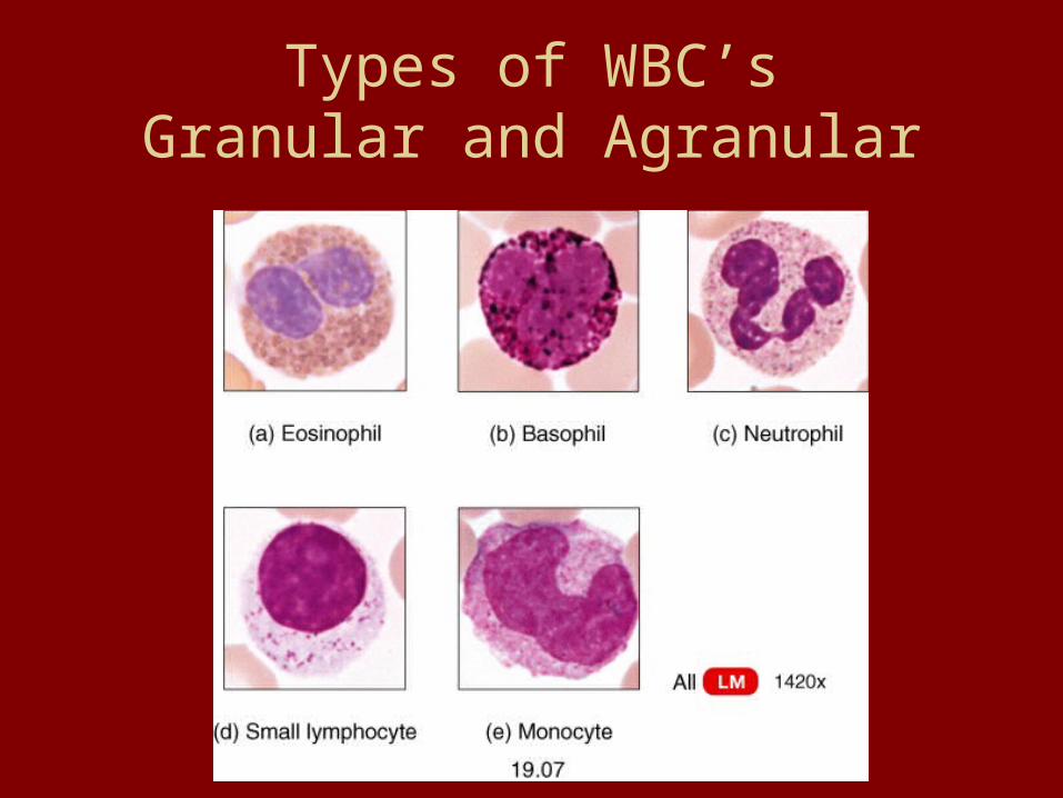
Types of WBC’sGranular and Agranular

Types of White Blood Cells
Leukocytes (WBCs)
- protect against disease
- blood transports WBCs to sites of infection
- 5 types are normally in circulation
-WBCs differ in size, nature of cytoplasm, nuclei shape, and staining
characteristics

Types of White Blood Cells
1. Granulocytes
- contain granular cytoplasm
- twice the size of RBCs
- develop in red bone marrow
- short life span (about 12 hours)

3 Types of Granulocytes
1. Neutrophils
- fine granules that stain pinkish in neutral stain
- nucleus is lobed and consists of two to five sections connected by thin strands of chromatin
- accounts for 54-62% of leukocytes in a typical blood sample
- phagocytizes small particles

2. Eosinophils- contains coarse uniformly sized cytoplasmic granules that stain deep red in acid stain- nucleus usually has ony two lobes (bilobed)- 1-3% of total number of circulating leukocytes- kills parasites and helps control inflammation and allergic
reactions

3. Basophils
- similar to eosinophils in size and in shape of their nuclei
- fewer more irregularly shaped cytoplasmic granules that stain deep blue in basic stain
- account for less than 1% of leukocytes
- releases heparin (blood-clotting-inhibiting substance) and histamine

Agranulocytes – without cytoplasmic granules
1. Monocytes- Arise from red bone marrow- Largest blood cells- 2 to 3 times greater in diameter than RBCs - Nuclei vary in shape and can be round,
kidney-shaped, oval, or lobed- 3-9% of leukocytes- Live for several weeks or even
months- Phagocytize large particle

2. Lymphocytes
- form in the organs of lymphatic system as well as red bone marrow
- slightly larger than RBCs
- large round nucleus with a thin rim of cytoplasm
- 25-33% of leukocytes
- may live for years
- provides immunity

WBC Counts
• WBCC normally includes 5,000-10,000 cells
• Rise accompanies some diseases• Exceeding 10,000 per mm3 constitutes
leukocytosis, acute infection such as appendicitis
• Below 5,000 per mm3 constitutes leukopenia, typhoid fever, influenza, measles, mumps, chicken pox, AIDS, poliomyelitis

WBC Counts cont….
• A differential white blood cell count (DIFF) list percentages of the types of leukocytes in a blood sample.
• The relative proportion of WBCs may change in particular diseases:
- Neutrophils increase during bacterial infections
- eosinophils increase during parasitic infections and allergic reactions
- In AIDS, T-lymphocytes and B-lymphocytes drop sharply.

Blood PlateletsThrombocytes (platelets) are not complete blood cells.
- arise from very large cells in red marrow, called megakaryocytes, that fragment like a shattered plate, releasing small sections of cytoplasm – the platelet – into circulation- The larger fragments shrink and also become platelets as they pass through blood vessels in the lungs.-lacks a nuclei and is less than half the size of RBCs-capable of amoeboid movement and may live for about 10 days-count varies from 130,000-360,000 per mm3-help close breaks in damaged blood vessels and initiate formation of blood clots

Blood Plasma
• Clear, straw-colored liquid
• 92% water
• Functions: – Transporting Nutrients, Gases, Vitamins– Helps regulate fluid and electrolyte balance– Maintains a favorable pH

Plasma Proteins
• Most abundant of the dissolved substances
• Remains in the blood and interstitial fluids
• Not used as energy1. Albumins – smallest; 60%, synthesiszed in
liver, help maintain blood osmotic pressure by regulating water movement between blood and tissue

Plasma Proteins cont….2. Globulins – 36%
a. Alpha globulins – synthesized in liver, transports lipids and fat-soluable vitamins
b. Beta globulins - synthesized in liver, transports lipids and fat-soluable vitaminsc. gamma globulins – synthesized in lymphatic tissue, constitutes a type of antibody
3. Fibrinogen – 4%, functions in blood coagulation, synthesized in liver, largest of the proteins

Nutrients and GasesPlasma Nutrients
1. Simple sugars2. amino acids3. nucleotides4. Lipids: must combine with proteins to form lipoproteins; lipids are less dense than a protein, therefore, as the proportion of lipids in a lipoprotein increases, the density of the particle decreases—conversely, as the proportion decreases, the density increases; lipoproteins are classified according to their density
a. Chylomicron – high concentration of triglycerides absorbed from the SI; transports dietary fats to muscles and adipose cells; after delivery the remnants are transferred to HDL molecules

Nutrients and Gases cont….
b. Very low-density lipoproteins (VLDL) – high concentrations of triglycerides; produced in the liver; transports to adipose tissue;
remnants break down into LDLc. Low-density lipoprotein (LDL) – high concentrations of cholesterol; formed from VLDL giving off their triglycerides; delivers cholesterol to various cells including liver cellsd. High-density lipoprotein (HDL) – high concentrations of protein and low lipids; transports to the liver remnants of chylomicrons that have given up their triglycerides

Nutrients and Gases cont….
Blood Gases
Oxygen and carbon dioxide, as well as a considerable amount of dissolved nitrogen, which has no physiological function. (Further discussion during respiration)
Non protein nitrogenous substances contain nitrogen atoms but are not proteins:
a. amino acids – come from protein digestion and amino acid absorption
b. urea & uric acid – products of protein and nucleic acid catabolism and are excreted in the urine

Nutrients and Gases cont….
Plasma Electrolytes- Absorbed from the intestine or released as a byproduct of cellular metabolism- include sodium, potassium, calcium, magnesium, chloride, bicarbonate, phosphate, and sulfate ions- sodium & chloride are most abundant-important in maintaining the osmotic pressure and pH of plasma-regulated so their blood concentrations remain relatively stable (further discussed in urinary system)

Hemostasis: Stopping of Bleeding1. Blood Vessel Spasm (vasospasm) – cutting of
breaking a blood vessel stimulates the smooth muscles in the vessel wall to contract, lessening blood loss almost immediately; may completely close the ends of a severed vessel; may last only a few minutes, but the effect usually continues for about 30 minutes which allows a platelet plug to form
2. Platelet Plug Formation – platelets adhere to the underlying collagen of the endothelial lining of the blood vessels; platelets also stick to each other forming a plug in the vascular break; they release serotonin which contracts smooth muscles in the blood vessel walls; controls blood loss for a small break but will require a clot for a large break

Hemostasis: Stopping of Bleeding cont….
3. Blood Coagulation – forms a blood clot; complex and utilizes many biochemicals called clotting factors; some of these factors promote coagulation and other inhibit it; damaged tissues release tissue thromboplastin, initiating a series of reactions resulting in production of prothrombin activator; this series of reactions depends on the presence of calcium ions as well as certain proteins and phospholipids for completion
1. Prothrombin – alpha globulin that the liver continually produces and thus is a constituent of plasma, which in the presence of calcium ions converts prothrombin into thrombin
2. Thrombin – catalyzes a reaction that fragments fibrinogen; the fragments join to form long threads of fibrin; once these threads form, they stick to the exposed surface of damaged blood vessels, creating a meshwork that entraps blood cells and platelets; resulting in a blood clott

• Once a blood clot begins to form, it promotes still more clotting because thrombin also acts on blood clotting factors other than fibrinogen, causing prothrombin to form still more thrombin. This is a positive feedback mechanism, in which the original actions stimulates more of the same type of action. Normally, blood flow throughout the body prevents formation of a massive clot within the cardiovascular system by rapidly carrying excess thrombin away and keeping concentrations too low to enhance further clotting.
1. Thrombus – a clot forming in a vessel abnormally; often associated with conditions that change endothelial linings of vessels2. Embolus – a dislodged or fragmented clot that iscarried away by the blood flow; emboli continue to move until they reach narrow places in vessels, where they lodge and may interfere with blood flow


Blood Groups
Antigens and Antibodies
1. Agglutination – the clumping of red blood cells following transfusion reaction; due to a reaction between:
a. Antigens – red blood cell surface molecule
b. Antibody – protein carried in plasma

ABO Blood Group• The ABO Blood Group is based on the presence (or
absence) of two major antigens on red blood cell membranes – antigen A or antigen B.
• A person’s erythrocytes contain one of four antigen combinations as a result of inheritance: only A, only B, A and B, neither A nor B.
• Certain antibodies are synthesized in plasma about 2 to 8 months following birth.
• When A is absent in red blood cells, an antibody called anti-A develops and ……An antibody of one type will react with an antigen of the same type and clump red blood cells; therefore, such combinations must be avoided


Blood Type Antigen Antibody Donate to Receive From
A A Anti-B A, AB A, O
B B Anti-A B, AB B, O
AB (universal
recipient)
A and B Neither
Anti-A or Anti-B
AB A, B, AB, O
O (universal
donor)
Neither A nor B
Both anti-A and
anti-B
A, B, AB, O
O


Transfusions
• If type O blood is given to a person with type A, B, or AB, it should be transfused slowly so that the recipient’s larger blood volume will dilute it, minimizing the chance of an adverse reaction.
• The same is true for a type AB person receiving type A, B, or, O.
• If transfused to rapidly it could agglutinate because each of these blood types contain an antibody.

Rh Blood Groups
• Named after the rhesus monkey, in which it was first studied.
• Includes several Rh antigens (factors)• The most important is antigen D• If antigen D or any other Rh antigen are
present on the red blood cell membrane, the blood is called Rh positive
• If there are no Rh antigens present the blood is referred to as Rh negative
• Presence or absences is inherited

Rh Blood Groups cont….
• Antibodies (anti-Rh) do not appear spontaneously, instead they form only in response to special stimulation:– If an Rh-negative person receives a transfusion of Rh-
positive blood, the Rh antigens stimulate the recipient’s antibody-producing cells to begin producing anti-Rh antibodies.
– This initial transfusion has no serious consequences, but if the Rh-negative person – who is now synthesized to Rh-positive blood – receives another transfusion of Rh-positive some months later, the donated red cells are likely to agglutinate.


Rh Blood Groups cont….
• A related conditions may occur when an Rh-negative woman is pregnant with an Rh-positive fetus for the first time.
• Such a pregnancy may be uneventful, however, at birth (or during a miscarriage or abortion), the placental membranes that separate the maternal blood from the fetal blood tear, resulting in some of the infant’s Rh-positive blood cells to enter maternal circulation.
• These Rh-positive cells may stimulate the maternal tissues to begin producing anti-Rh antibodies.

Rh Blood Groups cont….• If a woman who has already developed anti-Rh
antibodies becomes pregnant with a second Rh-positive fetus, that ant-Rh antibodies, called hemolysins, pass through the placental membrane and destroy the fetal red blood cells.
• The fetus then develops a condition called erythroblastosis fetalis which is extremely rare today because physicians carefully note Rh status.
• An Rh-negative women who might carry an Rh-positive fetus receives an injection of a drug called Rhogam.
• Rhogam is actually an anti-Rh antibody, which binds to and shields any Rh-positive fetal cells that might contact the woman’s cells, sensitizing her immune system.
• Rhogam must be given within 72 hours of possible contact with Rh-positive cells – such conditions include giving birth, terminating a pregnancy, miscarrying, or undergoing an amniocentesis.


1. What causes red blood cells to become sickle shaped?
2. Can there be an advantage to this?

AnemiasDetermine the cause of the following types of anemia.
• Iron deficiency
• Pernicious (Low B12)
• Hemorrhagic
• Hemolytic
• Thalassemia
• Aplastic (loss of marrow)

Words to KnowDiscuss each of the following words in regard to cause and affect on the human body.
• Hemophilia
• Leukemia
• Autologous transfusion
• Cyanosis
• Hemochromatosis
• Jaundice
• Septicemia


