Catholic Medical Center Laboratory 100 McGregor Street, Manchester, NH 03102 BLOOD COLLECTION TUBES LIGHT BLUE TOP TUBE – 2.7 ml draw and 1.8 ml draw Liquid 3.2% sodium citrate anticoagulant RED TOP TUBE – 4 ml draw Sprayed on clot activator GOLD TOP TUBE (SST) – 3.5 ml draw Sprayed on clot activator with gel for serum separation LIGHT GREEN TOP TUBE – 3 ml draw Lithium heparin anticoagulant with gel for plasma separation PURPLE TOP TUBE – 2 ml draw or 4ml draw or 6ml draw Sprayed on K2EDTA Anticoagulant The following tubes are available upon request through Catholic Medical Center’s Laboratory Customer Services: NAVY BLUE TOP TUBE – 6 ml draw (trace element) No additive NAVY BLUE TOP TUBE – 7 ml draw Na Heparin NAVY BLUE TOP TUBE – 6 ml draw K2EDTA anticoagulant GREEN TOP TUBE – 4 ml draw Sodium heparin anticoagulant GRAY TOP TUBE – 6 ml draw Powder sodium fluoride / potassium oxalate anticoagulant YELLOW TOP TUBE – 8.5 ml draw Liquid ACD solution A YELLOW TOP TUBE – 6 ml draw Liquid ACD solution B YELLOW TOP TUBE – 8.3 ml draw SPS (Sodium Polyanelthol Sulfonate) After specimen collection, all tubes - with the exception of the Light Blue Top tube (Sodium Citrate) - are to be gently inverted 8-10 times. This will ensure the proper distribution of additives with the specimen. The Light Blue Top Sodium Citrate tube should be gently inverted only 3-4 times to avoid activation of platelets which could skew test results. Gold and red top tubes should be allowed to clot for a minimum of 30 minutes and centrifuged within a maximum of 2 hours after collection. Serum for the red top tube must be removed as soon as possible from the cells to maintain the integrity of the specimen. Extra red top tubes that are collected in the emergency will only be viable for one hour on arrival in the lab.
Transcript
Catholic Medical Center Laboratory 100 McGregor Street, Manchester, NH 03102
BLOOD COLLECTION TUBES
LIGHT BLUE TOP TUBE – 2.7 ml draw and 1.8 ml draw
Liquid 3.2% sodium citrate anticoagulant
RED TOP TUBE – 4 ml draw
Sprayed on clot activator
GOLD TOP TUBE (SST) – 3.5 ml draw
Sprayed on clot activator with gel for serum separation
LIGHT GREEN TOP TUBE – 3 ml draw
Lithium heparin anticoagulant with gel for plasma separation
PURPLE TOP TUBE – 2 ml draw or 4ml draw or 6ml draw
Sprayed on K2EDTA Anticoagulant
The following tubes are available upon request through Catholic Medical Center’s Laboratory
After specimen collection, all tubes - with the exception of the Light Blue Top tube (Sodium
Citrate) - are to be gently inverted 8-10 times. This will ensure the proper distribution of
additives with the specimen. The Light Blue Top Sodium Citrate tube should be gently inverted
only 3-4 times to avoid activation of platelets which could skew test results.
Gold and red top tubes should be allowed to clot for a minimum of 30 minutes and centrifuged
within a maximum of 2 hours after collection. Serum for the red top tube must be removed as
soon as possible from the cells to maintain the integrity of the specimen. Extra red top tubes that
are collected in the emergency will only be viable for one hour on arrival in the lab.
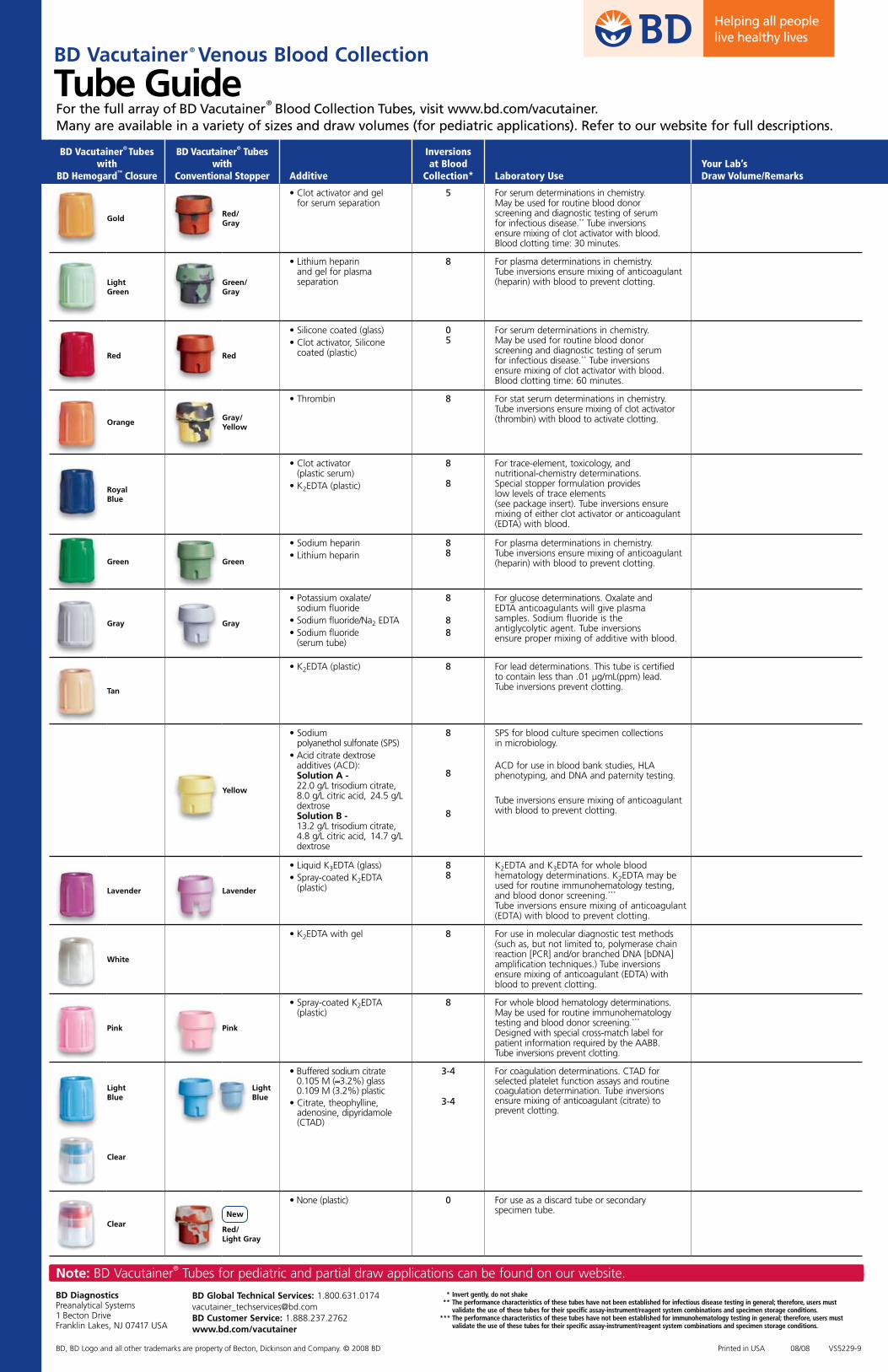
BD Vacutainer® Venous Blood Collection
Tube Guide
BD Diagnostics Preanalytical Systems 1 Becton Drive Franklin Lakes, NJ 07417 USA
* Invert gently, do not shake ** The performance characteristics of these tubes have not been established for infectious disease testing in general; therefore, users must
validate the use of these tubes for their specific assay-instrument/reagent system combinations and specimen storage conditions. *** The performance characteristics of these tubes have not been established for immunohematology testing in general; therefore, users must validate the use of these tubes for their specific assay-instrument/reagent system combinations and specimen storage conditions.
BD Global Technical Services: [email protected] Customer Service: 1.888.237.2762www.bd.com/vacutainer
5 For serum determinations in chemistry. May be used for routine blood donor screening and diagnostic testing of serum for infectious disease.** Tube inversions ensure mixing of clot activator with blood. Blood clotting time: 30 minutes.
Light Green
Green/Gray
• Lithium heparin and gel for plasma separation
8 For plasma determinations in chemistry. Tube inversions ensure mixing of anticoagulant (heparin) with blood to prevent clotting.
For serum determinations in chemistry. May be used for routine blood donor screening and diagnostic testing of serum for infectious disease.** Tube inversions ensure mixing of clot activator with blood. Blood clotting time: 60 minutes.
OrangeGray/Yellow
• Thrombin 8 For stat serum determinations in chemistry. Tube inversions ensure mixing of clot activator (thrombin) with blood to activate clotting.
Royal Blue
• Clot activator (plastic serum)
• K2EDTA (plastic)
8 8
For trace-element, toxicology, and nutritional-chemistry determinations. Special stopper formulation provides low levels of trace elements (see package insert). Tube inversions ensure mixing of either clot activator or anticoagulant (EDTA) with blood.
Green Green
• Sodium heparin• Lithium heparin
8 8
For plasma determinations in chemistry. Tube inversions ensure mixing of anticoagulant (heparin) with blood to prevent clotting.
Gray Gray
• Potassium oxalate/ sodium fluoride
• Sodium fluoride/Na2 EDTA• Sodium fluoride
(serum tube)
8
88
For glucose determinations. Oxalate and EDTA anticoagulants will give plasma samples. Sodium fluoride is the antiglycolytic agent. Tube inversions ensure proper mixing of additive with blood.
Tan
• K2EDTA (plastic) 8 For lead determinations. This tube is certified to contain less than .01 µg/mL(ppm) lead. Tube inversions prevent clotting.
SPS for blood culture specimen collections in microbiology. ACD for use in blood bank studies, HLA phenotyping, and DNA and paternity testing.
Tube inversions ensure mixing of anticoagulant with blood to prevent clotting.
Lavender Lavender
• Liquid K3EDTA (glass)• Spray-coated K2EDTA
(plastic)
8 8
K2EDTA and K3EDTA for whole blood hematology determinations. K2EDTA may be used for routine immunohematology testing, and blood donor screening.*** Tube inversions ensure mixing of anticoagulant (EDTA) with blood to prevent clotting.
White
• K2EDTA with gel 8 For use in molecular diagnostic test methods (such as, but not limited to, polymerase chain reaction [PCR] and/or branched DNA [bDNA] amplification techniques.) Tube inversions ensure mixing of anticoagulant (EDTA) with blood to prevent clotting.
Pink Pink
• Spray-coated K2EDTA (plastic)
8 For whole blood hematology determinations. May be used for routine immunohematology testing and blood donor screening.*** Designed with special cross-match label for patient information required by the AABB. Tube inversions prevent clotting.
Light Blue
Light Blue
• Buffered sodium citrate 0.105 M (≈3.2%) glass 0.109 M (3.2%) plastic
For coagulation determinations. CTAD for selected platelet function assays and routine coagulation determination. Tube inversions ensure mixing of anticoagulant (citrate) to prevent clotting.
Clear
ClearNew
Red/Light Gray
• None (plastic) 0 For use as a discard tube or secondary specimen tube.
Centrifuge at full speed
1100 – 1300g for 13 mm Plus Plastic tubes
1000 – 1300g for 16 mm Plus Plastic tubes
for 10 minutes in a swing-bucket unit or 15 minutes for a fixed-angle unit (balance tubes in centrifuge).
Gel barrier will form to separate plasma from red blood cells.
x 8
Ready for
Analysis
How to Prepare a Quality Sample
Spin
10Minutes
Invert
8-10Times
Using BD Vacutainer® PST™ Tubes
Centrifuge at1100 - 1300g
Gently invert 8 -10 times immediately after collection to mix lithium heparin anticoagulant with blood.
Insufficient mixing may lead to microclot and fibrin strand formation.
Use in laboratory for plasma determinations in chemistry.
BDPAS-PSTQualWC-7846.indd 1 3/6/06 12:27:10 PM
• Allow blood to clot for a minimum of 5 minutes in a vertical position.
• Observe a dense clot.
Spinas low as
3*
Minutes
Invert
5-6Times
Gently invert 5-6 times to mix clot activator with blood.
• Centrifuge at * 4000g for 3 minutes, 2000g for 4 minutes, or 1500–2000g for 10 minutes (balance tube in centrifuge), at 23-27°C.
• Barrier will form, separating serum specimen from clot.
• Transport spun tube to laboratory.
Clot
5Minutes
x 5-6
How to Prepare a Quality SampleBD Vacutainer® Rapid Serum Tube
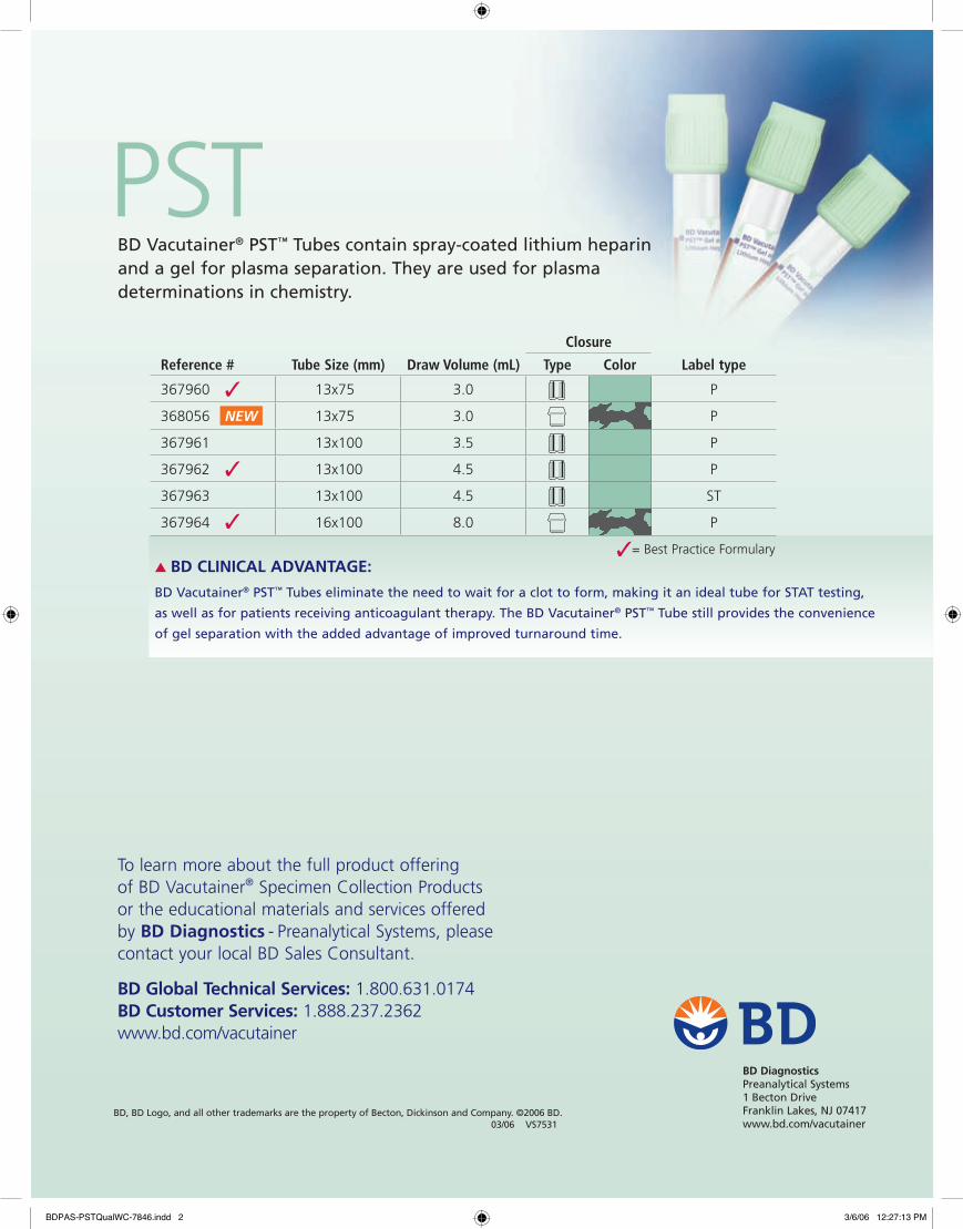
PST BD Vacutainer® PST™ Tubes contain spray-coated lithium heparin and a gel for plasma separation. They are used for plasma determinations in chemistry.
Closure
Reference # Tube Size (mm) Draw Volume (mL) Type Color Label type
367960 13x75 3.0 P
368056 13x75 3.0 P
367961 13x100 3.5 P
367962 13x100 4.5 P
367963 13x100 4.5 ST
367964 16x100 8.0 P
✓
✓
✓
NEW
To learn more about the full product offering of BD Vacutainer® Specimen Collection Products or the educational materials and services offered by BD Diagnostics - Preanalytical Systems, please contact your local BD Sales Consultant.
BD Global Technical Services: 1.800.631.0174BD Customer Services: 1.888.237.2362www.bd.com/vacutainer
▲ BD CLINICAL ADVANTAGE:
BD Vacutainer® PST™ Tubes eliminate the need to wait for a clot to form, making it an ideal tube for STAT testing,
as well as for patients receiving anticoagulant therapy. The BD Vacutainer® PST™ Tube still provides the convenience
of gel separation with the added advantage of improved turnaround time.
✓= Best Practice Formulary
BDPAS-PSTQualWC-7846.indd 2 3/6/06 12:27:13 PM
Maximum Fill*
Minimum FillIndicator
BD V
acut
aine
r® P
lus
Plas
tic
Citr
ate
Tub
e
Note: The quantity of blood drawn into evacuated tubes varies with altitude, ambient temperature, barometric pressure, tube age, venous pressure and filling technique.
Sufficient volume achieved if blood drawn falls above minimum fill indicator. For blood transfer, do not fill above illustrated dashed maximum line.
BD Vacutainer® Plus Plastic Citrate Tube Draw Volume Guide
* According to CLSI guideline, Dec. 2003, Doc. H1-A5, Vol. 23, No. 33.
2.7 mL Draw Tube
13 mm x 75 mm Full Draw
1.8 mL Draw Tube13 mm x 75 mmFull Draw
Ref. # 363083 Ref. # 363080
Represents minimum volume of blood
required for appropriate analysis
Now with Minimum Fill
Indicator
BD Diagnostics Preanalytical Systems 1 Becton Drive Franklin Lakes, NJ 07417BD, BD Logo and BD Vacutainer® are trademarks of Becton, Dickinson and Company.
• Assemble needle in holder; always fully seat and hold a citrate tube on the back end of the needle while filling.
• Allow the tube to fill until the vacuum is exhausted and blood flow ceases.
• Tubes should fill between ±10% of the stated draw volume of the tube (CLSI guideline, Dec. 2003, Doc. H1-A5, Vol. 23, No. 33).
• Minimum fill indicator represents the minimum volume of blood required for appropriate analysis.
• A discard tube (without additives) must be used if only a citrate tube is to be drawn using a winged blood collection set. It is important to remove the air from the blood collection set to ensure the proper blood volume is obtained in the tube.
• Do not fill tubes from other tubes or combine two partially filled citrate tubes.
• If the specimen is drawn with a syringe, do not fill the BD Vacutainer® Citrate Tube beyond the level as illustrated on the reverse side of this guide. Allow the tube to draw the blood from the syringe using a BD Vacutainer® Blood Transfer Device if available. Do not force blood into tube.
• Immediately after draw, gently invert tube 3 to 4 times. Do not shake.
BD V
acut
aine
r® P
lus
Plas
tic
Citr
ate
Tub
e Minimizing Preanalytical Variables for Coagulation Tests
Cat# Size Draw Citrate
363083 13 x 75 mm 2.7 mL 3.2% (0.109M)
363080 13 x 75 mm 1.8 mL 3.2% (0.109M)
BD Global Technical Services: 1.800.631.0174www.bd.com/vacutainer
BD Vacutainer® Order of Draw for Multiple Tube CollectionsDesigned for Your Safety
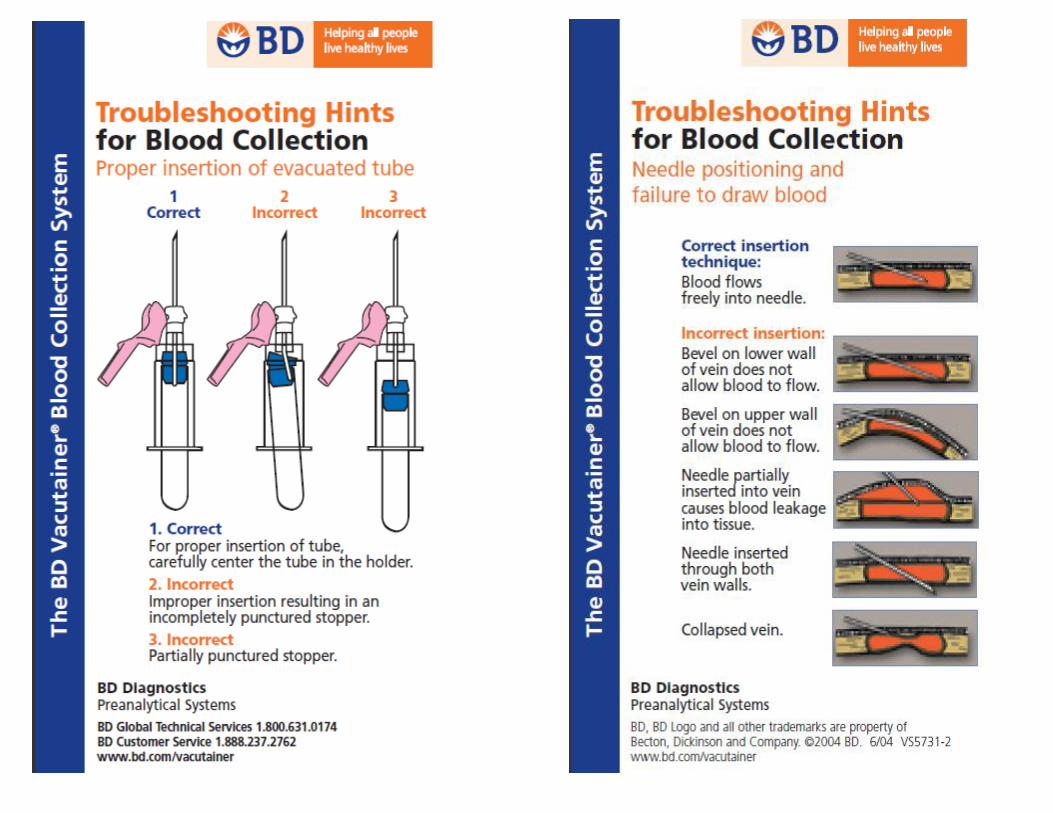
Handle all biologic samples and blood collection “sharps” (lancets, needles, luer adapters and blood collection sets) according to the policies and procedures of your facility. Obtain appropriate medical attention in the event of any exposure to biologic samples (for example, through a puncture injury) since they may transmit viral hepatitis, HIV (AIDS), or other infectious diseases. Utilize any built-in used needle protector if the blood collection device provides one. BD does not recommend reshielding used needles, but the policies and procedures of your facility may differ and must always be followed. Discard any blood collection “sharps” in biohazard containers approved for their disposal.
Note: Always follow your facility’s protocol for order of draw
BD Diagnostics Preanalytical Systems 1 Becton Drive Franklin Lakes, NJ 07417
www.bd.com/vacutainer
BD Global Technical Services
1.800.631.0174
BD Customer Service
1.888.237.2762www.bd.com/vacutainer
= 1 inversion
BD Vacutainer® Blood Collection Tubes (glass or plastic)
or
or
or
• Blood Cultures - SPS
• Citrate Tube*
• BD Vacutainer® SST™
Gel Separator Tube• Serum Tube
(glass or plastic)
• Heparin Tube
• BD Vacutainer® PST™
Gel Separator Tube With Heparin
• EDTA Tube
• Fluoride (glucose) Tube
8 to 10 times
3 to 4 times
5 times
5 times (plastic) none (glass)
8 to 10 times
8 to 10 times
8 to 10 times
8 to 10 times
Closure Color Collection Tube Mix by Inverting
* When using a winged blood collection set for venipuncture and a coagulation (citrate) tube is the first specimen tube to be drawn, a discard tube should be drawn first. The discard tube must be used to fill the blood collection set tubing’s “dead space” with blood but the discard tube does not need to be completely filled. This important step will ensure maintenance of the proper blood-to-additive ratio of the blood specimen. The discard tube should be a nonad-ditive or coagulation tube.
Reflects change in NCCLS recommended Order of Draw (NCCLS H3-A5, Vol 23, No 32, 8.10.2)
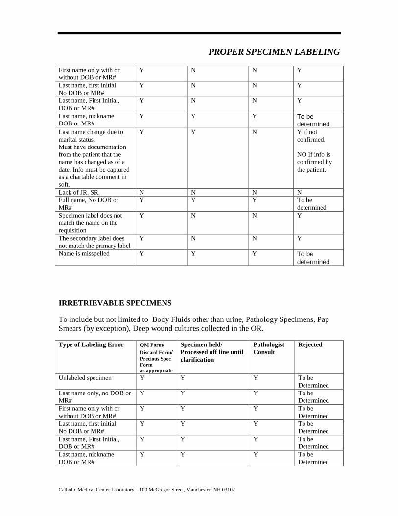
PROPER SPECIMEN LABELING
Catholic Medical Center Laboratory 100 McGregor Street, Manchester, NH 03102
PATIENT SAFETY IS IN YOUR HANDS.
Label all specimens in the presence of the patient
List the full legal name , First and Last List another unique identifier i.e. Date of
Birth or Medical Record Number List the date and time of the collection List the collectors ID
PROPER SPECIMEN LABELING
Catholic Medical Center Laboratory 100 McGregor Street, Manchester, NH 03102
LABELING All specimens arriving in the laboratory must be properly labeled to support Patient Safety. Two unique identifiers for the patient must be on the specimen. The first and last name of the patient is mandatory. Either a date of birth or the medical record number is acceptable as the second identifier. The patient’s social security is not necessary. The date and time of collection, as well as the ID of the collector should be noted if not captured electronically. The labeling information must be confirmed with the patient when ever possible before labeling the specimen by asking the patient to recite the spelling of their name and date of birth. If the patient is unable to confirm by spelling the name etc, the name of a care giver that provides confirmation of the identity of the patient should be listed. Any discrepancies must be reconciled. Information presented on the order request and specimen labels must be in agreement. Additionally, this information should be concurrent to the information present on the patient identification band. GUIDELINES FOR LABELING:
• Immediately after collection, legibly label the specimen in the presence of the patient. • For positive patient identification ask the patient to spell their first and last name
along with reciting their date of birth. Compare with the preprinted label if available. If no label is available, hand label the tube with the patient’s full name, date of birth, date and time of collection, and the code/initials of the collector. This information along with the patient’s demographics should also appear on the requisition. Patient Identification Label For Specimens
NAME DOB Phleb Initials Date Time
PROPER SPECIMEN LABELING
Catholic Medical Center Laboratory 100 McGregor Street, Manchester, NH 03102
Labels whether electronically or manually generated should be:
• Placed vertically over the tube’s paper label with the name seated near the cap of the tube.
• Placed on straight, not wrapped around the tube or tube bottom, or overlapping the tube cap
• Free of wrinkles and tears • Free of stray marks in the bar-coded area • Placed on the tube in single thickness (only a single label placed on the tube)
SPECIMEN REJECTION DUE TO LABELING ERROR The laboratory strives for 100% compliance when it comes to specimen labeling and Patient Safety. Specimens are considered mislabeled when there is a mismatch between patient specific identifiers and information accompanying the specimen. When insufficient, inconsistent, or inaccurate identification exists, the laboratory will recommend that a new specimen be obtained if feasible, based on the outline of the following charts (Retrievable Specimens, Irretrievable or Precious Specimens). LABELING GUIDELINES: It is expected that lab specimens will be labeled at the point of collection in the presence of the patient. The labels are required to have 2 unique patient identifiers, Date and Time of collection and the collectors ID. Immediate notification to the provider is imperative for all discrepancies. **************************************************************** RETRIEVABLE SPECIMENS For Retrievable Samples to include but not limited to: urines, blood collections, Stool, Semen, Swabs, (except those obtained in the OR). Blood Bank specimens must match exactly and have all of the labeling criteria or they will be rejected. * For specimens mailed to the laboratory by the patient, such as fecal occult blood cards, the second identifier can be the address and phone number of the patient as the card template displays. The requisit ion information accompanying the stool cards should match the name, address and phone number as w ritten on the stool card. Type of Labeling Error QM Form/
Discard Form/ Precious Spec Form as appropriate
Specimen held/ Processed off line until clarification
Pathologist Consult
Rejected
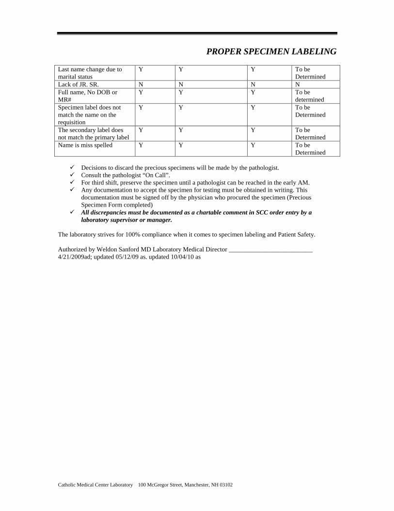
Unlabeled specimen Y N N Y Last name only, no DOB or MR#
Y N N Y
PROPER SPECIMEN LABELING
Catholic Medical Center Laboratory 100 McGregor Street, Manchester, NH 03102
First name only with or without DOB or MR#
Y N N Y
Last name, first initial No DOB or MR#
Y N N Y
Last name, First Initial, DOB or MR#
Y N N Y
Last name, nickname DOB or MR#
Y Y Y To be determined
Last name change due to marital status. Must have documentation from the patient that the name has changed as of a date. Info must be captured as a chartable comment in soft.
Y Y N Y if not confirmed. NO If info is confirmed by the patient.
Lack of JR. SR. N N N N Full name, No DOB or MR#
Y Y Y To be determined
Specimen label does not match the name on the requisition
Y N N Y
The secondary label does not match the primary label
Y N N
Y
Name is misspelled Y Y Y To be determined
IRRETRIEVABLE SPECIMENS To include but not limited to Body Fluids other than urine, Pathology Specimens, Pap Smears (by exception), Deep wound cultures collected in the OR. Type of Labeling Error QM Form/
Discard Form/ Precious Spec Form as appropriate
Specimen held/ Processed off line until clarification
Pathologist Consult
Rejected
Unlabeled specimen Y Y Y To be Determined
Last name only, no DOB or MR#
Y Y Y To be Determined
First name only with or without DOB or MR#
Y Y Y To be Determined
Last name, first initial No DOB or MR#
Y Y Y To be Determined
Last name, First Initial, DOB or MR#
Y Y Y To be Determined
Last name, nickname DOB or MR#
Y Y Y To be Determined
PROPER SPECIMEN LABELING
Catholic Medical Center Laboratory 100 McGregor Street, Manchester, NH 03102
Last name change due to marital status
Y Y Y To be Determined
Lack of JR. SR. N N N N Full name, No DOB or MR#
Y Y Y To be determined
Specimen label does not match the name on the requisition
Y Y Y To be Determined
The secondary label does not match the primary label
Y Y Y
To be Determined
Name is miss spelled Y Y Y To be Determined
Decisions to discard the precious specimens will be made by the pathologist. Consult the pathologist “On Call”. For third shift, preserve the specimen until a pathologist can be reached in the early AM. Any documentation to accept the specimen for testing must be obtained in writing. This
documentation must be signed off by the physician who procured the specimen (Precious Specimen Form completed)
All discrepancies must be documented as a chartable comment in SCC order entry by a laboratory supervisor or manager.
The laboratory strives for 100% compliance when it comes to specimen labeling and Patient Safety. Authorized by Weldon Sanford MD Laboratory Medical Director __________________________ 4/21/2009ad; updated 05/12/09 as. updated 10/04/10 as
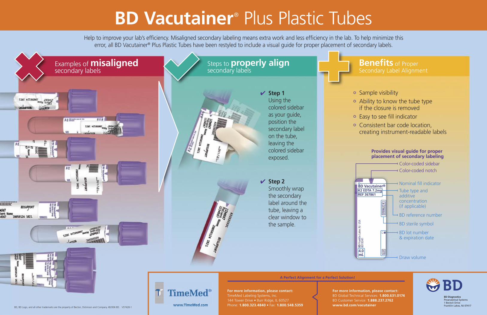
Benefi ts of Proper Secondary Label Alignment
Examples of misaligned secondary labels
Steps to properly alignsecondary labels
BD Vacutainer® Plus Plastic TubesHelp to improve your lab’s effi ciency. Misaligned secondary labeling means extra work and less effi ciency in the lab. To help minimize this
error, all BD Vacutainer® Plus Plastic Tubes have been restyled to include a visual guide for proper placement of secondary labels.
✔ Step 1Using the colored sidebar as your guide, position the secondary label on the tube, leaving the colored sidebar exposed.
✔ Step 2Smoothly wrap the secondary label around the tube, leaving a clear window to the sample.
Sample visibility Ability to know the tube type if the closure is removed
Easy to see fi ll indicator Consistent bar code location, creating instrument-readable labels
Draw volume
Tube type and additive concentration (if applicable)
BD lot number & expiration date
BD reference number
Color-coded sidebarColor-coded notch
Provides visual guide for proper placement of secondary labeling
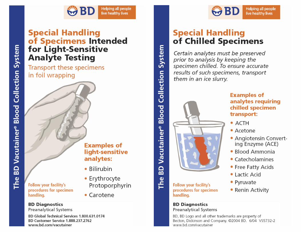
B12 and Folate 8 hrs- Room Temp or Refrig After 8 hrs protect from light and refrigerate C-Diff 48 hr Refrigerated CBC 24 hr- Room Temp Clotted samples or those containing clots, fibrin strands,
or platelet clumps, grossly hemolyzed specimens, insufficient volume, and those drawn above an IV are not acceptable. Sample stability is 24 hours at room temperature.
Cold Agglutinin Room temperature specimen clots at 37 degrees Cryoglobulin 3 days at Room temperature D-Dimer 24 hr- Room Temp or Refrig Direct Bili 24 hr- Room Temp or Refrig Protect from Light Total Bilirubin 24 hr- Room Temp or Refrig Protect from Light ED Cardiac Panel-Biosite 4 hrs- Room Temp
ESR 24 hr- Room Temp Insufficient volume (Tube must be 2/3 full), specimens more then 24 hours post-collection, hemolysis, clotted specimens, and improperly labeled specimens.
Fibrinogen 4 hr- Room Temp Patient plasma should be tested within 4 hours (centrifuged or uncentrifuged) if stored in an unopened tube at Room Temp Short draw tubes, clotted and hemolyzed specimens are never acceptable.
HIT- Heparin Dependent Antibodies Screen- IgGAM
Separated plasma: freeze immediately 6 months frozen at -20
Plasma should be separated as soon as possible after collection, this test cannot be added on.
Herpes Viral Culture 4 days refrigerated, 6 months frozen
Viral Culture Medium(VCM) only
Homocysteine 1 hour- on ice separate plasma and perform testing immediately, refrigerated plasma stable for 7 days
Ionized Calcium Room temperature Specimen should never have been uncapped. Kleihauer Betke 2 hrs - Room Temp
24 hours on ice
Lactic Acid 1 hour- on ice Must be collected on ice and performed within 1 hr of collection
Catholic Medical Center Laboratory 100 McGregor Street, Manchester, NH 03102
Test Name Specimen Stability from Collection Time Special Instructions
LDH Specimen kept at Room Temp, serum/plasma must be separated from cells within 2 hrs of collection.
O&P Screen 24 hrs - Room Temp if not in preservative 2 weeks if in preservative
Osmolality-Serum 3 hours at Room Temperature up to 10 hours refrigerated
Osmolality-Urine 3 hours at room temperature up to 24 hours refrigerated 1 week frozen
PFA 4 hrs- Room Temp Patient samples are stable for up to 4 hours and must be stored at room temperature. Do not refrigerate or centrifuge sample prior to analysis.Do not use the pneumatic tube; hand deliver to the lab via transport personnel. Hemolyzed specimens not accepted.
PT and/or PT Mixing Study
24 hr- Room Temp Patient plasma should be tested within 24 hours (centrifuged or uncentrifuged) if stored in an unopened tube at Room Temp Short draw tubes, clotted and hemolyzed specimens are never acceptable. Frozen platelet poor plasma good for 2 weeks at minus 20 degrees, and good for 12 months at minus 70 degrees.
PTT and/or PTT Mixing Study
4 hr- Room Temp Patient plasma should be tested within 4 hours (centrifuged or uncentrifuged) if stored in an unopened tube at Room Temp Short draw tubes, clotted and hemolyzed specimens are never acceptable. Frozen platelet poor plasma good for 2 weeks at minus 20 degrees, and good for 12 months at minus 70 degrees.
Retic 24 hr- Room Temp Clotted samples or those containing clots, fibrin strands, or platelelt clumps, grossly hemolyzed sepcimens, insufficient volume, and those drawn above an IV are not acceptable. Sample stability is 24 hours at room temperature.
TEG 2 hrs- Room Temp Specimens are stable at room temperature for two (2) hours. They must not be refrigerated, nor centrifuged. Do not use the pneumatic tube.


![l>lf·· E ·B; -I,:,C-·-1·1V · cat. no.i bd lj.657 bd lj.6]5 bd 4630 bd 4·627 bd 4628 bd 4886 bd 4546 bd 4·545 bd 4544 bd 4542 bd lj,588 bd lj.593 bd 0102 bd 4636 bd 4632 bd](https://static.documents.pub/doc/80x56/5f7c69bb7d840d18665ab1e6/llf-e-b-ic-11v-cat-noi-bd-lj657-bd-lj65-bd-4630-bd-4627-bd-4628-bd.jpg)


