Blood flow dynamics of one cardiac cycle and relationship tomechanotransduction and trabeculation during heart looping
Barbara Garita,1,2 Michael W. Jenkins,3 Mingda Han,1 Chao Zhou,4 Michael VanAuker,2
Andrew M. Rollins,3 Michiko Watanabe,5 J. G. Fujimoto,4 and Kersti K. Linask1
1Department of Pediatrics, The Children’s Research Institute, University of South Florida and All Children’s Hospital,St. Petersburg; and 2Department of Chemical Engineering, University of South Florida, Tampa, Florida; 3Department ofBiomedical Engineering, Case Western Reserve University, Cleveland, Ohio; 4Department of Electrical Engineeringand Computer Science and Research Laboratory of Electronics, Massachusetts Institute of Technology, Cambridge,Massachusetts; and 5Department of Pediatrics, Case Western Reserve University, and Rainbow Babies and Children’sHospital, Cleveland, Ohio
Submitted 3 May 2010; accepted in final form 5 January 2011
Garita B, Jenkins MW, Han M, Zhou C, VanAuker M, RollinsAM, Watanabe M, Fujimoto JG, Linask KK. Blood flow dynamicsof one cardiac cycle and relationship to mechanotransduction andtrabeculation during heart looping. Am J Physiol Heart Circ Physiol300: H879–H891, 2011. First published January 14, 2011;doi:10.1152/ajpheart.00433.2010.—Analyses of form-function rela-tionships during heart looping are directly related to technologicaladvances. Recent advances in four-dimensional optical coherencetomography (OCT) permit observations of cardiac dynamics at high-speed acquisition rates and high resolution. Real-time observation ofthe avian stage 13 looping heart reveals that interactions between theendocardial and myocardial compartments are more complex thanpreviously depicted. Here we applied four-dimensional OCT to elu-cidate the relationships of the endocardium, myocardium, and cardiacjelly compartments in a single cardiac cycle during looping. Sixcardiac levels along the longitudinal heart tube were each analyzed at15 time points from diastole to systole. Using image analyses, theorganization of mechanotransducing molecules, fibronectin, tenascinC, �-tubulin, and nonmuscle myosin II was correlated with specificcardiac regions defined by OCT data. Optical coherence microscopyhelped to visualize details of cardiac architectural development in theembryonic mouse heart. Throughout the cardiac cycle, the endocar-dium was consistently oriented between the midline of the ventralfloor of the foregut and the outer curvature of the myocardial wall,with multiple endocardial folds allowing high-volume capacities dur-ing filling. The cardiac area fractional shortening is much higher thanpreviously published. The in vivo profile captured by OCT revealedan interaction of the looping heart with the extra-embryonic splanch-nopleural membrane providing outside-in information. In summary,the combined dynamic and imaging data show the developing struc-tural capacity to accommodate increasing flow and the mechanotrans-ducing networks that organize to effectively facilitate formation of thetrabeculated four-chambered heart.
A CRITICAL TRANSITION PERIOD in cardiogenesis relates to thesingle-chambered, tubular heart undergoing dramatic morpho-genesis by looping to become a four-chambered structure withvalve leaflets, ventricular trabeculae, and double parallel bloodflow. Integration of complex changes associated with looping,starting with the relatively simple heart tube, has been the
subject of much study and speculation. The interplay betweenform and function is hypothesized to be critical for heartmorphogenesis (19). Mechanotransduction of the physicalforces of blood flow, along with cardiac tube elongation, aresuggested to regulate looping to generate the four-chamberedheart (27, 30). Using genetic and surgical experiments tocompromise cardiovascular function in the zebrafish (19),avian (15, 47, 54), and mouse models (32), investigators haveprovided evidence supporting the deduction that fluid forcesare an important factor in cardiac morphogenesis. What re-mains unknown is how cardiac function and mechanotransduc-tion systems are coordinated to transduce the changing fluidforces of blood across the cardiac jelly (CJ) compartment to themyocardium.
Using high-frequency ultrasound, the chicken embryo hearthas been imaged from stages 9 to 39 (37). Whereas high-frequency ultrasound is excellent at analyzing morphology andfunction in embryos with four-chambered hearts, the limitedresolution becomes an issue for investigating tubular heartdevelopment, especially before stage 20. An acoustic mediumalso is needed to transmit the ultrasound signal, making theprocess somewhat invasive and not ideal for longitudinalstudies. Confocal microscopy has the increased resolution forobserving the early stages of development (13), but the pene-tration depth is lacking and makes confocal incapable ofimaging important animal models that develop four-chamberedhearts (murine and avian) under physiological conditions. Op-tical coherence tomography (OCT) has both the resolution(2–15 �m) and imaging depth (1–2 mm) to investigate thetubular avian and murine heart under physiological conditions.The present studies analyzing a single complete cardiac cycleusing four-dimensional (4D) OCT directed focus on specificregions of the heart that appear to relate to areas of biophysicalsignificance in the development of form and function duringlooping. Additionally, optical coherence microscopy (OCM) ofthe looping murine heart, integrated with immunohistochemi-cal analysis of mechanotransducing molecules, provides newinsights as to mechanisms involved in relation to the endocar-dial, myocardial, and CJ compartments during looping and thedevelopment of the trabeculated cytoarchitecture of the four-chambered heart postlooping.
Our analysis of structure-function relationships of a singlecardiac cycle is presented in detail. The cardiac cycle is thebasic unit of function and, therefore, provides necessary infor-mation on cardiac parameters that need to be considered for
Address for reprint requests and other correspondence: K. K. Linask,USF/ACH Children’s Research Institute, 140 7th Aveneue South, St. Peters-burg, FL 33701 (e-mail: [email protected]).
Am J Physiol Heart Circ Physiol 300: H879–H891, 2011.First published January 14, 2011; doi:10.1152/ajpheart.00433.2010.
defining mechanisms of mechanotransduction, i.e., the conver-sion of physical forces to biochemical cellular directives. Thereproducibility of the cardiac cycle among different embryonichearts for the specific stage analyzed was consistent. Theobjectives of the described studies were to define, during onecardiac filling (diastole) and ejection of blood (systole), therelationships between the endocardium, myocardium, and CJin the living embryonic avian heart during mid-looping atHamburger and Hamilton (HH) stage 13. The results definedseveral mechanotransducing pathways that coordinate bloodflow information with the morphogenetic process, looping.
MATERIALS AND METHODS
OCT and Image Processing
Three quail (Coturnix coturnix japonica), embryonic stage 13hearts, �48–52 h (2), were monitored in ovo with the ultrahigh-speed4D OCT system. This stage is comparable to HH stage 13 chickembryos (17) with an intact circulatory system. At the stages reportedhere, there is little developmental difference between chick and quailembryos: both chick and quail embryos are the same size, and heartdevelopment is similar. The quail embryo was used because it has asmaller yolk and is thus easier to monitor underneath the microscope.Using OCT, we recorded data for three quail embryos. Visual com-parison of the recorded data demonstrated no differences in themechanics of the cardiac cycles of each embryo at this stage. There-fore, one cardiac cycle pattern was selected for structural and func-tional analyses that are described here. The stage 13 avian embryonicheart corresponds to the human heart at �3–4 wk postconception.
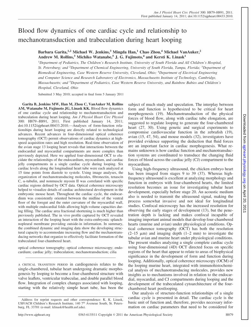
As previously described, the ultrahigh-speed, 4D OCT system in anenvironmental chamber was used to acquire in vivo data on theembryonic heart (14). A Fourier domain mode locked laser enabledultrahigh-speed OCT data collection at 235 frames/s (24). By applyingan image-based retrospective gating algorithm to the data, the recon-structed image set consisted of 90 volumes (equally spaced in time)spanning a single heartbeat (14). Each three-dimensional (3D) data setcomprised the complete C-loop of the embryonic heart. Figure 1 isprovided for orientation of the heart analyses, including direction ofblood flow, the levels (L1–L6) of the heart analyzed, and depiction ofthe cardiac compartments. For stage 13 avian hearts that cyclebetween 2–3 beats/s, one cardiac cycle corresponded to 90 volumes.
We investigated the relationships that exist between the myocar-dium, endocardium, and CJ at planes perpendicular to the axialdirection of the heart tube, and the blood flow path during systole anddiastole. Of the 90 volumes of data, we selected 15 for analysis at eachlevel of the heart tube. These 15 volumes were equally spaced in time,30 ms apart from each other. The temporal resolution of this systemensured that motion artifacts are negligible. The maximum displace-ment error of the system due to motion artifact would be 10 �m,whereas an OCT system imaging at 8 frames/s can have a motionartifact as high as 250 �m during systole (24). 4D image processingwas essential in determining the location of an orthogonal crosssection. The 15 volumes were imported into Amira 5.1 (VisageImaging, San Diego, CA). This image processing program allows themanipulation of the volumetric data, and any plane of view can beselected. Because our interest is focused on defining the changes ofthe myocardium and endocardium as the blood flows through the hearttube, we acquired cross sections cut perpendicular to the direction ofblood flow and analyzed the images.
A midcoronal plane that divided the ventral and dorsal sides wasdefined first (Fig. 1B), and its location was maintained for all volumes.Onto this midcoronal plane, intersecting planes were carefully se-lected at six different levels along the length of the heart and incorrespondence with the blood flow pattern. Levels 1 and 2 (L1 andL2) correspond to cuts made through the outflow and outflow/rightventricle (RV) boundary regions; levels 3, 4, and 5 (L3, L4, and L5)
are through the RV and left ventricular regions; and level 6 (L6) isrepresentative of the inflow region (Fig. 1C). This procedure resultedin 90 two-dimensional cross sections (15 selected time points duringa cardiac cycle at 6 levels) showing the boundaries of the myocar-dium, endocardium, and CJ areas. The endocardial lumen, the areaencompassing the inner myocardial wall, and the area encompassingthe outer myocardium were manually segmented for each image, andtheir areas quantified in pixels using MetaMorph 6.1 (MolecularDevices, Sunnyvale, CA) (Fig. 1D). From these segmentations, thearea of the CJ was derived by subtracting the endocardial lumen areafrom the inner myocardial area, and the thickness of the myocardialwall was calculated by subtracting the inner from the outer myocardialareas.
At the time of OCT monitoring of heart wall motion duringlooping, the splanchnopleural membrane was noted to be closelyapposed to the ventral side of the myocardial wall. As a result, we alsoanalyzed the relationship of the myocardial wall to the extracellularsplanchnopleural membrane, as this membrane was suggested to exerta mechanical stress during heart looping and aid in directionality (40),but it was not understood how this may occur. Our high-resolutionOCT observations on the relationship are included in Fig. 5.
Area Fractional Shortening Calculation
Area fractional shortening (AFS) is a measurement of the percentchange between areas of end relaxation to end contraction in crosssections of the heart wall and endocardial lumen.
OCM
Three wild-type and two heterozygous (NMHC-IIB�/� C57Bl6)mouse embryos on embryonic day (ED) 9.5 were isolated, fixed in3.5% paraformaldehyde in PBS, rinsed in PBS, and were sent to Dr.
Fig. 1. The embryonic heart as captured in real time and under physiologicalconditions with optical coherence tomography (OCT). A: the dashed white lineshows the direction of blood flow on a digitally reconstructed OCT radiograph.B: for all 15 volumes analyzed, the placement of the midcoronal plane (whiteline) was the same. C: three-dimensional (3D) reconstruction of Hamburgerand Hamilton (HH) stage 13 heart depicts endocardium (Endo) in dark gray,and myocardium (Myo) is transparent in light gray. Six cross-sectional cuts[levels 1–6 (L1–L6)] were defined perpendicular to blood flow. D: cross-sectional area measurements of the outer Myo area, endocardial lumen area,and cardiac jelly (CJ) area were performed with MetaMorph 6.1. The outlinesare shown in white. Number 18 on figure identifies frame that was used.Magnification bars in all panels � 100 �m.
H880 ANALYSIS OF A CARDIAC CYCLE DURING HEART LOOPING
Fujimoto’s laboratory at the Massachusetts Institute of Technology(MIT) for OCM imaging. As was earlier reported, the heterozygousembryos showed no morphological differences from the wild type(57). The OCM image of a representative wild-type embryonic heartis shown in Fig. 7 and in supplemental material (fly-through movie;the online version of this article contains supplemental data). Aprototype OCM imaging system (1) was developed at MIT andemployed for imaging fixed ED 9.5 mouse hearts. OCM is anotheroptical imaging modality based on low-coherence interferometry (23).Unlike confocal microscopy, OCM discriminates reflections in depthby both a confocal and a coherence gate, which increases the signal-to-noise ratio at greater depths (�2-fold increase in depth penetration)and can provide cellular resolution with the lateral resolution ap-proaching 1 �m (21). In this study, for comparison of the avianembryonic heart structure to that of the mammalian mouse embryo atcomparable stages, we compared the OCT imaging and analysis of asingle heartbeat in the avian embryo with OCM imaging of the mouseembryonic heart. Enhancement of the OCM images of fibrils and cellextensions was done using both Matlab and Amira 5.2.
Immunohistochemistry
The high-resolution immunohistochemical localization protocol forplastic sections that is routinely used in our laboratory has beenpublished (29). Specific extracellular matrix (ECM) and cytoskeletal-related proteins reported to be involved in mechanotransduction werelocalized in the HH stage 13 to stage 16 hearts during looping. Theantibodies used were against the ECM protein fibronectin (FN)(Sigma, St. Louis, MO), ciliar acetylated �-tubulin (58), cytoskeletal�-tubulin (Developmental Studies Hybridoma Bank, Iowa University,IA), tenascin C (TN-C) (IBL, Takasaki-sh, Gunma, Japan), andcytoskeletal nonmuscle myosin heavy chain (NMHC)-IIB (Covance,Emeryville, CA). This is not an exhaustive list, and other moleculescould have been considered for analysis, as, for example, fibrillins-1and -3 (44), fibulin-1 which our laboratory reported on earlier (28), orfibulin-2 (61). To encompass different cellular aspects of mechano-transduction (ECM, cytoskeletal, cilia), the listed subset of moleculeswas analyzed in this study.
Fluorescence and Confocal Microscopy
Fluorescence microscopy was done using a Nikon Optiphot IImicroscope. Optical serial sectioning was carried out using a LeicaTCS SP II laser scanning confocal microscope at the University ofSouth Florida College of Medicine Imaging Core Facility.
RESULTS
OCT Analysis: Multilevel Cross-sectional Analysisand ECM Relationships
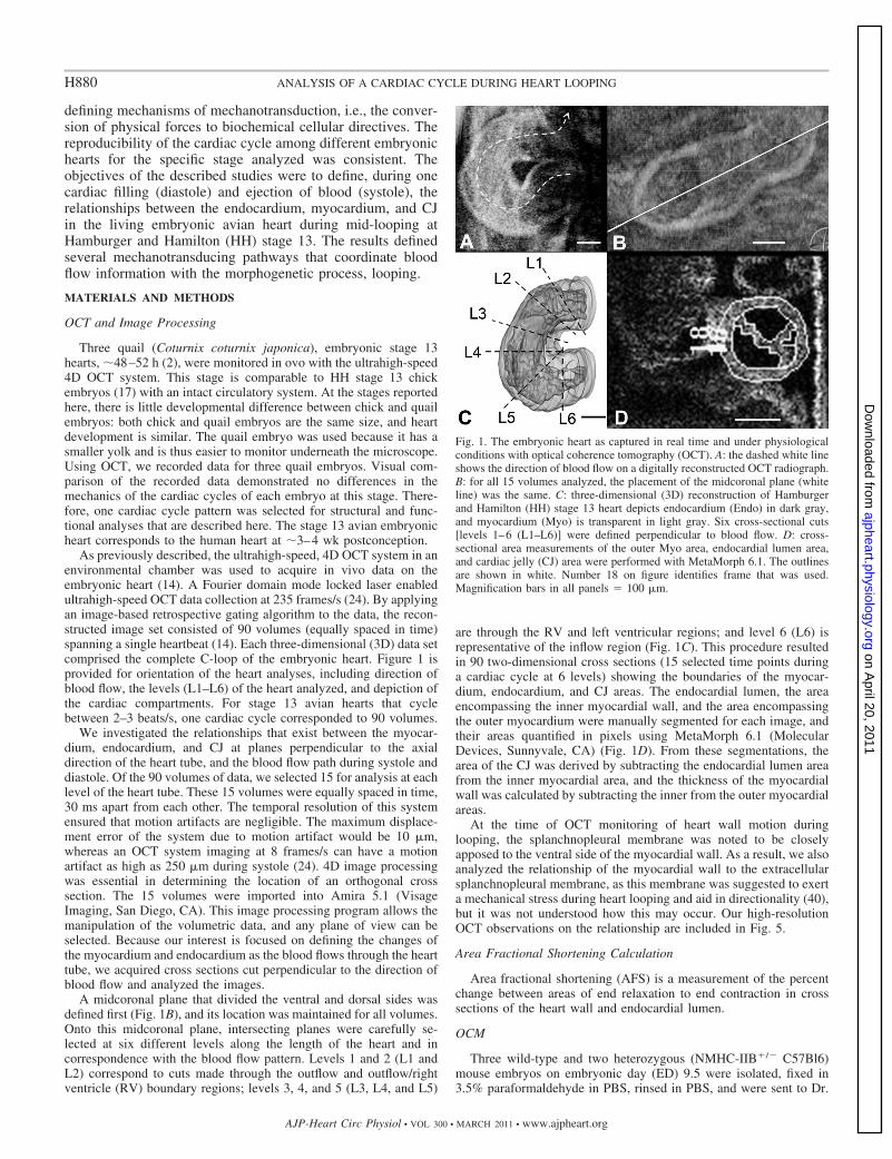
Multilevel cross-sectional analysis of the embryonic heartwas used to define tissue relationships of the myocardial,endocardial, and CJ compartments during a single cardiaccycle. The results of the analysis during a complete cardiaccycle are shown in Fig. 2. This figure depicts cross sectionsshown perpendicular to the blood flow at the above describedlevels L1–L6 of the heart, passing from anterior to posterior, at15 time points (t1–t15) during a cardiac cycle, and readingfrom left to right. Each level represents 30 ms. There is noprecedent to define end-diastole and end-systole for regions ofthe heart, as analyzed here, because these terms have been usedto define only the global relaxation-contraction states of theheart. We describe the patterns of relaxation and contractionthroughout the cardiac cycle at a specific level of the heart.Therefore, as the contraction wave was followed along theheart tube, the time when the area of the lumen was largest wasdefined as the end-relaxation for that specific level of the tube;the time when the area of the lumen was smallest was definedas the end-contraction. These sites of the end-relaxation/con-traction states are shaded in red for each level in Fig. 2. Forexample, at L2 in the RV region, end-relaxation occurred at t2,and end-contraction (underlined) at t8. There were notablecross-sectional area differences between the outflow, ventric-ular, and inflow regions. In relation to the whole heart, theoutflow region (L2) was smallest; the largest regions were theinflow (L6) and the midventricular region (L4). The myocar-dium was thicker in the inflow region than at any other regionin the heart tube. The greatest amount of CJ was in theventricular region, and the least amount in the outflow region.
Shape analysis in the cross sections revealed that the loopingheart myocardium was elliptical in relaxation and more circularduring contraction. Throughout one cardiac cycle, the endo-cardium was more irregularly shaped than any other compart-ment and displayed multiple folds (Fig. 2), which were espe-cially prominent in the ventricular region. These folds allowed
Fig. 2. Cross-sectional cuts are specified at 6different levels along the length of the hearttube for a HH stage 13 embryonic heartduring a cardiac cycle of 450-ms duration.Cuts L1 and L2 are through the outflowregion. L3, L4, and L5 cuts are in the ven-tricular region, and L6 is representative of theinflow region. The red fillings represent end-relaxation, and the underlined tracings indi-cate end-contraction. All outlines are alignedin a consistent orientation and in accordancewith the legend on the right corner. Attime point 15 (t15) and for all levels, theblue line demonstrates the change in theshape of the heart tube from inflow tooutflow during diastole. Orientation of theheart tube relative to the whole embryo isindicated. Magnification bar on bottom left offigure � 300 �m.
H881ANALYSIS OF A CARDIAC CYCLE DURING HEART LOOPING
the cross-sectional area of the lumen to expand from a smallarea into a relatively large area throughout the cardiac cycle.On average, the inflow (L6) and the midventricular (L4)regions had the largest lumen areas, whereas the outflow (L2)region had the smallest. Figure 2 shows that, during bothcontraction and relaxation in all cross sections, the endocar-dium and myocardium remained positioned in this orientation.There were consistent matrix-mediated associations that com-partmentalized the CJ to the lateral sides of the heart tube (seeFig. 3). During contraction, specifically in the ventricularregion, the endocardium also displayed folds or evaginations(levels L4 and L5 at time t5–t7). For all levels at time periodt15, a blue line placed along the long axis of the ellipticallyshaped heart, in relaxation phase, revealed changes in the axisorientation from inflow to outflow and suggested a twistingmotion to the heart.
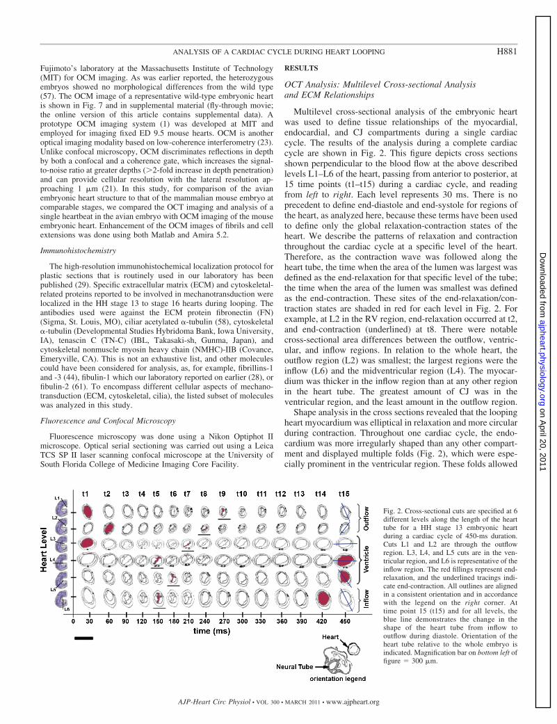
Immunohistochemical Analysis: ECM-mediated EndocardialAttachment Regions
In all regions of the heart tube as observed above, theendocardium was oriented in the direction of the ventral floorof the foregut and the outermost convex part of the myocar-dium during the cardiac cycle. This orientation was maintainedby ECM-mediated attachments (Fig. 3). To define the compo-sition of the ECM-mediated attachment regions, we usedimmunolocalization of specific proteins that are reported to beinvolved in mechanotransduction. The focus was directed tothe attachment sites between the myocardium and endocardiumthat were apparent within the specified six levels of the hearttube within one cardiac cycle. Color encoding in this plate usesgreen arrows to depict the ventral foregut attachment region,and orange arrows point to the outer curvature. Due to theseECM-mediated embryonic midline attachment sites, the endo-cardium has a slitlike appearance during contraction of theheart tube.
Sections were cut through a stage 13 (17) embryonic chickheart. Segmentation of the cardiac compartments was donemanually using Amira software to create a 3D reconstructionof the looping heart (Fig. 3, A–C). The endocardial luminalsurface (encoded blue) displayed numerous convolutions (Fig.3B). Figure 3C shows surface renderings of the myocardium(pink) and endocardium (blue), along with a cross-sectionslice. The myocardium is rendered transparent to visualize theendocardium (orange arrows). The specified midline attach-ment regions (green and orange arrows) were consistentlyobserved. These regions localized proteins, cytoplasmic matrixand ECM, which have been reported to be involved in thetransduction of mechanical forces, including FN that localizedalong the whole length of the heart (Fig. 3, top panel, fororientation of Fig. 3D at the foregut and Fig. 3E at the outercurvature), �-tubulin (Fig. 3, F and G, at higher magnification),and TN-C (Fig. 3, H and I, at higher magnification).
FN was highly expressed by the endocardium in both chickand mouse looping hearts (for chick, see Fig. 3D; for mouse,Fig. 7F, yellow arrows) and at the outer part of the elongatedendocardium at the outer curvature (Fig. 3E, orange arrows).The FN-mediated attachment at the outer curvature was moreapparent in anterior regions of the heart tube during diastole,where the endocardium is more closely associated with themyocardium, than at posterior regions in systole. In crosssections, FN (Fig. 3E, green signal, orange arrows) seemingly“tethered” the endocardial endothelial cells to the myocardialwall at the outer curvature region. FN also mediated an attach-ment of endocardial endothelial cells at the anterior intestinalportal region, as reported previously (28). Multiple FN fibrilsconsistently extended from the ventral floor of the foreguttoward the endocardium, as reported earlier (28). TN-C (seebelow) also colocalized with FN at the ventral foregut floorattachment region. �-Tubulin localization is shown at low andhigh magnification, respectively (Fig. 3, F and G). Evidence
Fig. 3. Mechanotransducing molecules at myocardial-endocardial attachment areas. On left, diagrams are tracings of the outlines of the Myo and Endo as capturedby OCT at the six levels (L1–L6) of the heart tube from anterior to posterior. The arrows are color-coded defining cardiac areas shown in the figures: orangearrows define outer curvature; green arrows, the ventral midline at floor of the foregut (FG). A–C: from serial sections (A), a 3D reconstruction of endocardialwall is shown in blue (B). C: endocardial wall is closely associated with the Myo (color-coded pink) in anterior regions (orange arrows). D and E: fibronectin(FN) localization. Top panel is for orientation, with boxes indicating regions shown in D and E panels. In D, FN (bright red signal) localized at attachment areaat the FG, and in E (green signal), at the outer curvature where FN tethers the Endo (arrows) to the Myo. F: �-tubulin is present throughout Myo. G: at the outercurvature region shown at higher magnification, �-tubulin is oriented in direction of strain exerted by the Endo at this site. H and I: tenascin-C (TN-C) localizationin myocardial basal lamina and within the Endo. I: 4 TN-C-mediated attachments (arrows) facilitate myocardial-endocardial communication at the outercurvature. NT, neural tube; N, notochord; DM, dorsal mesocardium; Spl, splanchnopleural. Magnifications bars � 180 �m (A, B, and C), 100 �m (D and E),76 �m (F and H), and 38 �m (G and I).
H882 ANALYSIS OF A CARDIAC CYCLE DURING HEART LOOPING
indicates that both �-tubulin and TN-C are influenced bymechanical stimuli (25, 46). �-Tubulin was present throughoutthe myocardium. At the outer curvature (orange arrows; Fig. 3,F and G), the cardiomyocyte microtubules were oriented in thedirection of strain that is exerted by the endocardium andwhere the attachment is mediated by FN and TN-C. In Fig. 3,H and I, four TN-C-mediated attachments extended to the sameouter curvature area, where the myocardium and endocardiuminteracted and where �-tubulin localized (compare Fig. 3, Fand G with H and I). Therefore, both FN and TN-C localizedto the same attachment areas at the outer curvature of the heartwall and to the ventral foregut in the HH stage 12–16 embryos.These matrix-cytoskeletal-mediated interactions were consis-tently maintained during looping. Because of these attachmentregions, the endocardium takes on the noted slitlike, elongatedappearance in end-systole leading into diastole. These attach-ments also serve to compartmentalize the CJ into left and rightsides.
The endocardium displayed convolutions and did not have asmooth central ellipsoid shape as often depicted. We observedthese lateral ridges related to fibrils radially connecting theendocardium and myocardium, as also seen in Fig. 3H withTN-C lateral fibril localization (white arrows). One part of alateral attachment was deeper in the CJ and hence out of planeof focus shown here, but apparent in adjacent sections.
OCT Analysis: Wave of Contraction Map
A map of the wave of contraction at the six levels is shownin Fig. 4A to demonstrate differences in relative time periods oflumen closure and opening along the length of the heart tube.Systole (defined by black squares) extended for 210 ms in the
outflow region (L1 and L2). This time period represents 46%of the total cardiac cycle time. In the inflow region, systoleextended for 150 ms, which is 33% of the total cardiac cycletime. These observations indicate that the inflow and outflowregions remain in a state of relative contraction for a consid-erable amount of time, with the outflow tract period remainingin contraction the longest by 60 ms. The RV and left ventric-ular regions (L3–L5) show synchronous contractions in lumenarea beginning at time t4, indicating a joint contribution to theejection of blood, as in the four-chambered fetal heart afterlooping.
Changes in Area for Myocardium and Endocardium.
The AFS is a measurement of the changes in area fromend-diastole to end-systole (Fig. 4B). The mean AFS for themyocardial wall and endocardium lumen was �46 � 2.4 and�86 � 3.5%, respectively; values are negative because theyrepresent reductions in areas.
OCT of Myocardial Wall Dynamics and Constraints of theSplanchopleural Membrane
The myocardial wall dynamics during contraction and re-laxation described above were analyzed also in relation to theextraembryonic splanchnopleural membrane apposed to themyocardial wall (Fig. 5A, see arrows). Monocilia have beenreported in the myocardium (33, 50). We also identified myo-cardial monocilia (Fig. 5B, white arrows) when using anantibody against acetylated �-tubulin, as used previously todefine endocardial monocilia (blue arrow) (58). We show fourframes from an OCT movie clip for illustration (Fig. 5, C–F),as the heart proceeds from diastole into systole in a peristalticwave of contraction discussed above. The movie clip is pro-vided in the supplemental material (Splanchno movie). Duringdiastole with endocardial filling with blood, the myocardialwall moved outwards and contacted the splanchnopleuralmembrane (Sp; red arrows define regions of contact). Withcontinued filling (Fig. 5C), the myocardium continued to ex-pand (Fig. 5D) and remained associated with the splanchno-pleural membrane, as seen over several movie frames (redarrows in Fig. 5, D and E). The myocardium moved along themembrane for a period of time (in Fig. 5E remaining in contactbetween the red arrows). During myocardial wall contraction(Fig. 5F), it pulled away from the extraembryonic membrane.The cycle repeated.
OCT Analysis: Behavior of CJ in One Cardiac Cycle
The behavior of the CJ was analyzed for a single cardiaccycle and at the same previously specified levels along thelength of the heart tube. The analyses included changes in CJcross-sectional area. Each chart in Fig. 6A represents levelsL1–L6 through the heart. As expected, there was a consistentdecrease in endocardial lumen cross-sectional area (dashedcurve) as the heart cycle progressed from end-relaxation toend-contraction. The behavior of the CJ is defined by thedotted line curve and its overall trend in gray. The data werenormalized by subtracting the lumen area by the minimumlumen area, and the result was divided by the maximum lumenarea. These graphs demonstrate that there was a progressiveincrease in the cross-sectional area of the CJ compartment atL1, L2, and L6, up to the time the endocardium had reached
Fig. 4. A: peristaltoid map. The S marks the location of end-contraction, andthe D of end-relaxation. Black blocks correspond to lumen areas within 25%of end-contraction. Light gray and dark gray blocks correspond to lumen areasthat are 50 and 75% of end-contraction, respectively. White corresponds tolumen areas within 25% of end-relaxation. B: area fractional shortening (AFS)measurements depict the percent reductions in area from end-relaxation toend-contraction for the heart and Endo lumen at the six heart levels analyzed.The myocardial AFS averages �46 � 2.4%, and the AFS of the endocardiallumen averages �86 � 3.5%.
H883ANALYSIS OF A CARDIAC CYCLE DURING HEART LOOPING
�50% of its end-contraction cross-sectional area; beyond thispoint, the CJ cross-sectional area began to decrease in parallelto that of the endocardium. The latter was in contrast to whatwas observed in L3, L4, and L5 (ventricular regions), wherethere was little change in the cross-sectional area of the CJ.
Figure 6B demonstrates that, at any one moment during thecardiac cycle, the total amount of CJ in the regions of the heartanalyzed was relatively constant (mean � SD, 0.87 � 0.07).There were, however, small changes in the amount of CJpresent at diastole (t1), compared with systole (t7). At diastole,when the endocardial lumen was expanded, there was less CJ(0.79), compared with when the heart was at systole (0.97).There was a nearly linear increase in the CJ as the heart shiftedtoward systole and a linear decrease as the heart shifted towarddiastole.
Immunohistochemical Analysis of the CJ at Looping Stages
During looping, the embryonic heart is compartmentalizedinto left and right sides by the consistent attachment areasdescribed earlier. At this time, fine fibrillar networks and cellnetworks are being organized within the CJ. These networksexist within the proteoglycan-rich jelly that undergoes somemovement and redistribution described above, as the cellsmaintain attachments with the myocardium and endocardium.These mechanosensing networks, as a result, undergo cyclicalcontracting and stretching, as the endocardium and myocar-dium pass through the contractile cycle.
In addition to TN-C and FN described above, we analyzedthe mechanotransducing molecule NMHC-IIB. In the ventric-ular part of the heart, long thin cell processes were observed inthe CJ, as defined by NMHC-II localization (Fig. 7). In Fig. 7A,a plastic cross section of a looping avian heart immunostainedwith NMHC-II antibody and serially cut at 4 �m along itslength is shown. Fig. 7B depicts NMHC-II-positive fine fibrilsor cell extensions color-coded in purple that were apparentwithin the CJ chiefly in the ventricular region. These sametypes of attachments and fine fibrils also were observed in afixed mouse heart on ED 9.5 of gestation, as visualized byOCM and digitally enhanced (Fig. 7C and in 7D shown athigher magnification). A fly-through movie of OCM imagesthrough the mouse heart is provided in the supplementalmaterial. In Fig. 7E, an ED 9.5 embryonic mouse heart wasimmunostained for NMHC-II (red signal), as well as for FN(7F, green signal). NMHC-IIB is expressed by the endocardialcells and by fibrillar-like extensions that connect with themyocardium. NMHC-IIB localized within a fine fibrillar net-work or cell processes within the CJ. FN also was associatedwith the endocardium, localized to fibrils extending throughthe CJ, and mediated attachments between the endocardialevaginations and the myocardial wall (yellow arrows). FN isalso expressed by the splanchnopleural membrane. In Fig. 7F,the avian endocardial lumen was 3D reconstructed from sec-tions, segmented, and color encoded in blue. The endocardialsurface, as described above, was not smooth, but displayedconvolutions. The ridges are depicted as darker blue regionsthat are closely apposed to the myocardial wall. The fineNMHC-II extensions (purple) reconstructed from the sections,when superimposed upon the endocardium, colocalized withthe endocardial ridges (yellow arrows in Fig. 7, F and G).These appear as attachment areas to the myocardial wall. Thelatter was further analyzed and described below.
CJ, Contractility, and Cardiomyocyte-derived MigratingCells Leading to Trabeculation
The changes in CJ area, as based on the OCT data, wesuggested reflected differential CJ movement and redistribu-tion along the heart tube during the cardiac cycle. Classically,the CJ compartment has been considered acellular. Recentdata, however, demonstrate this to be incorrect, because cellsbegin to move into the CJ during looping with the initiation oftrabeculation (41). Thus the cyclical dynamics of each heart-beat imply that any cells directionally migrating into the CJ andthat remain associated with the myocardial wall are alsosubjected to cyclical strain and movement.
To investigate whether cells moving into the CJ remainassociated with the myocardial wall, we prepared CJ prepara-
Fig. 5. Looping heart and relationship to Spl membrane (SPM). A: loopingheart (HT) enclosed in SPM (arrows). B: acetylated �-tubulin antibody (brightgreen signal) localized to monocilia in cardiomyocytes of a stage 17 chick Myo(white arrows). Blue arrows point to a monocilium associated with theendocardial endothelium cells. C–F: 4 frames of an OCT movie show themyocardial surface touching the SPM (green arrows) during diastole (C; redarrow indicates membrane contact area), then moving further along themembrane (D and E; red arrows delineate contact region), and then Myo pullsaway from membrane during systole (F; contraction). Magnification bar in B �76 �m. AIP, anterior intestinal portal.
H884 ANALYSIS OF A CARDIAC CYCLE DURING HEART LOOPING
tions from HH stage 14–16 looping hearts, using a previouslypublished protocol (39). After isolation, pieces of myocardialfragments and CJ were stained with Coomassie blue for gen-eral protein localization (Fig. 8A, 8B at higher magnification).The myocardial fragments were two cell layers thick, asexpected at this developmental stage, in contrast to the singlecell layer that is characteristic of the endocardium. At highermagnification, cells and their thin cell processes are visibleextending away from the wall (Fig. 8B) of the myocardialfragments. To confirm that these were indeed cells, we immu-nostained the CJ preparations with NMHC-II antibody andwith nuclear 4,6-diamidino-2-phenylindole (DAPI) stain (seeinset in Fig. 8B). NMHC-II-expressing cells, a doublet shownhere in the inset panel, were detectable that also colocalizedwith DAPI when superimposed. The nucleus of the right-handcell is easily seen in the inset, while most of the cell body is outof plane of focus in this image, but was easily detectable whenfocusing up and down. The analyses indicated that individualcells directionally migrate from the inner aspect of the myo-cardial wall into the CJ to initiate trabeculation, but remainedassociated with myocardial wall cardiomyocytes via long, thincell processes. These cells also displayed a spatial periodicity.
The presence of doublets indicated that these cells generatedaughter myocytes that remain interconnected and eventually,after multiple cell divisions, form a network of clonal cells, aswas reported by others (41). In Fig. 8C, a confocal micrographof an immunostained whole embryo, the myocardial walldisplays NMHC-II cell processes extending from the two-layered myocardium into the CJ.
As based on a temporal study, the relationship between thespatial organization of the migrating “pioneering cells” withinthe jelly adjacent to the myocardium and trabeculation becameevident: in sections of the looping avian heart, initially only afew cells were apparent near the myocardium in the CJ at chickHH stage 12 (Fig. 8D). As reported earlier (56), cells alsobegin to accumulate in the CJ adjacent to the endocardial side,as seen in sections of HH stages 13/14 hearts (Fig. 8E). By HHstage 24 (day 4), distinct trabeculae were present orientedradially and extending into the CJ from both the myocardialand endocardial compartments (Fig. 8F). The orientation of thetrabeculae was consistent with the radial arrangement of ECMmolecules and NMHC-II extensions, particularly in the ven-tricular regions that were discernible in the OCM images andin association with the endocardial ridges in the 3D reconstruc-
Fig. 6. A: multilevel analysis of the behavior of the CJ (dashed line with solid circles) as the Endo area (dotted line with triangles) changes from end-relaxationto end-contraction. L1–L6 on top of each curve refer to the heart level, whereas T1, T2, T5, T6, T7, T8, T9, T14, and T15 represent time during the cardiac cycleshown in Fig. 2. endRelax, end-relaxation; endCont, end-contraction. Gray lines represent trend lines for the behavior of the CJ. The data were normalized bysubtracting the lumen area by the minimum lumen area, and the result was divided by the maximum lumen area. B: normalized quantity of CJ in the heart atthe 15 time points during a cardiac cycle analyzed. The solid line is the average (0.87 � 0.07) for all time points.
H885ANALYSIS OF A CARDIAC CYCLE DURING HEART LOOPING
tion. By HH stage 36 (day 10) postlooping, in the four-chambered heart the ventricular region was highly trabeculated(Fig. 8G), and the trabeculae remain radially oriented. Direc-tion of strain during the heart wall dynamics is indicated bydouble-headed arrows in diagrams of a wedge of the heart atthe bottom of Fig. 8.
DISCUSSION
The avian and mouse embryonic hearts are reliable mod-els of the human heart at early stages of development.Imaging of the beating, looping, avian heart by 4D OCTprovided insights into the dynamic interrelationships thatexist between the three cardiac compartments at HH stage13, i.e., between the myocardium, CJ, and endocardium. Byanalyzing the behavior of these compartments perpendicularto the flow of blood, we found evidence that cardiac func-tion was coordinated closely with the assembly of an orga-nizational cellular-ECM network that appears vital for loop-ing and for transduction of biophysical forces to initiatecellular biochemical signaling pathways for organogenesis.A structural-functional relationship in cardiac growth andremodeling has been described as based on conotruncalmeasurements (26) and use of venous clips (52). We providenew data on how the biophysical changes can be mechano-transduced to affect morphogenesis.
Cardiac Compartment Changes and Relationships
The endocardium displays consistent attachment points dur-ing looping. The shape of the endocardium along the length ofthe tube is more complex than previously detected (34). Asshown in Fig. 2 (e.g., L4, t6), the endocardium forms folds thatextend toward the myocardium and attach to it at specificregions. We suggest these endocardial folds allow for the
endocardial cavity to expand and to accommodate the increas-ing volumes of blood that enter the heart during looping (20),while also providing structural stability to the endocardial tubevia the FN- and TN-C-mediated attachments that form betweenthe endocardium, the myocardium, and the midline of theventral floor of the foregut. Without the structural stabilityprovided through attachments to the myocardium, the endocar-dium tube could shift back and forth, preventing regularity inthe cardiac cycle.
Other investigators observing the shape of the endocardiumduring the cardiac cycle at a similar developmental stage haveconcluded that, during end-systole, the endocardium is slit-shaped, and, during end-diastole, it is elliptical (12, 34). Ingeneral, we agree with these conclusions. We extend theprevious observations, however, with identification of fibrousattachments that extend from the endocardial wall to themyocardium, providing a complex shape to the endocardium.Presumably, the endocardium requires these attachments tostabilize lumen orientation and to compartmentalize the CJlaterally. We also suggest that these attachments provide aform of structural communication, or mechano-communica-tion, between the myocardium and endocardium at specificsites that is critical for the developmental progression of theearly beating heart and for trabeculation (30, 35). The attach-ments at the midline of the ventral floor of the foregut and atthe outermost region of the convex part of the heart areconsistently maintained. The endocardial folds and attach-ments with the myocardium remain consistent, but patterningof folds along the heart tube appears to vary among differentembryos. The difference may reflect developmental age differ-ences among embryos and in the amount of blood flow. Theprecise developmental timing of analysis from embryo toembryo is not possible.
Fig. 7. Fine nonmuscle myosin heavy chain (NMHC)-IIB-expressing cell processes and fibrils extend between Myo and Endo (En). A and B: adjacent plasticsections through the ventricular region of a looping avian HH stage 13 heart. A: cell processes/fibrils (green arrow) extend radially through CJ between the Myoand Endo. Yellow arrow points to the undulating surface of the Endo. In B, the cell processes/fibrils were color-coded in purple. C and D: optical coherencemicroscopy (OCM) images of the looping mouse heart (embryonic day 9.5). Endocardial folds are present (yellow arrows), as well as a fine fibrillar networkin the CJ. Boxed region in C is shown at a higher magnification in D (green arrows point to cell processes and fibrils in the CJ; yellow arrow, an attachmentof the Endo to the Myo). E: red Cy-3 immunostaining of NMHC-IIB shows localization to endocardial associations with the myocardial wall and in finerfibrillar-like material in the CJ. F: endocardial evaginations associate with the mouse Myo via FN-mediated attachments (yellow arrows; green signal for FN).G: 3D reconstruction of the endocardial tube from serial sections as in A and B. The undulating surface of the avian Endo during looping is evident. The ventralFG wall is encoded in gray and white, here defining the embryonic midline. Dark blue denotes where endocardial wall has evaginated toward the Myo, formingfolds and outpocketings. H: superimposition of the segmented purple fibrils/processes with the Endo. The cell processes and fibrils colocalized at the endocardialridges. Magnification bars in A and for B � 100 �m; in C and for D � 150 �m. Sp, splanchnopleural membrane.
H886 ANALYSIS OF A CARDIAC CYCLE DURING HEART LOOPING
The elliptical shape of the heart and chamber orientation. Atend-diastole, the myocardium has an elliptical shape, and atend-systole it has more of a circular shape. This findingconflicts with previous reports (34), in which the heart wasdepicted as circular at end-diastole and elliptical at end-systole.These differences in interpretation most likely are due todifferences in imaging resolution and/or to the fact that weanalyzed cross-sectional cuts perpendicular to the direction offlow.
In observations of OCT movies and data from other hearts,as well as the detailed analyses shown in this report, we notethat the long axis of the elliptically shaped heart during diastoleis not oriented consistently in the same direction along thelength of the tube, but rather the direction of the long axischanges along the length of the tube (Fig. 2, t15). Therefore,we suggest that, as the tube contracts and its shape changesfrom elliptical to circular, it simulates a wringing motion,which constitutes a more effective physical basis for movingblood out of the heart. This early wringing motion is similar towhat has been described for torsion, described for the adultbeating heart (7, 48).
Differences in cardiac cross-sectional areas and wallthickness. Different regions or segments of the heart arisesequentially from the heart-forming fields. By stage 13, boththe RV and left ventricular regions and the future ventricularinlet (atrioventricular canal) region, as well as part of theoutflow associated with the RV, are present (8, 36). Theposterior sinoatrial region (inflow) and the anterior conus andtruncus of the outflow region continue to develop duringlooping into stages 14 and 17 (36). In the whole heart, theventricular region of the heart has the largest cross-sectionalarea, while the ventricular-outflow boundary region has thesmallest. This finding is expected, because the ventricularregion is where the maximum amount of blood is maintainedbefore being expulsed through a narrower ventricular-outflowboundary region. In addition, the inflow region has the largestmyocardial thickness, possibly because the myocardial con-tractile strength needs to be strongest in this region to maintainadequate cardiac output. The importance of the inflow regionfor cardiac output during early heart development was noted inour laboratory’s previous studies on the ED 11 mouse heartusing Doppler ultrasound (16) and subsequently by othersusing the chick heart and OCT analysis (24, 53).
Peristaltoid time periods. The timing map shown in Fig. 4Aillustrates end-systole (S) and end-diastole (D) at differentlevels along the length of the tube. This figure providesinformation on the differential time intervals of relative con-traction along the length of the tube. The outflow regionremains in a state of relative contraction (defined as the lumenarea being within 25% of the lumen area at end-systole) for 210ms, or 46% of the total cardiac cycle time. Of note, 50% of thistime period takes place during the beginning of diastole. Thisextended contraction state allows the ventricular region to fillwith blood. In turn, the ventricular region stays longer in arelaxed state, possibly allowing for pressure to increase andresulting in subsequent stronger ejection of blood. At time t4 inFig. 4A, the ventricular region (L3–L5) shows a synchronizedcontraction that should contribute to the strong ejection ofblood. Other investigators have measured very high flow ve-locities at these same stages (20). These blood flow velocitiescould be explained by a synchronized contraction of the ven-tricular regions of the early heart tube. These findings suggestthat, at these early stages in development, the heart needs toachieve a high ejection velocity of blood flow, and the stretch-activation response of the myocardium contributes to the per-formance of a beating heart (59) to achieve this goal. Patten etal. (43) described how the build-up of pressure in the ventric-ular region leads to high flow velocity. Additionally, thestretching of the cardiomyocytes as pressures build may benecessary for development of the adult myofibrillar organiza-
Fig. 8. Initiation of trabeculation is associated with looping stages andNMHC-II expressing cells. A and B: isolated myocardial fragments of HHstage 13/14 hearts show a two-layered Myo with cells extending from the wall,but remaining attached (yellow arrows). The cells in red boxed-in region in Aare shown at higher magnification in B. Superimposed images of immunolo-calization of NMHC-II with nuclear 4,6-diamidino-2-phenylindole staining(arrows pointing to such a doublet of cells in inset) demonstrate that these arecell structures within the CJ. C: confocal micrograph of a chick looping heartat stage 13 immunostained for NMHC-II shows fine cell extensions in CJextending from the Myo basal lamina toward the Endo. D–G: a cellularorganization characteristic of trabeculation becomes apparent between HHstages 12 and 16. D: a cell is present in the CJ near Myo at HH stage 13 withNMHC-IIB in CJ beginning to organize (arrows). E: similarly, cells areobserved in the CJ near Endo at HH stages 13/14. F: by day 4 postlooping,distinct trabeculae (arrows) are apparent, showing similar periodicity as “pi-oneer” cells in A. G: by day 10, the ventricular region is highly trabeculated(arrows), maintaining the spacing that was evident earlier. Bottom row:diagrams of a wedge through the ventricular region of a tubular heart shown atdifferent stages to illustrate our hypothesis: the initial pioneer cells lead tocellular clones of cells, forming a network in the CJ that postlooping hasformed the trabeculae. The double-headed arrow depicts the expected directionof strain within the CJ and cellular networks during systole and diastole.Magnification bar in A and C � 100 �m; B � 33 �m.
H887ANALYSIS OF A CARDIAC CYCLE DURING HEART LOOPING
tion (11, 42, 55). This stretching also may be important for theendocardial endothelial cell regulation of expression of keygenes (62). Indeed, the relationship between end-diastolic pres-sure (or volume) and pressure at end systole, known as theFrank-Starling mechanism in the adult heart, seems to bepresent already at the 13-somite stage in cardiac development,as was reported also for the later stages of 18, 24, and 29 chickembryos (60).
It would be interesting to correlate our data on time intervalsof contraction with the activation sequence revealed by opticalmapping data regarding the differences in time spent in con-traction via differential duration of action potential. However,little is known about stage 13 in terms of conduction patterns,as investigators analyzing stage 13 have not gotten goodsignals and indicate only slow conduction of 0.9 cm/s at thisstage (9).
AFS. Alexander Barry’s conclusions (3) in his often-citedpaper on the functional significance of the CJ still holds true.He, however, assumed the shortening of the myocardium was20% (3). Based on our OCT analysis, we calculated that theAFS for the myocardium is more than twofold higher, i.e.,46%, along the length of the tube. If the myocardium andendocardium tube were in direct contact with each other, theywould both have an AFS of 46%. We found that the AFS of theendocardial lumen is �86% along the entire length of the tube.This discrepancy in AFS between endocardium and myocar-dium is attributable to the presence of the CJ, and thus it allowsfor complete closure of the lumen at certain stages during thecardiac cycle, as was suggested also by Barry. In addition,results on modeling of strain patterns on the embryonic heartwall revealed that strain is higher in the inner layer of the earlyheart tube (6).
Myocardial Wall Dynamics and Constraints of theSplanchnopleural Membrane
The existence of myocardial microcilia was initially reportedby Manasek in 1968 (33). Several mechanosensing mutantshave been generated in which the mutations affect ciliarybiogenesis or mechanosensing pathways. These mutants in-clude the lrd�/� mutant with absent node cilia motility, thePkd2�/� mutant with defective ciliary mechanosensation, andthe Kif3a�/� mutant with complete absence of cilia (50). Onthe basis of histological descriptive analyses of embryonichearts immunostained for monocilia in transgenic mouse em-bryos with these mutations, it was concluded that a subset ofcilia, identified as cardiac cilia, is required in heart develop-ment that is separate from earlier ciliary function in lateralityspecification (50). The cited study, however, did not define arole for the presence of cardiac cilia. In a separate analysis, itwas shown that, when the splanchnopleural membrane on theventral surface of the embryo was removed from the chickembryo during looping, torsion was suppressed, a significantincrease in myocardial wall stiffness ensued, and looping wasaffected (40). The source by which the myocardium detectschanges in the mechanical force exerted by the splanchnopleu-ral membrane was not defined, but thought to be associated insome manner with the myocardiocyte cytoskeleton.
Our OCT data suggests a mechanism that interconnects theabove-cited observations. The myocardial wall during loopingtouches the splanchnopleural membrane and moves along it for
a distance with each heartbeat. As the myocardium rolls alongthe membrane, the deformation of the myocardial wall alongwith its microcilia can provide feedback on strains and posi-tional information from the outside in during looping andgrowth. As looping proceeds over time, the outside-in splanch-nopleural membrane strain information would be continuouslyrelayed to the myocardium, positionally slightly changing, asthe loop elongates and deepens. As the heart grows, differentparts of the myocardium, micron by micron, push against thesplanchnopleural membrane with each cardiac cycle. As ourdata would suggest, when ciliary mechanosensation is altered,abnormal heart development would be observed, as was notedalso for the above-described knockout models. The constraintof the extraembryonic membrane aids in maintaining myocar-dial integrity, the direction of looping, as well as spatiallyconfining the deepening of the loop with cardiac growth, toeventually result in the atria becoming situated cephalad to theventricles at the end of looping.
OCT Analysis of CJ Changes
As the heart contracts, the curve representing the behavior ofthe CJ at levels L1, L2, and L6 is distinctive relative to theother levels (Fig. 6A). At L2 and L6, the curves show a similarpeak in CJ cross-sectional area halfway through the contractionperiod. This peak signifies that there is an increase in CJcross-sectional area that accompanies an endocardial decreasein area during systole, but only up to a certain point in systole.After this point, the cross-sectional area of the CJ declines inparallel to that of the endocardial cross-sectional area. Thisfinding suggests that, during contraction, the CJ is recruitedinto L2 and L6 regions to allow for isovolumetric contractionand to prevent regurgitation. Figure 6B shows that the totalvolume of CJ increases as the cardiac cycle progresses duringsystole, further suggesting that there is a redistribution of CJduring systole. Both figures, taken together, indicate that thereis a greater movement of the CJ in L2 and L6 relative to theother regions we analyzed. Levels L2 and L6 correspond tothe valvelike regions of the tubular heart. We hypothesizethat these are the regions at which cardiac valves eventuallywill develop and presage the ultimate fate of these regionsas blood-flow regulatory areas. In contrast, L3, L4, and L5show small changes in CJ cross-sectional area as the endo-cardial lumen is closing, indicating little change of CJ in theventricular areas, compared with the inflow and outflowregions.
From projections of successive time-lapse film frames of alive embryonic heart, Patten et al. (43) described the presenceof endocardial “mounds” of CJ at specific locations in a 48- to50-h living embryo where the endocardial lumen is occluded:1) between the atrium and the ventricle (atrioventricular canal);and 2) at the ventricular conus to truncus arteriosus transitionarea. The previous authors described that CJ mounds “heap up”to obliterate the cardiac lumen and thereby prevent regurgita-tion of blood. We did not observe heaping of CJ at theselocations: instead, closure occurs by actual contraction of theheart (46% AFS). Thus the closure of the endocardium is dueto larger changes in myocardial shortening than previouslydescribed, and the closure is accompanied by reduction of CJin these regions (Fig. 6A). Together, these structural changesduring the cardiac cycle prevent the back flow, regurgitation, of
H888 ANALYSIS OF A CARDIAC CYCLE DURING HEART LOOPING
blood. Confirmatory evidence that heart function is importantfor valve development is provided by the zebrafish model inwhich it was shown that, in the homozygous silent heartembryos, which lack a heartbeat due to a mutation of cardiactroponin, the atrioventricular endocardial cushions did notform (4). It would be of interest to analyze whether there iscompression of the CJ at the inflow and outflow regions or asqueezing of the CJ into adjacent regions where the heart iscontinuous with the vasculature.
We report in this study that cells and long myocardial cellprocesses within the CJ expressing NMHC-II are situated in aradial orientation and were present in both chick and mousehearts. The OCM data on the mouse embryo confirmed thatmyocardial-endocardial radial associations exist also in theintact ED 9.5 looping mouse heart.
Our study adds new data for consideration in trabeculation(Fig. 8). Individual cells begin to migrate directionally into theCJ at HH stage 12 (day 2) and express NMHC-IIB, a moleculethat normally is part of the actomyosin cytoskeleton of themyocardiocyte, has a role in myofibrillogenesis (10) and in cellmotility (31), and has an important role in mechanotransduc-tion (5). As the myocardial wall transitions from an epitheliumto a trabeculated organ during looping, specific cardiomyo-cytes, in response to signals from the endocardium, direction-ally migrate toward the endocardium (51). Confirming ourobservations, the individual cells were reported to undergo celldivisions, forming a tight cluster or clone of cells that expressN-cadherin to maintain their homotypic interactions to even-tually give rise to a single or, at most, two trabeculae (41). Atthe same time as the myocardial side of trabeculation begins,endothelial cells are similarly leaving the endocardium to moveinto the CJ and have been shown to also play a role in theformation of trabeculae. The migration of both populationsorient along the radially aligned matrix molecules that we,using OCM, and others, using scanning electron microscopy,have described in the looping heart (22, 38). During the cardiaccycle, these cells and cellular networks that form continuouslyexperience cyclic strain and stretching (arrow, Fig. 8 diagrams)associated with rhythmic contractions during each cell cycle.Our data, taken together with the cited studies, suggest thatthese cells undergo myogenesis in response to the cyclicalstrain of each heartbeat. Cyclical movements of cardiomyo-cytes can alter gene expression (5, 49, 62) and, with theorganization of the ECM, help to shape the characteristic cellorganization of the trabeculated ventricular architecture of thefour-chambered heart after looping. The importance of hemo-dynamics in trabeculation is further suggested by a recentreport on testing the effects of hemodynamic unloading of theembryonic chick heart: this resulted in disorganized trabecula-tion and arrested growth (45).
Taken collectively, the cited descriptive studies of trans-genic mouse embryo models, as well as avian experimentalmanipulations, and our present OCT and immunohistochem-ical data indicate ECM-mediated mechanotransduction ofblood flow forces exists in 3D organized mechanosensingnetworks throughout the CJ associated with the myocardiumand endocardium along the length of the looping heart tube.
The described ECM and cellular networks form a vital partof transducing physical forces from not only the endocar-dium to the myocardium, but also external forces of thesplanchnopleural membrane to the myocardium, to coordi-nate development of form with function with each cardiaccycle.
ACKNOWLEDGMENTS
We appreciate the helpful discussions on fetal cardiac function with Dr.Serggio C. Lanata of Brown University, RI. The authors acknowledge Des-mond Adler and Robert Huber for technical contributions with the Fourierdomain mode locked laser. The authors also acknowledge David Wilson andMadhu Gargesha at Case Western Reserve University for technical contribu-tions to the image processing algorithms used to prepare the data for analy-sis.
GRANTS
This work was supported by the Suncoast Cardiovascular Research andEducation Foundation funded by Helen Harper Brown (K. K. Linask); theMason Chair (K. K. Linask) from the Foundation of the University of SouthFlorida and All Children’s Hospital; and partially funded by the AmericanHeart Association (K. K. Linask). This work was also funded by the USDepartment of the Army under Award no. W81XWH-05-1-0585 (M.VanAuker). The US Army Medical Research Acquisition Activity (820Chandler St., Fort Detrick, MD 21702-5014) is the awarding and admin-istering acquisition office. This research is supported in part by NationalInstitutes of Health (NIH) RO1 Awards HL083048 and HL096717 (A. M.Rollins), the Ohio Wright Center of Innovation and Biomedical Researchand Technology Transfer award: “The Biomedical Structure, Functionaland Molecular Imaging Enterprise”, and Interdisciplinary Biomedical Im-aging Training Program NIH T32EB007509. This investigation was con-ducted in a facility constructed with support from Research FacilitiesImprovement Program Grant no. C06 RR12463-01 from the NationalCenter of Research Resources, NIH. The OCM analysis was supported byNIH R01-CA075289-13 and by Air Force Office of Scientific ResearchContract FA9550-07-1-0101 (to J. G. Fujimoto).
DISCLAIMER
Information contained in this article does not necessarily reflect the positionor the policy of the government, and no official endorsement is inferred.
DISCLOSURES
J. G. Fujimoto receives royalties from intellectual property licensed by MITto Carl Zeiss and Lightlabs Imaging.
REFERENCES
1. Aguirre AD, Hsiung P, Ko TH, Hartl I, Fujimoto JG. High-resolutionoptical coherence microscopy for high-speed, in vivo cellular imaging.Opt Lett 28: 2064–2066, 2003.
2. Ainsworth S, Stanley R, Evans D. Developmental stages of the Japanesequail. J Anat 216: 3–15, 2010.
3. Barry A. The functional significance of the cardiac jelly in the tubularheart of the chick embryo. Anat Rec 102: 289–298, 1948.
4. Bartman T, Walsh EC, Wen KK, McKane M, Ren J, Alexander J,Rubenstein PA, Stainier DY. Early myocardial function affects endocar-dial cushion development in zebrafish. PLoS Biol 2: E129, 2004.
5. Clark K, Langeslag M, Figdor CG, Van Leeuwen FN. Myosin II andmechanotransduction: a balancing act. Trends Cell Biol 17: 178–186,2007.
6. Damon BJ, Remond MC, Bigelow MR, Trusk TC, Xie W, Per-ucchio R, Sedmera D, Denslow S, Thompson RP. Patterns of mus-cular strain in the embryonic heart wall. Dev Dyn 238: 1535–1546,2009.
7. Davis JS, Hassanzadeh S, Winitsky S, Wen H, Aletras A, Epstein ND.A gradient of myosin regulatory light-chain phosphorylation across theventricular wall supports cardiac torsion. Cold Spring Harb Symp QuantBiol 67: 345–352, 2002.
8. de la Cruz MV, Sanchez-Gomez C, Palomina MA. The primitivecardiac regions in the straight tube heart (Stage 9) and their anatomical
H889ANALYSIS OF A CARDIAC CYCLE DURING HEART LOOPING
expression in the mature heart: an experimental study in the chick embryo.J Anat 165: 121–131, 1989.
9. deJong F, Opthof T, Wilde A, Janse M, Charles R, Lamers W,Moorman A. Persisting zones of slow impulse conduction in developingchicken hearts. Circ Res 71: 240–250, 1992.
10. Du A, Sanger JM, Linask KK, Sanger JW. Myofibrillogenesis in thefirst cardiomyocytes formed from isolated quail precardiac mesoderm.Dev Biol 257: 382–394, 2003.
11. Epstein ND, Davis JS. Sensing stretch is fundamental. Cell 112: 147–150,2003.
12. Filas BA, Efimov IR, Taber LA. Optical coherence tomography as a toolfor measuring morphogenetic deformation of the looping heart. Anat Rec(Hoboken) 290: 1057–1068, 2007.
13. Forouhar AS, Liebling M, Hickerson A, Nasiraei-Moghaddam A, TsaiHJ, Hove JR, Fraser SE, Dickinson ME, Gharib M. The embryonicvertebrate heart tube is a dynamic suction pump. Science 312: 751–753,2006.
14. Gargesha M, Jenkins M, Wilson D, Rollins AM. High temporal reso-lution OCT using image-based retrospective gating. Opt Express 17:10786–10799, 2009.
15. Groenendijk BC, Hierck BP, Vrolijk J, Baiker M, Pourquie MJ,Gittenberger-de Groot AC, Poelmann RE. Changes in shear stress-related gene expression after experimentally altered venous return in thechicken embryo. Circ Res 96: 1291–1298, 2005.
16. Gui YH, Linask KK, Khowsathit P, Huhta JC. Doppler echocardiog-raphy of normal and abnormal embryonic mouse heart. Pediatr Res 40:633–642, 1996.
17. Hamburger V, Hamilton HL. A series of normal stages in the develop-ment of the chick embryo. J Morphol 88: 49–92, 1951.
18. Hogers B, DeRuiter M, Baasten A, Gittenberger-de Groot A,Poelmann R. Intracardiac blood flow patterns related to the yolk saccirculation of the chick embryo. Circ Res 76: 871–877, 1995.
19. Hove JR, Koster RW, Forouhar AS, Acevedo-Bolton G, Fraser SE,Gharib M. Intracardiac fluid forces are an essential epigenetic factor forembryonic cardiogenesis. Nature 142: 172–177, 2003.
20. Hu N, Clark EB. Hemodynamics of the stage 12 to stage 29 chickembryo. Circ Res 65: 1665–1670, 1989.
22. Hurle JM, Icardo JM, Ojeda JL. Compositional and structural hetero-genicity of the cardiac jelly of the chick embryo tubular heart: a TEM,SEM, and histochemical study. J Embryol Exp Morphol 56: 211–223,1980.
24. Jenkins MW, Adler DC, Gargesha M, Huber R, Rothenberg F,Belding J, Watanabe M, Wilson DL, Fugimoto JG, Rollins AM.Ultrahigh-speed optical coherence tomography imaging and visualizationof the embryonic avian heart using a buffered Fourier Domain ModeLocked laser. Opt Express 15: 6251–6259, 2007.
25. Jones PL, Chapados R, Baldwin HS, Raff GW, Vitvitsky EV, SprayTL, Gaynor JW. Altered hemodynamics controls matrix metalloprotei-nase activity and tenascin-C expression in neonatal pig lung. Am J PhysiolLung Cell Mol Physiol 282: L26–L35, 2002.
26. Keller BB, Hu N, Clark EB. Correlation of ventricular area, perimeter,and conotruncal diameter with ventricular mass and function in the chickembryo from stages 12 to 24. Circ Res 66: 109–114, 1990.
27. Linask KK. Regulation of heart morphology: current molecular andcellular perspectives on the coordinated emergence of cardiac form andfunction. Birth Defects Res C Embryo Today 69: 14–24, 2003.
28. Linask KK, Han M, Cai DH, Brauer PR, Manisastry SM. Cardiacmorphogenesis: matrix metalloproteinase coordination of cellular mecha-nisms underlying heart tube formation and directionality of heart looping.Dev Dyn 233: 739–753, 2005.
29. Linask KK, Tsuda T. Application of plastic embedding for sectioningwhole-mount immunostained early vertebrate embryos. Methods Mol Biol135: 165–173, 2000.
30. Linask KK, Vanauker M. A role for the cytoskeleton in heart looping.ScientificWorldJournal 7: 280–298, 2007.
31. Lo CM, Buxton DB, Chua GC, Dembo M, Adelstein RS, Wang YL.Nonmuscle myosin IIb is involved in the guidance of fibroblast migration.Mol Biol Cell 15: 982–989, 2004.
32. Lu W, Seeholzer SH, Han M, Arnold AS, Serrano MC, Garita B,Philp NJ, Farthing C, Steele P, Chen J, Linask KK. Cellular nonmusclemyosins NMHC-IIA and NMHC-IIB and vertebrate heart looping.. DevDyn 237: 3577–3590, 2008.
33. Manasek FJ. A light and electron microscopic study of myocardialdevelopment in the early chick embryo. J Morphol 125: 329–366, 1968.
34. Manner J, Thrane L, Norozi K, Yelbuz TM. High-resolution in vivoimaging of the cross-sectional deformations of contracting embryonicheart loops using optical coherence tomography. Dev Dyn 237: 953–961,2008.
35. Manner J, Thrane L, Norozi K, Yelbuz TM. In vivo imaging of thecyclic changes in cross-sectional shape of the ventricular segment ofpulsating embryonic chick hearts at stages 14 to 17. A contribution to theunderstanding of the ontogenesis of cardiac pumping function. Dev Dyn238: 3273–3284, 2009.
36. Markwald RR, Trusk R, Moreno-Rodriguez R. Formation and septa-tion of the tubular heart: integrating the dynamics of morphology withemerging molecular concepts. In: Living Morphogenesis of the Heart,edited by de la Cruz MV and Markwald RR. Boston, MA: Birkhauser,1998, p. 43–84.
37. McQuinn TC, Bratoeva M, Dealmeida A, Redmond M, ThompsonRP, Sedmera D. High-frequency ultrasonographic imaging of aviancardiovascular development. Dev Dyn 236: 3503–3513, 2007.
38. Nakamura A, Manasek F. Cardiac jelly fibrils: their distribution andorganization. In: Morphogenesis and Malformation of the CardiovascularSystem, edited by Rosenquist G and Bergsma D. New York: Liss, 1978, p.229–250.
39. Nakamura A, Manasek FJ. Experimental studies of the shape andstructure of isolated cardiac jelly. J Embryol Exp Morphol 43: 167–183,1978.
40. Nerukar N, Ramasubramanian A, Taber LA. Morphogenetic adapta-tion of the looping embryonic heart to altered mechanical loads. Dev Dyn235: 1822–1829, 2006.
41. Ong LL, Newrhee K, Mima T, Cohen-Gould L, Mikawa T. Trabecularmyocytes of the embryonic heart require N-cadherin for migratory unitidentity. Dev Biol 193: 1–9, 1998.
42. Pan J, Singh US, Takahashi T, Oka Y, Palm-Leis A, Herbelin BS,Baker KM. PKC mediates cyclic stretch-induced cardiac hypertrophythrough Rho family GTPases and mitogen-activated protein kinases incardiomyocytes. J Cell Physiol 202: 536–553, 2005.
43. Patten B, Kramer T, Barry A. Valvular action in the embryonic heart bylocalized apposition of endocardial masses. Anat Rec 102: 299–311, 1948.
44. Rossi A, Weber E, Sacchi G, Maestrini D, Di Cintio F, Gerli R.Mechanotransduction in lymphatic endothelial cells. Lymphology 40:102–113, 2007.
45. Sankova B, Machalek J, Sedmera D. Effects of mechanical loading onearly conduction system differentiation in the chick. Am J Physiol HeartCirc Physiol 298: H1571–H1576, 2010.
46. Schroder EA, Tobita K, Tinney JP, Foldes JK, Keller BB. Microtubuleinvolvement in the adaptation to altered mechanical load in developingchick myocardium. Circ Res 91: 353–359, 2002.
47. Sedmera D, Pexieder T, Rychterova V, Hu N, Clark EB. Remodelingof chick embryonic ventricular myoarchitecture under experimentallychanged loading conditions. Anat Rec 254: 238–252, 1999.
48. Sengupta P, Tajik A, Chandrasekaran K, Khandheria B. Twist me-chanics of the left ventricle: principles and application. JACC CardiovascImaging 1: 366–376, 2008.
49. Sil P, Gupta S, Young D, Sen S. Regulation of myotrophin gene bypressure overload and stretch. Mol Cell Biochem 262: 79–89, 2004.
50. Slough J, Cooney L, Brueckner M. Monocilia in the embryonic mouseheart suggest a direct role for cilia in cardiac morphogenesis. Dev Dyn237: 2304–2314, 2008.
51. Stankunas K, Hang C, Tsun ZY, Chen H, Lee N, Wu J, Shang C,Bayle J, Shou W, Iruela-Arispe M, Chang C. Endocardial Brg1 re-presses ADAMTS1 to maintain the microenvironment for myocardialmorphogenesis. Dev Cell 14: 298–311, 2008.
52. Stekelenburg-deVos S, Ursem NTC, Hop WCJ, Wladimiroff JW,Gittenberger-De Groot AC, Poelmann RE. Acutely altered hemody-namics following venous obstruction in the early chick embryo. J Exp Biol206: 1051–1057, 2003.
53. Taber LA, Zhang J, Perucchio R. Computational model for the transi-tion from peristaltic to pulsatile flow in the embryonic heart tube. JBiomech Eng 129: 441–449, 2007.
H890 ANALYSIS OF A CARDIAC CYCLE DURING HEART LOOPING
54. Tobita K, Schroder EA, Tinney JP, Garrison JB, Keller BB. Regionalpassive ventricular stress-strain relations during development of altered loadsin chick embryo. Am J Physiol Heart Circ Physiol 282: H2386–H2396, 2002.
55. Torsoni AS, Marin TM, Velloso LA, Franchini KG. RhoA/ROCKsignaling is critical to FAK activation by cyclic stretch in cardiacmyocytes. Am J Physiol Heart Circ Physiol 289: H1488 –H1496, 2005.
56. Toyofuku T, Zhang H, Kumanogoh A, Takegahara N, Yabuki M,Harada K, Hori M, Kikutani H. Guidance of myocardial patterning incardiac development by Sema6D reverse signalling. Nat Cell Biol 6:1204–1211, 2004.
57. Tullio AN, Accili D, Ferrans VJ, Yu ZX, Takeda K, Grinberg A,Westphal H, Preston YA, Adelstein RS. Nonmuscle myosin II-B isrequired for normal development of the mouse heart. Proc Natl Acad SciU S A 94: 12407–12412, 1997.
58. Van der Heiden K, Groenendijk BC, Hierck BP, Hogers B, KoertenHK, Mommaas AM, Gittenberger-de Groot AC, Poelmann RE. Mono-
cilia on chicken embryonic endocardium in low shear stress areas. DevDyn 235: 19–28, 2006.
59. Vemuri R, Lankford EB, Poetter K, Hassanzadeh S, Takeda K, YuZX, Ferrans VJ, Epstein ND. The stretch-activation response may becritical to the proper functioning of the mammalian heart. Proc Natl AcadSci U S A 96: 1048–1053, 1999.
60. Wagman A, Hu N, Clark E. Effect of changes in circulating bloodvolume on cardiac output and arterial and ventricular blood pressure in thestage 18, 24, and 29 chick embryo. Circ Res 67: 187–192, 1990.
61. Zhang HY, Chu ML, Pan TC, Sasaki T, Timpl R, Ekblom P. Extra-cellular matrix protein fibulin-2 is expressed in the embryonic endocardialcushion tissue and is a prominent component of valves in adult heart. DevBiol 167: 18–26, 1995.