1 BLUE LIGHT INDUCES RADICAL FORMATION AND AUTOPHOSPHORYLATION IN THE LIGHT-SENSITIVE DOMAIN OF CHLAMYDOMONAS CRYPTOCHROME* Dominik Immeln ╪,§ , Ramona Schlesinger ╪ , Joachim Heberle § , and Tilman Kottke ╪,§ From the ╪ INB-2: Molecular Biophysics, Research Center Jülich, 52425 Jülich, Germany and the § Department of Chemistry, Biophysical Chemistry, Bielefeld University, Universitätsstr. 25, 33615 Bielefeld, Germany Running Title: Blue Light Responses of Chlamydomonas Cryptochrome Address correspondence to: Tilman Kottke, Department of Chemistry, Bielefeld University, Universitätsstr. 25, 33615 Bielefeld, Germany, Tel. +49-521-106-2062; Fax: +49-521-106-2981; E-Mail: [email protected]Cryptochromes are sensory blue light receptors mediating various responses in plants and animals. Studies on the mechanism of plant cryptochromes have been focused on the flowering plant Arabidopsis. In the genome of the unicellular green alga Chlamydomonas reinhardtii, a single plant cryptochrome, Chlamydomonas photolyase homologue 1 (CPH1), has been identified. The N-terminal 500 amino acids comprise the light-sensitive domain of CPH1 linked to a C-terminal extension of similar size. We have expressed the light-sensitive domain heterologously in E. coli in high yield and purity. The 59 kDa protein bears exclusively flavin adenine dinucleotide in its oxidized state. Illumination with blue light induces formation of a neutral flavin radical with absorption maxima at 540 and 580 nm. The reaction proceeds aerobically even in the absence of an exogenous electron donor, which suggests that it reflects a physiological response. The process is completely reversible in the dark and exhibits a decay time constant of 200 s in the presence of oxygen. Binding of ATP strongly stabilizes the radical state after illumination and impedes the dark recovery. Thus, ATP binding has functional significance for plant cryptochromes and does not merely result from structural homology to DNA photolyase. The light-sensitive domain responds to illumination by an increase in phosphorylation. The autophosphorylation takes place although the protein is lacking its native C-terminal extension. This finding indicates that the extension is dispensable for autophosphorylation, despite the role it has been assigned in mediating signal transduction in Arabidopsis. Blue light governs many responses of organisms to environmental conditions. Cryptochromes have been shown to act as sensory blue light photoreceptors in plants and animals, with their action being as diverse as their origin (1). From sequence analysis, cryptochromes have been divided into three subgroups: animal, plant, and DASH cryptochromes (2). Animal cryptochrome synchronizes the circadian clock of Drosophila to the 24h rhythm (3). In mammals, cryptochrome has been suggested to be involved in circadian entrainment (4) and it functions independent of light as a main component of the biological clock. Arabidopsis cryptochromes 1 and 2 (AtCRY1 1 and AtCRY2) mediate de- etiolation responses, entrain the circadian clock, trigger programmed cell death induced by singlet oxygen, and regulate flowering time, stomatal opening, and production of anthocyanin (5-8). DASH cryptochromes are mostly found in bacteria, but also in Neurospora crassa, aquatic vertebrates (9) and Arabidopsis (AtCRY3) (10). Their putative role as sensory receptors has recently been challenged by showing that they act as repair enzymes for single-stranded DNA (11). The 500 N-terminal amino acids constitute the photolyase homology region (PHR) with reference to the DNA repairing enzyme. In this domain all cryptochromes characterized so far carry flavin adenine dinucleotide (FAD) as light-sensitive chromophore (2;12;13). Similarity in structure to the photolyases suggests that 5,10- methenyltetrahydrofolate (MTHF) might be bound as a second (antenna) chromophore. However, only few preparations of plant cryptochromes contain MTHF (14), which might be explained by a reduced affinity to the altered binding pocket (15). Plant cryptochromes are characterized by a http://www.jbc.org/cgi/doi/10.1074/jbc.M700849200 The latest version is at JBC Papers in Press. Published on June 4, 2007 as Manuscript M700849200 Copyright 2007 by The American Society for Biochemistry and Molecular Biology, Inc. by guest on April 3, 2020 http://www.jbc.org/ Downloaded from
Transcript
1
BLUE LIGHT INDUCES RADICAL FORMATION AND AUTOPHOSPHORYLATION IN THE LIGHT-SENSITIVE DOMAIN OF
CHLAMYDOMONAS CRYPTOCHROME* Dominik Immeln╪,§, Ramona Schlesinger╪, Joachim Heberle§, and Tilman Kottke╪,§
From the ╪INB-2: Molecular Biophysics, Research Center Jülich, 52425 Jülich, Germany and the §Department of Chemistry, Biophysical Chemistry, Bielefeld University, Universitätsstr. 25, 33615 Bielefeld, Germany
Running Title: Blue Light Responses of Chlamydomonas Cryptochrome Address correspondence to: Tilman Kottke, Department of Chemistry, Bielefeld University, Universitätsstr. 25, 33615 Bielefeld, Germany, Tel. +49-521-106-2062; Fax: +49-521-106-2981; E-Mail: [email protected]
Cryptochromes are sensory blue light receptors mediating various responses in plants and animals. Studies on the mechanism of plant cryptochromes have been focused on the flowering plant Arabidopsis. In the genome of the unicellular green alga Chlamydomonas reinhardtii, a single plant cryptochrome, Chlamydomonas photolyase homologue 1 (CPH1), has been identified. The N-terminal 500 amino acids comprise the light-sensitive domain of CPH1 linked to a C-terminal extension of similar size. We have expressed the light-sensitive domain heterologously in E. coli in high yield and purity. The 59 kDa protein bears exclusively flavin adenine dinucleotide in its oxidized state. Illumination with blue light induces formation of a neutral flavin radical with absorption maxima at 540 and 580 nm. The reaction proceeds aerobically even in the absence of an exogenous electron donor, which suggests that it reflects a physiological response. The process is completely reversible in the dark and exhibits a decay time constant of 200 s in the presence of oxygen. Binding of ATP strongly stabilizes the radical state after illumination and impedes the dark recovery. Thus, ATP binding has functional significance for plant cryptochromes and does not merely result from structural homology to DNA photolyase. The light-sensitive domain responds to illumination by an increase in phosphorylation. The autophosphorylation takes place although the protein is lacking its native C-terminal extension. This finding indicates that the extension is dispensable for autophosphorylation, despite the role it has been assigned in mediating signal transduction in Arabidopsis.
Blue light governs many responses of
organisms to environmental conditions. Cryptochromes have been shown to act as sensory blue light photoreceptors in plants and animals, with their action being as diverse as their origin (1). From sequence analysis, cryptochromes have been divided into three subgroups: animal, plant, and DASH cryptochromes (2). Animal cryptochrome synchronizes the circadian clock of Drosophila to the 24h rhythm (3). In mammals, cryptochrome has been suggested to be involved in circadian entrainment (4) and it functions independent of light as a main component of the biological clock. Arabidopsis cryptochromes 1 and 2 (AtCRY11 and AtCRY2) mediate de-etiolation responses, entrain the circadian clock, trigger programmed cell death induced by singlet oxygen, and regulate flowering time, stomatal opening, and production of anthocyanin (5-8). DASH cryptochromes are mostly found in bacteria, but also in Neurospora crassa, aquatic vertebrates (9) and Arabidopsis (AtCRY3) (10). Their putative role as sensory receptors has recently been challenged by showing that they act as repair enzymes for single-stranded DNA (11).
The 500 N-terminal amino acids constitute the photolyase homology region (PHR) with reference to the DNA repairing enzyme. In this domain all cryptochromes characterized so far carry flavin adenine dinucleotide (FAD) as light-sensitive chromophore (2;12;13). Similarity in structure to the photolyases suggests that 5,10-methenyltetrahydrofolate (MTHF) might be bound as a second (antenna) chromophore. However, only few preparations of plant cryptochromes contain MTHF (14), which might be explained by a reduced affinity to the altered binding pocket (15). Plant cryptochromes are characterized by a
http://www.jbc.org/cgi/doi/10.1074/jbc.M700849200The latest version is at JBC Papers in Press. Published on June 4, 2007 as Manuscript M700849200
Copyright 2007 by The American Society for Biochemistry and Molecular Biology, Inc.
long C-terminal extension, that varies considerably in size and sequence. Fusion of the extension with β-glucuronidase leads to a constitutively active phenotype for AtCRY1 and AtCRY2, which implies a role of this domain for signal transduction (16).
All spectroscopic data on the blue light response of plant cryptochromes have been recorded on Arabidopsis cryptochromes. Interpretation of the data has been facilitated by the availability of a crystal structure of the AtCRY1-PHR domain (15). In AtCRY1, absorption of blue light induces formation of a neutral flavin radical, which decays on a millisecond timescale (17). Results from fluorescence and electron paramagnetic resonance (EPR) experiments on whole insect cells containing AtCRY1 indicate that the radical is the active state of the receptor in vivo (18). This radical shows a unique spectroscopic signature in the visible and infrared spectral region, which distinguishes it from that of the homologous photolyases (19). Results from transient absorption spectroscopy and site-directed mutagenesis showed that a conserved tryptophan triad and a tyrosine residue are involved in the electron transfer process (17;20). It was concluded from infrared difference spectroscopy that a crucial step for photoactivation is the protonation of the chromophore by a nearby aspartic acid (19). Further downstream in the signal cascade, blue light induces autophosphorylation of AtCRY1 in vitro and in vivo (21;22) and was proposed to change the interaction with the E3 ubiquitin ligase COP1 (23;24).
A single putative member of the plant cryptochrome family has been identified in the unicellular green alga Chlamydomonas reinhardtii (Chlamydomonas photolyase homologue 1 CPH1). As a possible evolutionary precursor of plants (25), the CPH1 gene product is closely related to AtCRY1 with 49% identity in the first 500 amino acids (26). CPH1 contains by far the longest C-terminal extension (507 amino acids) compared to all known cryptochromes (7). Initial expression of full-length CPH1 yielded a 110 kDa product from E. coli but 126 and 143 kDa proteins from Chlamydomonas as revealed by Western Blot analysis (27). Size discrepancies were hypothesized to be due to posttranslational modifications. In vivo, CPH1 undergoes light-induced proteolysis (27), but the physiological
function of CPH1 remains elusive. It has been suggested that CPH1 is involved in regulation of gene transcription and/or circadian phototaxis rhythmicity (27).
Here, we report on a high yield production of the light-sensitive domain of C. reinhardtii cryptochrome in E. coli. Blue light induces formation of the radical state similar to full-length Arabidopsis CRY1. We demonstrate that ATP binding strongly influences the properties of the domain and enables light-induced auto-phosphosphorylation in the absence of the C-terminal domain.
Experimental Procedures
DNA manipulation, cloning and transformation
of E. coli were done by standard techniques (28). Oligonucleotides were purchased from MWG Biotech or Invitrogen. Restriction enzymes were from New England Biolabs, Gibco-BRL or Fermentas. Sequencing of DNA was performed by dideoxy sequencing using Thermo Sequenase fluorescent labeled primer cycle sequencing kit (Amersham Biosciences) and the LI-COR 4200 Gene ReadIR (MWG Biotech). 5, 10-Methenyltetrahydrofolate (MTHF) was a kind gift of Dr. R. Moser (Merck Eprova AG, Schaffhausen).
Construction of expression vector - The sequence encoding the N-terminal 504 amino acids of CPH1 from Chlamydomonas reinhardtii (Accession No. L07561) was amplified by PCR. A coding region for six histidines was added downstream to the start codon. The PCR product was ligated into pCR 2.1 (Invitrogen). E. coli strain TOP10 (Invitrogen) was used for genetic manipulations. The construct was cloned into a modified pET11a (Novagen) expression vector providing the coding sequence for a 3’-terminal Strep-tag II (WSHPQFEK) after an Nhe I site coding for alanine and serine.
Protein expression and purification - E. coli BL21 (DE 3) pLysE (Novagen) containing the expression construct was grown in LB medium supplemented with 200 mg/L ampicillin and 200 µM riboflavin at 37°C in the dark. When the optical density at 600 nm reached 0.5, the temperature was lowered to 18°C. At an optical density of 0.7, isopropyl-β-D-1-thiogalactoside (IPTG) was added to a final concentration of 10 µM. After 20 hrs, the cells were harvested by
centrifugation (6.000 g, 20 min, 4°C). The cell pellet was resuspended in 0.2 M Tris, pH 8, 0.3 M NaCl, 20% glycerol, 1 tablet of protease inhibitor cocktail (Complete EDTA-free, Roche) and DNase. The cells were disintegrated using French Press (3 times 1000 psi). The lysate was centrifuged (100.000 g, 1h, 4°C) and loaded onto a Strep-Tactin-Sepharose column (IBA). After washing with 0.2 M Tris, pH 8, 0.3 M NaCl, 20 % glycerol, the protein was eluted in the same buffer supplied with 2.5 mM D-desthiobiotin. The isolated holoprotein is stable for several days at 4°C.
Protein analysis - The construct was detected on Western Blot (28) using an antibody against the Strep-tag (IBA) and the Strep-tag HRP Detection Kit (IBA) or an antibody against the polyhistidine-tag (Sigma) and Super Signal West Pico Chemiluminescent substrate (Pierce). MALDI-TOF analysis was performed after in-gel digestion with trypsin as described previously (29). For determination of the protein concentration, the protein was diluted in 6 M guanidinium hydrochloride. The extinction coefficient of the apoprotein at 280 nm was calculated with ProtParam (www.expasy.org). A chromophore content of up to 70% was estimated by the ratio A280/A450 using an extinction coefficient for FAD in solution of 11300 M-1 cm-1 at 450 nm (30).
Folate assay - In a modified fluorescence assay for folate (31), CPH1-PHR was treated as follows: 0.2 µl of concentrated HCl was added to 25 µl of sample and heated for 3 min at 99 °C. The precipitate was removed by centrifugation and the supernatant diluted to 200 µl. In the next step, sodium hydroxide (10 M) was added until pH 12-13 was reached. Finally, the sample was oxidized by 10 µl of a KI / I2 (20 % / 10 %) solution and overnight incubation. After each step, fluorescence spectra were recorded with a Shimadzu RF 1501 spectrofluorophotometer.
Photoreduction and kinetics of recovery - UV-Vis absorption spectra were recorded with a Uvikon 943 (Kontron Instruments) or a Shimadzu 2401 PC spectrophotometer. The buffer was exchanged to 0.2 M Tris, pH 8, 0.3 M NaCl, 20% glycerol prior to photoreduction and the sample was kept at 10 °C. MgCl2 was added to 17 mM. The influence of ATP was investigated in the presence of 3.3 mM ATP, 3.3 mM GTP or 3.3 mM adenylyl imidodiphosphate (AMP-PNP). A 445 nm LED (22 nm full width at half maximum
(FWHM), Luxeon Star Lumileds) was used for illumination with 25 mW/cm2 at the sample. Between two illuminations, a dark time of four minutes was applied. For kinetic measurements, the change in absorbance at 450 nm was monitored. β-ME was added to 20 mM. Oxygen was bubbled through the sample for 5 min prior to 10 s of illumination with the 457 nm LED (20 nm FWHM, 40 mW/cm2). ATP was added to 3.3 mM and MgCl2 to 17 mM, if used.
Phosphorylation assay - The recombinant protein isolated from E. coli was dephosphorylated in the dark. Phosphatase treatment was performed at 4°C for 2.5 h in a buffer containing 36 µM CPH1-PHR, 40 mM Tris, 40 mM NaCl, 10 mM MgCl2, 8 % glycerol, 5 mM KCl, 10 µM ZnCl2, and 7.5 units of calf intestine alkaline phosphatase (CIAP, Fermentas). After the treatment, 10 µl of 1.7 mM Na3VO4 for inhibition of the CIAP (32), 10 µl of 100 mM MgCl2, and 10 µl of 33 mM ATP were added to 70 µl of the sample solution. All samples were kept at 25 °C and shaken in a thermomixer at 500-1000 rpm (Eppendorf). To prevent heating of the sample by irradiation, a 457 nm light emitting diode was used (Luxeon Star Lumileds, 20 nm FWHM). It provided 77 µW/cm² of blue light at the sample. Dark samples were covered with aluminum foil. Reactions were stopped by the addition of SDS-PAGE sample buffer (28) containing 0.4 M dithiothreitol, 8 M urea, 10% sodium dodecyl sulfate, and by boiling for 15 min at 95 °C. These harsh denaturing conditions were applied to ensure complete dissociation of ATP from the protein. After performing electrophoresis with a 12% polyacrylamide gel, phosphorylated protein was stained with the Pro-Q Diamond Phosphoprotein Gel Stain (Molecular Probes) (33) and detected with 302 nm UV light (Gel Doc, Bio-Rad). Total protein was made visible afterwards by Coomassie staining. Signal strength in the phosphostain was analyzed in reference to the background and normalized to the protein amount from the Coomassie stain by ImageJ software (NIH Image, http://rsb.info.nih.gov/nih-image/index.html).
RESULTS
Chlamydomonas photolyase homologue 1
(CPH1) from the unicellular green alga C. reinhardtii is a member of the plant cryptochrome family according to sequence analysis. Its function
and properties are unknown, therefore we set out to study the biochemical and spectroscopic characteristics of the CPH1 light-sensitive domain.
Construction of the expression vector - The 504 aa boundary of the N-terminal photolyase-like domain was determined using sequence alignment and available structural data of AtCRY1 and E. coli DNA photolyase in conjunction with secondary structure prediction and hydropathy analysis.
Sequencing of the cDNA revealed a deviation in the construct as compared to the published sequence. Codon 314 was found to be cgc instead of ggc, which would result in a mutation from Gly to Arg. In a control experiment, genomic DNA from a C. reinhardtii extract (P. Hegemann, Berlin, Germany) was used as a PCR template to amplify a part of the CPH1 sequence containing codon 314. The PCR product was cloned and analyzed by sequencing, confirming the deviation from the published sequence. This implies that amino acid at position 314 of CPH1 is indeed arginine.
Protein expression and purification - Using standard expression conditions for E. coli (37°C, 3h, 1mM IPTG), the protein was obtained in insoluble form as inclusion bodies. It was solubilized with 1% N-lauroylsarcosine and further purified. However, attempts to reconstitute the protein with FAD as a chromophore were not successful. Modification of the protocol to very mild expression conditions (see Experimental procedures) yielded up to 20 mg of a soluble, yellow colored protein per liter of induced cell culture. SDS-PAGE showed a band at about 60 kDa with a purity of > 90% (Fig. 1, lanes A and C). The experimentally determined molecular weight corresponds to the theoretical mass of the apoprotein of 59.13 kDa. The recombinant protein was detected using antibodies against the N-terminal polyhistidine tag and the C-terminal Strep-tag (Fig. 1, lanes B and D). The identity of the CPH1-PHR construct was further confirmed by MALDI-TOF mass spectrometry. The protein was subjected to tryptic digestion. Matched peptides covered 21% (109/520 aa) of the construct (data not shown).
Identification of the chromophores - A yellow protein was obtained after affinity chromatography. The UV-Vis spectrum showed a fine-structured (“three-finger”) absorption band in the 400-500 nm region, and a broad absorption in
the 300-400 nm region with maxima at 368 nm and 449 nm (Fig. 2A). These are typical features of an oxidized flavoprotein.
As heterologously expressed flavoproteins may incorporate different flavins such as riboflavin, FMN or FAD (34), the chemical nature of the cofactor was determined by fluorescence spectroscopy. CPH1-PHR was heat denatured and the precipitate was removed by centrifugation. Before and after denaturation, fluorescence excitation spectra with emission at 528 nm and emission spectra with excitation at 450 nm were recorded (Fig. 2B). Maxima were detected at 364 nm and 448 nm in the excitation spectrum and at 529 nm in the emission. Upon acidification with HCl the fluorescence signal of the free chromophore rose approximately by 4-fold (Fig. 2B). This rise is a peculiarity of FAD caused by a loss of interaction between the adenine and flavin ring systems in acidic solution (35). In a more quantitative approach, the chromophore composition was further analyzed by thin layer chromatography (TLC). Using authentic FAD as a standard, the isolated chromophore showed a signal at the same height without contributions from other flavin species (supplemental Fig. 1). The results from UV-Vis spectroscopy, fluorescence spectroscopy and TLC analysis identified oxidized FAD as the exclusive flavin chromophore of CPH1.
It has been reported that AtCRY1 contains MTHF as a second chromophore (14), which is absent in most preparations (15;19). As CPH1 is highly homologous to AtCRY1, CPH1 might also bind MTHF. The chromophore was separated from the CPH1-PHR apoprotein and investigated with a fluorescence assay for folates (31) (Fig. 3). Upon excitation at 350 nm, the fluorescence spectrum showed a prominent flavin emission at 528 nm. Additionally, a weak band at 441 nm was present, which is indicative for folates. The supernatant was basicified by sodium hydroxide and finally oxidized by adding KI/I2. The decay of the 441 nm fluorescence after addition of base and the subsequent strong increase by oxidation are strong indications for the presence of a folate as a second chromophore in CPH1. Bases catalyse the hydrolysis of the methenyl bridge of MTHF to the nonfluorescent 10-formyltetrahydrofolate (31). The oxidizing agent then produces fluorescent products. It should be pointed out, however, that
the amount of detected folate was minute and it was only observed in few preparations.
The response of the chromophore to blue light illumination - The effect of blue light on CPH1-PHR was followed by UV-Vis spectroscopy under aerobic conditions without external electron donor. Illumination at λ = 445 nm caused a decrease of the absorption band at 449 nm, which shows the disappearance of oxidized flavin (Fig. 4A). Simultaneously, formation of a broad absorption band between 500 and 600 nm with maxima at 540 and 580 nm was observed. This absorption is indicative for a blue neutral flavoprotein radical (36). Two isosbestic points at 354 nm and 494 nm were obtained, implying the presence of two species in the sample, i.e., the flavoprotein in its oxidized and radical state. The isosbestic point at 354 nm was lost after illumination for more than 1 minute, indicating partial formation of the fully reduced state of CPH1-PHR. Even under anaerobic conditions, prolonged illumination (> 1 min) was necessary to initiate the production of the fully reduced state (data not shown). It has been reported that AtCRY1 binds ATP (21) to the PHR domain (15). Addition of ATP to CPH1-PHR lead to a strong enhancement in light-induced radical formation (Fig. 4B). Isosbestic points did not change even after 300 s of illumination. The presence of AMP-PNP, a nonhydrolyzable analog of ATP, resulted in an identical light response as with ATP (Fig. 4C). This result shows that the effect is due to ATP binding to the protein and not related to protein phosphorylation. The enhancement in radical formation is specific for ATP, because it was not inducible by addition of GTP (data not shown).
The decay of the CPH1-PHR radical in the dark was investigated in a separate series of experiments. The sample was illuminated for 10 s with 457 nm blue light and the recovery of the oxidized state was monitored through the absorption at 450 nm (Fig. 5A). The kinetic measurements were performed aerobically in the presence of the exogenous electron donor 2-mercaptoethanol. Initially, time traces were not fully reproducible and the sample did not fully recover back to the oxidized state. These problems were attributed to a deprivation of oxygen in the sample during illumination. Treatment with oxygen prior to illumination lead to recovery kinetics of the oxidized state, which follow a monoexponential shape with a time constant of ca.
200 s. The UV-Vis spectrum obtained after recovery was identical to the one recorded before illumination (Fig. 5A, inlet), implying that the light-induced reaction was fully reversible. Addition of ATP to the sample strongly impeded the radical decay (Fig. 5B). Only a slight recovery of the 450 nm absorption of the oxidized flavin was observed within the experimental time window of 30 min, which is illustrated by comparison with a theoretical curve with a time constant of 200s (Fig. 5B, dashed line). Similarly, the decay kinetics was strongly slowed down under anaerobic conditions, and was not interpretable by a simple kinetic scheme (data not shown). These findings indicate that ATP and oxygen concentration are major determinants of the decay kinetics of the CPH1-PHR radical.
The response of the domain to blue light: a phosphorylation assay - We hypothesized that CPH1 undergoes a blue-light-dependent autophosphorylation as it has been claimed for AtCRY1 (21;22). The phosphorylation assay demonstrated that the CPH1-PHR is isolated from E. coli in a strongly phosphorylated state (Fig. 6A, lane 1). To determine blue-light-induced phosphorylation, it was necessary to initially dephosphorylate the protein. This was achieved by incubation with calf intestine alkaline phosphatase (CIAP) at 4 °C in the dark, by which CPH1-PHR was dephosphorylated to a basal level (Fig. 6A, lane 2). In the next step, the phosphatase was inhibited by the addition of Na3VO4. ATP and MgCl2 were added as substrate and cosubstrate in the phosphorylation reaction. Illumination with blue light for 10, 20, and 30 min induced a significant increase of the signal at 60 kDa on the phosphostain gel as compared to the dark-treated samples, incubated for the same time (Fig. 6A). Equal loading of the protein was visualized by Coomassie staining (Fig. 6B). It is concluded that the illuminated samples are stronger phosphorylated than the dark treated samples, which implies that CPH1-PHR undergoes a blue-light-dependent autophosphorylation. Incubation at 25 °C in the dark did not increase the phosphorylation level within the accuracy of the detection.
When illuminated, the sample tended to aggregate and therefore was detected at higher masses and in the stacking gel, especially at longer illumination times (supplemental Fig. 2). These aggregates are highly phosphorylated and could
not be fully separated on the gel. An additional band was detected between the 60 kDa band of CPH1-PHR and the 66 kDa band of the standard (Fig. 6). This band is assigned to CIAP, which is active as a dimer of two 65 kDa subunits (37). Faint bands of the CIAP are observed in the Coomassie-stained gel, which give strong signals on the phosphostain. These signals are attributed to a fraction of strongly phosphorylated CIAP from the catalyzed hydrolysis of ATP, despite the inhibition by Na3VO4. This interpretation was supported by a control experiment without CPH1-PHR (supplemental Fig. 3). Even before the addition of ATP, CIAP was detected as a weak signal on the phosphostain gel (Fig. 6, lane 2). In this case, the substrate for CIAP might be phosphorylated CPH1-PHR.
DISCUSSION
We report on the successful heterologous
overexpression of a plant cryptochrome in E. coli. Until now, only Arabidopsis CRY1-PHR has been obtained from E. coli as a fusion to maltose binding protein (14;15), whereas full-length plant cryptochromes remain inaccessible from prokaryotic expression systems. The N-terminal photolyase homology region of Chlamydomonas reinhardtii CPH1 forms a neutral flavoprotein radical in response to blue light concomitant with an increase in phosphorylation (Fig. 7). The recovery of oxidized FAD proceeds in the order of minutes in the dark and is strongly delayed by the presence of ATP or the absence of oxygen.
CPH1 is a member of the plant cryptochrome family - Phylogenetic analysis groups Chlamydomonas CPH1 into the family of plant cryptochromes, in which CPH1 might play an ancestral role (25). This assignment is supported by the fact that only one plant cryptochrome is found in the algal genome, whereas other cryptogams such as Physcomitrella or Adiantum have two and five genes, respectively (38). The PHR domain of CPH1 shows a 49% identity to AtCRY1 (26), and 31% to E. coli DNA photolyase. Its physical and chemical properties identify CPH1 as a plant cryptochrome: The flavin chromophore is found exclusively in its oxidized state after purification, as it is in AtCry1 (13). In contrast, photolyases and members of the Cry-DASH family contain FAD in its oxidized, radical, or fully reduced form depending on enzyme and
preparation procedure (39-41). Illumination of CPH1-PHR with blue light leads to formation of a neutral flavoprotein radical indicated by a broad absorption band with maxima at 540 at 580 nm. The maximum at 580 nm is strongly blue-shifted by more than 40 nm in comparison to the spectra of the radicals in E. coli photolyase (42) or DASH cryptochrome AtCRY3 (39). This shift is characteristic for plant cryptochromes, as it has been shown for AtCRY1 (19), and is even more pronounced in CPH1. In comparison to other plant cryptochromes, the C-terminal extension does not contain the conserved DQXVP-acidic-STAES motif (7;43), nor does it show any known motifs besides a high amount of Gly (25%) and Ala (16%) residues.
The chromophores in CPH1 - The recombinant N-terminal domain of CPH1 purified from E. coli bears FAD in its oxidized state. The presence of riboflavin or FMN in the binding pocket of CPH1 is excluded on the basis of TLC analysis (supplemental Fig. 1). This points to an important contribution of the adenine moiety of FAD to binding of the chromophore in cryptochromes. It has been shown for the homologous DNA photolyase from E. coli that riboflavin and FMN fail to bind to the apoprotein (44). These findings are in contrast to other blue light receptor domains such as the LOV domains of phototropin, where reconstitution with flavin analogues has been achieved (45), or the sensors of blue light using FAD (BLUF) domain, where expression conditions determine the chromophore composition (34).
Low amounts of a folate were detected by fluorescence analysis in a few preparations of CPH1-PHR (Fig. 3). It has been reported that AtCRY1 contains MTHF as a second chromophore (14). Preservation of functional MTHF during the preparation procedure is complicated by the fact that it decomposes once released from the apoprotein at pH 8 (31). Gel filtration experiments on CPH1-PHR showed that folate is predominantly present in aggregates (data not shown). We suggest that multimerization facilitates retaining the MTHF chromophore in CPH1. However, it is not clear yet if plant cryptochromes bear functional MTHF at all. The high extinction coefficient of oxidized flavin might allow for blue light sensing without the necessity for MTHF as an antenna chromophore.
Radical formation in CPH1-PHR upon blue light illumination - The response to blue light differs among the cryptochromes making it necessary to discuss the subgroups individually. In the plant cryptochrome AtCRY1, blue light induces a transition from the oxidized state of flavin to the neutral radical FADH• (13;17), whereas in the animal cryptochrome dCRY from Drosophila an anionic radical FAD⎯• is formed (46). In the DASH cryptochrome AtCRY3, the reaction proceeds via the neutral radical to the fully reduced state of flavin FADH2 (47). CPH1-PHR responded very similar to illumination as AtCRY1: During the illumination process two species were present exclusively in the sample, i.e., the oxidized and the neutral radical state of the flavoprotein (Fig. 4). Only prolonged, intense illumination produced the fully reduced state of CPH1-PHR.
Radical formation under anaerobic conditions and with external electron donors is a common feature of flavoproteins (40) and does not allow to draw any meaningful conclusion on physiological processes. In contrast, the present experiments on CPH1-PHR were performed aerobically without any exogenous electron donor (Fig. 4). Formation of a stable radical was observed despite the fact that the experimental conditions were favoring the oxidized state of the flavin. Therefore, we propose in analogy to Arabidopsis cryptochromes that the oxidized state is the native dark state. We consider the flavoprotein radical as being physiologically active in Chlamydomonas as has been claimed for AtCRY1 and AtCRY2 (18-20;48). The critical residues that have been proposed to be involved in signal transduction in AtCRY1, i.e., W324 (17), W400 (20), and D396 (19), are all conserved in CPH1. For cry-DASH proteins, such as CRY1 from Vibrio cholerae, the fully reduced state has been proposed to represent the dark form (40). A clear differentiation between plant and DASH cryptochrome is necessary considering the recent finding that cry-DASH members act as photolyases for single-stranded DNA (11).
The decay of the radical in the dark proceeds on the order of minutes. In a solution saturated with oxygen and in the presence of 2-ME, the time constant was determined to be ~200 s. This is comparable to the dark recovery of AtCRY2 with a time constant of 360 s (t1/2 =250 s) (48). For AtCRY1, Giovani et al. (17) demonstrated that the neutral flavin radical decayed already in
milliseconds, but did not resolve a contribution with a half live of > 100 ms. Time-resolved measurements in the sub-second time regime need to be conducted on CPH1 to resolve this issue. The concentration of oxygen is a limiting factor for the decay of the radical in CPH1-PHR. If the sample is depleted with oxygen during illumination, the recovery kinetics of the oxidized state is slowed down, because oxygen has to diffuse from the atmosphere into the sample solution and further into the chromophore binding pocket.
Effect of ATP binding to the blue light receptor domain - The crystal structure is available of the AtCRY1-PHR domain with bound AMP-PNP (15). The nucleotide occupies the site of substrate binding postulated for DNA photolyases. The analysis did not reveal any structural changes upon binding. The effect of ATP on the cryptochrome light reaction has not been studied before. The observed strong enhancement in light-induced radical formation of CPH1-PHR is primarily an effect of a slower dark recovery and not of an increase in quantum yield of formation of the radical. A small stabilizing effect of the substrate on the enzyme has been reported for DNA photolyase (49). Upon binding of the cyclobutane pyrimidine dimer, the reduced state of the enzyme is more resistant to air oxidation due to a rise in reduction potential by 65 mV. In contrast, ATP binding leads to a strong stabilization of the radical state of CPH1 with reference to both, oxidized and reduced states. This can be deduced from the much slower recovery of oxidized flavin (Fig. 5B), and the absence of any fully reduced state in the sequence of light-induced absorption spectra (Fig. 4B), respectively. Together with the observed autophosphorylation (see below), these findings indicate that nucleotide binding is of functional significance for plant cryptochromes and not merely caused by structural similarity in the binding pocket to DNA photolyase. ATP is abundant in cells and shows high affinity to PHR domains with a dissociation constant (Kd) of 20 μM (as determined for AtCRY1) (21). As a consequence, experiments on plant cryptochromes require to be conducted in the presence of ATP or ATP analogs in order to draw meaningful conclusions with regard to the in vivo situation.
Light-dependent autophosphorylation of CPH1-PHR - We have shown that CPH1-PHR performs blue-light-dependent autophos-phorylation under in vitro conditions (Fig. 6). This
reaction is surprising because the native C-terminal extension is missing in the construct. It is not clear if the observed autophosphorylation of the PHR domain reflects a specific response of the full-length protein in vivo. However, the level of phosphorylation detected speaks against an unspecific over-phosphorylation as found for example in rhodopsin in vitro (50). Autophosphorylation has already been demonstrated for full-length AtCRY1 and AtCRY2 in vitro and in vivo (21;22;51) despite the fact that they do not contain sequence similarities to any known kinase. It is closely associated with the function or regulation of the receptors (22;51). The PHR domain is sufficient for this response in AtCRY12. Our results imply that the C-terminal extension is dispensable for the phosphorylation response and that the ability to undergo light-induced autophosphorylation is a characteristic feature of plant cryptochromes. The lack of homology of the C-terminal extensions between algal and flowering plant cryptochromes indicates that a conserved phosphorylation response might indeed be limited to the PHR domain.
A recent study has challenged the influence of light to the autophosphorylation process in AtCRY1 (52). The authors determined only a minor influence of light. In comparison to our study on CPH1-PHR, illumination conditions were applied with 31 μW/cm2 as compared to 77 μW/cm2 intensity and 360 nm light instead of 445 nm. Particularly the shorter wavelength might strongly reduce the reaction efficiency via enhanced internal conversion as was found in the flavin-containing LOV domains of Chlamydomonas phot (53). Furthermore, detection of a phosphorylation increase by radiostaining instead of using a fluorescent dye might be limited, if proteins are already phosphorylated to significant levels prior to the study (54).
From the crystal structure analysis of the AtCRY1-PHR domain with bound ATP analog it was suggested that the nature of the autophosphorylation reaction is intermolecular,
implying a dimerization reaction with a second AtCRY1 molecule (15). Dimerization might be necessary to shorten the distance of the γ-phosphate of ATP to a putative acceptor, which is in the monomer about 11 Å. In support of this proposal, we observed a light-induced multimerization of CPH1-PHR (supplemental Fig. 2). It is tempting to speculate that the blue-light-induced autophosphorylation and aggregation of CPH1-PHR we observed in vitro are correlated with the physiological function in Chlamydomonas. CPH1 undergoes light-induced proteolysis in vivo, which is eliminated by the addition of kinase inhibitors (27). From this observation it was concluded that phosphorylation of CPH1 might be required for light-induced degradation. The photosensory function of CPH1 in Chlamydomonas is yet unknown. A study using an RNAi approach is in preparation.
Conclusions - More than 11 years have passed since the first absorption spectrum of a plant cryptochrome was published (13). Since then, the understanding of the molecular processes after blue light illumination has only gradually improved. This is partly due to demanding sample preparation. The Chlamydomonas CPH1-PHR domain is now available in sufficient yield and purity from heterologous expression for investigation with biophysical techniques such as ultrafast spectroscopy or Fourier transform infrared spectroscopy. This approach has been very fruitful for the blue light receptor phototropin, where many details of the reaction mechanism have been elucidated within a few years. We have demonstrated that the C-terminal extension of cryptochromes is dispensable for light-induced autophosphorylation. Therefore, our eukaryotic model domain with its high homology to AtCRY1 is a good candidate to study two fundamental light-induced processes of full-length plant cryptochromes: radical formation and autophosphorylation.
REFERENCES 1. Cashmore, A. R. (2003) Cell 114, 537-543 2. Brudler, R., Hitomi, K., Daiyasu, H., Toh, H., Kucho, K., Ishiura, M., Kanehisa, M., Roberts, V. A.,
Todo, T., Tainer, J. A., and Getzoff, E. D. (2003) Mol. Cell 11, 59-67 3. Stanewsky, R., Kaneko, M., Emery, P., Beretta, B., Wager-Smith, K., Kay, S. A., Rosbash, M., and
Hall, J. C. (1998) Cell 95, 681-692 4. Partch, C. L. and Sancar, A. (2005) Methods Enzymol. 393, 726-745 5. Mao, J., Zhang, Y. C., Sang, Y., Li, Q. H., and Yang, H. Q. (2005) Proc. Natl. Acad. Sci. U. S. A
102, 12270-12275 6. Ahmad, M., Lin, C., and Cashmore, A. R. (1995) Plant J. 8, 653-658 7. Lin, C. and Shalitin, D. (2003) Annu. Rev. Plant Biol. 54, 469-496 8. Danon, A., Coll, N. S., and Apel, K. (2006) Proc. Natl. Acad. Sci. U. S. A 103, 17036-17041 9. Daiyasu, H., Ishikawa, T., Kuma, K., Iwai, S., Todo, T., and Toh, H. (2004) Genes Cells 9, 479-495 10. Kleine, T., Lockhart, P., and Batschauer, A. (2003) Plant J 35, 93-103 11. Selby, C. P. and Sancar, A. (2006) Proc. Natl. Acad. Sci. U. S. A 103, 17696-17700 12. Hsu, D. S., Zhao, X., Zhao, S., Kazantsev, A., Wang, R. P., Todo, T., Wei, Y. F., and Sancar, A.
(1996) Biochemistry 35, 13871-13877 13. Lin, C., Robertson, D. E., Ahmad, M., Raibekas, A. A., Jorns, M. S., Dutton, P. L., and Cashmore,
A. R. (1995) Science 269, 968-970 14. Malhotra, K., Kim, S. T., Batschauer, A., Dawut, L., and Sancar, A. (1995) Biochemistry 34, 6892-
6899 15. Brautigam, C. A., Smith, B. S., Ma, Z., Palnitkar, M., Tomchick, D. R., Machius, M., and
Deisenhofer, J. (2004) Proc. Natl. Acad. Sci. USA 101, 12142-12147 16. Yang, H. Q., Wu, Y. J., Tang, R. H., Liu, D., Liu, Y., and Cashmore, A. R. (2000) Cell 103, 815-
827 17. Giovani, B., Byrdin, M., Ahmad, M., and Brettel, K. (2003) Nat. Struct. Biol. 10, 489-490 18. Bouly, J. P., Schleicher, E., Dionisio-Sese, M., Vandenbussche, F., Van Der Straeten, D., Bakrim,
N., Meier, S., Batschauer, A., Galland, P., Bittl, R., and Ahmad, M. (2007) J. Biol. Chem. 282, 9383-9391
19. Kottke, T., Batschauer, A., Ahmad, M., and Heberle, J. (2006) Biochemistry 45, 2472-2479 20. Zeugner, A., Byrdin, M., Bouly, J. P., Bakrim, N., Giovani, B., Brettel, K., and Ahmad, M. (2005)
J. Biol. Chem. 280, 19437-19440 21. Bouly, J. P., Giovani, B., Djamei, A., Mueller, M., Zeugner, A., Dudkin, E. A., Batschauer, A., and
Ahmad, M. (2003) Eur. J. Biochem. 270, 2921-2928 22. Shalitin, D., Yu, X., Maymon, M., Mockler, T., and Lin, C. (2003) Plant Cell 15, 2421-2429 23. Yang, H. Q., Tang, R. H., and Cashmore, A. R. (2001) Plant Cell 13, 2573-2587 24. Wang, H., Ma, L. G., Li, J. M., Zhao, H. Y., and Deng, X. W. (2001) Science 294, 154-158 25. Lariguet, P. and Dunand, C. (2005) J. Mol. Evol. 61, 559-569 26. Small, G. D., Min, B., and Lefebvre, P. A. (1995) Plant Mol. Biol. 28, 443-454 27. Reisdorph, N. A. and Small, G. D. (2004) Plant Physiol 134, 1546-1554 28. Sambrook, J. and Russell, D. W. (2001) Molecular Cloning - A Laboratory Manual, 3rd Ed., CSHL
Press, Cold Spring, NY 29. Schaffer, S., Weil, B., Nguyen, V. D., Dongmann, G., Gunther, K., Nickolaus, M., Hermann, T.,
and Bott, M. (2001) Electrophoresis 22, 4404-4422 30. Siegel, L. M. (1978) Methods Enzymol. 53, 419-429 31. Johnson, J. L., Hamm-Alvarez, S., Payne, G., Sancar, G. B., Rajagopalan, K. V., and Sancar, A.
(1988) Proc. Natl. Acad. Sci. U. S. A 85, 2046-2050 32. Zueva, N. N., Dalev, P. G., and Lazarova, D. L. (1993) Biokhimiia. 58, 1009-1023 33. Schulenberg, B., Aggeler, R., Beechem, J. M., Capaldi, R. A., and Patton, W. F. (2003) J. Biol.
34. Laan, W., Bednarz, T., Heberle, J., and Hellingwerf, K. J. (2004) Photochem. Photobiol. Sci. 3, 1011-1016
35. Faeder, E. J. and Siegel, L. M. (1973) Anal. Biochem. 53, 332-336 36. Massey, V. and Palmer, G. (1966) Biochemistry 5, 3181-3189 37. Lorenz, B. and Schröder, H. C. (2001) Biochim. Biophys. Acta 1547, 254-261 38. Suetsugu, N. and Wada, M. (2003) Curr. Opin. Plant Biol. 6, 91-96 39. Pokorny, R., Klar, T., Essen, L. O., and Batschauer, A. (2005) Acta Cryst. F61, 935-938 40. Worthington, E. N., Kavakli, I. H., Berrocal-Tito, G., Bondo, B. E., and Sancar, A. (2003) J. Biol.
Chem. 278, 39143-39154 41. Sancar, A. (2003) Chem. Rev. 103, 2203-2237 42. Jorns, M. S., Sancar, G. B., and Sancar, A. (1984) Biochemistry 23, 2673-2679 43. Imaizumi, T., Kanegae, T., and Wada, M. (2000) Plant Cell 12, 81-96 44. Payne, G., Wills, M., Walsh, C., and Sancar, A. (1990) Biochemistry 29, 5706-5711 45. Dürr, H., Salomon, M., and Rüdiger, W. (2005) Biochemistry 44, 3050-3055 46. Berndt, A., Kottke, T., Breitkreuz, H., Dvorsky, R., Hennig, S., Alexander, M., and Wolf, E. (2007)
J. Biol. Chem. 282, 13011-13021 47. Song, S. H., Dick, B., Penzkofer, A., Pokorny, R., Batschauer, A., and Essen, L. O. (2006) J.
Photochem. Photobiol. B 85, 1-16 48. Banerjee, R., Schleicher, E., Meier, S., Munoz, V. R., Pokorny, R., Ahmad, M., Bittl, R., and
Batschauer, A. (2007) J. Biol. Chem., in press 49. Gindt, Y. M., Schelvis, J. P., Thoren, K. L., and Huang, T. H. (2005) J. Am. Chem. Soc. 127,
10472-10473 50. Maeda, T., Imanishi, Y., and Palczewski, K. (2003) Prog. Retin. Eye Res. 22, 417-434 51. Shalitin, D., Yang, H., Mockler, T. C., Maymon, M., Guo, H., Whitelam, G. C., and Lin, C. (2002)
Nature 417, 763-767 52. Özgür, S. and Sancar, A. (2006) Biochemistry 45, 13369-13374 53. Holzer, W., Penzkofer, A., and Hegemann, P. (2005) J. Lumin. 112, 444-448 54. Schulenberg, B., Aggeler, R., Beechem, J. M., Capaldi, R. A., and Patton, W. F. (2003) J. Biol.
Chem. 278, 27251-27255
FOOTNOTES
*We thank Georg Büldt (FZ Jülich) for generous support, Peter Hegemann (HU Berlin) for CPH1 cDNA and valuable discussions, Boris Zorin (HU Berlin) for C. reinhardtii gDNA, and J. Schweitzer (FZ Jülich) for the MALDI-TOF analysis. D. I. and T. K. acknowledge support by the Helmholtz Gemeinschaft (VH-NG-014), and J. H. by the Deutsche Forschungsgemeinschaft (FOR 526). 1The abbreviations used are: AMP-PNP, adenylyl imidodiphosphate; AtCRY, Arabidopsis thaliana cryptochrome; BLUF, sensor of blue light using FAD; 2-ME, 2-mercaptoethanol; CIAP, calf intestine alkaline phosphatase; CPH1, Chlamydomonas photolyase homolog 1; EDTA, ethylenediaminetetraacetic acid; EPR, electron paramagnetic resonance; FAD, flavin adenine dinucleotide; FMN, flavin mononucleotide; FWHM, full width at half maximum; LOV, light-, oxygen-, and voltage-sensitive; MTHF, 5-,10-methenyltetrahydrofolate; PHR, photolyase homology region; TLC, thin layer chromatography. 2M. Ahmad, personal communication.
FIGURE LEGENDS
Fig. 1: SDS-PAGE and immunoblotting of the light-sensitive domain of the Chlamydomonas reinhardtii cryptochrome CPH1 after purification. A/C, Coomassie staining shows a band close to the theoretical size
of 59 kDa. The corresponding immunoblots against the N-terminal polyhistidine tag (B) and the C-terminal Strep-tag (D) confirm the identity of the construct. Faint bands at higher masses are attributed to oligomers of the domain.
Fig. 2: Spectroscopic characterization of the purified CPH1-PHR in its dark form. A, the UV-Vis absorption spectrum with absorption maxima at 368 and 449 nm identifies CPH1 as a flavoprotein carrying flavin in its oxidized state. B, fluorescence excitation spectra (left) and emission spectra (right) of CPH1-PHR in its native state (solid line), after heat denaturation (dashed line), and after subsequent acidification with HCl (dotted line). The rise in emission is a peculiarity of flavin adenine dinucleotide. Excitation spectra were recorded by monitoring the emission at 528 nm. Emission spectra were obtained with excitation at 450 nm.
Fig. 3: Fluorescence assay for folates in CPH1-PHR. Excitation spectra (left) were recorded by monitoring emission at 450 nm. Emission spectra (right) were obtained with excitation at 350 nm. The solid lines depict the fluorescence of the supernatant after denaturation and centrifugation. Addition of sodium hydroxide leads to a decay of the signal (dashed lines). The dotted lines represent the spectra after oxidation with KI/I2 and incubation overnight. The decay and rise of fluorescence in the assay is characteristic for folates such as MTHF.
Fig. 4: Blue-light illumination of CPH1-PHR under aerobic conditions in the absence of exogenous electron donors. From a series of UV-Vis spectra, selected traces are shown recorded in the dark and after the indicated times of illumination with ca. 40 mW/cm2 of 445 nm light. The reaction was carried out in presence of Mg2+ (A), after addition of ATP and Mg2+ (B), and in presence of the nonhydrolyzable ATP analog AMP-PNP and Mg2+ (C). The rise of a band between 500 and 600 nm shows the formation of a neutral flavoprotein radical. Light-induced radical formation was strongly enhanced by addition of ATP as well as AMP-PNP.
Fig. 5: Decay of the neutral flavoprotein radical to the oxidized state. The sample was purged with oxygen and illuminated for 10 s under aerobic conditions in the presence of the electron donor 2-mercaptoethanol. The recovery of absorption of oxidized flavin in the dark was monitored at 450 nm. A, analysis of the recovery (solid line) with a monoexponential function (dashed line) yielded a time constant of 200 s at 10°C. The inset shows UV-Vis spectra before (solid line) and after illumination (dashed line), and after dark recovery (solid line). The reaction was fully reversible. B, the dark recovery is strongly impeded by the presence of ATP (solid line). For comparison, a simulated curve (dashed line) illustrates a hypothetical recovery with a time constant of 200s.
Fig. 6: Phosphorylation assay on the effect of blue light on CPH1-PHR. A: Pro-Q Diamond phosphoprotein gel stain. The protein was obtained strongly phosphorylated after purification (lane 1). Treatment with calf intestine alkaline phosphatase (CIAP) lead to dephosphorylation (lane 2). Mg2+, ATP and the phosphatase inhibitor Na3VO4 were added. Equal amounts of the protein were then illuminated with blue light for the indicated time intervals (lane 4, 6, 8), whereas the reference samples were kept in the dark (lanes 3, 5, 7). For illustration, the signal strength in reference to the background was analyzed by ImageJ software. The increase in signal points to a blue-light-induced autophosphorylation. B: Coomassie stain of the gel. In addition to the signals detected in the phosphostain, the molecular weight standard is visible in lane 9 (unphosphorylated bovine serum albumine, 66.2 kDa).
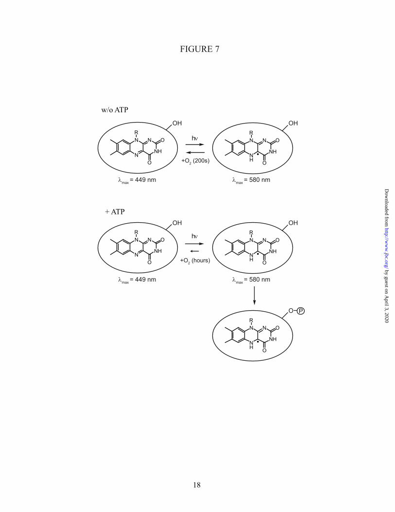
Fig. 7: The light-sensitive domain of Chlamydomonas cryptochrome carries oxidized flavin in the dark. Without ATP, illumination with blue light leads to formation of the neutral flavin radical, the putative signaling state. In presence of oxygen, the dark form is fully regained with a time constant of 200 s. Binding of ATP leads to stabilization of the radical state with respect to the oxidized and fully reduced states. Moreover, the presence of ATP enables light-induced autophosphorylation of the domain.