For peer review only SHOCK WAVE THERAPY, ASSOCIATED TO ECCENTRIC STRENGTHENING VERSUS ISOLATED ECCENTRIC STRENGTHENING FOR ACHILLES INSERTIONAL TENDINOPATHY TREATMENT: A DOUBLE BLINDED RANDOMIZED CLINICAL TRIAL Journal: BMJ Open Manuscript ID bmjopen-2016-013332 Article Type: Protocol Date Submitted by the Author: 13-Jul-2016 Complete List of Authors: Mansur, Nacime; Universidade Federal de Sao Paulo, Orthopaedics Faloppa, Flavio; Federal University of São Paulo (UNIFESP/EPM), Orthopedics and Traumatology - Division of Hand Surgery and Upper Limb Belloti, João ; Federal University of São Paulo (UNIFESP/EPM), Orthopedics and Traumatology - Division of Hand Surgery and Upper Limb Ingham, Sheila; Universidade Federal de Sao Paulo, Orthopaedics Matsunaga, Fabio; Federal University of São Paulo (UNIFESP/EPM), Orthopedics and Traumatology - Division of Hand Surgery and Upper Limb Santos, Paulo; Universidade Federal de Sao Paulo, Orthopaedics Santos, Bruno; Universidade Federal de Sao Paulo, Orthopaedics Carrazzone, Oreste; Universidade Federal de Sao Paulo, Orthopaedics Peixoto, Gabriel; Universidade Federal de Sao Paulo, Orthopaedics Ayoama, Bruno; Universidade Federal de Sao Paulo, Orthopaedics Tamaoki, Marcel Jun; Universidade Federal de Sao Paulo, Orthopaedics <b>Primary Subject Heading</b>: Evidence based practice Secondary Subject Heading: Sports and exercise medicine, Research methods, Rehabilitation medicine, Public health, Occupational and environmental medicine Keywords: achilles, tendinopathy, insertional, shock wave, Foot & ankle < ORTHOPAEDIC & TRAUMA SURGERY, eccentric For peer review only - http://bmjopen.bmj.com/site/about/guidelines.xhtml BMJ Open on 12 July 2018 by guest. Protected by copyright. http://bmjopen.bmj.com/ BMJ Open: first published as 10.1136/bmjopen-2016-013332 on 27 January 2017. Downloaded from
Transcript
For peer review only
SHOCK WAVE THERAPY, ASSOCIATED TO ECCENTRIC
STRENGTHENING VERSUS ISOLATED ECCENTRIC
STRENGTHENING FOR ACHILLES INSERTIONAL
TENDINOPATHY TREATMENT: A DOUBLE BLINDED
RANDOMIZED CLINICAL TRIAL
Journal: BMJ Open
Manuscript ID bmjopen-2016-013332
Article Type: Protocol
Date Submitted by the Author: 13-Jul-2016
Complete List of Authors: Mansur, Nacime; Universidade Federal de Sao Paulo, Orthopaedics Faloppa, Flavio; Federal University of São Paulo (UNIFESP/EPM), Orthopedics and Traumatology - Division of Hand Surgery and Upper Limb Belloti, João ; Federal University of São Paulo (UNIFESP/EPM), Orthopedics and Traumatology - Division of Hand Surgery and Upper Limb Ingham, Sheila; Universidade Federal de Sao Paulo, Orthopaedics Matsunaga, Fabio; Federal University of São Paulo (UNIFESP/EPM), Orthopedics and Traumatology - Division of Hand Surgery and Upper Limb Santos, Paulo; Universidade Federal de Sao Paulo, Orthopaedics Santos, Bruno; Universidade Federal de Sao Paulo, Orthopaedics Carrazzone, Oreste; Universidade Federal de Sao Paulo, Orthopaedics Peixoto, Gabriel; Universidade Federal de Sao Paulo, Orthopaedics Ayoama, Bruno; Universidade Federal de Sao Paulo, Orthopaedics Tamaoki, Marcel Jun; Universidade Federal de Sao Paulo, Orthopaedics
<b>Primary Subject Heading</b>:
Evidence based practice
Secondary Subject Heading: Sports and exercise medicine, Research methods, Rehabilitation medicine, Public health, Occupational and environmental medicine
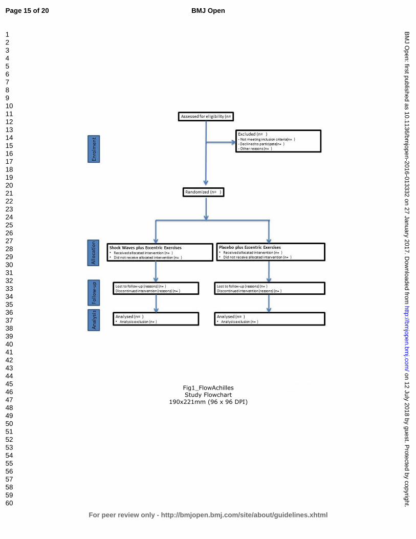
Methods and Analysis: Nine-three patients, referred from health care services, will be assessed and enrolled in
this study. They will be divided in two groups (randomized by sequentially numbered identical envelopes), one
containing the combination of shock wave and eccentric exercises as treatment, and the other comprehending
the exercises and the placebo treatment (apparatus placed in the therapeutic head). The assessments will occur in
2, 4, 6, 12 and 24 weeks. Patients will be evaluated primarily by the Victorian Institute of Sport Assessment-
Achilles questionnaire (VISA-A) and secondarily by the Visual Analogue Scale (VAS), Algometry, the
American Orthopedic Foot and Ankle Society (AOFAS) scale and the 12 Item Short Form Health Survey (SF-
12). We will use Comparison of Two Proportions via relative frequency analysis, the Pearson Correlation the
Chi-Square test and the ANOVA for statistical analyses.
Discussion: This study intends to demonstrate if the association of the eccentric exercise program with the
shock wave therapy can produce good results. In an attempt to prevent the high costs and complications
associated with surgery, we will try to prove this combination as a viable therapeutic option in the management
of this disease. The strengths of the study are the design and the combination of methods.
Ethics and Dissemination: The study is registered in the Clinical Trials database and approved by the
University Ethics Committe.
The study is registered in the Clinical Trials database (protocol number: 8094833648737701) and approved by the University Ethics Committee (number: 1373481).
1. Introduction
Calcaneus tendinopathy can be classified according to its anatomic site, as insertional and non-
insertional tendinopathy. It is characterized by intratendinous degenerations, secondary to low grade
inflammatory signs and erratic biological healing (Hartog 2009, Irwin 2010, Magnan et al 2014). The insertional
tendinopathy occurs in the Achilles attachment to the tuberosity of the calcaneus bone and up to 2 cm proximal
to the tuberosity. It is generally associated with a traction enthesophyte (upper spur), Haglund deformity (pump
bump) and with pre- and retro-achilles bursitis. The diagnosis is made based on clinical evaluation; ancillary
tests, such as X-Ray and Ultra Sound, are done only to confirm the lesion and to exclude differential diagnoses
(stress fractures, tumours). The clinical diagnosis consists of checking the level of pain via palpation of the
tendinous insertion region in the calcaneus bone (and up to 2cm around this region). The occurrence of volume
increase and mild hyperemia also supports the diagnosis (Hartog 2009, Irwin 2010, Magnan et al 2014, Kearney
et al 2010).
Page 2 of 16
For peer review only - http://bmjopen.bmj.com/site/about/guidelines.xhtml
on 12 July 2018 by guest. Protected by copyright. http://bmjopen.bmj.com/ BMJ Open: first published as 10.1136/bmjopen-2016-013332 on 27 January 2017. Downloaded from
11b If relevant, description of the similarity of interventions 6
Statistical methods 12a Statistical methods used to compare groups for primary and secondary outcomes 8
12b Methods for additional analyses, such as subgroup analyses and adjusted analyses 8
Results
Participant flow (a
diagram is strongly
recommended)
13a For each group, the numbers of participants who were randomly assigned, received intended treatment, and
were analysed for the primary outcome
x
13b For each group, losses and exclusions after randomisation, together with reasons x
Recruitment 14a Dates defining the periods of recruitment and follow-up x
14b Why the trial ended or was stopped x
Baseline data 15 A table showing baseline demographic and clinical characteristics for each group x
Numbers analysed 16 For each group, number of participants (denominator) included in each analysis and whether the analysis was
by original assigned groups
x
Outcomes and
estimation
17a For each primary and secondary outcome, results for each group, and the estimated effect size and its
precision (such as 95% confidence interval)
x
17b For binary outcomes, presentation of both absolute and relative effect sizes is recommended x
Ancillary analyses 18 Results of any other analyses performed, including subgroup analyses and adjusted analyses, distinguishing
pre-specified from exploratory
x
Harms 19 All important harms or unintended effects in each group (for specific guidance see CONSORT for harms) X
Discussion
Limitations 20 Trial limitations, addressing sources of potential bias, imprecision, and, if relevant, multiplicity of analyses 9
Generalisability 21 Generalisability (external validity, applicability) of the trial findings 9
Interpretation 22 Interpretation consistent with results, balancing benefits and harms, and considering other relevant evidence x
Other information
Registration 23 Registration number and name of trial registry 11
Protocol 24 Where the full trial protocol can be accessed, if available 11
Funding 25 Sources of funding and other support (such as supply of drugs), role of funders 1
*We strongly recommend reading this statement in conjunction with the CONSORT 2010 Explanation and Elaboration for important clarifications on all the items. If relevant, we also
recommend reading CONSORT extensions for cluster randomised trials, non-inferiority and equivalence trials, non-pharmacological treatments, herbal interventions, and pragmatic trials.
Additional extensions are forthcoming: for those and for up to date references relevant to this checklist, see www.consort-statement.org.
Page 16 of 16
For peer review only - http://bmjopen.bmj.com/site/about/guidelines.xhtml
on 12 July 2018 by guest. Protected by copyright. http://bmjopen.bmj.com/ BMJ Open: first published as 10.1136/bmjopen-2016-013332 on 27 January 2017. Downloaded from
Complete List of Authors: Mansur, Nacime; Universidade Federal de Sao Paulo, Orthopaedics Faloppa, Flavio; Federal University of São Paulo (UNIFESP/EPM), Orthopedics and Traumatology - Division of Hand Surgery and Upper Limb Belloti, João ; Federal University of São Paulo (UNIFESP/EPM), Orthopedics and Traumatology - Division of Hand Surgery and Upper Limb Ingham, Sheila; Universidade Federal de Sao Paulo, Orthopaedics Matsunaga, Fabio; Federal University of São Paulo (UNIFESP/EPM), Orthopedics and Traumatology - Division of Hand Surgery and Upper Limb Santos, Paulo; Universidade Federal de Sao Paulo, Orthopaedics Santos, Bruno; Universidade Federal de Sao Paulo, Orthopaedics Carrazzone, Oreste; Universidade Federal de Sao Paulo, Orthopaedics Peixoto, Gabriel; Universidade Federal de Sao Paulo, Orthopaedics Ayoama, Bruno; Universidade Federal de Sao Paulo, Orthopaedics Tamaoki, Marcel Jun; Universidade Federal de Sao Paulo, Orthopaedics
<b>Primary Subject Heading</b>:
Evidence based practice
Secondary Subject Heading: Sports and exercise medicine, Research methods, Rehabilitation medicine, Public health, Occupational and environmental medicine
SHOCK WAVE THERAPHY, ASSOCIATED TO ECCENTRIC STRENGTHENING VERSUS ISOLATED ECCENTRIC STRENGTHENING FOR ACHILLES INSERTIONAL TENDINOPATHY TREATMENT: A DOUBLE BLINDED RANDOMIZED CLINICAL TRIAL PROTOCOL.
Abstract
Background: There is no consensus regarding the treatment of Achilles insertional tendinopathies. Eccentric training remains the main choice in the conservative treatment of this illness; however, the good results in the management of non-insertional Achilles tendinopathy were not replicated in the insertional disease. Low energy shock wave therapy has been described as an alternative to these patients,
but has yet to be empirically tested.
Hypothesis: Shock wave therapy, adjunctive to eccentric strengthening protocol, will improve measures of pain and function.
Materials and Methods: Nine-three patients with a diagnosis of chronic insertional tendinopathy, referred from primary or secondary health care services, will be assessed and enrolled in this study. They will be divided in two groups (randomized by sequentially numbered identical envelopes, which will be administered serially to participants), one containing the combination of low energy shock wave and eccentric exercises, as treatment, and the other comprehending the exercises and the placebo treatment (an apparatus placed in the therapeutic head). The assessments will occur in 2, 4, 6, 12 and 24 weeks. Patients will be evaluated primarily by the Victorian Institute of Sport Assessment-Achilles questionnaire (VISA-A) and secondarily by the Visual Analogue Scale (VAS), Algometry, the American Orthopedic Foot and Ankle Society (AOFAS) scale, the Foot and Ankle Outcome Score (FAOS) and the 12 Item Short Form Health Survey (SF-12). We will use Comparison of Two Proportions via relative frequency analysis, the Pearson Correlation the Chi-Square test and the ANOVA for statistical analyses.
Discussion: This study intends to demonstrate if the association of the eccentric exercise program with the shock wave therapy can produce good results regarding the treatment of the Achilles insertional tendinopathy. In an attempt to prevent the high costs and complications associated with the surgical
intervention, we will try to prove this combination as a viable therapeutic option in the conservative management of this prevalent disease.
The strengths of the study are the design and the novelty of the combination of methods. The main limitation is the short follow-up course.
The study is registered in the Clinical Trials database (protocol number: 8094833648737701) and approved by the University Ethics Committee (number: 1373481).
1. Introduction
Calcaneus tendinopathy can be classified according to its anatomic site, as insertional and non-
insertional tendinopathy. It is characterized by intratendinous degenerations, secondary to low grade
inflammatory signs and erratic biological healing (Hartog 2009, Irwin 2010, Magnan et al 2014). The insertional
tendinopathy occurs in the Achilles attachment to the tuberosity of the calcaneus bone and up to 2 cm proximal
to the tuberosity. It is generally associated with a traction enthesophyte (upper spur), Haglund deformity (pump
bump) and with pre- and retro-achilles bursitis. The diagnosis is made based on clinical evaluation; ancillary
tests, such as X-Ray and Ultra Sound, are done only to confirm the lesion and to exclude differential diagnoses
Page 2 of 20
For peer review only - http://bmjopen.bmj.com/site/about/guidelines.xhtml
SPIRIT 2013 Checklist: Recommended items to address in a clinical trial protocol and related documents*
Section/item Item No
Description Addressed on page number
Administrative information
Title 1 Descriptive title identifying the study design, population, interventions, and, if applicable, trial acronym _____________
Trial registration 2a Trial identifier and registry name. If not yet registered, name of intended registry _____________
2b All items from the World Health Organization Trial Registration Data Set _____________
Protocol version 3 Date and version identifier _____________
Funding 4 Sources and types of financial, material, and other support _____________
Roles and responsibilities
5a Names, affiliations, and roles of protocol contributors _____________
5b Name and contact information for the trial sponsor _____________
5c Role of study sponsor and funders, if any, in study design; collection, management, analysis, and interpretation of data; writing of the report; and the decision to submit the report for publication, including whether they will have ultimate authority over any of these activities
_____________
5d Composition, roles, and responsibilities of the coordinating centre, steering committee, endpoint adjudication committee, data management team, and other individuals or groups overseeing the trial, if applicable (see Item 21a for data monitoring committee)
_____________
Page 16 of 20
For peer review only - http://bmjopen.bmj.com/site/about/guidelines.xhtml
6a Description of research question and justification for undertaking the trial, including summary of relevant studies (published and unpublished) examining benefits and harms for each intervention
_____________
6b Explanation for choice of comparators _____________
Objectives 7 Specific objectives or hypotheses _____________
Trial design 8 Description of trial design including type of trial (eg, parallel group, crossover, factorial, single group), allocation ratio, and framework (eg, superiority, equivalence, noninferiority, exploratory)
_____________
Methods: Participants, interventions, and outcomes
Study setting 9 Description of study settings (eg, community clinic, academic hospital) and list of countries where data will be collected. Reference to where list of study sites can be obtained
_____________
Eligibility criteria 10 Inclusion and exclusion criteria for participants. If applicable, eligibility criteria for study centres and individuals who will perform the interventions (eg, surgeons, psychotherapists)
_____________
Interventions 11a Interventions for each group with sufficient detail to allow replication, including how and when they will be administered
_____________
11b Criteria for discontinuing or modifying allocated interventions for a given trial participant (eg, drug dose change in response to harms, participant request, or improving/worsening disease)
_____________
11c Strategies to improve adherence to intervention protocols, and any procedures for monitoring adherence (eg, drug tablet return, laboratory tests)
_____________
11d Relevant concomitant care and interventions that are permitted or prohibited during the trial _____________
Outcomes 12 Primary, secondary, and other outcomes, including the specific measurement variable (eg, systolic blood pressure), analysis metric (eg, change from baseline, final value, time to event), method of aggregation (eg, median, proportion), and time point for each outcome. Explanation of the clinical relevance of chosen efficacy and harm outcomes is strongly recommended
_____________
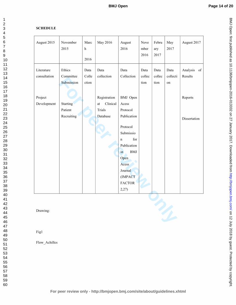
Participant timeline 13 Time schedule of enrolment, interventions (including any run-ins and washouts), assessments, and visits for participants. A schematic diagram is highly recommended (see Figure)
_____________
Page 17 of 20
For peer review only - http://bmjopen.bmj.com/site/about/guidelines.xhtml
Sample size 14 Estimated number of participants needed to achieve study objectives and how it was determined, including clinical and statistical assumptions supporting any sample size calculations
_____________
Recruitment 15 Strategies for achieving adequate participant enrolment to reach target sample size _____________
Methods: Assignment of interventions (for controlled trials)
Allocation:
Sequence generation
16a Method of generating the allocation sequence (eg, computer-generated random numbers), and list of any factors for stratification. To reduce predictability of a random sequence, details of any planned restriction (eg, blocking) should be provided in a separate document that is unavailable to those who enrol participants or assign interventions
_____________
Allocation concealment mechanism
16b Mechanism of implementing the allocation sequence (eg, central telephone; sequentially numbered, opaque, sealed envelopes), describing any steps to conceal the sequence until interventions are assigned
_____________
Implementation 16c Who will generate the allocation sequence, who will enrol participants, and who will assign participants to interventions
_____________
Blinding (masking) 17a Who will be blinded after assignment to interventions (eg, trial participants, care providers, outcome assessors, data analysts), and how
_____________
17b If blinded, circumstances under which unblinding is permissible, and procedure for revealing a participant’s allocated intervention during the trial
_____________
Methods: Data collection, management, and analysis
Data collection methods
18a Plans for assessment and collection of outcome, baseline, and other trial data, including any related processes to promote data quality (eg, duplicate measurements, training of assessors) and a description of study instruments (eg, questionnaires, laboratory tests) along with their reliability and validity, if known. Reference to where data collection forms can be found, if not in the protocol
_____________
18b Plans to promote participant retention and complete follow-up, including list of any outcome data to be collected for participants who discontinue or deviate from intervention protocols
_____________
Page 18 of 20
For peer review only - http://bmjopen.bmj.com/site/about/guidelines.xhtml
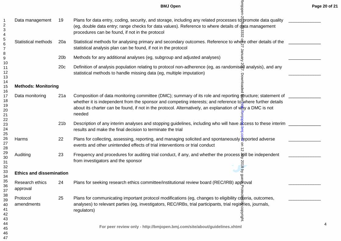
Data management 19 Plans for data entry, coding, security, and storage, including any related processes to promote data quality (eg, double data entry; range checks for data values). Reference to where details of data management procedures can be found, if not in the protocol
_____________
Statistical methods 20a Statistical methods for analysing primary and secondary outcomes. Reference to where other details of the statistical analysis plan can be found, if not in the protocol
_____________
20b Methods for any additional analyses (eg, subgroup and adjusted analyses) _____________
20c Definition of analysis population relating to protocol non-adherence (eg, as randomised analysis), and any statistical methods to handle missing data (eg, multiple imputation)
_____________
Methods: Monitoring
Data monitoring 21a Composition of data monitoring committee (DMC); summary of its role and reporting structure; statement of whether it is independent from the sponsor and competing interests; and reference to where further details about its charter can be found, if not in the protocol. Alternatively, an explanation of why a DMC is not needed
_____________
21b Description of any interim analyses and stopping guidelines, including who will have access to these interim results and make the final decision to terminate the trial
_____________
Harms 22 Plans for collecting, assessing, reporting, and managing solicited and spontaneously reported adverse events and other unintended effects of trial interventions or trial conduct
_____________
Auditing 23 Frequency and procedures for auditing trial conduct, if any, and whether the process will be independent from investigators and the sponsor
_____________
Ethics and dissemination
Research ethics approval
24 Plans for seeking research ethics committee/institutional review board (REC/IRB) approval _____________
Protocol amendments
25 Plans for communicating important protocol modifications (eg, changes to eligibility criteria, outcomes, analyses) to relevant parties (eg, investigators, REC/IRBs, trial participants, trial registries, journals, regulators)
_____________
Page 19 of 20
For peer review only - http://bmjopen.bmj.com/site/about/guidelines.xhtml
Consent or assent 26a Who will obtain informed consent or assent from potential trial participants or authorised surrogates, and how (see Item 32)
_____________
26b Additional consent provisions for collection and use of participant data and biological specimens in ancillary studies, if applicable
_____________
Confidentiality 27 How personal information about potential and enrolled participants will be collected, shared, and maintained in order to protect confidentiality before, during, and after the trial
_____________
Declaration of interests
28 Financial and other competing interests for principal investigators for the overall trial and each study site _____________
Access to data 29 Statement of who will have access to the final trial dataset, and disclosure of contractual agreements that limit such access for investigators
_____________
Ancillary and post-trial care
30 Provisions, if any, for ancillary and post-trial care, and for compensation to those who suffer harm from trial participation
_____________
Dissemination policy 31a Plans for investigators and sponsor to communicate trial results to participants, healthcare professionals, the public, and other relevant groups (eg, via publication, reporting in results databases, or other data sharing arrangements), including any publication restrictions
_____________
31b Authorship eligibility guidelines and any intended use of professional writers _____________
31c Plans, if any, for granting public access to the full protocol, participant-level dataset, and statistical code _____________
Appendices
Informed consent materials
32 Model consent form and other related documentation given to participants and authorised surrogates _____________
Biological specimens
33 Plans for collection, laboratory evaluation, and storage of biological specimens for genetic or molecular analysis in the current trial and for future use in ancillary studies, if applicable
_____________
*It is strongly recommended that this checklist be read in conjunction with the SPIRIT 2013 Explanation & Elaboration for important clarification on the items. Amendments to the protocol should be tracked and dated. The SPIRIT checklist is copyrighted by the SPIRIT Group under the Creative Commons “Attribution-NonCommercial-NoDerivs 3.0 Unported” license.
Page 20 of 20
For peer review only - http://bmjopen.bmj.com/site/about/guidelines.xhtml
Complete List of Authors: Mansur, Nacime; Universidade Federal de Sao Paulo, Orthopaedics Faloppa, Flavio; Federal University of São Paulo (UNIFESP/EPM), Orthopedics and Traumatology - Division of Hand Surgery and Upper Limb Belloti, João ; Federal University of São Paulo (UNIFESP/EPM), Orthopedics and Traumatology - Division of Hand Surgery and Upper Limb Ingham, Sheila; Universidade Federal de Sao Paulo, Orthopaedics Matsunaga, Fabio; Federal University of São Paulo (UNIFESP/EPM), Orthopedics and Traumatology - Division of Hand Surgery and Upper Limb Santos, Paulo; Universidade Federal de Sao Paulo, Orthopaedics Santos, Bruno; Universidade Federal de Sao Paulo, Orthopaedics Carrazzone, Oreste; Universidade Federal de Sao Paulo, Orthopaedics Peixoto, Gabriel; Universidade Federal de Sao Paulo, Orthopaedics Ayoama, Bruno; Universidade Federal de Sao Paulo, Orthopaedics Tamaoki, Marcel Jun; Universidade Federal de Sao Paulo, Orthopaedics
<b>Primary Subject Heading</b>:
Evidence based practice
Secondary Subject Heading: Sports and exercise medicine, Research methods, Rehabilitation medicine, Public health, Occupational and environmental medicine
SHOCK WAVE THERAPHY, ASSOCIATED TO ECCENTRIC STRENGTHENING VERSUS ISOLATED ECCENTRIC STRENGTHENING FOR ACHILLES INSERTIONAL TENDINOPATHY TREATMENT: A DOUBLE BLINDED RANDOMIZED CLINICAL TRIAL PROTOCOL.
Abstract
Background: There is no consensus regarding the treatment of Achilles insertional tendinopathies. Eccentric training remains the main choice in the conservative treatment of this illness; however, the good results in the management of non-insertional Achilles tendinopathy were not replicated in the insertional conditon. Low energy shock wave therapy has been described as an alternative to these
patients, but has yet to be empirically tested.
Hypothesis: Shock wave therapy, adjunctive to eccentric strengthening protocol, will improve measures of pain and function.
Materials and Methods: Nine-three patients with a diagnosis of chronic insertional tendinopathy, referred from primary or secondary health care services, will be assessed and enrolled in this study. They will be divided in two groups (randomized by sequentially numbered identical envelopes, which will be administered serially to participants), one containing the combination of low energy shock wave and eccentric exercises, as treatment, and the other comprehending the exercises and the placebo treatment (an apparatus placed in the therapeutic head). The assessments will occur in 2, 4, 6, 12 and 24 weeks. Patients will be evaluated primarily by the Victorian Institute of Sport Assessment-Achilles questionnaire (VISA-A) and secondarily by the Visual Analogue Scale (VAS), Algometry, the American Orthopedic Foot and Ankle Society (AOFAS) scale, the Foot and Ankle Outcome Score (FAOS) and the 12 Item Short Form Health Survey (SF-12). We will use Comparison of Two Proportions via relative frequency analysis, the Pearson Correlation the Chi-Square test and the ANOVA for statistical analyses.
Discussion: This study intends to demonstrate if the association of the eccentric exercise program with the shock wave therapy can produce good results regarding the treatment of the Achilles insertional tendinopathy. In an attempt to prevent the high costs and complications associated with the surgical
intervention, we will try to prove this combination as a viable therapeutic option in the conservative management of this prevalent condition.
The strengths of the study are the design and the novelty of the combination of methods. The main limitation is the short follow-up course.
The study is registered in the Clinical Trials database (protocol number: 8094833648737701) and approved by the University Ethics Committee (number: 1373481).
1. Introduction
Calcaneus tendinopathy can be classified according to its anatomic site, as insertional and non-
insertional tendinopathy. It is characterized by intratendinous degenerations, secondary to low grade
inflammatory signs and erratic biological healing.1-3
The insertional tendinopathy occurs in the Achilles
attachment to the tuberosity of the calcaneus bone and up to 2 cm proximal to the tuberosity. It is generally
associated with a traction enthesophyte (upper spur), Haglund deformity (pump bump) and with pre- and retro-
Page 2 of 20
For peer review only - http://bmjopen.bmj.com/site/about/guidelines.xhtml
SPIRIT 2013 Checklist: Recommended items to address in a clinical trial protocol and related documents*
Section/item Item No
Description Addressed on page number
Administrative information
Title 1 Descriptive title identifying the study design, population, interventions, and, if applicable, trial acronym _____________
Trial registration 2a Trial identifier and registry name. If not yet registered, name of intended registry _____________
2b All items from the World Health Organization Trial Registration Data Set _____________
Protocol version 3 Date and version identifier _____________
Funding 4 Sources and types of financial, material, and other support _____________
Roles and responsibilities
5a Names, affiliations, and roles of protocol contributors _____________
5b Name and contact information for the trial sponsor _____________
5c Role of study sponsor and funders, if any, in study design; collection, management, analysis, and interpretation of data; writing of the report; and the decision to submit the report for publication, including whether they will have ultimate authority over any of these activities
_____________
5d Composition, roles, and responsibilities of the coordinating centre, steering committee, endpoint adjudication committee, data management team, and other individuals or groups overseeing the trial, if applicable (see Item 21a for data monitoring committee)
_____________
Page 16 of 20
For peer review only - http://bmjopen.bmj.com/site/about/guidelines.xhtml
6a Description of research question and justification for undertaking the trial, including summary of relevant studies (published and unpublished) examining benefits and harms for each intervention
_____________
6b Explanation for choice of comparators _____________
Objectives 7 Specific objectives or hypotheses _____________
Trial design 8 Description of trial design including type of trial (eg, parallel group, crossover, factorial, single group), allocation ratio, and framework (eg, superiority, equivalence, noninferiority, exploratory)
_____________
Methods: Participants, interventions, and outcomes
Study setting 9 Description of study settings (eg, community clinic, academic hospital) and list of countries where data will be collected. Reference to where list of study sites can be obtained
_____________
Eligibility criteria 10 Inclusion and exclusion criteria for participants. If applicable, eligibility criteria for study centres and individuals who will perform the interventions (eg, surgeons, psychotherapists)
_____________
Interventions 11a Interventions for each group with sufficient detail to allow replication, including how and when they will be administered
_____________
11b Criteria for discontinuing or modifying allocated interventions for a given trial participant (eg, drug dose change in response to harms, participant request, or improving/worsening disease)
_____________
11c Strategies to improve adherence to intervention protocols, and any procedures for monitoring adherence (eg, drug tablet return, laboratory tests)
_____________
11d Relevant concomitant care and interventions that are permitted or prohibited during the trial _____________
Outcomes 12 Primary, secondary, and other outcomes, including the specific measurement variable (eg, systolic blood pressure), analysis metric (eg, change from baseline, final value, time to event), method of aggregation (eg, median, proportion), and time point for each outcome. Explanation of the clinical relevance of chosen efficacy and harm outcomes is strongly recommended
_____________
Participant timeline 13 Time schedule of enrolment, interventions (including any run-ins and washouts), assessments, and visits for participants. A schematic diagram is highly recommended (see Figure)
_____________
Page 17 of 20
For peer review only - http://bmjopen.bmj.com/site/about/guidelines.xhtml
Sample size 14 Estimated number of participants needed to achieve study objectives and how it was determined, including clinical and statistical assumptions supporting any sample size calculations
_____________
Recruitment 15 Strategies for achieving adequate participant enrolment to reach target sample size _____________
Methods: Assignment of interventions (for controlled trials)
Allocation:
Sequence generation
16a Method of generating the allocation sequence (eg, computer-generated random numbers), and list of any factors for stratification. To reduce predictability of a random sequence, details of any planned restriction (eg, blocking) should be provided in a separate document that is unavailable to those who enrol participants or assign interventions
_____________
Allocation concealment mechanism
16b Mechanism of implementing the allocation sequence (eg, central telephone; sequentially numbered, opaque, sealed envelopes), describing any steps to conceal the sequence until interventions are assigned
_____________
Implementation 16c Who will generate the allocation sequence, who will enrol participants, and who will assign participants to interventions
_____________
Blinding (masking) 17a Who will be blinded after assignment to interventions (eg, trial participants, care providers, outcome assessors, data analysts), and how
_____________
17b If blinded, circumstances under which unblinding is permissible, and procedure for revealing a participant’s allocated intervention during the trial
_____________
Methods: Data collection, management, and analysis
Data collection methods
18a Plans for assessment and collection of outcome, baseline, and other trial data, including any related processes to promote data quality (eg, duplicate measurements, training of assessors) and a description of study instruments (eg, questionnaires, laboratory tests) along with their reliability and validity, if known. Reference to where data collection forms can be found, if not in the protocol
_____________
18b Plans to promote participant retention and complete follow-up, including list of any outcome data to be collected for participants who discontinue or deviate from intervention protocols
_____________
Page 18 of 20
For peer review only - http://bmjopen.bmj.com/site/about/guidelines.xhtml
Data management 19 Plans for data entry, coding, security, and storage, including any related processes to promote data quality (eg, double data entry; range checks for data values). Reference to where details of data management procedures can be found, if not in the protocol
_____________
Statistical methods 20a Statistical methods for analysing primary and secondary outcomes. Reference to where other details of the statistical analysis plan can be found, if not in the protocol
_____________
20b Methods for any additional analyses (eg, subgroup and adjusted analyses) _____________
20c Definition of analysis population relating to protocol non-adherence (eg, as randomised analysis), and any statistical methods to handle missing data (eg, multiple imputation)
_____________
Methods: Monitoring
Data monitoring 21a Composition of data monitoring committee (DMC); summary of its role and reporting structure; statement of whether it is independent from the sponsor and competing interests; and reference to where further details about its charter can be found, if not in the protocol. Alternatively, an explanation of why a DMC is not needed
_____________
21b Description of any interim analyses and stopping guidelines, including who will have access to these interim results and make the final decision to terminate the trial
_____________
Harms 22 Plans for collecting, assessing, reporting, and managing solicited and spontaneously reported adverse events and other unintended effects of trial interventions or trial conduct
_____________
Auditing 23 Frequency and procedures for auditing trial conduct, if any, and whether the process will be independent from investigators and the sponsor
_____________
Ethics and dissemination
Research ethics approval
24 Plans for seeking research ethics committee/institutional review board (REC/IRB) approval _____________
Protocol amendments
25 Plans for communicating important protocol modifications (eg, changes to eligibility criteria, outcomes, analyses) to relevant parties (eg, investigators, REC/IRBs, trial participants, trial registries, journals, regulators)
_____________
Page 19 of 20
For peer review only - http://bmjopen.bmj.com/site/about/guidelines.xhtml
Consent or assent 26a Who will obtain informed consent or assent from potential trial participants or authorised surrogates, and how (see Item 32)
_____________
26b Additional consent provisions for collection and use of participant data and biological specimens in ancillary studies, if applicable
_____________
Confidentiality 27 How personal information about potential and enrolled participants will be collected, shared, and maintained in order to protect confidentiality before, during, and after the trial
_____________
Declaration of interests
28 Financial and other competing interests for principal investigators for the overall trial and each study site _____________
Access to data 29 Statement of who will have access to the final trial dataset, and disclosure of contractual agreements that limit such access for investigators
_____________
Ancillary and post-trial care
30 Provisions, if any, for ancillary and post-trial care, and for compensation to those who suffer harm from trial participation
_____________
Dissemination policy 31a Plans for investigators and sponsor to communicate trial results to participants, healthcare professionals, the public, and other relevant groups (eg, via publication, reporting in results databases, or other data sharing arrangements), including any publication restrictions
_____________
31b Authorship eligibility guidelines and any intended use of professional writers _____________
31c Plans, if any, for granting public access to the full protocol, participant-level dataset, and statistical code _____________
Appendices
Informed consent materials
32 Model consent form and other related documentation given to participants and authorised surrogates _____________
Biological specimens
33 Plans for collection, laboratory evaluation, and storage of biological specimens for genetic or molecular analysis in the current trial and for future use in ancillary studies, if applicable
_____________
*It is strongly recommended that this checklist be read in conjunction with the SPIRIT 2013 Explanation & Elaboration for important clarification on the items. Amendments to the protocol should be tracked and dated. The SPIRIT checklist is copyrighted by the SPIRIT Group under the Creative Commons “Attribution-NonCommercial-NoDerivs 3.0 Unported” license.
Page 20 of 20
For peer review only - http://bmjopen.bmj.com/site/about/guidelines.xhtml
Complete List of Authors: Mansur, Nacime; Universidade Federal de Sao Paulo, Orthopaedics Faloppa, Flavio; Federal University of São Paulo (UNIFESP/EPM), Orthopedics and Traumatology - Division of Hand Surgery and Upper Limb Belloti, João ; Federal University of São Paulo (UNIFESP/EPM), Orthopedics and Traumatology - Division of Hand Surgery and Upper Limb Ingham, Sheila; Universidade Federal de Sao Paulo, Orthopaedics Matsunaga, Fabio; Federal University of São Paulo (UNIFESP/EPM), Orthopedics and Traumatology - Division of Hand Surgery and Upper Limb Santos, Paulo; Universidade Federal de Sao Paulo, Orthopaedics Santos, Bruno; Universidade Federal de Sao Paulo, Orthopaedics Carrazzone, Oreste; Universidade Federal de Sao Paulo, Orthopaedics Peixoto, Gabriel; Universidade Federal de Sao Paulo, Orthopaedics Ayoama, Bruno; Universidade Federal de Sao Paulo, Orthopaedics Tamaoki, Marcel Jun; Universidade Federal de Sao Paulo, Orthopaedics
<b>Primary Subject Heading</b>:
Evidence based practice
Secondary Subject Heading: Sports and exercise medicine, Research methods, Rehabilitation medicine, Public health, Occupational and environmental medicine
SHOCK WAVE THERAPHY, ASSOCIATED TO ECCENTRIC STRENGTHENING VERSUS ISOLATED ECCENTRIC STRENGTHENING FOR ACHILLES INSERTIONAL TENDINOPATHY TREATMENT: A DOUBLE BLINDED RANDOMIZED CLINICAL TRIAL PROTOCOL.
Abstract
Background: There is no consensus regarding the treatment of Achilles insertional tendinopathies.
Eccentric training remains the main choice in the conservative treatment of this illness; however, the good results in the management of non-insertional Achilles tendinopathy were not replicated in the insertional condition. Low energy shock wave therapy has been described as an alternative to these patients, but has yet to be empirically tested.
Hypothesis: Shock wave therapy, adjunctive to eccentric strengthening protocol, will improve measures of pain and function.
Materials and Methods: Nine-three patients with a diagnosis of chronic insertional tendinopathy, referred from primary or secondary health care services, will be assessed and enrolled in this study. They will be divided in two groups (randomized by sequentially numbered identical envelopes, which will be
administered serially to participants), one containing the combination of low energy shock wave and eccentric exercises, as treatment, and the other comprehending the exercises and the placebo treatment (an apparatus placed in the therapeutic head). The assessments will occur in 2, 4, 6, 12 and 24 weeks. Patients will be evaluated primarily by the Victorian Institute of Sport Assessment-Achilles questionnaire (VISA-A) and secondarily by the Visual Analogue Scale (VAS), Algometry, the American Orthopedic
Foot and Ankle Society (AOFAS) scale, the Foot and Ankle Outcome Score (FAOS) and the 12 Item Short Form Health Survey (SF-12). We will use Comparison of Two Proportions via relative frequency analysis, the Pearson Correlation the Chi-Square test and the ANOVA for statistical analyses.
Discussion: This study intends to demonstrate if the association of the eccentric exercise program with the shock wave therapy can produce good results regarding the treatment of the Achilles insertional tendinopathy. In an attempt to prevent the high costs and complications associated with the surgical intervention, we will try to prove this combination as a viable therapeutic option in the conservative management of this prevalent condition. The strengths of the study are the design and the novelty of the combination of methods. The main limitation is the short follow-up course.
Ethics and Dissemination: The study is registered in the Clinical Trials database (protocol number: 8094833648737701) and was approved by the University Ethics Committee (number: 1373481).
Strengths and Limitations
The strengths of the study:
• Study design is ideal for treatment recommendations.
SPIRIT 2013 Checklist: Recommended items to address in a clinical trial protocol and related documents*
Section/item Item No
Description Addressed on page number
Administrative information
Title 1 Descriptive title identifying the study design, population, interventions, and, if applicable, trial acronym _____________
Trial registration 2a Trial identifier and registry name. If not yet registered, name of intended registry _____________
2b All items from the World Health Organization Trial Registration Data Set _____________
Protocol version 3 Date and version identifier _____________
Funding 4 Sources and types of financial, material, and other support _____________
Roles and responsibilities
5a Names, affiliations, and roles of protocol contributors _____________
5b Name and contact information for the trial sponsor _____________
5c Role of study sponsor and funders, if any, in study design; collection, management, analysis, and interpretation of data; writing of the report; and the decision to submit the report for publication, including whether they will have ultimate authority over any of these activities
_____________
5d Composition, roles, and responsibilities of the coordinating centre, steering committee, endpoint adjudication committee, data management team, and other individuals or groups overseeing the trial, if applicable (see Item 21a for data monitoring committee)
_____________
Page 17 of 21
For peer review only - http://bmjopen.bmj.com/site/about/guidelines.xhtml
6a Description of research question and justification for undertaking the trial, including summary of relevant studies (published and unpublished) examining benefits and harms for each intervention
_____________
6b Explanation for choice of comparators _____________
Objectives 7 Specific objectives or hypotheses _____________
Trial design 8 Description of trial design including type of trial (eg, parallel group, crossover, factorial, single group), allocation ratio, and framework (eg, superiority, equivalence, noninferiority, exploratory)
_____________
Methods: Participants, interventions, and outcomes
Study setting 9 Description of study settings (eg, community clinic, academic hospital) and list of countries where data will be collected. Reference to where list of study sites can be obtained
_____________
Eligibility criteria 10 Inclusion and exclusion criteria for participants. If applicable, eligibility criteria for study centres and individuals who will perform the interventions (eg, surgeons, psychotherapists)
_____________
Interventions 11a Interventions for each group with sufficient detail to allow replication, including how and when they will be administered
_____________
11b Criteria for discontinuing or modifying allocated interventions for a given trial participant (eg, drug dose change in response to harms, participant request, or improving/worsening disease)
_____________
11c Strategies to improve adherence to intervention protocols, and any procedures for monitoring adherence (eg, drug tablet return, laboratory tests)
_____________
11d Relevant concomitant care and interventions that are permitted or prohibited during the trial _____________
Outcomes 12 Primary, secondary, and other outcomes, including the specific measurement variable (eg, systolic blood pressure), analysis metric (eg, change from baseline, final value, time to event), method of aggregation (eg, median, proportion), and time point for each outcome. Explanation of the clinical relevance of chosen efficacy and harm outcomes is strongly recommended
_____________
Participant timeline 13 Time schedule of enrolment, interventions (including any run-ins and washouts), assessments, and visits for participants. A schematic diagram is highly recommended (see Figure)
_____________
Page 18 of 21
For peer review only - http://bmjopen.bmj.com/site/about/guidelines.xhtml
Sample size 14 Estimated number of participants needed to achieve study objectives and how it was determined, including clinical and statistical assumptions supporting any sample size calculations
_____________
Recruitment 15 Strategies for achieving adequate participant enrolment to reach target sample size _____________
Methods: Assignment of interventions (for controlled trials)
Allocation:
Sequence generation
16a Method of generating the allocation sequence (eg, computer-generated random numbers), and list of any factors for stratification. To reduce predictability of a random sequence, details of any planned restriction (eg, blocking) should be provided in a separate document that is unavailable to those who enrol participants or assign interventions
_____________
Allocation concealment mechanism
16b Mechanism of implementing the allocation sequence (eg, central telephone; sequentially numbered, opaque, sealed envelopes), describing any steps to conceal the sequence until interventions are assigned
_____________
Implementation 16c Who will generate the allocation sequence, who will enrol participants, and who will assign participants to interventions
_____________
Blinding (masking) 17a Who will be blinded after assignment to interventions (eg, trial participants, care providers, outcome assessors, data analysts), and how
_____________
17b If blinded, circumstances under which unblinding is permissible, and procedure for revealing a participant’s allocated intervention during the trial
_____________
Methods: Data collection, management, and analysis
Data collection methods
18a Plans for assessment and collection of outcome, baseline, and other trial data, including any related processes to promote data quality (eg, duplicate measurements, training of assessors) and a description of study instruments (eg, questionnaires, laboratory tests) along with their reliability and validity, if known. Reference to where data collection forms can be found, if not in the protocol
_____________
18b Plans to promote participant retention and complete follow-up, including list of any outcome data to be collected for participants who discontinue or deviate from intervention protocols
_____________
Page 19 of 21
For peer review only - http://bmjopen.bmj.com/site/about/guidelines.xhtml
Data management 19 Plans for data entry, coding, security, and storage, including any related processes to promote data quality (eg, double data entry; range checks for data values). Reference to where details of data management procedures can be found, if not in the protocol
_____________
Statistical methods 20a Statistical methods for analysing primary and secondary outcomes. Reference to where other details of the statistical analysis plan can be found, if not in the protocol
_____________
20b Methods for any additional analyses (eg, subgroup and adjusted analyses) _____________
20c Definition of analysis population relating to protocol non-adherence (eg, as randomised analysis), and any statistical methods to handle missing data (eg, multiple imputation)
_____________
Methods: Monitoring
Data monitoring 21a Composition of data monitoring committee (DMC); summary of its role and reporting structure; statement of whether it is independent from the sponsor and competing interests; and reference to where further details about its charter can be found, if not in the protocol. Alternatively, an explanation of why a DMC is not needed
_____________
21b Description of any interim analyses and stopping guidelines, including who will have access to these interim results and make the final decision to terminate the trial
_____________
Harms 22 Plans for collecting, assessing, reporting, and managing solicited and spontaneously reported adverse events and other unintended effects of trial interventions or trial conduct
_____________
Auditing 23 Frequency and procedures for auditing trial conduct, if any, and whether the process will be independent from investigators and the sponsor
_____________
Ethics and dissemination
Research ethics approval
24 Plans for seeking research ethics committee/institutional review board (REC/IRB) approval _____________
Protocol amendments
25 Plans for communicating important protocol modifications (eg, changes to eligibility criteria, outcomes, analyses) to relevant parties (eg, investigators, REC/IRBs, trial participants, trial registries, journals, regulators)
_____________
Page 20 of 21
For peer review only - http://bmjopen.bmj.com/site/about/guidelines.xhtml
Consent or assent 26a Who will obtain informed consent or assent from potential trial participants or authorised surrogates, and how (see Item 32)
_____________
26b Additional consent provisions for collection and use of participant data and biological specimens in ancillary studies, if applicable
_____________
Confidentiality 27 How personal information about potential and enrolled participants will be collected, shared, and maintained in order to protect confidentiality before, during, and after the trial
_____________
Declaration of interests
28 Financial and other competing interests for principal investigators for the overall trial and each study site _____________
Access to data 29 Statement of who will have access to the final trial dataset, and disclosure of contractual agreements that limit such access for investigators
_____________
Ancillary and post-trial care
30 Provisions, if any, for ancillary and post-trial care, and for compensation to those who suffer harm from trial participation
_____________
Dissemination policy 31a Plans for investigators and sponsor to communicate trial results to participants, healthcare professionals, the public, and other relevant groups (eg, via publication, reporting in results databases, or other data sharing arrangements), including any publication restrictions
_____________
31b Authorship eligibility guidelines and any intended use of professional writers _____________
31c Plans, if any, for granting public access to the full protocol, participant-level dataset, and statistical code _____________
Appendices
Informed consent materials
32 Model consent form and other related documentation given to participants and authorised surrogates _____________
Biological specimens
33 Plans for collection, laboratory evaluation, and storage of biological specimens for genetic or molecular analysis in the current trial and for future use in ancillary studies, if applicable
_____________
*It is strongly recommended that this checklist be read in conjunction with the SPIRIT 2013 Explanation & Elaboration for important clarification on the items. Amendments to the protocol should be tracked and dated. The SPIRIT checklist is copyrighted by the SPIRIT Group under the Creative Commons “Attribution-NonCommercial-NoDerivs 3.0 Unported” license.
Page 21 of 21
For peer review only - http://bmjopen.bmj.com/site/about/guidelines.xhtml