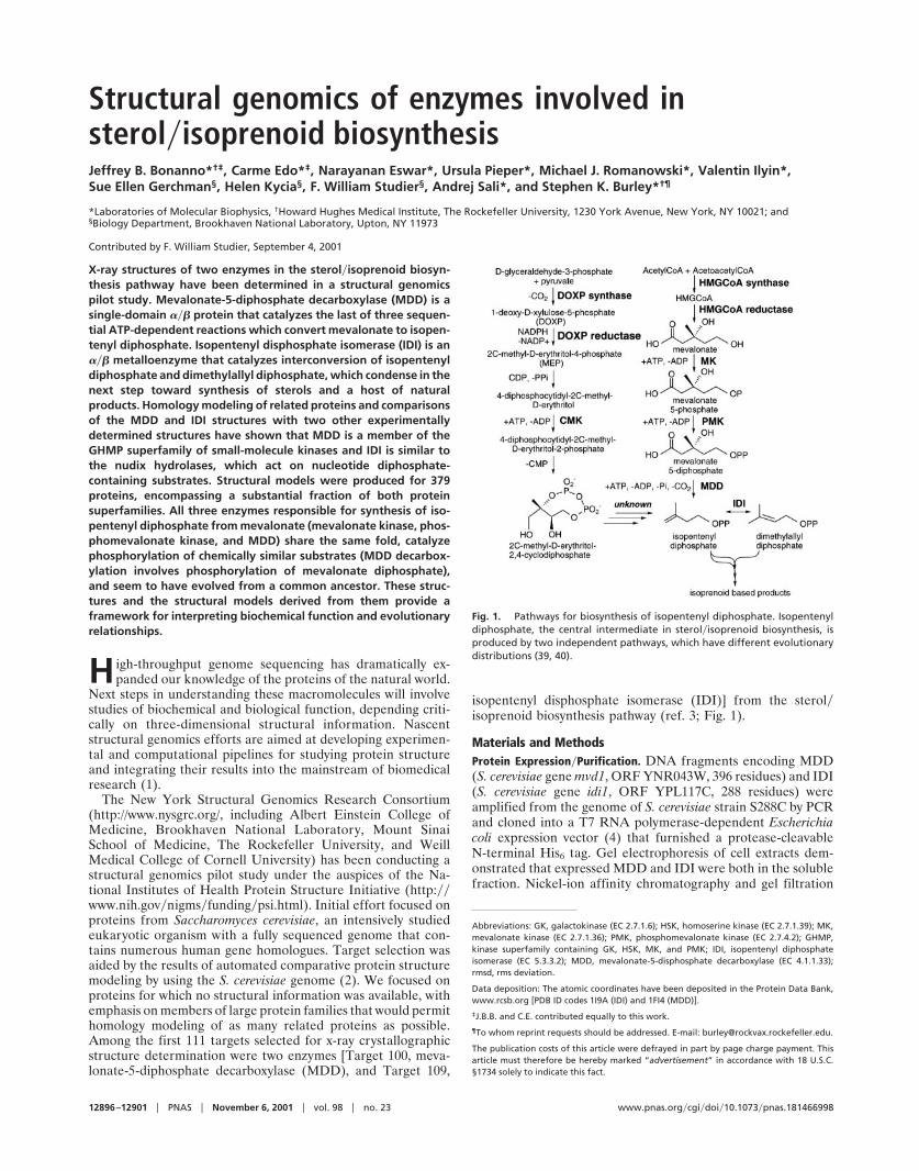
Structural genomics of enzymes involved in sterolisoprenoid biosynthesis Jeffrey B. Bonanno* †‡ , Carme Edo* ‡ , Narayanan Eswar*, Ursula Pieper*, Michael J. Romanowski*, Valentin Ilyin*, Sue Ellen Gerchman § , Helen Kycia § , F. William Studier § , Andrej Sali*, and Stephen K. Burley* †¶ *Laboratories of Molecular Biophysics, † Howard Hughes Medical Institute, The Rockefeller University, 1230 York Avenue, New York, NY 10021; and § Biology Department, Brookhaven National Laboratory, Upton, NY 11973 Contributed by F. William Studier, September 4, 2001 X-ray structures of two enzymes in the sterolisoprenoid biosyn- thesis pathway have been determined in a structural genomics pilot study. Mevalonate-5-diphosphate decarboxylase (MDD) is a single-domain protein that catalyzes the last of three sequen- tial ATP-dependent reactions which convert mevalonate to isopen- tenyl diphosphate. Isopentenyl disphosphate isomerase (IDI) is an metalloenzyme that catalyzes interconversion of isopentenyl diphosphate and dimethylallyl diphosphate, which condense in the next step toward synthesis of sterols and a host of natural products. Homology modeling of related proteins and comparisons of the MDD and IDI structures with two other experimentally determined structures have shown that MDD is a member of the GHMP superfamily of small-molecule kinases and IDI is similar to the nudix hydrolases, which act on nucleotide diphosphate- containing substrates. Structural models were produced for 379 proteins, encompassing a substantial fraction of both protein superfamilies. All three enzymes responsible for synthesis of iso- pentenyl diphosphate from mevalonate (mevalonate kinase, phos- phomevalonate kinase, and MDD) share the same fold, catalyze phosphorylation of chemically similar substrates (MDD decarbox- ylation involves phosphorylation of mevalonate diphosphate), and seem to have evolved from a common ancestor. These struc- tures and the structural models derived from them provide a framework for interpreting biochemical function and evolutionary relationships. H igh-throughput genome sequencing has dramatically ex- panded our knowledge of the proteins of the natural world. Next steps in understanding these macromolecules will involve studies of biochemical and biological function, depending criti- cally on three-dimensional structural information. Nascent structural genomics efforts are aimed at developing experimen- tal and computational pipelines for studying protein structure and integrating their results into the mainstream of biomedical research (1). The New York Structural Genomics Research Consortium (http://www.nysgrc.org/, including Albert Einstein College of Medicine, Brookhaven National Laboratory, Mount Sinai School of Medicine, The Rockefeller University, and Weill Medical College of Cornell University) has been conducting a structural genomics pilot study under the auspices of the Na- tional Institutes of Health Protein Structure Initiative (http:www.nih.govnigmsfundingpsi.html). Initial effort focused on proteins from Saccharomyces cerevisiae, an intensively studied eukaryotic organism with a fully sequenced genome that con- tains numerous human gene homologues. Target selection was aided by the results of automated comparative protein structure modeling by using the S. cerevisiae genome (2). We focused on proteins for which no structural information was available, with emphasis on members of large protein families that would permit homology modeling of as many related proteins as possible. Among the first 111 targets selected for x-ray crystallographic structure determination were two enzymes [Target 100, meva- lonate-5-diphosphate decarboxylase (MDD), and Target 109, isopentenyl disphosphate isomerase (IDI)] from the sterolisoprenoid biosynthesis pathway (ref. 3; Fig. 1). Materials and Methods Protein ExpressionPurification. DNA fragments encoding MDD (S. cerevisiae gene mvd1, ORF YNR043W, 396 residues) and IDI (S. cerevisiae gene idi1, ORF YPL117C, 288 residues) were amplified from the genome of S. cerevisiae strain S288C by PCR and cloned into a T7 RNA polymerase-dependent Escherichia coli expression vector (4) that furnished a protease-cleavable N-terminal His 6 tag. Gel electrophoresis of cell extracts dem- onstrated that expressed MDD and IDI were both in the soluble fraction. Nickel-ion affinity chromatography and gel filtration Abbreviations: GK, galactokinase (EC 2.7.1.6); HSK, homoserine kinase (EC 2.7.1.39); MK, mevalonate kinase (EC 2.7.1.36); PMK, phosphomevalonate kinase (EC 2.7.4.2); GHMP, kinase superfamily containing GK, HSK, MK, and PMK; IDI, isopentenyl diphosphate isomerase (EC 5.3.3.2); MDD, mevalonate-5-disphosphate decarboxylase (EC 4.1.1.33); rmsd, rms deviation. Data deposition: The atomic coordinates have been deposited in the Protein Data Bank, www.rcsb.org [PDB ID codes 1I9A (IDI) and 1FI4 (MDD)]. ‡ J.B.B. and C.E. contributed equally to this work. ¶ To whom reprint requests should be addressed. E-mail: [email protected]. The publication costs of this article were defrayed in part by page charge payment. This article must therefore be hereby marked “advertisement” in accordance with 18 U.S.C. §1734 solely to indicate this fact. Fig. 1. Pathways for biosynthesis of isopentenyl diphosphate. Isopentenyl diphosphate, the central intermediate in sterolisoprenoid biosynthesis, is produced by two independent pathways, which have different evolutionary distributions (39, 40). 12896 –12901 PNAS November 6, 2001 vol. 98 no. 23 www.pnas.orgcgidoi10.1073pnas.181466998
Transcript
Structural genomics of enzymes involved insterol�isoprenoid biosynthesisJeffrey B. Bonanno*†‡, Carme Edo*‡, Narayanan Eswar*, Ursula Pieper*, Michael J. Romanowski*, Valentin Ilyin*,Sue Ellen Gerchman§, Helen Kycia§, F. William Studier§, Andrej Sali*, and Stephen K. Burley*†¶
*Laboratories of Molecular Biophysics, †Howard Hughes Medical Institute, The Rockefeller University, 1230 York Avenue, New York, NY 10021; and§Biology Department, Brookhaven National Laboratory, Upton, NY 11973
Contributed by F. William Studier, September 4, 2001
X-ray structures of two enzymes in the sterol�isoprenoid biosyn-thesis pathway have been determined in a structural genomicspilot study. Mevalonate-5-diphosphate decarboxylase (MDD) is asingle-domain ��� protein that catalyzes the last of three sequen-tial ATP-dependent reactions which convert mevalonate to isopen-tenyl diphosphate. Isopentenyl disphosphate isomerase (IDI) is an��� metalloenzyme that catalyzes interconversion of isopentenyldiphosphate and dimethylallyl diphosphate, which condense in thenext step toward synthesis of sterols and a host of naturalproducts. Homology modeling of related proteins and comparisonsof the MDD and IDI structures with two other experimentallydetermined structures have shown that MDD is a member of theGHMP superfamily of small-molecule kinases and IDI is similar tothe nudix hydrolases, which act on nucleotide diphosphate-containing substrates. Structural models were produced for 379proteins, encompassing a substantial fraction of both proteinsuperfamilies. All three enzymes responsible for synthesis of iso-pentenyl diphosphate from mevalonate (mevalonate kinase, phos-phomevalonate kinase, and MDD) share the same fold, catalyzephosphorylation of chemically similar substrates (MDD decarbox-ylation involves phosphorylation of mevalonate diphosphate),and seem to have evolved from a common ancestor. These struc-tures and the structural models derived from them provide aframework for interpreting biochemical function and evolutionaryrelationships.
H igh-throughput genome sequencing has dramatically ex-panded our knowledge of the proteins of the natural world.
Next steps in understanding these macromolecules will involvestudies of biochemical and biological function, depending criti-cally on three-dimensional structural information. Nascentstructural genomics efforts are aimed at developing experimen-tal and computational pipelines for studying protein structureand integrating their results into the mainstream of biomedicalresearch (1).
The New York Structural Genomics Research Consortium(http://www.nysgrc.org/, including Albert Einstein College ofMedicine, Brookhaven National Laboratory, Mount SinaiSchool of Medicine, The Rockefeller University, and WeillMedical College of Cornell University) has been conducting astructural genomics pilot study under the auspices of the Na-tional Institutes of Health Protein Structure Initiative (http:��www.nih.gov�nigms�funding�psi.html). Initial effort focused onproteins from Saccharomyces cerevisiae, an intensively studiedeukaryotic organism with a fully sequenced genome that con-tains numerous human gene homologues. Target selection wasaided by the results of automated comparative protein structuremodeling by using the S. cerevisiae genome (2). We focused onproteins for which no structural information was available, withemphasis on members of large protein families that would permithomology modeling of as many related proteins as possible.Among the first 111 targets selected for x-ray crystallographicstructure determination were two enzymes [Target 100, meva-lonate-5-diphosphate decarboxylase (MDD), and Target 109,
isopentenyl disphosphate isomerase (IDI)] from the sterol�isoprenoid biosynthesis pathway (ref. 3; Fig. 1).
Materials and MethodsProtein Expression�Purification. DNA fragments encoding MDD(S. cerevisiae gene mvd1, ORF YNR043W, 396 residues) and IDI(S. cerevisiae gene idi1, ORF YPL117C, 288 residues) wereamplified from the genome of S. cerevisiae strain S288C by PCRand cloned into a T7 RNA polymerase-dependent Escherichiacoli expression vector (4) that furnished a protease-cleavableN-terminal His6 tag. Gel electrophoresis of cell extracts dem-onstrated that expressed MDD and IDI were both in the solublefraction. Nickel-ion affinity chromatography and gel filtration
Data deposition: The atomic coordinates have been deposited in the Protein Data Bank,www.rcsb.org [PDB ID codes 1I9A (IDI) and 1FI4 (MDD)].
‡J.B.B. and C.E. contributed equally to this work.
¶To whom reprint requests should be addressed. E-mail: [email protected].
The publication costs of this article were defrayed in part by page charge payment. Thisarticle must therefore be hereby marked “advertisement” in accordance with 18 U.S.C.§1734 solely to indicate this fact.
Fig. 1. Pathways for biosynthesis of isopentenyl diphosphate. Isopentenyldiphosphate, the central intermediate in sterol�isoprenoid biosynthesis, isproduced by two independent pathways, which have different evolutionarydistributions (39, 40).
yielded highly purified proteins, as judged by mass spectrometry.After difficulties with S. cerevisiae IDI crystallizability, E. coliIDI (gene idi, 182 residues) was cloned from UT5600 genomicDNA, expressed as a glutathione S-transferase fusion, andpurified to homogeneity by using published procedures (5).
Crystallizability Screening and Crystallization. Conformationallystable, monodisperse macromolecular preparations representgood crystallization candidates (6, 7). Conformational hetero-geneity can be detected by using fluorescence and circulardichroism spectroscopy or proteolysis combined with MS (8).Polydispersity (nonspecific aggregation) can be detected byusing dynamic light scattering (9). S. cerevisiae MDD behaved asa monodisperse dimer with an apparent molecular mass of 110kDa (predicted mass for the dimer is 94 kDa) in aqueoussolution. S. cerevisiae IDI was aggregated under similar condi-tions and was abandoned in favor of the more suitable homo-logue from E. coli, which behaves as a monodisperse monomer.
As expected from the results of crystallizability screening,hanging-drop�vapor-diffusion trials with S. cerevisiae MDD andE. coli IDI succeeded with minimal effort. MDD producedtrapezoidal crystals in 100 mM Tris�HCl, pH 8.5�15% (wt/vol)polyethylene glycol (PEG) 4K�1 M NaCl�5% (vol/vol) ethyleneglycol (space group, P21212; unit cell: a � 78.9 Å, b � 126.5 Å,c � 47.3 Å; 1 molecule per asymmetric unit). IDI yielded squarepyramidal crystals in 100 mM MES, pH 6.5�15% (wt/vol) PEG8K�8% (wt/vol) PEG 1K�100 mM NaCl (space group, P41212;unit cell: a � 72.4 Å, c � 204.4 Å; 2 molecules per asymmetricunit). Selenium-methionine proteins were produced and crys-tallized by using essentially identical procedures.
Data Collection and Structure Determination. X-ray diffraction datafor MDD and IDI were recorded under standard cryogenicconditions at Beamline X25 at the National Synchrotron LightSource and Beamline F2 at the Cornell High Energy Synchro-tron Source, respectively, and processed by using DENZO�SCALEPACK (10). Structures of MDD and IDI were obtained withthe multiwavelength anomalous dispersion method (11) fromdiffraction data recorded at 3 (MDD) or 2 (IDI) x-ray wave-lengths (Table 1). Selenium atomic positions (9 and 6 seleniumsites, respectively) were located with SNB (12). MLPHARE phasingfollowed by density modification (13) yielded high-quality ex-perimental phases for both systems. Electron density map in-terpretation (14) and refinement (15) produced structures of
MDD (391 residues, 164 waters, R factor � 23.9%, R free �26.8%, 2.3 Å resolution) and IDI (354 residues, 261 waters, Rfactor � 22.9%, R free � 27.9% 2.5 Å resolution).
Multiple Sequence Alignments. Proteins similar to S. cerevisiaeMDD and E. coli IDI (E value �10�4) were identified by using�-BLAST (16). Multiple sequence alignments were prepared byusing CLUSTAL (17), and conservation was calculated with BLO-SUM62 (18). Alignments of selected MDDs and IDIs are given inFigs. 2 and 3, respectively.
Homology Modeling. Automated comparative protein structuremodeling (hereafter referred to as ‘‘homology modeling’’) withMODPIPE (19) was carried out by using each experimentallydetermined structure (hereafter referred to as ‘‘structures’’) asa modeling template. Candidates for modeling were obtainedfrom the National Center for Biotechnology Information (ftp:��ftp.ncbi.nlm.nih.gov�blast�db) by using an iterative �-BLASTsearch (E value �100 for at least 30 residues) with the sequenceof the modeling template. Homology models were computedwith the �-BLAST alignments by means of satisfaction of spatialrestraints, as implemented in MODELLER (20), and assessed bycomputing a model score that uses a statistical energy function,sequence similarity with the modeling template, and a measureof structural compactness (2). Tests with known structures haveshown that models with scores from 0.7 to 1.0 have the correctfold at a 95% confidence level (2). All models discussed belowhave scores �0.7.
ResultsMDD Structural Overview. MDD is a single ��� domain (Fig. 4A,dimensions 75 Å � 42 Å � 38 Å) with a deep cleft. The orderof secondary structural elements is �1-�2-�3-�4-�5-�6-�A-�7-�B-�C-�D-�8-�9-�10-�E-�F-�G-�H-�11-�12-�I-�J-�13-�14,and the structure consists of three antiparallel �-sheets (�1-�6-�7-�14, �2-�5-�8-�9, �10-�11-�12-�13) and three sets of �-helices (�A-D, �E-G, �H-J). The crystallographic two-foldsymmetry axis parallel to the c axis generates a symmetric dimer,which presumably corresponds to the oligomerization statedetected by dynamic light scattering.
MDD alignments (Fig. 2) show conserved segments thatsurround a deep cleft (Fig. 4A). Within this surface concavity,Pro-113-Ala-122 (outlined in red in Fig. 2 and colored red in Fig.4A) resembles P loops responsible for ATP binding in other
Table 1. Statistics of the crystallographic analysis
All �1 data 18.0–2.3 0.239 0.268rmsd Bond lengths, 0.007 Å Bond angles, 1.30°
IDI refinementAll �1 data 20.0–2.5 0.229 0.279rmsd Bond lengths, 0.006 Å Bond angles, 1.39°
Rsym � ��I � �I����I, where I, observed intensity; �I�, average intensity obtained from multiple observations ofsymmetry related reflections. rms bond lengths and rms bond angles are the respective rmsds from ideal values.R free was calculated with 10% of data omitted from the refinement.
Bonanno et al. PNAS � November 6, 2001 � vol. 98 � no. 23 � 12897
BIO
CHEM
ISTR
Y
enzymes (21). GRASP calculations (22) revealed a surface patchwith a positive electrostatic potential within the cleft (data notshown), which represents an excellent candidate for binding theanionic substrate, mevalonate-5-diphosphate.
Homology Modeling with MDD. Comparison of S. cerevisiae MDDwith the Protein Data Bank (23) in November 2000 revealed nosimilar structures, as judged by DALI (24). Homology modelingwith S. cerevisiae MDD gave models for the other MDDs plusvarious GHMP kinases (21), including the canonical galactoki-
nases (GK), homoserine kinases (HSK), mevalonate kinases(MK; Fig. 1), and phosphomevalonate kinases (PMK; Fig. 1),plus diphosphocytidyl-2-C-methyl-D-erythritol kinases [CMK(EC 2.7.1.-), an enzyme in the 1-deoxy-D-xylulose-5-phosphatepathway to isopentenyl diphosphate; Fig. 1], and some hypo-thetical proteins. A kinase mechanism of action for MDD is notunreasonable. MDD phosphorylates the substrate C-3 hydroxylgroup, followed by elimination of both the added phosphate andcarboxylate groups to give isopentenyl diphosphate, ADP, andPi. Our modeling results suggest that all three enzymes respon-
Fig. 2. MDD sequence alignment. Secondary structural elements of S. cerevisiae MDD are shown with cylinders (�-helices) and arrows (�-strands). Gray dotsdenote poorly resolved residues in the final electron density map. Color-coding denotes sequence conservation among MDDs (white3 green ramp, 303 100%similarly). Red box denotes the putative ATP-binding P loop.
Fig. 3. IDI sequence alignment. Secondary structural elements, poorly resolved residues, and sequence conservation are denoted as in Fig. 2. #, metal-bindingresidues; *, conserved residues in the cleft; o � the active-site Cys. Some of the N- and C-terminal residues of IDI sequences other than E. coli have been excludedfor clarity.
12898 � www.pnas.org�cgi�doi�10.1073�pnas.181466998 Bonanno et al.
sible for sequential conversion of mevalonate to isopentenyldiphosphate (MK, PMK, and MDD; Fig. 1) have the same fold,indicating that they arose from a common precursor and mayrepresent an example of retrograde evolution (25). It is remark-able that diphosphocytidyl-2-C-methyl-D-erythritol kinase(CMK), which is part of the mevalonate-independent pathwayfor sterol�isoprenoid biosynthesis in plastids and bacteria, alsoseems to have evolved from the same ancestral small-moleculekinase.
Following our initial analyses, a bona fide GHMP kinasestructure was reported (ref. 26; Methanococcus jannaschii HSK,Protein Data Bank ID code 1FWL). The only significant struc-tural differences between S. cerevisiae MDD and M. jannaschiiHSK are two insertions within MDD (corresponding to �3 plus�4 and �J), providing direct experimental confirmation thatMDD is indeed a GHMP kinase superfamily member [Fig. 4; 276�-carbon pairs, rms deviation (rmsd) � 3.0 Å, 13% identity, Zscore � 21.9; all rmsds and Z scores reported herein wereobtained with DALI]. The P loop within HSK (colored red in Fig.4B) was shown to bind ADP, albeit in a distinct orientation fromthat seen in other P loop-containing enzymes (26).
By using both structures (MDD and HSK), MODPIPE producedmodels (length � 200 residues) for 181 proteins (as of March2001)—113 with both templates, 60 with HSK only, and 8 withMDD only. MDD and HSK each yielded models for the other.The HSK model derived from the MDD template vs. thestructure of HSK gave an rmsd of 3.8 Å (241 �-carbon pairs, 13%identity, Z score � 15.0). The MDD model derived from theHSK template vs. the structure of MDD gave an rmsd of 3.4 Å(272 �-carbon pairs, 16% identity, Z score � 17.7). The corre-spondence between our computed models and the structures ofMDD and HSK provides further evidence of the reliability ofhomology modeling with MODPIPE. In cases where models for thesame sequence were obtained from both structural templates,the average rmsd between alternative models was 3.4 Å with �Zscore� � 16.2 3.0.
Models were produced for members of all subgroups of theGHMP kinase superfamily, including 22 MDDs, 31 GKs, 33HSKs, 25 MKs, 9 PMKs, 25 diphosphocytidyl-2-C-methyl-D-erythritol kinases (CMK), 7 archael shikimate kinases (27), and8 D-glycero-D-manno-heptose 7-phosphate kinases (28). Theremaining 21 structural models fall into 4 sequence-similaritygroups (one of which contains 11 members), suggesting thatadditional enzyme activities are encompassed within the GHMP
kinase superfamily. Models covered at least 80% of the fulllength for 172 of 181 (95%) proteins modeled, suggesting thatthe GHMP kinase superfamily encompasses a rather narrowrange of variations on the basic fold. Modeling summary statis-tics are given in Tables 2–5, which are published as supportinginformation on the PNAS web site, www.pnas.org, and atomiccoordinate files for each of the models can be downloaded fromMODBASE (http:��guitar.rockefeller.edu�modbase�; select 1FI4and 1FWL datasets on the advanced search page).
IDI Structural Overview. IDI is a single ��� domain (Fig. 5A,dimensions 28 Å � 40 Å � 27 Å) with a divalent metal ion boundwithin a deep cleft. The order of secondary structural elementsis �1-�2-�A-�3-�4-�5-�6-�7-�B-�8-�9-�10-�11-�12-�C-�D-�E-�F, and the structure consists of two mixed polarity (�2-�1-�3-�10-�9, �7-�4-�11-�8) and one anti-parallel (�12-�5-�6)�-sheets. The second and third �-sheets are backed by �-helices.
Conserved metal-binding residues (His-25, His-32, His-69,Glu-114, and Glu-116; Fig. 3) form a distorted square pyramidalcoordination with a water molecule near the open coordinationsite (Mn2 � H2O � 5.5 Å; Fig. 5A). The metal ion was treatedas Mn2 for the purpose of crystallographic refinement, but itmay be Mg2 in vivo (29). Conserved polar residues (Lys-21,Arg-51, Lys-55, Arg-82, Arg-83, Glu-87, and Glu-135; Fig. 3) linethe remainder of the cleft (Fig. 5A), and we presume that someof the basic side chains contribute to binding of the anionicsubstrates isomerized by IDI. Experimental support for ourassignment of the IDI active site is provided by the results ofinhibition studies, which showed that nearby Cys-67 (Fig. 5A) isessential for catalysis (29).
IDI Resembles MutT. Comparison of our IDI structure with thecontents of the Protein Data Bank (PDB) in January 2001revealed one relative, E. coli MutT (PDB ID code 1TUM; 108�-carbon pairs, rmsd � 3.0 Å, 13% identity, Z score � 6.7). Thesolution NMR structure of MutT (Fig. 5B) resembles the portionof IDI containing the �3-�10-�9 and �7-�4-�11-�8 sheets andtwo of the flanking �-helices (�B and �F).
MutT is thought to guard against DNA incorporation of amutagenic product of oxidative damage by hydrolyzing 7,8-dihydro-8-oxoguanine-triphosphate to the monophosphate (30).It is a member of the nudix superfamily of enzymes that act onvarious substrates, most of which contain a nucleotide diphos-phate (31). A conserved motif characteristic of the nudix family
Fig. 4. S. cerevisiae MDD and M. jannaschii HSK. Ribbon drawings of MDD (A) and HSK (B) in the same orientation. The two-fold rotational symmetry axis thatgenerates the MDD homodimer is indicated. Putative P loops are colored red.
Bonanno et al. PNAS � November 6, 2001 � vol. 98 � no. 23 � 12899
BIO
CHEM
ISTR
Y
is Gly-X5-Glu-X7-Arg-Glu-�-X-Glu-Glu-X2-�, where X is anyresidue and � denotes a large hydrophobic side chain (31). Thecorresponding segment within the IDIs contains a similar butdifferent conserved motif Gly-X3-Ala-X2-Arg-Arg�Lys-�-X2-Glu-Leu-Gly-� (residues 75–90; Fig. 3), where Arg-Arg�Lys inIDI corresponds to Arg-Glu in MutT, and X-Glu in IDI corre-sponds to Glu-Glu in MutT. The positions of these conservedmotifs in the IDI and MutT structures are shown in Fig. 5(colored magenta).
Homology Modeling with IDI and MutT. By using the structures ofIDI and MutT as modeling templates, MODPIPE produced models(as of April 2001) for 202 nonredundant protein sequences(length � 100 residues)—62 with both templates, 103 with IDIonly, and 37 with MutT only. The MutT model derived from IDIvs. the solution NMR structure of MutT gave an rmsd of 3.0 Å(69 �-carbon pairs, 18% identity, Z score � 2.7). No model forIDI (score � 0.7) was produced with the structure of MutT as atemplate. In cases where models for the same sequence wereobtained from both structural templates (IDI and MutT), theaverage rmsd between alternative models was 3.6 Å with �Zscore� � 5.6 1.0. This �Z score� is considerably lower thanthe corresponding value obtained by comparing MDD- andHSK-derived alternative models, and presumably reflects thegreater structural difference between IDI and MutT. Nonethe-less, our structural comparison and modeling results do confirmthe recently proposed similarity of the IDIs and the nudixproteins (32).
The 202 nudix�IDI superfamily members for which MODPIPEproduced models were grouped on the basis of their amino acidsequences: 156 proteins fall into one of 15 groups in which atleast 1 member was annotated with a discrete enzymatic activity(including 37 IDIs), 42 proteins were singletons or fall into smallgroups with no attributed enzymatic activity, and 4 were largerproteins thought to possess more than 1 biochemical function. Incontrast to the GHMP kinase superfamily, the nudix�IDI su-perfamily fold seems to be rather frequently combined with
other structural features or domains. Models based on either IDIor MutT covered at least 80% of the full length for only 109 of202 (54%) proteins modeled, and less than 50% of the full lengthfor 35 proteins (17%). Modeling summary statistics are given inTables 2–5, and atomic coordinate files for each of the modelscan be downloaded from MODBASE (http:��guitar.rock-efeller.edu�modbase�; select 1I9A and 1MUT datasets on theadvanced search page).
DiscussionOne of the goals of the New York Structural Genomics ResearchConsortium is to generate experimentally determined structureswith sufficient diversity to support homology modeling of largenumbers of protein sequences. Recent analyses by Vitkup et al.(33) have suggested that determination of as few as 16,000selected structures could enable production of ‘‘accurate’’ ho-mology models for 90% of all proteins found in nature (seebelow).
Homology modeling can be distinguished from all othermethods for analyzing relationships among protein sequencesbecause it yields atomic coordinates suitable for direct compar-isons with x-ray and solution NMR structures. Acceptablemodels can be divided into three accuracy classes, characterizedwith blind tests by using known structures (34). Models based on�50% sequence identity with the template are comparable inaccuracy to 3 Å resolution x-ray structures or medium-resolutionsolution NMR structures (10 long-range restraints per residue).Models obtained with less similar templates (30–50% identity)typically have �85% of their �-carbons within 3.5 Å of thecorrect position. When sequence identities fall below 30%,models with acceptable model scores (�0.7) may contain sig-nificant errors arising from ambiguities in the sequence align-ment of the modeling candidate with the template, but cannevertheless be used for protein fold identification.
Targets 100 (MDD) and 109 (IDI) were selected for experi-mental structure determination to provide modeling templatesfor two enzyme families in the sterol�isoprenoid biosynthesis
Fig. 5. E. coli IDI and MutT. Ribbon drawings IDI (A) and MutT (B) in the same orientation. Ball-and-stick representations of the divalent metal ion and putativeactive-site residues are given for IDI (atom type code: C, green; S, yellow; N, blue; O, red; Mn2, gray). The positions of the equivalent conserved motifs of thetwo proteins are colored magenta.
12900 � www.pnas.org�cgi�doi�10.1073�pnas.181466998 Bonanno et al.
pathway and, indeed, we obtained models for 21 MDDs and 36IDIs. Greater modeling coverage was, however, possible becauseboth MDD and IDI proved to be members of distinct proteinsuperfamilies encompassing a range of different enzyme activ-ities and proteins of unknown biochemical function. Even widercoverage was afforded by the availability of a second modelingtemplate from each of these protein sequence�structure families.Of the 379 sequences modeled with one or more of the MDD,HSK, IDI, and MutT templates, 3 (�1%) fall into the highestaccuracy range (�50% identity), 53 (14%) in the mediumaccuracy range (30–50% identity), and 323 (85%) in the lowestaccuracy range (�30% identity). Virtually all of the 56 proteinswith medium or highest accuracy models (�30% identity) arethought to have the same enzymatic function as the template(with two possible exceptions in the nudix�IDI superfamily). Inclustering the members of these two superfamilies into groups ofthe most closely related proteins (by means of sequence com-parisons), proteins of unknown function were often groupedwith proteins of known enzymatic activity, allowing tentativeassignment of biochemical function (Tables 2–5). Isolated casesof patently incorrect functional annotation were also found(Tables 2–5).
The modeling coverage of the GHMP kinase superfamilyprovided some insights into the problem of target selection forstructural genomics. The structure of S. cerevisiae MDD pro-duced medium or high accuracy models for 80% of the modeledsequences thought to have MDD activity, but coverage of theHSK family provided by the M. jannaschii HSK template was notas broad (only 41% of HSK models are medium or highaccuracy). Current GHMP kinase superfamily members can beclustered into 19 discrete subfamilies with at least 4 members,encompassing the MDDs, 3 clusters of GKs, 4 clusters of HSKs,3 clusters of MKs, the PMKs, the D-glycero-D-manno-heptose7-phosphate kinases, 3 clusters of diphosphocytidyl-2-C-methyl-D-erythritol kinases (CMKs), the archael shikimate kinases, and2 distinct clusters of hypothetical proteins. We estimate that 17additional experimentally determined structures will be required
to produce medium to high accuracy models for most members(�80%) of each detectable GHMP kinase subfamily. The nudix�IDI superfamily, on the other hand, seems to have much greaterstructural diversity, and achieving equivalent coverage by me-dium to high accuracy models may present a greater logisticalchallenge.
Structures of GHMP kinase superfamily members and result-ing homology models may guide experimentation aimed atdefining enzymatic function, cofactor requirements, and mech-anism(s) of action, which should allow us to understand betterhow this family of structurally similar yet functionally diverseenzymes evolved from a common ancestor. The GHMP kinasemodels may be of some medical relevance in understanding thestructural bases of a number of diseases caused by single-nucleotide polymorphisms in coding regions. Impairment ofhuman GK function by missense mutations within the enzymeleads to galactosemia and cataract formation in newborns (35),which can be reversed by restricting dietary galactose. Mutationsin the human gene encoding MK lead to either hyperIgD andperiodic fever syndrome or mevalonic aciduria, a severe andfrequently fatal malady (36, 37). Finally, mutations in both MKand PMK have been implicated in the development of Zellwegersyndrome, a genetic disorder characterized by defects in perox-isome biogenesis (38).
This article is dedicated to the memory of Konrad Bloch. We are mostgrateful to Drs. M. Becker, L. Berman, and D. Thiel, and the MacChessstaff for synchrotron beamline access. We thank Drs. M. Goger and S. S.Ray for assistance with biophysical measurements; Ms. T. Niven formanuscript preparation; and Drs. M. S. Brown, J. L. Goldstein, D.Jeruzalmi, K. Kamada, G. A. Petsko, and members of the New YorkStructural Genomics Research Consortium for help and useful discus-sions. This work was supported by National Institutes of Health GrantsP50 GM62529 (New York Structural Genomics Research Consortium)and R29 GM54762 (A.S.), The Rockefeller University (S.K.B. and A.S.),the Mathers Foundation and Merck Genome Research Institute (A.S.),and the Office of Biological and Environmental Research of theDepartment of Energy (F.W.S.). S.K.B. is an Investigator in the HowardHughes Medical Institute. A.S. is an Irma T. Hirschl Career Scientist.
1. Burley, S. K., Almo, S. C., Bonanno, J. B., Capel, M., Chance, M. R.,Gaasterland, T., Lin, D., Sali, A., Studier, F. W. & Swaminathan, S. (1999) Nat.Genet. 23, 151–157.
2. Sanchez, R. & Sali, A. (1998) Proc. Natl. Acad. Sci. USA 95, 13597–13602.3. Goldstein, J. L. & Brown, M. S. (1990) Nature (London) 343, 425–430.4. Studier, F. W., Rosenberg, A. H., Dunn, J. J. & Dubendorff, J. W. (1990)
Methods Enzymol. 185, 60–89.5. Roll-Mecak, A., Cao, C., Dever, T. E. & Burley, S. K. (2000) Cell 103, 781–792.6. D’Arcy, A. (1994) Acta Crystallogr. D 50, 469–471.7. Ferre-D’Amare, A. R. & Burley, S. K. (1997) Methods Enzymol. 276, 157–166.8. Cohen, S. L., Ferre-D’Amare, A. R., Burley, S. K. & Chait, B. T. (1995) Protein
Sci. 4, 1088–1099.9. Schmitz, K. S. (1990) An Introduction to Dynamic Light Scattering by Macro-
molecules (Academic, San Diego).10. Otwinowski, Z. & Minor, W. (1997) Methods Enzymol. 276, 307–326.11. Hendrickson, W. (1991) Science 254, 51–58.12. Weeks, C. M. & Miller, R. (1999) J. Appl. Crystallogr. 32, 120–124.13. Dodson, E. J., Winn, M. & Ralph, A. (1997) Methods Enzymol. 277, 620–633.14. Jones, T. A. & Kjeldgaard, M. (1997) Methods Enzymol. 277, 173–208.15. Brunger, A. T., Adams, P. D., Clore, G. M., Gros, P., Grosse-Kuntsleve, R. W.,
Jiang, J.-S., Kuszewski, J., Nilges, M., Pannu, N. S. & Read, R. J. (1998) ActaCrystallogr. D 54, 905–921.
16. Altschul, S. F., Madden, T. L., Schaffer, A. A., Zhang, J. Z., Miller, W. &Lipman, D. J. (1997) Nucleic Acids Res. 25, 3389–3402.
17. Higgins, D. G., Bleasby, A. J. & Fuchs, R. (1992) Comput. Appl. Biosci. 8,189–191.
18. Henikoff, S. & Henikoff, J. G. (1992) Proc. Natl. Acad. Sci. USA 89,10915–10919.
19. Sanchez, R., Pieper, U., Mirkovic, N., de Bakker, P. I., Wittenstein, E. & Sali,A. (2000) Nucleic Acids Res. 28, 250–253.
20. Sali, A. & Blundell, T. L. (1993) J. Mol. Biol. 234, 779–815.21. Bork, P., Sander, C. & Valencia, A. (1993) Protein Sci. 2, 31–40.22. Gilson, M., Sharp, K. & Honig, B. (1988) J. Comput. Chem. 9, 327–335.
23. Berman, H. M., Westbrook, J., Feng, Z., Gilliland, G., Bhat, T. N., Weissig, H.,Shindyalov, I. N. & Bourne, P. E. (2000) Nucleic Acids Res. 28, 235–242.
24. Holm, L. & Sander, C. (1993) J. Mol. Biol. 233, 123–138.25. Horowitz, N. H. (1945) Proc. Natl. Acad. Sci. USA 31, 153–157.26. Zhou, T., Daugherty, M., Grishin, N. V., Osterman, A. L. & Zhang, H. (2000)
Structure (London) 8, 1247–1257.27. Daugherty, M., Vonstein, V., Overbeek, R. & Osterman, A. (2001) J. Bacteriol.
183, 292–300.28. Kneidinger, B., Graninger, M., Puchberger, M., Kosma, P. & Messner, P.
(2001) J. Biol. Chem. 276, 20935–20944.29. Hahn, F. M., Hurlburt, A. P. & Poulter, C. D. (1999) J. Bacteriol. 181,
4499–4504.30. Maki, H. & Sekiguchi, M. (1992) Nature (London) 355, 273–275.31. Bessman, M. J., Frick, D. N. & O’Handley, S. F. (1996) J. Biol. Chem. 271,
25059–25062.32. Smit, A. & Mushegian, A. (2000) Genome Res. 10, 1468–1484.33. Vitkup, D., Melamud, E., Moult, J. & Sander, C. (2001) Nat. Struct. Biol. 8,
559–566.34. Marti-Renom, M. A., Stuart, A., Fiser, A., Sanchez, R., Melo, F. & Sali, A.
(2000) Annu. Rev. Biophys. Biomol. Struct. 29, 291–325.35. Novelli, G. & Reichardt, J. K. (2000) Mol. Genet. Metab. 71, 62–65.36. Houten, S. M., Koster, J., Romeijn, G.-J., Frenkel, J., Di Rocco, M., Caruso,
U., Landrieu, P., Kelly, R. I., Kuis, W., Poll-The, B. T., et al. (2001) Eur. J. Hum.Genet. 9, 253–259.
37. Cuisset, L., Drenth, J. P. H., Simon, A., Vincent, M. F., van der Velde Visser,S., van der Meer, J. W. M., Grateau, G. & Delpech, M. (2001) Eur. J. Hum.Genet. 9, 260–266.
38. Wanders, R. J. & Romeijn, G. J. (1998) Biochem. Biophys. Res. Commun. 247,663–667.
39. Lange, B. M., Rujan, T., Martin, W. & Croteau, R. (2000) Proc. Natl. Acad. Sci.USA 97, 13172–13177. (First Published November 14, 2000; 10.1073�pnas.240454797)
40. Eisenreich, W., Rohdich, F. & Bacher, A. (2001) Trends Plant Sci. 6, 78–84.
Bonanno et al. PNAS � November 6, 2001 � vol. 98 � no. 23 � 12901