Bone Morph ogenetic Protein Signalin g and Olig1/2 Interact to Regulate the Differentiation and Maturation of Adu lt Oligodendrocyte Precursor Cells Xiaoxin Cheng a,b , Yaping Wang a,b,d , Qian He a,b , Mengsheng Qiu a,c , Scott R. Whittemore a,b,c , and Qilin Cao a,b a Kentucky Spinal Cord Injury Research Center, University of Louisville School of Medicine, Louisville, Kentucky, USA b Department of Neurological Surgery, University of Louisville School of Medicine, Louisville, Kentucky, USA c Department of Anatomical Sciences and Neurobiology, University of Louisville School of Medicine, Louisville, Kentucky, USA d Department of Anesthesiology, Second Xian-Ya Hospital of South Central University, Changsha, Hunan, People's Republic of China Ab st rac t Promotion of remyelination is an important therapeutic strategy for the treatment of the demyelinating neurological disorders. Adult oligodendrocyte precursor cells (OPCs), which normally reside quiescently in the adult central nervous system (CNS), become activated andproliferative after demyelinating lesions. However, the extent of endogenous remyelination is limitedbecause of the fai lure of adult OPCs to mat ure into myelina ting oligodendrocytes (OLs) in t he demyelinated CNS. Understanding the molecular mechanisms that regulate the differentiation ofadult OPCs could lead to new therapeutic strategies to treat these disorders. In this study, we established a stable culture of adult spinal cord OPCs and developed a reliable in vitro protocol to induce their sequential differentiation. Adult OPCs expressed bone morphogenetic protein (BMP) type Ia, Ib, and II receptor subunits, which are required for BMP signal transduction. BMP2 and 4 promoted dose-dependent astrocyte differentiatio n of adult OPCs with concurrent suppression of OL differentiation. Treat ment of OPCs with BMP2 and 4 increased ID4 expression and decreased the expression of olig1 and olig2. Overexpression of olig1 or olig2 blocked the astrocyte differentiati on of adult OPCs induced by BMP2 and 4. Furthermore, overexpression of both olig1 and olig2, but not olig1 or olig2 alone, rescued OL differentiation from inhibition by BMP2 and 4. Our results demonstrated that downregulation of olig1 and olig2 is an important mechanism by which BMP2 and 4 inhibit OL differentiation of adult OPCs. These data suggest that blocking BMP signaling combined with olig1/2 overexpression could be a useful therapeutic strategy to enhance endogenous remyelination and facilitate functional recovery in CNS demyelinated disorders. Correspondence: Qilin Cao, M.D., Kentucky Spinal Cord Injury Research Center, Department of Neurological Surgery, MDR 616, 511 South Floyd Street, University of Louisville School of Medicine, Louisville, Kentucky 40202, USA. Telephone: 502-852-0284; Fax: 502-852-514 8; [email protected]. Disclosure of Potential Conflicts of Interest:The authors indicate no potential conflicts of interest. Supplementary Material: Supplementary material can be found at: http://www.StemCells.com/cgi/content/full/2007-0284/DC1 NIH Public Access Author Manuscript Stem Cells. Author manuscript; available in PMC 2009 September 14. Published in final edited form as: Stem Cells. 2007 December ; 25(12): 3204–3214. doi:10.1634/stemcells.2007-0284. IPAAu tho ra u scrip tI- PAAu tho ra u scrip tI- PAAu tho ra u scrip t
Transcript
8/11/2019 Bone Morphogenetic Protein Signaling and Interact to Regulate the Differentiation and Maturation of Adult Oligode…
Bone Morphogenetic Protein Signaling and Olig1/2 Interact to
Regulate the Differentiation and Maturation of Adult
Oligodendrocyte Precursor Cells
Xiaoxin Chenga,b, Yaping Wanga,b,d, Qian Hea,b, Mengsheng Qiua,c, Scott R.
Whittemorea,b,c, and Qilin Caoa,b
aKentucky Spinal Cord Injury Research Center, University of Louisville School of Medicine,
Louisville, Kentucky, USA
bDepartment of Neurological Surgery, University of Louisville School of Medicine, Louisville,
Kentucky, USA
cDepartment of Anatomical Sciences and Neurobiology, University of Louisville School of Medicine,
Louisville, Kentucky, USA
dDepartment of Anesthesiology, Second Xian-Ya Hospital of South Central University, Changsha,
Hunan, People's Republic of China
Abstract
Promotion of remyelination is an important therapeutic strategy for the treatment of the
demyelinating neurological disorders. Adult oligodendrocyte precursor cells (OPCs), which
normally reside quiescently in the adult central nervous system (CNS), become activated and
proliferative after demyelinating lesions. However, the extent of endogenous remyelination is limited
because of the failure of adult OPCs to mature into myelinating oligodendrocytes (OLs) in the
demyelinated CNS. Understanding the molecular mechanisms that regulate the differentiation of
adult OPCs could lead to new therapeutic strategies to treat these disorders. In this study, weestablished a stable culture of adult spinal cord OPCs and developed a reliable in vitro protocol to
induce their sequential differentiation. Adult OPCs expressed bone morphogenetic protein (BMP)
type Ia, Ib, and II receptor subunits, which are required for BMP signal transduction. BMP2 and 4
promoted dose-dependent astrocyte differentiation of adult OPCs with concurrent suppression of OL
differentiation. Treatment of OPCs with BMP2 and 4 increased ID4 expression and decreased the
expression of olig1 and olig2. Overexpression of olig1 or olig2 blocked the astrocyte differentiation
of adult OPCs induced by BMP2 and 4. Furthermore, overexpression of both olig1 and olig2, but
not olig1 or olig2 alone, rescued OL differentiation from inhibition by BMP2 and 4. Our results
demonstrated that downregulation of olig1 and olig2 is an important mechanism by which BMP2
and 4 inhibit OL differentiation of adult OPCs. These data suggest that blocking BMP signaling
combined with olig1/2 overexpression could be a useful therapeutic strategy to enhance endogenous
remyelination and facilitate functional recovery in CNS demyelinated disorders.
Correspondence: Qilin Cao, M.D., Kentucky Spinal Cord Injury Research Center, Department of Neurological Surgery, MDR 616, 511South Floyd Street, University of Louisville School of Medicine, Louisville, Kentucky 40202, USA. Telephone: 502-852-0284; Fax:502-852-5148; [email protected].
Disclosure of Potential Conflicts of Interest: The authors indicate no potential conflicts of interest.
Supplementary Material: Supplementary material can be found at: http://www.StemCells.com/cgi/content/full/2007-0284/DC1
NIH Public AccessAuthor ManuscriptStem Cells. Author manuscript; available in PMC 2009 September 14.
Published in final edited form as:
Stem Cells. 2007 December ; 25(12): 3204–3214. doi:10.1634/stemcells.2007-0284.
In spite of the ubiquitous distribution in the mature central nervous system (CNS), the majority
of oligodendrocytes (OLs) originate from relatively discrete ventral regions along the neural
axis during early embryogenesis. They subsequently migrate laterally and dorsally to populate
all parts of the developing CNS before differentiating into myelin-forming OLs [1,2]. The
secreted glycoprotein Sonic Hedgehog (Shh), released from the notochord and floor plate, is
primarily responsible for the initial specification of oligodendrocyte precursor cells (OPCs) in
the ventral spinal cord [3–5]. More recent studies show that a small number of OLs are
generated from the dorsal spinal cord or forebrain during later developmental stages in a Shh-
independent manner [6–8]. Irrespective of their ventral or dorsal origin, however, two basic-
helix-loop-helix (bHLH) transcription factors, olig1 and olig2, are ultimately required for OL
development, since OLs completely fail to develop in olig1 and olig2 double-null mice [9,
10]. During CNS development, bone morphogenetic proteins (BMPs), members of the
transforming growth factor- β protein family of extracellular ligands, repressoligodendrogenesis while enhancing the development of astrocytes [11,12]. Implantation of
noggin-producing cells into the early developing chicken spinal cord or anti-BMP antibody
coated beads into developing Xenopus promotes the subsequent appearance of OL progenitors
in dorsal neural tube [13,14], suggesting that endogenous dorsally expressed BMPs inhibit
oligodendrogenesis. Conversely, elevated BMP expression inhibits the appearance of ventral
OL progenitors [13,14], and overexpression of BMP4 enhances astrocytic lineage commitment
in vivo and significantly inhibits the generation of OLs [15]. In vitro, BMP signaling promotes
astrocytic differentiation at the expense of OLs from embryonic or postnatal multipotential
NSCs [16–20] or OPCs [21,22]. However, the mechanism(s) by which BMP signaling inhibits
OL development remain to be elucidated.
OPCs reside in the white matter of the adult CNS [23,24]. Although these cells remain relatively
quiescent in the normal CNS, they become activated and proliferate in response to CNS injury,including demyelination [25–28]. More importantly, these cells have the potential to
differentiate into mature OLs and myelinate axons both in vitro and in vivo [29–31]. However,
endogenous remyelination by adult OPCs is very limited in a number of demyelinated
disorders, including multiple sclerosis [32,33] and spinal cord injury (SCI) [34,35]. The failure
of endogenous remyelination may be due to the inhibition of adult OPC differentiation and
maturation into myelinating OLs. Although OPCs are present in the demyelinated adult CNS
and even contact demyelinated axons, they fail to differentiate into mature OLs that remyelinate
these axons [28,36–38]. Understanding the molecular mechanism(s) that regulate the
differentiation of adult OPCs may lead to new therapeutic strategies for demyelinated
disorders. Most previous studies investigating the response of OPCs to environmental factors
present within demyelinating lesions have used embryonic and neonatal progenitor cells.
However, the proliferation and differentiation of adult and perinatal OPCs in response to
growth factors are different [39–41]. The molecular mechanism(s) that control their
differentiation may also be different. For example, olig1 is not required for OL generation and
myelination during CNS development but is essential for remyelination following
demyelinating lesions in adult mice [42].
Cheng et al. Page 2
Stem Cells. Author manuscript; available in PMC 2009 September 14.
NI H-P A A
ut h or Manus c r i pt
NI H-P A A ut h or Manus c r i pt
NI H-P A A ut h or
Manus c r i pt
8/11/2019 Bone Morphogenetic Protein Signaling and Interact to Regulate the Differentiation and Maturation of Adult Oligode…
In the present study, we established an in vitro model to directly study the proliferation and
differentiation of adult spinal cord OPCs. We provide evidence that the interaction between
BMP signaling and olig1 and olig2 regulates the differentiation of adult OPCs.
Materials and Methods
All animal care and surgical interventions were undertaken in strict accordance with the Public
Health Service Policy on Humane Care and Use of Laboratory Animals and with the approvalof the University of Louisville Institutional Animal Care and Use Committee and the
Institutional Biosafety Committee.
Isolation of OPCs from Adult Spinal Cord
OPCs were immunopanned from adult spinal cords of Fischer rats (3–4 months old) with an
O4 antibody using a protocol modified from a previous study [39]. Briefly, the dissected spinal
cords were minced into 1-mm3 pieces and incubated in Hanks' balanced salt solution containing
0.1% papain, 0.1% neutral protease, and 0.01% DNase for 30 minutes at 37°C. The digestion
was stopped by the addition of an equal volume of Dulbecco's modified Eagle's medium
(DMEM) containing 20% fetal bovine serum (FBS). Tissues were dissociated by repeated
trituration with fire-polished Pasteur pipettes and were filtered through 70- μm nylon mesh.
The cells were incubated at 37°C on an anti-RAN-2 antibody-coated dish for 30 minutes to
deplete type 1 astrocytes and meningeal cells and then transferred to an O4-coated dish for 45minutes to select OPCs. The purified OPCs were removed with trypsin and cultured in poly-
L-lysine/laminin (P/L)-coated dishes with DMEM/Ham's F-12 medium containing 1 × N2 and
growth factor aa (PDGFaa), 5 μg/ml insulin, and 0.1% bovine serum albumin. Cells were fed
with fresh growth medium every other day. In all cases, an aliquot of cells was analyzed
immunohistochemically or by fluorescence-activated cell sorting the next day to determine the
efficiency of the immunopanning. Only those cell preparations in which >95% of the bound
cells expressed O4 were used in the subsequent experiments.
OL Differentiation and Maturation of Adult OPCs
Passage 4–7 adult OPCs were plated on P/L-coated 60-mm culture dishes for Western blot or
24-well plates for immunohistochemical analyses. The following day, adult OPCs wereinduced to differentiate by withdrawal of FGF2 and PDGFaa from the growth medium for 3
days with or without BMP2 and 4 (R&D Systems Inc., Minneapolis,
http://www.rndsystems.com). To examine the effects of BMP2 and 4 on OL maturation of
adult OPCs, cells were allowed to differentiate into O1+ OLs by withdrawal of FGF2 and
PDGFaa for 2 days and then cultured for 3 more days with or without BMPs. The concentration
of BMP2 and 4 was 10 ng/ml except where otherwise indicated.
Olig1 and Olig2 Overexpression
Olig1, olig2, and enhanced green fluorescence protein (EGFP) cDNAs were cloned into the
LZRS retroviral vector [43]. To generate high titer virus, Φ NX cells (provided by Dr. Gary
Nolan, Stanford University) were transfected using GenePorter II (Gene Therapy Systems,
Inc., San Diego, http://www.genlantis.com), which routinely gave transfection efficiencies of
50%–65%. Selection with 2 μg/ml puromycin began 48 hours later. To harvest viral
supernatant, medium was changed to the appropriate serum-free medium overnight and
harvested the next day. We routinely obtained titers of 5 × 10⁵ to 5 × 10⁶ transforming units/
ml. Cells were treated with 1 μg/ml polybrene for 1 hour, followed by a wash and subsequent
replacement of half of the medium with viral stock. After incubation at 37°C for 6 hours, the
medium was changed to the appropriate growth medium. With this protocol, approximately
Cheng et al. Page 3
Stem Cells. Author manuscript; available in PMC 2009 September 14.
50% of the cells were labeled. Two days after infection, adult OPCs were induced to
differentiate and mature with or without BMP2 and 4, as described above.
olig1 and olig2 Small Interference RNA
A retrovirus vector carrying small interference RNAs (siRNAs) targeting the mouse olig1 or
olig2 gene was prepared using the BD Knockout RNAi System (Clontech, Palo Alto, CA,
http://www.clontech.com). The polyacrylamide gel electrophoresis-purified complementary
oligonucleotide pair for the hairpin siRNA was synthesized to target the coding region of mouseolig1 or olig2 mRNA as follows: 5′-gatccGCCACGAGTACAAACATCAATT-
basic protein (MBP; 1:50; Chemicon, Temecula, CA, http://www.chemicon.com), glial
fibrillary acidic protein (GFAP; 1:400; Sigma-Aldrich, St. Louis,
http://www.sigmaaldrich.com), or proteolipid protein (PLP; 1:200; Serotec, Serotec, Raleigh,
NC, http://www.serotec.com) or rabbit polyclonal antibodies against olig1 (1:100; Chemicon),
Olig2 (1:2,000; a generous gift from Dr. Charles Stiles, Harvard University), or chicken
polyclonal antibody anti-EGFP (1:500; Chemicon) were applied overnight at 4°C. Then, the
appropriate fluorophore-conjugated secondary antibodies (1:200; Jackson Immunoresearch
Laboratories, West Grove, PA, http://www.jacksonimmuno.com) were applied, and the nuclei
were counterstained with 4,6-diamidino-2-phenylindole. Negative control experiments
performed with the appropriate species-specific IgG or with inappropriate secondary
antibodies showed negligible background. Total cellular counts for each experimental well
were obtained in 10 fields with a ×20 objective from three independent culture wells. The resultfor each experimental condition was verified a minimum of three times.
Western Blot Experiments
Cells were harvested in ice-cold lysis buffer. Equivalent amounts of total protein extract from
each sample were mixed with sample buffer, boiled, and loaded onto SDS polyacrylamide gels.
Cheng et al. Page 4
Stem Cells. Author manuscript; available in PMC 2009 September 14.
overnight at 4°C. Then, the appropriate fluorophore-conjugated secondary antibodies (1:200;
Jackson Immunoresearch Laboratories) were applied. A Nikon C1 confocal microscope
(Nikon, Tokyo, http://www.nikon.com) was used to capture representative images.
Photomicrographs were assembled using Adobe Photoshop and Adobe Illustrator software
(Adobe Systems Inc., San Jose, CA, http://www.adobe.com).
Results
Characterization of Adult OPC
In the presence of FGF2 and PDGFaa, O4+ OPCs proliferated and could be passaged many
times. Proliferating OPCs displayed a characteristic adult OPC morphology, with several small
short processes emanating from a small round cell body (Fig. 1A), as shown in the intact adult
spinal cord by NG2 immunostaining [23,46]. More than 95% of these cells were stained by
antibodies directed against O4 (Fig. 1B, 1F), A2B5 (Fig. 1C, 1F), or NG2 (Fig. 1D, 1F). Fewer
than 5% of the adult OPCs expressed the more mature OL-specific proteins O1 or CNPase and
they did not express MBP, PLP, GFAP, or vimentin (data not shown). To examine their
proliferative capacity, the adult OPCs were cultured in serum-free medium containing FGF2
and PDGFaa for 5 days, and then 10 μM 5-bromo-2′-deoxyuridine (BrdU) was added overnight.
More than 50% of adult OPCs were BrdU+ (Fig. 1B, 1C), indicating that they were mitotically
active. After proliferating for 2–8 passages, the percentage of adult OPCs expressing A2B5,
NG2, or O4 did not significantly change (Fig. 1F), indicating that adult OPCs can proliferateextensively in vitro without phenotypic drift. When cultured in serum-free medium containing
insulin and ciliary neurotrophic factor but lacking FGF2 and PDGFaa for 3 days, more than
85% of cells differentiated to express O1 and developed the typical process-bearing
morphology of OLs (Fig. 1G). Moreover, after 6 days of differentiation, more than 80% of the
cells developed into CNPase+ (Fig. 1H) or MBP+ (Fig. 1I) mature OLs with obvious membrane
sheet. No GFAP+ astrocytes were observed (data not shown). In contrast, when cultured in the
Cheng et al. Page 5
Stem Cells. Author manuscript; available in PMC 2009 September 14.
same differentiation medium but containing 10% FBS, more than 60% of the cells
differentiated into GFAP+ astrocytes that coexpressed A2B5 with typical stellate morphology
of type 2 astrocytes (Fig. 1J). These results showed that adult spinal cord OPCs were bipotential
and differentiated into OLs in the absence of serum and into type 2 astrocytes in its presence.
Expression of BMP Receptors by OPCs and OLs
OL differentiation in cell culture can be roughly divided into three stages: A2B5+/O4+ OPCs,
O1+ immature OLs, and MBP+ mature OLs. As described above, adult OPCs readilydifferentiated into O1+ and MBP+ OLs after withdrawal of FGF2 and PDGFaa for 3 and 5
days, respectively. To determine whether cells in the three stages of OL development can
respond to BMP, we examined their expression of BMP receptors Ia, Ib, and II using
immunohistochemistry and Western blot. Adult OPCs expressed BMP receptors BMPR Ia
(Fig. 2Aa), BMPR Ib (Fig. 2Ab), and BMPR II (Fig. 2Ac). With slight downregulation,
immature O1+ and mature MBP+ OLs still persistently expressed these receptors (Fig. 2Ad).
These findings suggest that OPCs in the different stages of OL differentiation all have the
potential to respond to BMP.
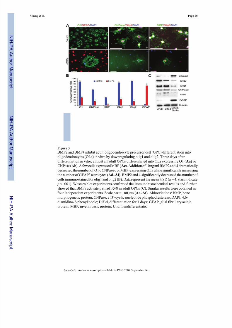
BMP2 and BMP4 Suppress OL Differentiation and Promote Astrocyte Differentiation by Adult
OPCs
Previous studies showed that either BMP2 or BMP4 increased astrocyte differentiation and inhibited OL differentiation of neural stem cells or OPCs isolating from embryonic or postnatal
developing CNS [17–19,21,22]. Whether BMPs have similar effects on adult OPCs has not
been determined. When adult OPCs were differentiated without BMP2 or BMP4 for 3 days,
almost all of the cells differentiated into O1+ OLs. No astrocyte differentiation was observed
(Fig. 2Ba). After addition of BMP2, BMP4, or both in the differentiation medium, the number
of OPCs that differentiated into O1+ OLs dramatically decreased, with concurrent increases
of GFAP+ astrocytes (Fig. 2Bb–2Bd). The number of OPCs that differentiated into mature
MBP+ OLs also decreased (Fig. 2Be–2Bh). To determine dose-dependent effects on the
differentiation of adult OPCs, increasing concentrations of BMP2, 4, or both 2 and 4 were
added to the medium. As shown in Figure 2C, when differentiated for 3 days in the presence
of BMP2 at concentrations of 1, 2, 5, or 10 ng/ml, the percentage of adult OPCs that
differentiated into GFAP+ astrocytes significantly increased from 0.4% at control to 14%, 18%,
25%, and 46%, respectively ( p < .001 at all four concentrations). The percentage of O1+ OLsdecreased from 77% to 60%, 45%, 34%, and 19% ( p < .001), respectively, and MBP+ OLs
also were suppressed from 58% to 60%, 45%, 34%, and 19% ( p < .01), respectively. BMP4
was more potent than BMP2 in the inhibition of OL differentiation and promotion of astrocyte
differentiation. In the presence of BMP4 at 1, 2, 5, and 10 ng/ml, the number of OPCs that
differentiated into GFAP+ astrocytes were 35%, 44%, 66%, and 75%, respectively, all being
significantly increased compared with 0.4% in control cultures (all p < .001). The number of
O1+ and MBP+ OLs were 20%, 13%, 6%, 1% and 21%, 9%, 2%, 1%, respectively, all
significantly decreased compared with 77% and 58% (all p < .001). In the presence of BMP2
and 4 at the same concentrations, further increases in the number of GFAP+ astrocytes and
decreases of O1+ or MBP+ OLs were observed (Fig. 2C). These findings were confirmed by
Western blot experiments (Fig. 2D). Moreover, we found that the expression of pSmad1/5/8
and ID4, two intracellular downstream targets of BMP signaling, was also increased in
proportion with the dosage of BMP2, 4, or 2 and 4, suggesting that BMP signaling inhibits OLdifferentiation and promotes astrocyte differentiation of adult OPCs through Smad-dependent
pathways.
Cheng et al. Page 6
Stem Cells. Author manuscript; available in PMC 2009 September 14.
NI H-P A A
ut h or Manus c r i pt
NI H-P A A ut h or Manus c r i pt
NI H-P A A ut h or
Manus c r i pt
8/11/2019 Bone Morphogenetic Protein Signaling and Interact to Regulate the Differentiation and Maturation of Adult Oligode…
BMP2 and BMP4 Downregulate the Expression of o lig1 and o lig2 During Adult OPC
Differentiation
To determine the potential mechanism through which BMPs affect the differentiation of adult
OPCs, we examined the expression of olig1 and olig2, two bHLH transcription factors
important for OL development. When differentiating for 3 days in the absence of BMP2 and
4, the majority of cells developed into OLs (Fig. 3Aa) that expressed olig1 (Fig. 3Ab) and olig2
(Fig. 3Ac). Addition of BMP2 and 4, however, significantly decreased the number of olig1-
expressing cells from 79% to 15% ( p < .001) and olig2-expressing cells from 76% to 15% ( p< .001) in addition to dramatically increasing the number of GFAP+ astrocytes and decreasing
O1+, CNPase+, or MBP+ OLs (Fig. 3Ad–3Af, 3B). Western blot experiments confirmed that
addition of BMP2 and 4 in the differentiating medium activated Smad1/5/8 to inhibit the
expression of olig1, olig2, and MBP and to promote the expression of GFAP (Fig. 3C).
Previous studies have shown that adult OPCs change their responses to growth factors after
culturing for a long time in the presence of FGF2 and PDGFaa [41]. To address this possibility,
we tested the effects of BMP2 and 4 on differentiation of freshly isolated OPCs from adult
spinal cord. As shown in supplemental online Figure 1, all OPCs expressed A2B5
(supplemental online Fig. 1A) and O4 (supplemental online Fig. 1B). After differentiation
without FGF2 and PDGFaa for 3 days, most OPCs differentiated into OLs expressing O1
online Fig. 2E). No GFAP+ astrocytes were observed (supplemental online Fig. 2A). Additionof BMP2 and 4 dramatically increased the number of GFAP+ astrocytes (supplemental online
Fig. 2B) and simultaneously decreased the number of O1+ (supplemental online Fig. 2B),
CNPase+ (supplemental online Fig. 2D), or MBP+ (supplemental online Fig. 2E) OLs. The
number of cells expressing Olig2 (supplemental online Fig. 2C, 2D) or Olig1 (supplemental
online Fig. 2E, 2F) was also significantly decreased by the treatment of BMP2 and 4. These
data indicate that the effects of BMP2 and 4 on the differentiation of freshly isolated adult
OPCs are identical to their effects on OPCs of passages 4–7, which we used in most of our
present study. Thus, exposure to FGF2 and PDGFaa in vitro does not change the responses of
adult OPCs to BMP2 and 4.
BMP2 and 4 Inhibit OL Maturation
The findings that immature O1+ OLs expressed BMP receptors (Fig. 2) and that the number of MBP+ OL decreased after the treatment with BMP2 and 4 suggested that BMP might also
inhibit the maturation of OL from immature O1+ to more mature MBP+ stages. To determine
the effects of BMP2 and 4 on OL maturation, adult OPCs were allowed to differentiate into
O1+ OLs by withdrawing FGF2 and PDGFaa for 2 days and then continued to mature for 3
more days in the absence or presence of BMP2 and 4. As shown previously, when
differentiating for 2 days after withdrawal of FGF2 and PDGFaa, more than 75% of adult OPCs
developed into O1+ OLs; after 3 more days, more than 95% of cells became O1+ OLs (Fig.
4Aa), and 90% of OPCs matured into CNPase+ (Fig. 4Ab) or MBP+ (Fig. 4Ac) OLs. When
10 ng/ml BMP2 and 4 were added for the last 3 days of differentiation, a significant decrease
in the number of OLs expressing either MBP+ (from 95% to 64%) or CNPase (from 93% to
58%; p < .001) was observed (Fig. 4Ad–4Af, 4B). However, the number of O1+ OLs (from
98–89%; p > .05) or GFAP+ astrocytes (from 1.1% to 5.6%; p > .05) did not significantly
change (Fig. 4Aa, 4Ad, 4B). These results showed that BMP2 and 4 inhibited the maturationof O1+ into MBP+ or CNPase+ OLs.
We further examined the expression of olig1 and olig2 during the maturation of adult OPCs.
When differentiating in the presence of BMP2 and 4, the number of olig1+ cells significantly
decreased from 79% at 5 days after differentiation without BMP2 and 4 to 49% ( p < .01) (Fig.
4Ab, 4Ae, 4B, 4C). The number of olig2+ cells was also decreased from 80% at control to
Cheng et al. Page 7
Stem Cells. Author manuscript; available in PMC 2009 September 14.
NI H-P A A
ut h or Manus c r i pt
NI H-P A A ut h or Manus c r i pt
NI H-P A A ut h or
Manus c r i pt
8/11/2019 Bone Morphogenetic Protein Signaling and Interact to Regulate the Differentiation and Maturation of Adult Oligode…
45% ( p < .05) (Fig. 4Ac, 4Af, 4B, 4C). BMP2 and 4 also inhibited the development of both
CNPase- or MBP-expressing mature OLs, as shown by both immunohistochemistry (Fig. 4A,
4B) and Western blot (Fig. 4C). Our results showed that BMP2 and 4 specifically decreased
the expression of both olig1 and olig2 and subsequently inhibited OL differentiation and
maturation of adult OPCs.
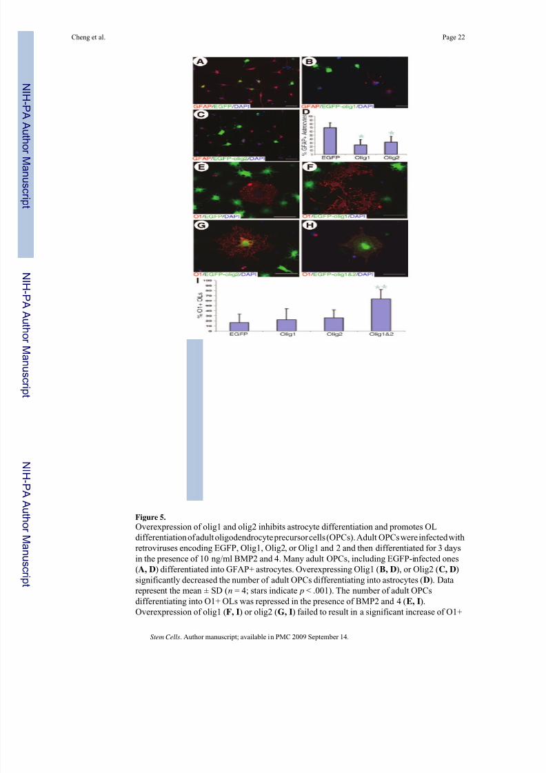
Overexpression of olig1 and ol ig2 Blocks the BMP-Mediated Inhibition of Adult OPC
DifferentiationWhen differentiating in the presence of BMP2 and 4 for 3 days, 70% of EGFP-expressing
OPCs became GFAP+ astrocytes (Fig. 5A, 5D). Astrocyte differentiation in olig1- or olig2-
expressing OPCs was dramatically decreased to 25% ( p < .001) and 32% ( p < .001),
respectively (Fig. 5B–5D). However, the percentage of O1+ OLs derived from olig1- or olig2-
expressing OPCs (22% and 26%, respectively) after 3 days of differentiation in the presence
of BMP2 and 4 was slightly but not significantly increased compared with 17% of EGFP-OPCs
(both p > .05) (Fig. 5E–5I). These results indicate that overexpression of olig1 or olig2 is
sufficient to block the astrocyte differentiation of adult OPCs induced by BMP2 and 4.
However, overexpression of either olig1 or olig2 failed to block the inhibition of BMP2 and 4
on OL differentiation of adult OPCs. Since BMP2 and 4 downregulate the expression of both
olig1 and olig2 in the adult OPCs, overexpression of both olig1 and olig2, not olig1 or olig2
alone, may be necessary to overcome the inhibition of BMP2 and 4 on OL differentiation of
adult OPCs. To test this hypothesis, we infected adult OPCs with retroviruses encoding both
olig1 and olig2 and then differentiated the cells for 3 days in the presence of BMP2 and 4. As
shown in Figure 5H and 5I, the percentage of O1+ OLs in olig1/olig2-expressing adult OPCs
was 63%, which was significantly increased compared with 17%, 22% or 26% in EGFP-,
olig1-, or olig2-expressing adult OPCs, respectively (all p < .001).
Expression of Both o lig1 and olig2 Is Necessary for OL Differentiation o f Adul t OPCs
To further elucidate the role(s) of olig1 and olig2 in the differentiation of adult OPCs, we tested
whether downregulation of olig1 or olig2 by specific siRNA would inhibit OL differentiation
of adult OPCs. We first tested whether siRNAs were able to efficiently downregulate the
expression of olig1 or olig2 in the adult OPCs. Expression of either olig2 (Fig. 6A–6C) or olig1
(Fig. 6D–6F) in adult OPCs infected with retroviruses encoding control scrambled siRNA did
not change. However, the expression of olig2 was dramatically decreased in these adult OPCsinfected with retroviruses encoding olig2 siRNA (Fig. 6A′ –6C′). Expression of olig1, however,
did not change in these olig2 siRNA OPCs (data not shown). Similarly, adult OPCs expressing
olig1 siRNA significantly downregulated its expression of olig1 (Fig. 6D′ –6F′) but not olig2
(data not shown). These results demonstrated that olig1 or olig2 siRNA specifically knocked
down the expression of olig1 and olig2 in adult OPCs, respectively. We then examined whether
OL differentiation of adult OPCs was inhibited following the downregulation of olig1 or olig2
with specific siRNA. Adult OPCs infected with retroviruses encoding either control scrambled,
olig1, or olig2 siRNA were differentiated for 3 days. In the control group, more than 90% of
infected adult OPCs differentiated into O1+ OLs (Fig. 6G, 6J). O1+ OLs, however,
significantly decreased in olig1 siRNA OPCs (35%; p < .001) (Fig. 6I, 6J) or in olig2 siRNA
OPCs (59%; p < .05) (Fig. 6H, 6J). These results showed that both olig1 and olig2 were
necessary for OL differentiation of adult OPCs. No GFAP+ astrocyte differentiation was
observed in either EGFP, olig1, or olig2 siRNA OPCs after 3 days differentiation withoutBMP2 and 4 (data not shown).
Expression of BMP2 and 4 and olig1 and 2 in the Injured Spinal Cord
Obvious expression of BMP2 or BMP4 was not observed in the normal adult spinal cord
(supplemental online Fig. 3A, 3D). At 7 days after contusion, expression of BMP2 and BMP4
Cheng et al. Page 8
Stem Cells. Author manuscript; available in PMC 2009 September 14.
NI H-P A A
ut h or Manus c r i pt
NI H-P A A ut h or Manus c r i pt
NI H-P A A ut h or
Manus c r i pt
8/11/2019 Bone Morphogenetic Protein Signaling and Interact to Regulate the Differentiation and Maturation of Adult Oligode…
online Fig. 4B) and 1 month (supplemental online Fig. 4C) after the contusion, the number of NG+ OPCs significantly increased in the injured spinal cord, but the number of olig1+ cells
decreased. Most of NG2+ OPCs did not express olig1 (supplemental online Fig. 4B, 4C, inset,
arrowheads), whereas some OPCs remained olig1-positive (supplemental online Fig. 4B, 4C,
inset, arrows). OPCs in the adult spinal cord also expressed olig2 in their nuclei (supplemental
online Fig. 4D, inset, arrow). At 1 week postinjury, the number of olig2+ cells increased in the
injured spinal cord, and its expression in some cells was upregulated (supplemental online Fig.
4E). However, many adult OPCs did not express olig2 (supplemental online Fig. 4E, inset,
arrowheads). The number of olig2+ cells and its expression level decreased in the injured spinal
cord at 1 month postinjury compared with 1 week postinjury (supplemental online Fig. 4F).
More OPCs did not express olig2 (supplemental online Fig. 4F, inset, arrowheads).
Discussion
OPCs in the Adult Spinal Cord
We obtained highly purified OPCs from the adult rat spinal cord that share many fundamental
properties with OPCs from perinatal and adult optic nerve or subcortical white matter. They
express similar phenotype-specific antigens, O4, A2B5, and NG2. OPCs cultured from adult
rat optical nerve [47] or human cortical white matter [48,49] express O4. Approximately 80%
of acutely isolated cycling progenitor cells in the adult mammalian white matter are O4+
[50]. OPCs in the adult spinal cord also express A2B5 and NG2, which is in accord with
previous results from adult optic nerve [39,47]. In the intact adult spinal cord, more than 70%
of BrdU+ cycling precursor cells express NG2 [23]. Therefore, the purified A2B5+/NG2+/O4
+ OPCs in this study are the most actively proliferative precursor cells in the normal adult
spinal cord.
Consistent with present data with adult spinal cord OPCs, OPCs from postnatal seven days ratoptic nerve can proliferate for many passages without differentiation in the presence of PDGFaa
and in the absence of thyroid hormone [51,52]. FGF2 can inhibit the differentiation of OPCs
and promote their proliferation [49,53] and can convert slowly dividing adult OPCs to rapidly
dividing cells with characteristics of perinatal OPCs [41]. Moreover, the adult spinal cord OPCs
differentiate into OLs in the serum-free medium and into type 2 astrocytes in medium
containing serum, a property of OPCs from other CNS regions [54,55]. The fact that adult
spinal cord OPCs can be induced to differentiate into different developmental stages of OLs
by withdrawing FGF2 and PDGFaa for certain times (Fig. 1) establishes a stable adult spinal
cord OPC model to investigate potential molecular mechanism(s) by which adult OPCs
proliferate and differentiate in vitro.
BMP Signaling Inhibits Oligodendrogenesis and Promotes Astrogliogenesis from Adult
OPCs
In vitro, BMP signaling enhances astrocyte differentiation and inhibits OL differentiation from
cultured neural stem cells or OPCs isolated from embryonic or perinatal CNS [16,17,21,22,
56]. In vivo, BMP signaling also represses OL development [11,13,57]. Furthermore,
overexpression of BMP4 in transgenic mice under the control of the neuron-specific enolase
promoter results in a significant decrease in OLs, with a concurrent increase in astrocytes
Cheng et al. Page 9
Stem Cells. Author manuscript; available in PMC 2009 September 14.
NI H-P A A
ut h or Manus c r i pt
NI H-P A A ut h or Manus c r i pt
NI H-P A A ut h or
Manus c r i pt
8/11/2019 Bone Morphogenetic Protein Signaling and Interact to Regulate the Differentiation and Maturation of Adult Oligode…
[15]. The present study extends these previous findings to show that BMP signaling also
inhibits the differentiation and maturation of adult OPCs, suggesting that BMP signaling may
play an important role in the regulation of remyelination of the adult CNS following traumatic
or demyelinating injuries.
Importantly, present data document the molecular mechanism(s) by which BMP signaling
inhibits OPC differentiation into mature OLs. One possible mechanism is the inhibition of
olig1/2 activity by ID4 (Fig. 2), as suggested by a previous study [20]. Although ID4 and ID2are expressed in proliferating OPCs, their expression decreases progressively as OPCs
differentiate and mature [20,58,59]. Overexpression of ID2 or ID4 in OPCs inhibits OL
differentiation, whereas downregulation of their expression induces premature differentiation
in vitro [20,58,59]. ID2 and ID4 bind olig1 and olig2 in the cytoplasm of OPCs to prevent their
translocation to nucleus, thereby preventing olig1 and olig2 from binding target DNA [20].
The present study further suggests that an additional mechanism by which BMP signaling
inhibits OL differentiation from adult OPC is the downregulation of olig1 and olig2 (Figs. 2–
5). That overexpression of olig1 and olig2 reverses the inhibitory effects of BMP signaling on
OL differentiation of OPCs further confirms the suggestion that BMP signaling inhibits
oligodendrogenesis from OPCs by downregulating olig1 and olig2. Therefore, downregulation
of olig1 and olig2 expression and inhibition of olig1/2 activity by increased ID4 may work
synergistically to inhibit OL differentiation and to enhance astrogliogenesis (Fig. 7).
Olig1 and olig2 in the Differentiation o f Adult OPCs
Transcriptional regulation orchestrates oligodendrogenesis during CNS development [1,2,
60]. Olig1 and olig2 play important roles in generating OLs during embryogenesis [9,10,61,
62]. Olig2 is required for OL lineage specification during CNS development, whereas olig1
contributes more to OL differentiation and maturation [9,10,63,64]. However, olig2
compensates for the function of olig1 during OL development, since OL differentiation and
myelination are delayed but eventually reach normal level in olig1-null mice [9,10]. In contrast
to embryonic development, adult OPCs require both olig1 and olig2 for their differentiation
and maturation. Downregulation of either olig1 or olig2 by siRNA inhibits adult OPCs
differentiation into OLs (Fig. 6). Overexpression of olig1 or olig2 alone fails to overcome the
inhibition of BMP to promote OL differentiation since BMP signaling decreases the expression
of both olig1 and olig2. However, overexpression of both olig1 and olig2 enhances OL
differentiation even in the presence of BMP2 and 4 (Fig. 5). These results are consistent with
data from Arnett et al. [42], which show that olig1 is required not for the development of OLs
but for remyelination in adult mice following demyelination. Taken together, these data
indicate that olig1 is required for the differentiation and maturation of adult OPCs and
endogenous remyelination after demyelination in adult CNS. Whether olig2 plays an important
role in the differentiation and maturation and myelination of OLs during development is not
known, since olig2-null mice die during the perinatal period. Interestingly, olig2 expression is
retained during OL maturation, suggesting its possible role in OL differentiation. Consistent
with this idea, our in vitro analyses show that olig2 is also required for OL differentiation and
maturation from adult OPCs. These data also suggest that it may have a potential role during
remyelination. Conditional olig2 knockout mice will be very useful to study its effects on
remyelination in vivo.
In addition to OL development, olig1 and olig2 also play an important role in astrogliogenesis.
Overexpression of olig1 or olig2 significantly inhibits astrocyte differentiation of adult OPCs
induced by BMP signaling, suggesting that either olig1 or olig2 is sufficient to repress
astrogliogenesis. These results are in good accord with the previous observation of Lu et al.
[10], which show that the null mutation of either olig1 or olig2 alone is not sufficient to increase
astrogliogenesis in the spinal cord. The double-null mutation of olig1 and olig2, however,
Cheng et al. Page 10
Stem Cells. Author manuscript; available in PMC 2009 September 14.
NI H-P A A
ut h or Manus c r i pt
NI H-P A A ut h or Manus c r i pt
NI H-P A A ut h or
Manus c r i pt
8/11/2019 Bone Morphogenetic Protein Signaling and Interact to Regulate the Differentiation and Maturation of Adult Oligode…
results in an apparent increase in astrogliogenesis, with a complete failure of OL development
[9]. Thus, downregulation of both olig1 and olig2 is required for OPCs to differentiate into
astrocytes since olig1 and olig2 could complement the functions of one another sufficient to
inhibit astrocyte differentiation. Consistent with this notion, we observe that both olig1 and
olig2 are dramatically downregulated when astrocyte differentiation from adult OPCs is
induced by BMP signaling. Our results further show that downregulation of olig1 or olig2
alone by siRNA fails to result in enhanced astrocyte differentiation even though OL
differentiation is inhibited, suggesting that repression of OL differentiation alone is notsufficient to cause the astrocyte differentiation from adult OPCs if BMP signaling is not present.
BMP signaling activates Smad proteins, which translocate into the nucleus to form a complex
with p300 to directly activate the GFAP promoter [65,66]. Olig2 interacts with p300 to interrupt
the complex of p300 and Smad1 and thereby represses the astrocyte-specific GFAP promoter
[67]. Because of their structural similarity, it is possible that olig1 may use the same mechanism
to inhibit astrocyte differentiation. This would suggest that BMP signaling is able to induce
adult OPCs to differentiate into astrocytes through the activation of GFAP promoter by the
Smad-p300 complex only when olig1 and olig2 are also down-regulated (Fig. 7).
Downregulation of both olig1 and olig2 is required, but not sufficient, for astrocyte
differentiation from adult OPCs.
Implications for Remyelination
OPCs reside quiescently in the white matter of the adult CNS [23,24]. They become activated
and proliferate in response to CNS injury, including demyelination [25–28]. However, adult
OPCs fail to differentiate and mature into OL capable of remyelinating demyelinated axons in
multiple sclerosis lesions even though endogenous OPCs are present in the demyelinated areas
and some even closely contact the affected axons [36,38,68–70]. Inhibitory signals that prevent
OPCs from differentiation and maturation are present in the demyelinated lesions [71,72]. We
suggest that BMP signaling may be an important factor that inhibits OL differentiation and
maturation in the injured CNS. In fact, expression of BMPs dramatically increases after CNS
injury [73,74]. Moreover, neuronal and OL differentiation from engrafted neural stem cells in
the injured spinal cord is increased after blocking BMP signaling by expressing noggin [73].
Consistent with those findings, we also observed a significant increase of BMP2 and 4 in the
hypertrophic astrocytes after traumatic SCI (supplemental online Fig. 3), and expression of
olig1 and olig2 in the OPCs of the injured spinal cord decreased (supplemental online Fig. 4).Manipulation of BMP signaling may provide a new therapeutic strategy to enhance
remyelination from endogenous or/and grafted OPCs after multiple sclerosis or SCI.
Supplementary Material
Refer to Web version on PubMed Central for supplementary material.
Acknowledgments
This work was supported by National Center for Research Resources Grant 5P20 RR 015576 (to Q.C. and S.R.W.),
NIH R01 NS37717 (to M.Q.), the Kentucky Spinal Cord and Head Injury Research Trust (to Q.C. and S.R.W.), Norton
Healthcare, the Commonwealth of Kentucky Research Challenge for Excellence Trust Fund, and The Christopher
Reeve Paralysis Foundation (to S.R.W.). We thank George Harding of the Anatomical Sciences and Neurobiology,
University of Louisville School of Medicine, for expert technical help.
References
1. Miller RH. Regulation of oligodendrocyte development in the vertebrate CNS. Prog Neurobiol
2002;67:451–467. [PubMed: 12385864]
2. Rowitch DH. Glial specification in the vertebrate neural tube. Nat Rev Neurosci 2004;5:409–419.
[PubMed: 15100723]
Cheng et al. Page 11
Stem Cells. Author manuscript; available in PMC 2009 September 14.
NI H-P A A
ut h or Manus c r i pt
NI H-P A A ut h or Manus c r i pt
NI H-P A A ut h or
Manus c r i pt
8/11/2019 Bone Morphogenetic Protein Signaling and Interact to Regulate the Differentiation and Maturation of Adult Oligode…
11. Hall AK, Miller RH. Emerging roles for bone morphogenetic proteins in central nervous system glial
biology. J Neurosci Res 2004;76:1–8. [PubMed: 15048925]
12. Agius E, Soukkarieh C, Danesin C, et al. Converse control of oligodendrocyte and astrocyte lineagedevelopment by Sonic hedgehog in the chick spinal cord. Dev Biol 2004;270:308–321. [PubMed:
15183716]
13. Mekki-Dauriac S, Agius E, Kan P, et al. Bone morphogenetic proteins negatively control
oligodendrocyte precursor specification in the chick spinal cord. Development 2002;129:5117–5130.
[PubMed: 12399304]
14. Miller RH, Dinsio K, Wang R, et al. Patterning of spinal cord oligodendrocyte development by
dorsally derived BMP4. J Neurosci Res 2004;76:9–19. [PubMed: 15048926]
15. Gomes WA, Mehler MF, Kessler JA. Transgenic overexpression of BMP4 increases astroglial and
decreases oligodendroglial lineage commitment. Dev Biol 2003;255:164–177. [PubMed: 12618141]
16. Gross RE, Mehler MF, Mabie PC, et al. Bone morphogenetic proteins promote astroglial lineage
commitment by mammalian subventricular zone progenitor cells. Neuron 1996;17:595–606.
[PubMed: 8893018]
17. Mabie PC, Mehler MF, Kessler JA. Multiple roles of bone morphogenetic protein signaling in theregulation of cortical cell number and phenotype. J Neurosci 1999;19:7077–7088. [PubMed:
10436062]
18. Zhu G, Mehler MF, Zhao J, et al. Sonic hedgehog and BMP2 exert opposing actions on proliferation
and differentiation of embryonic neural progenitor cells. Dev Biol 1999;215:118–129. [PubMed:
10525354]
19. Nakashima K, Takizawa T, Ochiai W, et al. BMP2-mediated alteration in the developmental pathway
of fetal mouse brain cells from neurogenesis to astrocytogenesis. Proc Natl Acad Sci U S A
2001;98:5868–5873. [PubMed: 11331769]
20. Samanta J, Kessler JA. Interactions between ID and OLIG proteins mediate the inhibitory effects of
BMP4 on oligodendroglial differentiation. Development 2004;131:4131–4142. [PubMed:
15280210]
21. Mabie PC, Mehler MF, Marmur R, et al. Bone morphogenetic proteins induce astroglial differentiation
of oligodendroglial-astroglial progenitor cells. J Neurosci 1997;17:4112–4120. [PubMed: 9151728]22. Grinspan JB, Edell E, Carpio DF, et al. Stage-specific effects of bone morphogenetic proteins on the
44. Cao Q, Zhang YP, Iannotti C, et al. Functional and electrophysiological changes after graded traumaticspinal cord injury in adult rat. Exp Neurol 2005;191(suppl 1):S3–S16. [PubMed: 15629760]
45. Cao Q, Xu XM, DeVries WH, et al. Functional recovery in traumatic spinal cord injury after
transplantation of multineurotrophin-expressing glial-restricted precursor cells. J Neurosci
2005;25:6947–6957. [PubMed: 16049170]
46. McTigue DM, Wei P, Stokes BT. Proliferation of NG2-positive cells and altered oligodendrocyte
numbers in the contused rat spinal cord. J Neurosci 2001;21:3392–3400. [PubMed: 11331369]
Cheng et al. Page 13
Stem Cells. Author manuscript; available in PMC 2009 September 14.
NI H-P A A
ut h or Manus c r i pt
NI H-P A A ut h or Manus c r i pt
NI H-P A A ut h or
Manus c r i pt
8/11/2019 Bone Morphogenetic Protein Signaling and Interact to Regulate the Differentiation and Maturation of Adult Oligode…
53. Grinspan JB, Reeves MF, Coulaloglou MJ, et al. Re-entry into the cell cycle is required for bFGF-
induced oligodendroglial dedifferentiation and survival. J Neurosci Res 1996;46:456–464. [PubMed:
8950705]
54. Noble M, Wolswijk G, Wren D. The complex relationship between cell division and the control of
differentiation in oligodendrocyte-type-2 astrocyte progenitor cells isolated from perinatal and adult
rat optic nerves. Prog Growth Factor Res 1989;1:179–194. [PubMed: 2491261]
55. Raff MC, Miller RH, Noble M. A glial progenitor cell that develops in vitro into an astrocyte or an
oligodendrocyte depending on culture medium. Nature 1983;303:390–396. [PubMed: 6304520]56. See J, Zhang X, Eraydin N, et al. Oligodendrocyte maturation is inhibited by bone morphogenetic
62. Lu QR, Yuk D, Alberta JA, et al. Sonic hedgehog–regulated oligodendrocyte lineage genes encoding bHLH proteins in the mammalian central nervous system. Neuron 2000;25:317–329. [PubMed:
10719888]
63. Lu QR, Cai L, Rowitch D, et al. Ectopic expression of Olig1 promotes oligodendrocyte formation
and reduces neuronal survival in developing mouse cortex. Nat Neurosci 2001;4:973–974. [PubMed:
11574831]
64. Takebayashi H, Nabeshima Y, Yoshida S, et al. The basic helix-loop-helix factor olig2 is essential
for the development of motoneuron and oligodendrocyte lineages. Curr Biol 2002;12:1157–1163.
[PubMed: 12121626]
65. Nakashima K, Yanagisawa M, Arakawa H, et al. Synergistic signaling in fetal brain by STAT3-Smad1
complex bridged by p300. Science 1999;284:479–482. [PubMed: 10205054]
66. Fukuda S, Taga T. Cell fate determination regulated by a transcriptional signal network in the
developing mouse brain. Anat Sci Int 2005;80:12–18. [PubMed: 15794126]
67. Fukuda S, Kondo T, Takebayashi H, et al. Negative regulatory effect of an oligodendrocytic bHLHfactor OLIG2 on the astrocytic differentiation pathway. Cell Death Differ 2004;11:196–202.
[PubMed: 14576772]
68. Chang A, Tourtellotte WW, Rudick R, et al. Premyelinating oligodendrocytes in chronic lesions of
multiple sclerosis. N Engl J Med 2002;346:165–173. [PubMed: 11796850]
69. Wolswijk G. Chronic stage multiple sclerosis lesions contain a relatively quiescent population of
Isolation and differentiation of adult oligodendrocyte precursor cells (OPCs) in vitro. Adult
OPCs were purified from the spinal cord of adult rats by immunopanning with the O4 antibody.The proliferating precursors displayed a characteristic adult OPC morphology with several
small short processes emanating from a small round cell body (A). All cells expressed O4
(B), A2B5 (B), and NG2 (D, E). In the presence of fibroblast growth factor 2 (FGF2) and
platelet-derived growth factor aa (PDGFaa), adult OPCs divided and are readily labeled by
BrdU (B, C). These OPCs proliferated for multiple passages without changing phenotypes
(F). Data in (F) were the mean ± SD of four independent experiments. Three days after
withdrawal of FGF2 and PDGFaa, OPCs constitutively differentiated into OLs expressing O1
Cheng et al. Page 16
Stem Cells. Author manuscript; available in PMC 2009 September 14.
NI H-P A A
ut h or Manus c r i pt
NI H-P A A ut h or Manus c r i pt
NI H-P A A ut h or
Manus c r i pt
8/11/2019 Bone Morphogenetic Protein Signaling and Interact to Regulate the Differentiation and Maturation of Adult Oligode…
Inhibition of oligodendrocyte (OL) differentiation and promotion of astrocyte differentiation
of adult oligodendrocyte precursor cells (OPCs) by BMP2 and 4. Adult OPCs expressed BMPR Ia (Aa), Ib (Ab), and II (Ac). Although they were slightly downregulated, all three BMPRs
were persistently expressed in immature O1+ and more mature MBP+ OLs after Dif3d and
Dif5d, respectively (Ad). Treatment with BMP2, 4, or both decreased the number of O1+ OLs,
with concurrent increases in the number of GFAP+ astrocytes (Ba – Bd). The number of MBP
+ OLs was also decreased (Be – Bh). Quantification of cells immunostained for GFAP, O1, or
MBP showed a dose-dependent increase in the number of GFAP+ astrocytes and decrease of
O1+ or MBP+ OLs differentiated from adult OPCs by BMP2, 4, or both, respectively (C). Data
Cheng et al. Page 18
Stem Cells. Author manuscript; available in PMC 2009 September 14.
NI H-P A A
ut h or Manus c r i pt
NI H-P A A ut h or Manus c r i pt
NI H-P A A ut h or
Manus c r i pt
8/11/2019 Bone Morphogenetic Protein Signaling and Interact to Regulate the Differentiation and Maturation of Adult Oligode…
Overexpression of olig1 and olig2 inhibits astrocyte differentiation and promotes OL
differentiation of adult oligodendrocyte precursor cells (OPCs). Adult OPCs were infected withretroviruses encoding EGFP, Olig1, Olig2, or Olig1 and 2 and then differentiated for 3 days
in the presence of 10 ng/ml BMP2 and 4. Many adult OPCs, including EGFP-infected ones
(A, D) differentiated into GFAP+ astrocytes. Overexpressing Olig1 (B, D), or Olig2 (C, D)
significantly decreased the number of adult OPCs differentiating into astrocytes (D). Data
represent the mean ± SD (n = 4; stars indicate p < .001). The number of adult OPCs
differentiating into O1+ OLs was repressed in the presence of BMP2 and 4 (E, I).
Overexpression of olig1 (F, I) or olig2 (G, I) failed to result in a significant increase of O1+
Cheng et al. Page 22
Stem Cells. Author manuscript; available in PMC 2009 September 14.
NI H-P A A
ut h or Manus c r i pt
NI H-P A A ut h or Manus c r i pt
NI H-P A A ut h or
Manus c r i pt
8/11/2019 Bone Morphogenetic Protein Signaling and Interact to Regulate the Differentiation and Maturation of Adult Oligode…
Inhibition of adult oligodendrocyte precursor cell (OPC) differentiation into oligodendrocytes
(OLs) following downregulation of Olig2 and Olig1. Olig2 and Olig1 siRNA downregulated
the expression of Olig2 and Olig1 in adult OPCs. Adult OPCs were infected with retrovirusesthat express either scrambled negative control (A–F), Olig2 (A′–C′), or Olig1 (D′–F′) siRNA.
Two days later, control adult OPCs did not alter their expression of Olig2 ([A–C], arrows) or
Olig1 ([D–G], arrows). However, adult OPCs infected with retroviruses expressing either
Olig2 or Olig1 siRNA downregulated their expression of Olig2 ([A′–C′], arrows) or Olig1
([D′–G′], arrows), respectively. Adult OPCs were differentiated for 3 days following infection
with retroviruses encoding negative control, Olig2, or Olig1 siRNA. Uninfected OPCs ([G–
Cheng et al. Page 24
Stem Cells. Author manuscript; available in PMC 2009 September 14.
NI H-P A A
ut h or Manus c r i pt
NI H-P A A ut h or Manus c r i pt
NI H-P A A ut h or
Manus c r i pt
8/11/2019 Bone Morphogenetic Protein Signaling and Interact to Regulate the Differentiation and Maturation of Adult Oligode…
A schematic illustration of the regulation of adult oligodendrocyte precursor cell (OPC)
differentiation by the interaction of BMP signaling and olig1/2. BMPs upregulate or downregulate the expression of ID4 and olig1/2, respectively, by Smad-dependent or -
independent pathways. Increasing the expression of ID4, which blocks the translocation of
olig1 and 2 to the nucleus, works synergistically with the downregulation of olig1 and 2 to
inhibit oligodendrocyte (OL) differentiation and permit and/or potentiate astrocyte
differentiation following BMP signaling. Olig1 and 2 are necessary to promote OL
differentiation and sufficient to inhibit astrocyte differentiation by binding p300 to interrupt
the formation of the Smad/p300 complex, which activates the GFAP promoter. Therefore,
overexpression of olig1 and 2 overcomes the effects of BMP signaling on adult OPCs to
promote OL differentiation and inhibit astrocyte differentiation. Abbreviations: BMP, bone
morphogenetic protein; BMPR, bone morphogenetic protein receptor.
Cheng et al. Page 26
Stem Cells. Author manuscript; available in PMC 2009 September 14.