Bone status in genetic syndromes: A review Stefano Stagi, 1 Chiara Iurato, 1 Elisabetta Lapi, 2 Loredana Cavalli, 3 Maria Luisa Brandi, 3 Maurizio de Martino 1 1 Health Sciences Department, University of Florence, Anna Meyer Children’s University Hospital, 2 Genetics and Molecular Medicine Unit, Anna Meyer Children’s University Hospital, 3 Department of Internal Medicine, Endocrinology Unit, University of Florence, Florence, Italy ABSTRACT More and more data seem to indicate the presence of an increasing number of syndromes and genetic diseases characterized by impaired bone mass and quality. Meanwhile, the improve- ment of etiopathogenetic knowledge and the employment of more adequate treatments have generated a significant increase in survival related to these syndromes and diseases. It is thus important to identify and treat bone impairment in these patients in order to assure a better quality of life. This review provides an updated overview of bone pathophysiology and charac- teristics in patients with Down, Turner, Klinefelter, Marfan, Williams, Prader-Willi, Noonan, and 22q11 deletions syndrome. In addition, some options for the treatment of the bone status impairment in these patients will be briefly discussed. Key words: Bone mineral density, Fragility fractures, Genetic syndrome, Osteoporosis, Syndromes Review HORMONES 2015, 14(1):19-31 Address for correspondence: Stefano Stagi, MD, Department of Health Sciences, University of Florence, Anna Meyer Children’s University Hospital, Florence, Italy; Tel.: +39-055-8451210; Fax: +39-055-8451211; E-mail: [email protected]Received: 14-09-2014, Accepted: 13-01-2015 because it enables increased survival and enhanced quality of life. The process of bone turnover, which is crucial to ensure optimal bone quality and mass, is regulated by local or/and systemic factors. 1,2 An impairment of bone resorption or a defect in quality or quantity of neo-synthesis processes is, by contrast, likely to lead to a condition of reduced gain or loss of bone mass. 1,2 Indeed, many data show that bone diseases may originate at very early stages of life. 3 In fact, from the first weeks of intrauterine life, bone presents a progressive increase until reaching a maximum value called peak bone mass (PBM) from 16-18 years (e.g., for the spine and femoral neck) and up to 35 years (for the skull). 4,5 The achievement of this value is influenced by the INTRODUCTION Improved research and scientific data, particularly in the last few years, have helped to explore the presence of altered bone density and quality in an increasing number of syndromes and genetic diseases. These developments, associated with increasing etiopathoge- netic knowledge and more adequate treatments, have led to emphasis being placed on the importance of identifying and treating bone impairments as early as possible. This is, of course, of particular significance
Transcript
Bone status in genetic syndromes: A review
Stefano Stagi,1 Chiara Iurato,1 Elisabetta Lapi,2 Loredana Cavalli,3 Maria Luisa Brandi,3 Maurizio de Martino1
1Health Sciences Department, University of Florence, Anna Meyer Children’s University Hospital, 2Genetics and Molecular Medicine Unit, Anna Meyer Children’s University Hospital, 3Department of Internal Medicine, Endocrinology Unit, University of Florence, Florence, Italy
AbstrAct
More and more data seem to indicate the presence of an increasing number of syndromes and genetic diseases characterized by impaired bone mass and quality. Meanwhile, the improve-ment of etiopathogenetic knowledge and the employment of more adequate treatments have generated a significant increase in survival related to these syndromes and diseases. It is thus important to identify and treat bone impairment in these patients in order to assure a better quality of life. This review provides an updated overview of bone pathophysiology and charac-teristics in patients with down, Turner, Klinefelter, Marfan, Williams, Prader-Willi, Noonan, and 22q11 deletions syndrome. In addition, some options for the treatment of the bone status impairment in these patients will be briefly discussed.
Key words: Bone mineral density, Fragility fractures, Genetic syndrome, Osteoporosis, Syndromes
Review
HORMONES 2015, 14(1):19-31
Address for correspondence:Stefano Stagi, MD, Department of Health Sciences, University of Florence, Anna Meyer Children’s University Hospital, Florence, Italy; Tel.: +39-055-8451210; Fax: +39-055-8451211; E-mail: [email protected]: 14-09-2014, Accepted: 13-01-2015
because it enables increased survival and enhanced quality of life.
The process of bone turnover, which is crucial to ensure optimal bone quality and mass, is regulated by local or/and systemic factors.1,2 An impairment of bone resorption or a defect in quality or quantity of neo-synthesis processes is, by contrast, likely to lead to a condition of reduced gain or loss of bone mass.1,2
Indeed, many data show that bone diseases may originate at very early stages of life.3 In fact, from the first weeks of intrauterine life, bone presents a progressive increase until reaching a maximum value called peak bone mass (PBM) from 16-18 years (e.g., for the spine and femoral neck) and up to 35 years (for the skull).4,5
The achievement of this value is influenced by the
INTrodUcTIoN
Improved research and scientific data, particularly in the last few years, have helped to explore the presence of altered bone density and quality in an increasing number of syndromes and genetic diseases. These developments, associated with increasing etiopathoge-netic knowledge and more adequate treatments, have led to emphasis being placed on the importance of identifying and treating bone impairments as early as possible. This is, of course, of particular significance
20 S. STAGI ET AL
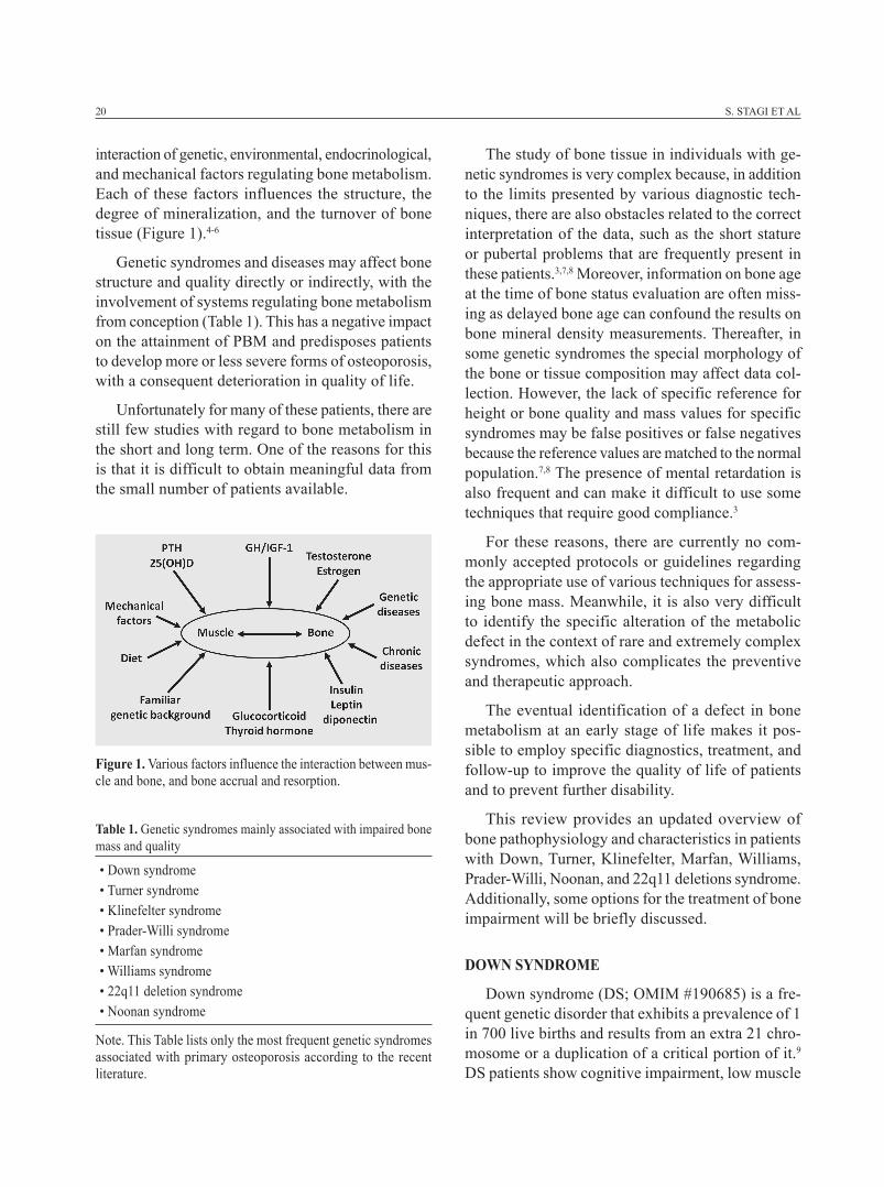
interaction of genetic, environmental, endocrinological, and mechanical factors regulating bone metabolism. Each of these factors influences the structure, the degree of mineralization, and the turnover of bone tissue (Figure 1).4-6
Genetic syndromes and diseases may affect bone structure and quality directly or indirectly, with the involvement of systems regulating bone metabolism from conception (Table 1). This has a negative impact on the attainment of PBM and predisposes patients to develop more or less severe forms of osteoporosis, with a consequent deterioration in quality of life.
Unfortunately for many of these patients, there are still few studies with regard to bone metabolism in the short and long term. One of the reasons for this is that it is difficult to obtain meaningful data from the small number of patients available.
The study of bone tissue in individuals with ge-netic syndromes is very complex because, in addition to the limits presented by various diagnostic tech-niques, there are also obstacles related to the correct interpretation of the data, such as the short stature or pubertal problems that are frequently present in these patients.3,7,8 Moreover, information on bone age at the time of bone status evaluation are often miss-ing as delayed bone age can confound the results on bone mineral density measurements. Thereafter, in some genetic syndromes the special morphology of the bone or tissue composition may affect data col-lection. However, the lack of specific reference for height or bone quality and mass values for specific syndromes may be false positives or false negatives because the reference values are matched to the normal population.7,8 The presence of mental retardation is also frequent and can make it difficult to use some techniques that require good compliance.3
For these reasons, there are currently no com-monly accepted protocols or guidelines regarding the appropriate use of various techniques for assess-ing bone mass. Meanwhile, it is also very difficult to identify the specific alteration of the metabolic defect in the context of rare and extremely complex syndromes, which also complicates the preventive and therapeutic approach.
The eventual identification of a defect in bone metabolism at an early stage of life makes it pos-sible to employ specific diagnostics, treatment, and follow-up to improve the quality of life of patients and to prevent further disability.
This review provides an updated overview of bone pathophysiology and characteristics in patients with Down, Turner, Klinefelter, Marfan, Williams, Prader-Willi, Noonan, and 22q11 deletions syndrome. Additionally, some options for the treatment of bone impairment will be briefly discussed.
doWN syNdroMe
Down syndrome (DS; OMIM #190685) is a fre-quent genetic disorder that exhibits a prevalence of 1 in 700 live births and results from an extra 21 chro-mosome or a duplication of a critical portion of it.9 DS patients show cognitive impairment, low muscle
Figure 1. various factors influence the interaction between mus-cle and bone, and bone accrual and resorption.
Table 1. Genetic syndromes mainly associated with impaired bone mass and quality
• Down syndrome• Turner syndrome• Klinefelter syndrome• Prader-Willi syndrome• Marfan syndrome• Williams syndrome• 22q11 deletion syndrome• Noonan syndrome
Note. This Table lists only the most frequent genetic syndromes associated with primary osteoporosis according to the recent literature.
Syndromic osteoporosis 21
tone, and craniofacial alterations that affect many systems including bone.10 Because of the increased life expectancy today of DS patients, the acquisition of a good PBM may represent an important factor in the prevention of osteoporosis, bone fragility, and related fractures in these patients.11
Several studies have reported that DS patients have a lower level of bone mass than their peers without DS, but it is unclear whether this is due to specific effects of chromosome 21 genes or their lifestyle factors (Table 2).12,13
Gonzalez-Aguero et al report lower values of bone mineral density (BMD) in children with DS with respect to unaffected siblings. Moreover, the differ-ences in the observed sexual dimorphism indicate a non-standard bone development in this specific population.10 However, McKelvey et al measured BMD by dual-energy X-ray absorptiometry (DXA) in a group of 30 patients with DS and revealed that 53.3% showed a BMD ≤-2 SDS. The authors also demonstrated that both bone formation and resorp-tion were suppressed in DS compared with controls, indicating low bone formation and decreased bone turnover as the primary causes of the low bone mass observed in these patients.13 Wu compared BMD by DXA between preadolescent boys with and without DS and confirmed that patients with DS have lower BMD and bone mineral content (BMC), also sug-gesting that the pelvis may be the first site to show bone impairment in DS.14 These results were partially confirmed by peripheral quantitative computed to-mography (pQCT) where a higher volumetric bone mineral density (vBMD) was observed in determined skeletal sites.15 Nevertheless, adolescents with DS have a higher risk of suffering bone fractures due to a decreased bone resistance to load bending or torsion.15
In DS, factors possibly related to low bone mass are hypotonia, low amounts of physical activity, poor calcium and vitamin D intake, celiac disease, and hor-monal factors.16 Hypotonia not only is a characteristic of DS, but it represents a limit to physical activity.17 However, some authors studying the effect of physical activity alone or of increase of calcium intake alone did not demonstrate a significant effect on BMD. On the contrary, studies carried out on patients with DS who received an increase of calcium intake and who were subjected to a specific program of physical activity have shown that these two elements have a beneficial synergistic effect on BMD.18 Moreover, hypovitaminosis D seems to be very frequent in DS, in particular in subjects with obesity and autoimmune diseases.19 DS patients with obesity and autoimmune diseases could require higher cholecalciferol sup-plementation.19 Nevertheless, no data about vitamin D intake and BMD are reported in DS.
Finally, adolescents with DS in the high physical activity tertile showed lower risk of developing future osteoporosis by having a higher BMD Z-score at the hip; this demonstrates the importance of accumulated minutes of physical activity on bone health in ado-lescents with DS.17
Nevertheless, other authors demonstrated that one year of training significantly increased BMC values at the lumbar spine and total hip and BMD values only at the lumbar spine, whereas changes in broadband ultrasound attenuation (BUA) and speed-of-sound (SOS) values were not evident following training but showed a lower bone improvement compared to that reported.20
Thus, impaired bone mass and quality is a frequent characteristic of DS probably related to some skeletal segments compared to others. In these patients, many factors affect correct bone turnover and gain, and therefore a multidisciplinary approach that comprises constant physical activity and an optimal calcium and vitamin D intake is mandatory.
TUrNer syNdroMe
Turner syndrome (TS) is the most common gonadal dysgenesis in females and occurs in about 1 per 2,000 liveborn females.21 This syndrome is due to the total
Table 2. Mechanisms contributing to reduced bone mass in Down syndrome
• Down syndrome related mechanisms• Hypotonia• Low physical activity levels• Low intake of calcium • Low vitamin D levels• Hormonal factors• Autoimmune diseases
22 S. STAGI ET AL
absence of one X chromosome, mosaicisms, or its structural abnormalities.21
The classic TS phenotype includes short stature, gonadal dysgenesis with primary amenorrhea and hypergonadotropic hypogonadism, facial and body dysmorphisms, skeletal abnormalities, cardiovascular and renal malformations, and predisposition to auto-immune diseases, such as thyroid diseases, diabetes mellitus, celiac disease, and vitiligo.21 Short stature is always present and is caused by several factors, such as Short Stature Homeobox (SHOX) haploinsuf-ficiency and related skeletal dysplasia, intrauterine growth retardation, poor growth in early childhood, absence of the growth spurt, and skeletal dysplasia.21
TS is a state of reduced sensitivity to growth hor-mone (GH) rather than a GH deficiency, and treatment with high doses of GH can increase growth velocity and final height. In puberty, estrogen therapy should be coordinated with GH use.21 However, optimization of bone health in girls with TS also requires maintain-ing a healthy, active lifestyle with adequate calcium and vitamin D intake.22
Despite only limited reports of a greater number of fractures during childhood or adulthood, osteo-porosis historically has been described as a feature in TS. However, data of prevalence of osteoporosis in TS are not uniform and the causes have not been fully elucidated (Table 3).
Several studies have concluded that TS patients have low BMD.22 For example, Lisà et al described a reduced bone density in 25% of TS associated with reduced osteocalcin and alkaline phosphatase values, indicating a reduced osteoblast activity.23 However, Benetti-Pinto et al studied BMD by DXA in 38 women
and showed a marked decrease in BMD of the lumbar spine (90%) and femoral neck (55%). Interestingly, the authors have found a positive correlation with BMD in the lumbar spine and the duration of estrogen therapy but not with the age at which therapy was initiated.24
More recently, Nadeem calculated bone mineral apparent density (BMAD) for correction of misdiag-nosed BMD due to short stature and demonstrated that TS patients have lower BMAD values at the lumbar spine compared with an age- and sex-matched general population. According to their study, puberty had a positive impact on BMAD irrespective of the time of its commence. In addition, they noted that it is the stage of puberty, not the age at initiation of estrogen therapy, which is important for BMAD. No influence of karyotype on BMAD was demonstrated.25
However, using new techniques such as high-res-olution peripheral quantitative computed tomography (HR-pQCT), Hansen et al evaluated 32 TS patients and 32 controls matched respective to age and body-mass index and showed that TS had compromised trabecular microarchitecture and lower bone strength at the radius and tibia, which may partly account for the increased risk of fracture observed in these patients.25
Nonetheless, other studies have displayed con-flicting results. For example Shaw et al conducted a longitudinal study examining BMD at the lumbar spine in 18 girls with TS aged 4-17 years by DXA several times over a 2.5-year period. They reported only one girl with a significant bone density reduc-tion in comparison with controls. No advantage was found for any form of treatment in optimizing bone mineralization.27
Estrogen therapy is very important to avoid a rapid decrease in BMD and to induce maximal peak bone mass in TS adolescents. Bertelloni et al stud-ied 26 young women with TS at final heights. The areal BMD (aBMD) and the vBMD measured by DXA demonstrated that final heights and aBMD were significantly higher in TS treated with estrogen and GH therapy than those in patients treated only with estrogen therapy. Nevertheless, vBMD was higher, though not significantly different between the groups.28 These data may suggest that in TS, GH administra-tion improves final height and aBMD, but it does not significantly increase vBMD; aBMD reduction in
Table 3. Mechanisms contributing to reduced bone mass in Turner syndrome
• Turner syndrome related mechanisms• Puberty delay• Estrogen deficiency• Low intake of vitamin D• Vitamin D receptor gene polymorphisms• Hormonal factors (i.e., thyroid disorders)• Autoimmune diseases
Syndromic osteoporosis 23
of hormonal factors, including the growth-regulating hormones and the gonadal steroids. However, ad-ditional measures to prevent osteoporosis must be taken, such as ensuring adequate calcium, vitamin D intake, ample weight-bearing activities, and the precocious diagnosis of autoimmune diseases such as celiac disease.
KlINeFelTer syNdroMe
Klinefelter syndrome (KS) is a chromosomal disor-der with a prevalence of 1/500-1/600 newborn males and is characterized by the presence of one extra X chromosome resulting in a 47,XXY karyotype. However, other karyotypes have also sometimes been observed (10%), such as 48,XXYY; 48,XXXY; and 49,XXXXY.33
Classical KS is characterized by primary hypog-onadism resulting in a progressive testicular failure that begins during pubertal development. Clinical phenotype includes increased stature, gynecomastia, small testes, and infertility.34 However, a less dis-tinctive phenotype has been observed. In fact, KS is an underdiagnosed condition; only 25% of patients are diagnosed, and only a minority of these before puberty,35 although the number of prenatal diagnosis are increasing.
Presence of metabolic bone disorders in KS has been described (Table 4). In fact, testosterone regulates male bone metabolism directly by the androgen receptor on the osteoblast and indirectly by aromatization to estrogens, which promotes periosteal bone formation during puberty and reduces bone resorption in adult life. However, trabecular bone is rich in androgen receptors. Androgens increase the volume of trabecular bone and therefore increase trabecular number, which impedes resorption and reduces intertrabecular space.36 At puberty, testosterone is important for bone matu-ration and for achieving adequate peak bone mass. Decreased bone mass in KS has been traditionally related to low testosterone levels.
Jo et al described 18 KS adults treated with tes-tosterone by measuring BMD by DXA at the lumbar spine and at the femoral neck. The BMD at the lum-bar spine increased significantly after testosterone treatment.38 In contrast, testosterone therapy does not
Turner syndrome is likely due to the impaired growth and reduced bone size.
In these patients, there are many data that show a beneficial dose-dependent GH effect on BMD. Neely et al, for example, evaluated the bone mineral status of 19 TS adolescents by DXA at the lumbar spine and demonstrated that BMC and BMD as well as BMAD in TS on GH therapy are normal and lumbar bone mineral is greater in younger TS than in controls; this difference could be related to GH therapy.29 However, in a longitudinal study, Sas et al measured BMD by phalangeal radiographic absorptiometry in 68 previ-ously untreated TS and divided them into three groups according to the dose of GH. During 7 years of GH treatment, the BMD Z-score showed a significant increase respective to the baseline when BMD values were within the normal range of healthy girls. The increment in BMD SDS, consisting predominantly of cortical bone, was significantly higher in the group receiving a higher GH dosage.30
Unfortunately, there are no uniform data from current studies on bone metabolism. This is due to the difficulty in recruiting a significant number of untreated TS patients to compare with treated subjects and/or with healthy subjects. In addition, the BMD measurement techniques commonly used today are limited by the presence of short stature in TS and also by the absence of TS-specific BMD reference values.
In TS, the presence of co-morbidity may further compromise normal bone metabolism. Celiac disease is a common secondary cause of metabolic bone dis-ease, and the incidence of this autoimmune disorder is increased in TS. A good vitamin D status is very important for bone, but in TS it may be compromised by celiac disease.31
A study of the relationship between vitamin D re-ceptor gene polymorphisms in TS shows that patients carrying genotype BsmI or FokI had lower BMD than those with other genotypes. Moreover, in TS the presence of low BMD and the increased fracture risk may be due to a genetic defect of the vitamin D receptor aggravated by hormonal deficiency.32
Thus, the pathogenesis of the skeletal deminerali-zation in TS support the concept of an intrinsic bone defect that is subsequently exacerbated by a number
24 S. STAGI ET AL
Table 4. Possible mechanisms contributing to reduced bone mass in Klinefelter syndromeMechanisms Bone structure involvedLow 25(OH)D levels bone mass in totoUnfavourable muscle/fat ratio bone mass in totoLow testosterone levels bone mass in totoLow androgen receptor expression (AR) bone mass in totoNonrandom X-chromosome inactivation trabecular boneAR CAG length trabecular boneLow insulin-like growth factor-3 (IGFBP-3) levels trabecular boneEstrogen deficiency cortical boneModified by Ferlin et al, 2011.37
necessarily increase bone mass, and low bone mass was also observed in patients with normal testosterone levels. In fact, Ferlin et al evaluated 112 KS patients and showed that 42.5% were affected by osteοpenia/osteoporosis. No significant relationship was observed between testosterone levels and bone mass, and the prevalence of osteopenia/osteoporosis was similar in subjects with normal and low testosterone levels. Furthermore, the androgen receptor CAG polymor-phism study showed that the CAG polymorphism of the androgen receptor contributes to a decreased bone mass.34 These results are in contrast to a precedent study of Zitzmann et al that measured bone density with a phalangeal bone ultrasound in 77 KS patients untreated with testosterone and which observed that the CAG polymorphism is negatively and indepen-dently associated with BMD.39
In another study, Bojesen et al analyzed 70 KS patients (50% of whom had been treated with tes-tosterone) by DXA and studied the impact of the androgen receptor polymorphism and skewed X in-activation on phenotype. These authors did not find any tendency for the allele with the shortest CAGn to be preferentially inactivated. Also, they observed no impact of skewed X-chromosome inactivation either on hormonal levels or on bone mass.40
Considering the presence of these unclear data, Ferlin et al suggested that variable degrees of puberty among KS subjects might lead to differences in the achievement of the BMD at PBM, thus resulting in different BMD in adult life. In this setting, differences in BMD in adulthood might reflect the timing of onset and the duration of the hypogonadism state.34 More studies are certainly needed.
Other systems have been studied to uncover the possible cause of reduced bone mass observed in KS patients. In fact, testosterone acts on vitamin D me-tabolism. Francis et al described a correlation between testosterone levels and activity of renal 25-hydroxilase which would indicate that reduced testosterone levels are related to reduction of active vitamin D with a negative effect on bone metabolism.41
Stepan et al showed that 25-OH-vitamin D are in the low-normal range in KS subjects, and the rate of BMD gain at the femoral neck after ibandronate therapy was inversely related to 25-OH-vitamin D levels, but that the effects were adversely influenced by vitamin D insufficiency or deficiency.42 Furthermore, Ferlin observed reduced 25-OH-vitamin D levels in KS subjects compared with controls.34
In an interesting study, Aksglaede et al analyzed the muscle/fat ratio and BMC in 18 untreated participants and 6 KS boys receiving androgen and observed an unfavourable muscle/fat ratio with normal lumbar BMD and whole body BMC. These results suggest that the unfavourable metabolic profile seen in adult KS may already be present in childhood, as evidenced by the increased fat mass, whereas the reported low BMD seems to develop after puberty.43 It would be interesting to investigate whether, and if so how, the considerable presence of an unfavourable muscle/fat ratio affects hormone metabolism in these patients.
In conclusion, in KS the hypogonadism, or the reduced testosterone levels in particular if started early in life, represent one of the most important causes of osteoporosis. However, possible new causes for osteoporosis in KS might be related to androgen
Syndromic osteoporosis 25
receptor function. Additional measures to prevent osteoporosis must be implemented, such as ensuring adequate calcium and vitamin D intake and ample weight-bearing activities as well as the precocious diagnosis of autoimmune diseases.
PrAder-WIllI syNdroMe
Prader-Willi syndrome (PWS; OMIM #176270) is a complex genetic disorder that is due to one of the following: a deletion in the 15q11.2 region of the paternal allele, the presence of maternal 15q11 disomy, or an imprinting disorder located on chromosome 15 (q11-13). PWS patients are characterized by neonatal hypotonia, feeding problems, growth retardation, and developmental delay followed by hyperphagia, severe obesity, short stature, hypogonadism, and cognitive difficulties after 2-3 years of age. Diabetes, osteopo-rosis, and scoliosis are common.44,45
Increased fat mass and decreased lean body mass are characteristics of PWS. Muscle mass is decreased by 25-37%, which might partly explain the hypotonia. However, there are also structural and functional muscle abnormalities, and cortical motor areas are hypo-excitable in PWS patients.46
As demonstrated by several studies, PWS adult patients have a lower BMD than normal subjects.
Marked hypotonia may be a cause of reduced BMD according to the “mechanostat theory”, which postulates that bone adapts to the mechanical forces to which it is subjected to keep the strain on the bone at a constant set point.47 Hoybye et al studied bone metabolism and body composition of 19 PWS adults and found that BMD at the lumbar spine was significantly lower, with 4 patients (21%) exhibiting osteoporosis and 11 osteopenia.48 Similar data have also been reported by Butler et al, who compared 21 obese subjects with PWS. These authors showed that PWS had a significantly lower BMD and BMC and higher unrinary N-telopeptides levels. They con-cluded that decreased BMD in PWS may be related to the lack of depositing bone mineral during growth, possibly because of decreased production of sex or growth hormones and hypotonia.49
vestergaard et al compared bone mineral status, body composition, and biochemical markers of bone
turnover of eight PWS adults with age-, sex- and body mass index-matched controls and showed that BMD and BMC in the PWS group were significantly lower than in the control group. They postulated that these data were caused by an increased bone turnover probably linked to sex steroid deficiency because the resorptive and formative bone markers were significantly elevated and plasma testosterone was low in PWS males.50
However, several studies in PWS children reported that in infancy there are normal BMD values at the lumbar spine and reduced lean mass with low motor performance. These data are limited because children affected with PWS, with short stature and obesity, are very difficult to compare with an appropriate control group. Edouard et al studied muscle-bone characteristics in 17 PWS children (untreated with GH) compared to height-matched controls. They used DXA to study the lumbar spine, radius, and tibia by pQCT. BMD in PWS patients was normal in the axial and appendicular skeleton. Compared to age- or height-matched controls, PWS patients had lower maximal muscle force and power relative to body mass during jumping. PWS patients had similar absolute maximal muscle force but lower absolute maximal power compared to age- or height-matched controls. Relationships between bone mass and muscle size and force were similar in PWS patients and in healthy subjects.51
Other studies have focused on the effect of GH in PWS. Carrel et al studied the GH effects in PWS children and reported that GH early treatment (begun prior to 2 years of age) improves body composition, motor function, height, and lipid profiles.52 These gains are associated with increased BMD during GH treatment. For two years De Lind van Wijngaarden et al studied BMD in 46 PWS prepubertal children randomized in a group treated with GH and in a control group. They showed that at 24 months total body and lumbar spine BMD and BMAD did not significantly differ between the groups and that BMD was normal. GH treatment caused temporary decrease of total body BMD in the first 6 months of therapy, but at 24 months there were no effects. However, it is interesting that the values of IGF-1 were positively correlated with total body and lumbar spine BMD but not with BMAD.53 Similar data have been reported
26 S. STAGI ET AL
by Colmenares et al after 36 months of GH supple-mentation in 36 children with PWS.45
In a recent paper, Khare et al studied GH effects on BMD in the different genetic subtypes of PWS, and they demonstrated that in 44% total body, hip, and spine BMD was <-1 SDS and in 10% was <-2 SDS. BMD Z-scores and total BMD in the spine were significantly higher in the GH-treated group than in controls. In patients with uniparental disomy there was higher BMD compared with patients with classi-cal deletions, but this difference was not statistically significant.54
In conclusion, although the present data are not uniform, the importance of monitoring bone miner-alization status in PWS is clear. GH treatment has beneficial effects on the auxological and metabolic conditions in subjects with PWS and probably the increase in lean body mass, and the consequent de-crease of hypotonia has a beneficial effect on bone.
NooNAN syNdroMe
Noonan syndrome (NS; OMIM #163950) is an autosomal dominant disorder that is characterized by dysmorphic facies, short stature, delayed puberty, cardiac defects, and skeletal deformities. About 50% of NS patients have a missense PTPN11 (Protein-Tyrosine Phosphatase, Non-receptor Type 11; OMIM *176876) mutation, whereas other mutations exhibited are in the KRAS (V-KI-RAS2 Kirsten Rat Sarcoma Viral Oncogene Homolog; OMIM *190070), SOS1 (Son Of Sevenless, Drosophila, Homolog 1; OMIM *182530), NRAS (Neuroblastoma RAS Viral Oncogene Homolog; OMIM *164790), and RAF1 (V-RAF-1 Murine Leukemia Viral Oncogene Homolog 1; OMIM *164760) genes.55,56
Approximately 50-70% of individuals with NS have short stature, even though some individuals have normal growth and stature.57 However, puberty is typically, but not universally, delayed for both NS boys and girls with a reduced pubertal growth spurt.56
PTPN11 encodes a Src homology 2 domain con-taining tyrosine phosphatase 2, a protein acting in the RAS-mitogen activated protein kinase (MAPK) signal transduction pathway.57 However, NS is also a disorder of the RAS-MAPK pathway. While clinical
features such as short stature and skeletal deformi-ties suggest an underlying skeletal dysplasia, little information is available on bone mineralization and metabolism. For example, Stevenson et al studied bone resorption markers in 49 patients with RAS-MAPK pathway disorders including 14 NS, and they sug-gested an increased bone resorption in NS compared to controls.58 Takagi59 and Reinker et al60 reported 4 NS adults with osteopenia, although a limited amount of information was provided regarding two of the patients described by Reinker et al.60 Noordam et al evaluated 16 NS children before and after GH therapy, and they reported low trabecular vBMD at baseline with improvement following GH therapy.61
Generally, 50% of NS subjects had a low bone mass as measured by DXA. Of these, mean BMC Z-score was marginally decreased (-0.89 SDS), whereas mean total body BMD z-score was significantly reduced to -1.87 SDS (-0.82 SDS if calculated on the basis of height). Biochemical evaluation of bone turnover was unremarkable, with the exception of serum IGF- I and IGF-BP3 levels, which were low-normal for age.62
The exact role of the RAS-MAPK pathway in the regulation of bone homeostasis is not clear. In NF1, another genetic disorder associated with dysregulation of the RAS-MAPK pathway, the NF1 gene encodes for neurofibromin which negatively regulates the activity of intracellular signaling molecule RAS by functioning as a GTPase-activating protein.63 Haploinsufficiency, or a deficiency of NF1, results in a dose-dependent increase in RAS activity, which in turn can activate a variety of downstream signaling pathways. Several data demonstrate that osteoprogenitors and osteoblasts from Nf1+/− mice and NF1 haploinsufficient human embryonic bone cells showed reduced expression of bone markers and/or produced less mineralized matrix when grown under osteogenic conditions.64
Thus, metabolic bone disease present in NS may result from a subtle variation in the interplay of os-teoclast and osteoblast activity without clear abnor-malities being defined in the metabolism of either. The clinical significance of this finding needs to be validated by longitudinal studies. However, data about familiar history of bone diseases, exercise, and vitamin D intake, which would be helpful to gain in-sight into the etiology, are lacking today. Importantly,
Syndromic osteoporosis 27
histomorphometric analysis of bone tissue obtained from patients with NS or mouse models of NS may allow us to establish associations between a molecular defect in the RAS-MAP kinase pathway and altera-tions in the function of osteoblasts and osteoclasts on a tissue level.
MArFAN syNdroMe
Marfan syndrome (MFS; OMIM #154700) is an autosomal dominant disorder with a prevalence of 1 per 10,000 subjects exhibiting variable skeletal, cardiovascular, and ocular involvement.65 MFS is caused by Fibrillin 1 gene mutations (FBN1; OMIM *134797).66,67 In MFS, the phenotype is characterized by a prolapsed mitral valve, an aortic root diameter at the upper limits of normal for body size, skin stretch marks unrelated to weight gain or loss, and skeletal features, including scoliosis, chest wall deformities, and joint hypermobility.65
The loss of the fibrillin 1 protein has a subsequent effect on the pool of TGF-β, a factor that plays a criti-cal role in bone health.66 TGF-β positively regulates osteoblast proliferation and differentiation in vitro.67 In fact, TGF-β-/- mice exhibited low bone mass and quality.68 In addition, TGF-β may induce bone mes-enchymal stem cells migration, which differentiate into osteoblasts. Moreover, Fbn1mgR/mgR mice exhibited decreased bone volume and density due to TGF-β driven osteoclastogenesis.68
In adults with MFS, reduced axial and peripheral BMD have been observed, suggesting that these patients may be at higher risk of fractures.69-77 Con-versely, osteopenia has not been shown in some cases.78 Nonetheless, there is a paucity of data regarding bone mineral status of MFS patients during childhood in the literature. For example, in a study involving 16 MFS patients, a low BMD at the femoral neck and lumbar spine were reported.78,79 Other data indicated that MFS children have significantly lower BMC as well as whole-body and lumbar-spine BMD compared with controls.73 In contrast, another study reported normal BMD at the lumbar spine and femoral neck in 21 MFS children.79
Consequently, the ethiopathogenesis of impaired bone mass and quality in MFS is uncertain, though it
is most likely a primary osteoporosis. Large amounts of data are necessary to better delineate the bone status of this syndrome.
WIllIAMs syNdroMe
Williams-Beuren syndrome (WBS; #194050) is a well-recognized disorder that affects 1:10000 live births and is characterized by typical facial dys-morphisms, heart defects, short stature, and mental retardation.80 Over 90% of patients present a 1.5 Mb interstizial hemizygous deletion at 7q11.23 compris-ing the elastine gene.80
Furthermore, despite the fact that 5% to 50% of WBS cases present as one or more episodes of hypercalcemia, a disorder once named ‘calcium ho-meostasis disorder’, data about bone status in WBS are scarce in the literature.
Patients with WBS showed signs and symptoms related to premature aging. Also, bone status may be involved. Nevertheless, there are not many data about bone mass and quality reported in these patients. In one study, nine of 20 (45%) adults had decreased bone mineral density at multiple sites on DXA.81 In the same study, all of the WBS patients had normal blood calcium levels, except for one patient, a 41-year old man with a high calcium level.81 Therefore, in these patients vitamin D or calcium supplementation should be used with caution.82
However, since decreased bone density is more prevalent among adults with developmental disabili-ties than among adults in the general population, this finding may be nonspecific. It is interesting to note the case reported by Knudtzon et al83 concerning two brothers with WBS, one of whom showed an osteo-clerotic skeleton at birth and became osteoporotic at the age of 2 years. However, in this syndrome the possible appearance of celiac disease and thyroid dysfunction, such as early and fast puberty, may contribute to the impaired bone status.
22q11 deleTIoN syNdroMe
22q11.2 deletion syndrome (22q11DS) comprises many different phenotypes, including DiGeorge syn-drome (DGS; OMIM #188400), conotruncal anomaly face syndrome (CTAFS; OMIM #217095), and velo-
28 S. STAGI ET AL
cardio-facial syndrome (vCFS; #192430).84 In West-ern countries, chromosome 22q11.2 deletion occurs once in every 3,000 to 9,000 people.84 Since clinical phenotypes of DGS, CTAFS, and vCFS are overlap-ping in many cases, it is difficult to differentiate the syndromes using clinical phenotypes alone.84,85
Most of the patients were diagnosed based on slightly and severely decreased immunity, facial de-fects, and heart anomaly, and some patients have kidney abnormalities, hypoparathyroidism, hypothyroidism, and developmental retardation.84,85
Hypocalcemia caused by hypoparathyroidism is exhibited in 40-60% of patients with chromosome 22q11.2 deletion,84,86 in 60% of patients with DGS, and in 20% of patients with vCFS.86 However, although hypoparathyroidism with hypocalcaemia is one of the most frequent clinical features of 22q11DS, bone mass and metabolism have rarely been assessed in these patients.86
Kaufman et al87 identified chromosomal regions associated with bone status and showed that the 22q11 region was one of those associated with a lumbar spine BMD. However, we demonstrated that patients with 22q11DS show a significantly reduced BMAD Z-score compared with controls associated with a significantly lower ionized and total calcium levels as well as lower PTH levels.86 In particular, children and young patients with 22q11DS had significantly lower serum osteocalcin levels, BSAP levels, and urinary deoxypyridinoline concentrations. The above results suggest that these subjects present slow bone modeling and remodeling. This may cause reduced bone gain with a reduced PBM predispose the patient to a higher risk of impaired bone mass and quality in adulthood. However, Lee et al88 reported a female patient, with no untoward medical history other than a history of osteoporosis, who was diagnosed as hav-ing CATCH 22; this confirmed data about adults with 22q11DS who showed a high frequency of impaired bone status.86 In these patients, teriparatide treatment may potentially cause a gain in bone mineral density.89
It is therefore clear that subjects with 22q11DS may have a significant reduction in bone mass. More data and longitudinal studies are necessary to assess bone status and the parameters influencing it in this syndrome.
coNclUsIoNs
Syndromic and genetic disorders are frequently associated with a high risk of osteoporosis or impaired bone status. Pediatricians should therefore be aware of the risk for bone loss in children, adolescents, and adults with genetic syndromes in order to prevent fractures in the more severe cases or to ameliorate PBM in the less severe ones. This is crucial for reduc-ing the risk of adult-onset osteoporosis.
As we have seen, many factors are responsible for the achievement of better PBM. The influence of exercise-induced loading is well known, and the ability to perform consistent physical activity at in-tensity compatible with the basic condition should always be recommended and explained to the fam-ily. Evaluations of 25-hydroxyvitamin D levels and PTH should be performed for patients with these syndromes well before the patients develop osteo-porosis or osteopenia. However, although no direct correlation with fracture risk has been detected, it is surmised that vitamin D supplementation in children with osteopenia and osteoporosis is advisable and may indeed reduce morbidity. The same should be considered for calcium intake.
As the exact pathogenic mechanisms of impaired bone status in many genetic syndromes is not as yet known, it is difficult to proceed with rational drug discovery to specifically address them. Frequently, bis-phosphonate treatment represents the first-line therapy for many disorders and should also be considered for these patients. Thus, randomized controlled trials are strongly recommended. Notably, other new drugs such as recombinant human parathyroid hormone (rhPTH) may prove useful in the treatment of osteoporosis, if necessary. In particular, the use of the rhPTH in 22q11 deletions syndrome patients may represent a useful option if future studies confirm the presence of impaired bone status.
declArATIoN oF INTeresT
The authors declare that there are no conflicts of interest that could be perceived as prejudicing the impartiality of the research reported.
FUNdINg
This research did not receive any specific grant
Syndromic osteoporosis 29
from any funding agency in the public, commercial, or not-for-profit sector.
reFereNces1. Ma NS, Gordon CM, 2012 Pediatric osteoporosis: where
are we now? J Pediatr 161: 983-990.2. Stagi S, Cavalli L, Iurato C, Seminara S, Brandi ML,
de Martino M, 2013 Bone metabolism in children and adolescents: main characteristics of the determinants of peak bone mass. Clin Cases Miner Bone Metab 10: 172-179.
3. Stagi S, Cavalli L, Seminara S, de Martino M, Brandi ML, 2014 The ever-expanding conundrum of primary osteoporosis: aetiopathogenesis, diagnosis, and treat-ment. Ital J Pediatr 40: 55.
4. Ott SM, 1990 Attainment of peak bone mass. J Clin Endocrinol Metab 71: 1082A-1082C.
5. Kelly PJ, Twomey L, Sambrook PN, Eisman JA, 1990 Sex differences in peak adult bone mineral density. J Bone Miner Res 5: 1169-1175.
6. von Scheven E, Corbin KJ, Stagi S, Cimaz R, 2014 Glucocorticoid-associated osteoporosis in chronic inflam-matory diseases: epidemiology, mechanisms, diagnosis, and treatment. Curr Osteoporos Rep 12: 289-299.
7. Stagi S, Cavalli L, Iurato C, Seminara S, Brandi ML, de Martino M, 2013 Bone health in children and ado-lescents: the available imaging techniques. Clin Cases Miner Bone Metab 10: 166-171.
8. Bogunovic L, Doyle SM, vogiatzi MG, 2009 Measure-ment of bone density in the pediatric population. Curr Opin Pediatr 21: 77-82.
9. Weijerman ME, van Furth AM, vonk Noordegraaf A, van Wouwe JP, Broers CJ, Gemke RJ, 2008 Prevalence, neonatal characteristics, and first-year mortality of Down syndrome: a national study. J Pediatr 152: 15-19.
10. González-Agüero A, vicente-Rodríguez G, Moreno LA, Casajús JA, 2011 Bone mass in male and female children and adolescents with Down syndrome. Osteoporos Int 22: 2151-2157.
11. González-Agüero A, vicente-Rodríguez G, Gómez-Cabello A, Ara I, Moreno LA, Casajús JA, 2012 A 21-week bone deposition promoting exercise programme increases bone mass in young people with Down syn-drome. Dev Med Child Neurol 54: 552-556.
12. Fowler TW, McKelvey KD, Akel NS, et al, 2012 Low bone turnover and low BMD in Down syndrome: effect of intermittent PTH treatment. PLoS One 7: e42967.
13. McKelvey KD, Fowler TW, Akel NS, et al, 2013 Low bone turnover and low bone density in a cohort of adults with Down syndrome. Osteoporos Int 24: 1333-1338.
14. Wu J, 2013 Bone mass and density in preadolescent boys with and without Down syndrome. Osteoporos Int 24: 2847-2854.
15. González-Agüero A, vicente-Rodríguez G, Gómez-Cabello A, Casajús JA, 2013 Cortical and trabecular bone at the radius and tibia in male and female adoles-
cents with Down syndrome: a peripheral quantitative computed tomography (pQCT) study. Osteoporos Int 24: 1035-1044.
16. Hawli Y, Nasrallah M, El-Hajj Fuleihan G, 2009 En-docrine and musculoskeletal abnormalities in patients with Down syndrome. Nat Rev Endocrinol 5: 327-334.
17. Matute-Llorente A, González-Agüero A, Gómez-Cabello A, vicente-Rodríguez G, Casajús JA, 2013 Decreased levels of physical activity in adolescents with Down syndrome are related with low bone mineral density: a cross-sectional study. BMC Endocr Disord 13: 22.
18. Reza SM, Rasool H, Mansour S, Abdollah H, 2013 Effects of calcium and training on the development of bone density in children with Down syndrome. Res Dev Disabil 34: 4304-4309.
19. Stagi S, Lapi E, Romano S, et al, 2015 Determinants of vitamin D levels in children and adolescents with Down syndrome. Int J Endocrinol [Ahead of print]
20. Ferry B, Gavris M, Tifrea C, et al, 2014 The bone tissue of children and adolescents with Down syndrome is sensitive to mechanical stress in certain skeletal loca-tions: A 1-year physical training program study. Res Dev Disabil 35: 2077-2084.
21. Gravholt CH, 2004 Epidemiological, endocrine and metabolic features in Turner syndrome. Eur J Endocrinol 151: 657-687.
22. Hjerrild BE, Mortensen KH, Gravholt CH, 2008 Turner syndrome and clinical treatment. Br Med Bull 86: 77-93.
23. Lisá L, Neradilová M, Tomásová H, Kouba M, Preucil P, 1996 Changes in bone metabolism in girls with Turner’s syndrome. Cas Lek Cesk 135: 178-180.
24. Benetti-Pinto CL, Bedone A, Magna LA, Marques-Neto JF, 2002 Factors associated with the reduction of bone density in patients with gonadal dysgenesis. Fertil Steril 77: 571-575.
25. Nadeem M, Roche EF, 2012 Bone health in children and adolescent with Turner syndrome. J Pediatr Endocrinol Metab 25: 823-833.
26. Hansen S, Brixen K, Gravholt CH, 2012 Compromised trabecular microarchitecture and lower finite element estimates of radius and tibia bone strength in adults with turner syndrome: a cross-sectional study using high-resolution-pQCT. J Bone Miner Res 27: 1794-1803.
27. Shaw NJ, Rehan vK, Husain S, Marshall T, Smith CS, 1997 Bone mineral density in Turner’s syndrome-a longitudinal study. Clin Endocrinol (Oxf) 47: 367-370.
28. Bertelloni S, Cinquanta L, Baroncelli GI, Simi P, Rossi S, Saggese G, 2000 volumetric bone mineral density in young women with Turner’s syndrome treated with estrogens or estrogens plus growth hormone. Horm Res 53: 72-76.
29. Neely EK, Marcus R, Rosenfeld RG, Bachrach LK, 1993 Turner syndrome adolescents receiving growth hormone are not osteopenic. J Clin Endocrinol Metab 76: 861-866.
30. Sas Tc, de Muinck Keizer-Schrama SM, Stijnen T, et al, 2001 Bone mineral density assessed by phalangeal
30 S. STAGI ET AL
radiographic absorptiometry before and during long-term growth hormone treatment in girls with Turner’s syndrome participating in a randomized dose-response study. Pediatr Res 50: 417-422.
31. Lleo A, Moroni L, Caliari L, Invernizzi P, 2012 Au-toimmunity and Turner’s syndrome. Autoimmun Rev 11: A538-A543.
32. Peralta López M, Miras M, Silvano L, et al, 2011 vitamin D receptor genotypes are associated with bone mass in patients with Turner syndrome. J Pediatr Endocrinol Metab 24: 307-312.
33. visootsak J, Graham JM Jr, 2006 Klinefelter syndrome and other sex chromosomal aneuploidies. Orphanet J Rare Dis 1: 42.
34. Ferlin A, Schipilliti M, vinanzi C, et al, 2011 Bone mass in subjects with Klinefelter syndrome: role of testosterone levels and androgen receptor gene CAG polymorphism. J Clin Endocrinol Metab 96: E739-E745.
35. Bojesen A, Gravholt CH, 2007 Klinefelter syndrome in clinical practice. Nat Clin Pract Urol 4: 192-204.
36. Marcus R, Leary D, Schneider DL, Shane E, Favus M, Quigley CA, 2000 The contribution of testosterone to skeletal development and maintenance: lessons from the androgen insensitivity syndrome. J Clin Endocrinol Metab 85: 1032-1037.
37. Ferlin A, Schipilliti M, Foresta C, 2011 Bone density and risk of osteoporosis in Klinefelter syndrome. Acta Paediatr 100: 878-884.
38. Jo DG, Lee HS, Joo YM, Seo JT, 2013 Effect of tes-tosterone replacement therapy on bone mineral density in patients with Klinefelter syndrome. Yonsei Med J 54: 1331-1335.
39. Zitzmann M, Depenbusch M, Gromoll J, Nieschlag E, 2004 X-chromosome inactivation patterns and androgen receptor functionality influence phenotype and social characteristics as well as pharmacogenetics of testos-terone therapy in Klinefelter patients. J Clin Endocrinol Metab 89: 6208-6217.
40. Bojesen A, Hertz JM, Gravholt CH, 2011 Genotype and phenotype in Klinefelter syndrome - impact of androgen receptor polymorphism and skewed X inactivation. Int J Androl 34: e642-e648.
41. Francis RM, Peacock M, Aaron JE, et al, 1986 Osteo-porosis in hypogonadal men: role of decreased plasma 1,25-dihydroxyvitamin D, calcium malabsorption, and low bone formation. Bone 7: 261-268.
42. Stepan JJ, Burckhardt P, Hána v, 2003 The effects of three-month intravenous ibandronate on bone mineral density and bone remodeling in Klinefelter’s syndrome: the influence of vitamin D deficiency and hormonal status. Bone 33: 589-596.
43. Aksglaede L, Molgaard C, Skakkebaek NE, Juul A, 2008 Normal bone mineral content but unfavourable muscle/fat ratio in Klinefelter syndrome. Arch Dis Child 93: 30-34.
44. Buiting K, 2010 Prader-Willi syndrome and Angel-man syndrome. Am J Med Genet C Semin Med Genet
154C: 365-376.45. Colmenares A, Pinto G, Taupin P, et al, 2011 Effects on
growth and metabolism of growth hormone treatment for 3 years in 36 children with Prader-Willi syndrome. Horm Res Paediatr 75: 123-130.
46. Reus L, Zwarts M, van vlimmeren LA, et al, 2011 Motor problems in Prader-Willi syndrome: a systematic review on body composition and neuromuscular functioning. Neurosci Biobehav Rev 35: 956-969.
47. Frost HM, 2003 Bone’s mechanostat: a 2003 update. Anat Rec A Discov Mol Cell Evol Biol 275: 1081-1101.
48. Höybye C, Hilding A, Jacobsson H, Thorén M, 2002 Metabolic profile and body composition in adults with Prader-Willi syndrome and severe obesity. J Clin En-docrinol Metab 87: 3590-3597.
49. Butler MG, Haber L, Mernaugh R, Carlson MG, Price R, Feurer ID, 2001 Decreased bone mineral density in Prader-Willi syndrome: comparison with obese subjects. Am J Med Genet 103: 216-222.
50. vestergaard P, Kristensen K, Bruun JM, et al, 2004 Reduced bone mineral density and increased bone turn-over in Prader-Willi syndrome compared with controls matched for sex and body mass index—a cross-sectional study. J Pediatr 144: 614-619.
51. Edouard T, Deal C, van vliet G, et al, 2012 Muscle-bone characteristics in children with Prader-Willi syndrome. J Clin Endocrinol Metab 97: E275-E281.
52. Carrel AL, Myers SE, Whitman BY, Eickhoff J, Allen DB, 2010 Long-term growth hormone therapy changes the natural history of body composition and motor function in children with prader-willi syndrome. J Clin Endocrinol Metab 95: 1131-1136.
53. de Lind van Wijngaarden RF, Festen DA, Otten BJ, et al, 2009 Bone mineral density and effects of growth hormone treatment in prepubertal children with Prader-Willi syndrome: a randomized controlled trial. J Clin Endocrinol Metab 94: 3763-3771.
54. Khare M, Gold JA, Wencel M, et al, 2014 Effect of genetic subtypes and growth hormone treatment on bone mineral density in Prader-Willi syndrome. J Pediatr Endocrinol Metab 27: 511-518.
55. Gelb BD, Tartaglia M, 2006 Noonan syndrome and related disorders: dysregulated RAS-mitogen activated protein kinase signal transduction. Hum Mol Genet 15 Spec No 2: R220-R226.
56. Romano AA, Allanson JE, Dahlgren J, et al, 2010 Noonan syndrome: clinical features, diagnosis, and management guidelines. Pediatrics 126: 746-759.
57. Tartaglia M, Martinelli S, Cazzaniga G, et al, 2004 Genetic evidence for lineage-related and differentiation stage-related contribution of somatic PTPN11 mutations to leukemogenesis in childhood acute leukemia. Blood 104: 307-313.
58. Stevenson DA, Schwarz EL, Carey JC, et al, 2011 Bone resorption in syndromes of the Ras/MAPK pathway. Clin Genet 80: 566-573.
59. Takagi M, Miyashita Y, Koga M, Ebara S, Arita N,
Syndromic osteoporosis 31
Kasayama S, 2000 Estrogen deficiency is a potential cause for osteopenia in adult male patients with Noonan’s syndrome. Calcif Tissue Int 66: 200-203.
60. Noordam C, Span J, van Rijn RR, Gomes-Jardin E, van Kuijk C, Otten BJ, 2002 Bone mineral density and body composition in Noonan’s syndrome: effects of growth hormone treatment. J Pediatr Endocrinol Metab 15: 81-87.
61. Reinker KA, Stevenson DA, Tsung A, 2011 Orthopaedic conditions in Ras/MAPK related disorders. J Pediatr Orthop 31: 599-605.
62. Choudhry KS, Grover M, Tran AA, O’Brian Smith E, Ellis KJ, Lee BH, 2012 Decreased bone mineralization in children with Noonan syndrome: another consequence of dysregulated RAS MAPKinase pathway? Mol Genet Metab 106: 237-240.
63. McCormick F, 1995 Ras signaling and NF1. Curr Opin Genet Dev 5: 51-55.
64. Yu X, Chen S, Potter OL, et al, 2005 Neurofibromin and its inactivation of Ras are prerequisites for osteoblast functioning. Bone 36: 793-802.
65. Gray JR, Bridges AB, Faed MJ, et al, 1994 Ascertain-ment and severity of Marfan syndrome in a Scottish population. J Med Genet 31: 51-54.
66. Tang Y, Wu X, Lei W, et al, 2009 TGF-beta1-induced migration of bone mesenchymal stem cells couples bone resorption with formation. Nat Med 15: 757-765.
67. Mohammad KS, Chen CG, Balooch G, et al, 2009 Phar-macologic inhibition of the TGF-beta type I receptor kinase has anabolic and anti-catabolic effects on bone. PLoS One 4: e5275.
68. Nistala H, Lee-Arteaga S, Carta L, et al, 2010 Differ-ential effects of alendronate and losartan therapy on osteopenia and aortic aneurysm in mice with severe Marfan syndrome. Hum Mol Genet 19: 4790-4798.
69. Kohlmeier L, Gasner C, Bachrach LK, Marcus R, 1995 The bone mineral status of patients with Marfan syn-drome. J Bone Miner Res 10: 1550-1555.
70. Kohlmeier L, Gasner C, Marcus R, 1993 Bone mineral status of women with Marfan syndrome. Am J Med 95: 568-572.
71. Tobias JH, Dalzell N, Child AH, 1995 Assessment of bone mineral density in women with Marfan syndrome. Br J Rheumatol 34: 516-519.
72. Carter N, Duncan E, Wordsworth P, 2000 Bone mineral density in adults with Marfan syndrome. Rheumatology (Oxford) 39: 307-309.
73. Le Parc JM, Plantin P, Jondeau G, Goldschild M, Al-bert M, Boileau C, 1999 Bone mineral density in sixty adult patients with Marfan syndrome. Osteoporos Int 10: 475-479.
74. Moura B, Tubach F, Sulpice M, et al, 2006 Bone mineral density in Marfan syndrome. A large case-control study. Joint Bone Spine 73: 733-735.
75. Nickerson E, Greenberg F, Keating MT, McCaskill
C, Shaffer LG, 1995 Deletions of the elastin gene at 7q11.23 occur in approximately 90% of patients with Williams syndrome. Am J Hum Genet 56: 1156-1161.
76. Giampietro PF, Peterson M, Schneider R, et al, 2003 As-sessment of bone mineral density in adults and children with Marfan syndrome. Osteoporos Int 14: 559-563.
77. Giampietro PF, Peterson MG, Schneider R, et al, 2007 Bone mineral density determinations by dual-energy x-ray absorptiometry in the management of patients with Marfan syndrome–some factors which affect the measurement. HSS J 3: 89-92.
78. Gray JR, Bridges AB, Mole PA, Pringle T, Boxer M, Paterson CR, 1993 Osteoporosis and the Marfan syn-drome. Postgrad Med J 69: 373-375.
79. Grover M, Brunetti-Pierri N, Belmont J, et al, 2012 Assessment of bone mineral status in children with Marfan syndrome. Am J Med Genet A 158A: 2221-2224.
80. Grimm T, Wesselhoeft H, 1980 The genetic aspects of Williams-Beuren syndrome and the isolated form of the supravalvular aortic stenosis. Investigation of 128 families. Z Kardiol 69: 168-172.
81. Cherniske EM, Carpenter TO, Klaiman C, et al, 2004 Multisystem study of 20 older adults with Williams syndrome. Am J Med Genet A 131: 255-264.
82. Pober BR, 2010 Williams-Beuren syndrome. N Engl J Med 362: 239-252.
83. Knudtzon J, Aksnes L, Akslen LA, Aarskog D, 1987 Elevated 1,25-dihydroxyvitamin D and normocalcaemia in presumed familial Williams syndrome. Clin Genet 32: 369-374.
84. Maggadottir SM, Sullivan KE, 2013 The Diverse Clinical Features of Chromosome 22q11.2 Deletion Syndrome (DiGeorge Syndrome). J Allergy Clin Immunol Pract 1: 589-594.
85. Gao S, Li X, Amendt BA, 2013 Understanding the role of Tbx1 as a candidate gene for 22q11.2 deletion syndrome. Curr Allergy Asthma Rep 13: 613-621.
86. Stagi S, Lapi E, Gambineri E, et al, 2010 Bone den-sity and metabolism in subjects with microdeletion of chromosome 22q11 (del22q11). Eur J Endocrinol 163: 329-337.
87. Kaufman JM, Ostertag A, Saint-Pierre A, et al, 2008 Genome-wide linkage screen of bone mineral density (BMD) in European pedigrees ascertained through a male relative with low BMD values: evidence for quantitative trait loci on 17q21-23, 11q12-13, 13q12-14, and 22q11. J Clin Endocrinol Metab 93: 3755-3762.
88. Lee SK, Lee MJ, Lee HJ, Kim BK, Sohn YB, Chung YS, 2013 A Case of CATCH22 Syndrome Diagnosed in Postmenopausal Woman. J Bone Metab 20: 57-60.
89. Díaz-Soto G, Mora-Porta M, Nicolau J, Perea v, Halperin I, Puig-Domingo M, 2012 Efficacy and safety of long term treatment of unresponsive hypoparathyroidism using multipulse subcutaneous infusion of teriparatide. Horm Metab Res 44: 708-710.