REVIEW Bone tissue engineering therapeutics: controlled drug delivery in three-dimensional scaffolds Viviana Mourin ˜ o and Aldo R. Boccaccini* Department of Materials, Imperial College London, Prince Consort Road, London SW7 2BP, UK This paper provides an extensive overview of published studies on the development and applications of three-dimensional bone tissue engineering (TE) scaffolds with potential capa- bility for the controlled delivery of therapeutic drugs. Typical drugs considered include gentamicin and other antibiotics generally used to combat osteomyelitis, as well as anti-inflam- matory drugs and bisphosphonates, but delivery of growth factors is not covered in this review. In each case reviewed, special attention has been given to the technology used for controlling the release of the loaded drugs. The possibility of designing multifunctional three-dimensional bone TE scaffolds for the emerging field of bone TE therapeutics is discussed. A detailed sum- mary of drugs included in three-dimensional scaffolds and the several approaches developed to combine bioceramics with various polymeric biomaterials in composites for drug-delivery systems is included. The main results presented in the literature are discussed and the remaining challenges in the field are summarized with suggestions for future research directions. Keywords: bone tissue engineering; drug release; drug-delivery system; scaffolds; biodegradable materials; bioactive glass 1. INTRODUCTION Even though osseous tissue has the unique internal repair capacity to heal and remodel without scarring, there are several conditions, both congenital and acquired, where bone replacement is needed (Buckwalter et al. 1996a,b). Furthermore, the clinical need to effectively treat bone defects is expected to increase as population ageing continues to grow. Autologous grafting is the therapeutically obvious gold standard in reconstructive surgery owing to high immunocompatibility (Reichert et al. 2009). However, this concept is bound by several constraints (e.g. requirement of secondary surgery, lim- ited amount of tissue that can be harvested, increased risk of infection or recurrent pain) (Younger & Chapman 1989; Arosarena 2004). Allograft or xenograft graftings are optional treatments, but each process has its own dis- advantages including the possibility of graft rejection by the immune system, risk of infections and transmission of donor pathogens (Kumar 2006). To overcome these problems, tissue engineering approaches are emerging as convenient alternatives to promote the regenerative ability of the host body (Goessler et al. 2007; Lee & Shin 2007; Bran et al. 2008; Kanczler & Oreffo 2008). One of the most important stages of bone tissue engin- eering (TE) is the design and processing of a porous, biodegradable three-dimensional structure called a ‘scaffold’, exhibiting high porosity, high pore intercon- nectivity and uniform pore distribution (Hutmacher 2000). These scaffolds should provide structural support for cells and the new tissue being formed, acting as a temporary extracellular matrix inducing the natural pro- cesses of tissue regeneration and development (Lee & Shin 2007). Thus, the improvement of the biological activity and performance of bone-substitute materials and scaffolds is one of the main concerns in bone regeneration (Le Bolay et al. 2009). Further, bone tissue requires the action of growth factors that provide signals at local injury sites allowing progenitors and inflammatory cells to migrate and trig- ger the healing process (Furth et al. 2007; Lee & Shin 2007). Several attempts have been made to include growth factors and morphogens within bioactive scaf- folds to stimulate cellular adhesion, proliferation and differentiation, in order to promote bone regeneration (Habraken et al. 2007; Lee & Shin 2007). In addition, enhancing further the functionality of these already complex matrices by loading drugs into them to treat bone disorders or to act on the surrounding tissues with an adequate therapeutic concentration level and for a desired time frame is recognized as being highly beneficial, hence the increasing interest in incor- porating a drug-delivery function in TE applications *Author for correspondence ([email protected]). J. R. Soc. Interface (2010) 7, 209–227 doi:10.1098/rsif.2009.0379 Published online 28 October 2009 Received 28 August 2009 Accepted 29 September 2009 209 This journal is # 2009 The Royal Society on November 18, 2018 http://rsif.royalsocietypublishing.org/ Downloaded from
Transcript
J. R. Soc. Interface (2010) 7, 209–227
on November 18, 2018http://rsif.royalsocietypublishing.org/Downloaded from
doi:10.1098/rsif.2009.0379Published online 28 October 2009
REVIEW
*Author for c
Received 28 AAccepted 29 S
Bone tissue engineering therapeutics:controlled drug delivery inthree-dimensional scaffolds
Viviana Mourino and Aldo R. Boccaccini*
Department of Materials, Imperial College London, Prince Consort Road,London SW7 2BP, UK
This paper provides an extensive overview of published studies on the development andapplications of three-dimensional bone tissue engineering (TE) scaffolds with potential capa-bility for the controlled delivery of therapeutic drugs. Typical drugs considered includegentamicin and other antibiotics generally used to combat osteomyelitis, as well as anti-inflam-matory drugs and bisphosphonates, but delivery of growth factors is not covered in this review.In each case reviewed, special attention has been given to the technology used for controllingthe release of the loaded drugs. The possibility of designing multifunctional three-dimensionalbone TE scaffolds for the emerging field of bone TE therapeutics is discussed. A detailed sum-mary of drugs included in three-dimensional scaffolds and the several approaches developedto combine bioceramics with various polymeric biomaterials in composites for drug-deliverysystems is included. The main results presented in the literature are discussed and theremaining challenges in the field are summarized with suggestions for future research directions.
Keywords: bone tissue engineering; drug release; drug-delivery system; scaffolds;biodegradable materials; bioactive glass
1. INTRODUCTION
Even though osseous tissue has the unique internal repaircapacity to heal and remodel without scarring, there areseveral conditions, both congenital and acquired, wherebone replacement is needed (Buckwalter et al. 1996a,b).Furthermore, the clinical need to effectively treatbone defects is expected to increase as populationageing continues to grow. Autologous grafting is thetherapeutically obvious gold standard in reconstructivesurgery owing to high immunocompatibility (Reichertet al. 2009). However, this concept is bound by severalconstraints (e.g. requirement of secondary surgery, lim-ited amount of tissue that can be harvested, increasedrisk of infection or recurrent pain) (Younger & Chapman1989; Arosarena 2004). Allograft or xenograft graftingsare optional treatments, but each process has its own dis-advantages including the possibility of graft rejection bythe immune system, risk of infections and transmissionof donor pathogens (Kumar 2006). To overcome theseproblems, tissue engineering approaches are emergingas convenient alternatives to promote the regenerativeability of the host body (Goessler et al. 2007; Lee &Shin 2007; Bran et al. 2008; Kanczler & Oreffo 2008).One of the most important stages of bone tissue engin-eering (TE) is the design and processing of a porous,
biodegradable three-dimensional structure called a‘scaffold’, exhibiting high porosity, high pore intercon-nectivity and uniform pore distribution (Hutmacher2000). These scaffolds should provide structural supportfor cells and the new tissue being formed, acting as atemporary extracellular matrix inducing the natural pro-cesses of tissue regeneration and development (Lee &Shin 2007). Thus, the improvement of the biologicalactivity and performance of bone-substitute materialsand scaffolds is one of the main concerns in boneregeneration (Le Bolay et al. 2009).
Further, bone tissue requires the action of growthfactors that provide signals at local injury sites allowingprogenitors and inflammatory cells to migrate and trig-ger the healing process (Furth et al. 2007; Lee & Shin2007). Several attempts have been made to includegrowth factors and morphogens within bioactive scaf-folds to stimulate cellular adhesion, proliferation anddifferentiation, in order to promote bone regeneration(Habraken et al. 2007; Lee & Shin 2007). In addition,enhancing further the functionality of these alreadycomplex matrices by loading drugs into them to treatbone disorders or to act on the surrounding tissueswith an adequate therapeutic concentration leveland for a desired time frame is recognized as beinghighly beneficial, hence the increasing interest in incor-porating a drug-delivery function in TE applications
210 Review. Bone TE therapeutics V. Mourino and A. R. Boccaccini
on November 18, 2018http://rsif.royalsocietypublishing.org/Downloaded from
(Gomes & Reis 2004; Duarte et al. 2009a). The increas-ing volume of work dealing with this approach isleading to the establishment of an emerging fieldwhich has been termed TE therapeutics (Baroli 2009).
Three-dimensional bioactive bone scaffolds can befabricated from bioceramics, biodegradable polymersand their composites (Rezwan et al. 2006). Bioactiveceramic scaffolds alone used in bone TE can serve asa delivery vehicle of drugs but the drug release patternsare difficult to control (Habraken et al. 2007). On theother hand, biodegradable polymeric materials suchas poly(lactic-co-glycolic acid) (PLGA) (Garvin &Feschuk 2005) and poly(propylene glycol-fumerate)/methylmethacrylate (Gerhart et al. 1993) can be usedto control the local delivery of drugs; however, theyshow impaired osteoconduction and sometimesthey provoke an adverse tissue response owing toinflammation as a consequence of acidic degradation(Bostman & Pihlajamaki 2000). Thus, the smart com-bination of bioceramics (including calcium phosphates(CaP), hydroxyapatite (HA) and silicate bioactiveglasses (BGs)) and biodegradable polymers can notonly improve the degradability of the inorganic materialand alter its mechanical/physical properties (Rezwanet al. 2006), but also drug-release profiles can becontrolled to a greater extent than on pure ceramics(Habraken et al. 2007). There is a wide range of differ-ent polymers that can be selected for this application,which show different degradation rates and mechanisms(Nair & Laurencin 2007), and a wide range ofbioceramic/biopolymer composite scaffolds is availablefor bone TE (Rezwan et al. 2006; Guarino et al. 2007;Yunos et al. 2008).
There are different possible routes of administrationfor most drugs, which can be broadly divided into twocategories: local and systemic (Somayaji et al. 1998).Drugs delivered systemically are absorbed into theblood stream and distributed throughout the body viathe circulatory system, which can result in systemictoxicity with associated renal and liver complications,poor penetration into the targeted tissue and the needfor hospitalized monitoring (Price et al. 1996; Somayajiet al. 1998; Ruszczak & Friess 2003). When a drug isdelivered locally, the side effects and risk of overdoseof systemic administration can be limited and there isa higher concentration of medication effectively reach-ing the targeted site (Somayaji et al. 1998). Moreover,controlled release systems might be helpful to improvedrug bioavailability because they could be designedto deliver a drug molecule at a specific rate and fora specific period of time at the desired location(Roman & Madruga 1989). The rate and manner inwhich a particular drug is released are determined bythe matrix into which the drug is loaded, the type ofdrug and its clearance rate (Wu & Grainger 2006;Zilberman & Elsner 2008; Baroli 2009). Different strat-egies followed to deliver specific growth factors (e.g.cytokines and hormones); morphogens and proteinsusing TE scaffolds to stimulate cellular adhesion, pro-liferation and differentiation, thus promoting boneregeneration, have been proposed and these have beenreviewed in the literature (Saltzman & Olbricht 2002;Chung & Park 2007; Habraken et al. 2007; Lee &
J. R. Soc. Interface (2010)
Shin 2007; Baroli 2009). However, there has been noprevious review, to the authors’ knowledge, focusingexclusively on TE approaches to deliver therapeutic(synthetic) drugs. The review, thus, focuses specificallyon the rationale to load different therapeutic drugs rel-evant for treating pathologies associated with the bonerepair process in three-dimensional scaffolds used inbone TE. Specific attention has been given to themost recent developments related to controlling therelease rate of the relevant therapeutic drugs, which isthe marker that defines the degree of success of agiven strategy. Approaches followed to deliver growthfactors and morphogens will not be covered in thisreview. Moreover, the review focuses on TE therapeuticstrategies based only on solid, three-dimensionalscaffolds, e.g. injectable systems are not included. Thepaper is organized in the following manner: §2 brieflydiscusses the key biomaterials used for bone tissuescaffolds, presenting their relative advantages anddisadvantages. This section also covers generalapproaches used for the appropriate formulation of scaf-folds with a focus on processing methods of relevance toscaffolds as drug-delivery carriers. Sections 3 and 4present the specific strategies adopted to develop TEscaffolds with drug-delivery capability for treatingrelevant pathologies associated with bone, focusing onantimicrobial agents (§3) and on other relevant useddrugs such as anti-inflammatory and antiresorptivepharmacotherapeutics (§4). Subsequently, §5 focuseson the key variables needed to be taken into accountto investigate in vitro drug release from particularscaffolds used. Finally, remaining challenges in thefield are summarized in §6, where directions for futureresearch efforts are also highlighted.
2. BIOACTIVE BONE SCAFFOLDS ASTHERAPEUTIC DRUG CARRIERS:THE ADDED VALUE
2.1. Scaffold materials
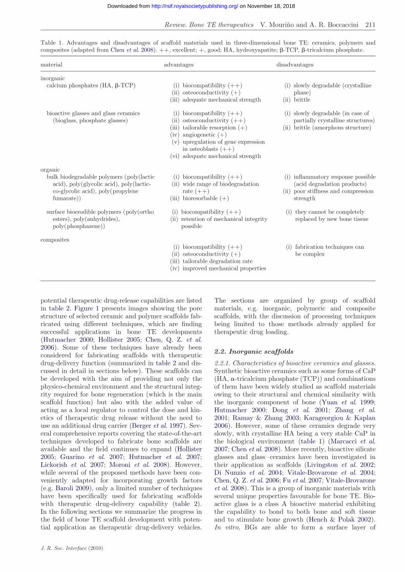
An optimal scaffold for bone TE should be osteocon-ductive and angiogenic and it should serve as athree-dimensional template to provide structural sup-port to the newly formed bone through aninterpenetrating network of pores (at least 100 mmwide) to allow cell migration, tissue in-growth and vas-cularization (Freyman et al. 2001; Hutmacher 2001;Guarino et al. 2007; Moroni et al. 2008). Analysis ofthe literature demonstrates that a wide range of three-dimensional bioactive scaffolds has been developedwhich can be potentially used as delivery systems fortherapeutic drugs relevant for bone repair processes.Basically, three major scaffold materials are used:ceramics, polymers and composites and their maincharacteristics are described briefly below. Comprehen-sive descriptions of scaffold fabrication techniques areavailable in the specialized literature (Hutmacher2000; Guarino et al. 2007; Chen et al. 2008; Moroniet al. 2008). Table 1 (Chen et al. 2008) summarizesthe advantages and disadvantages of the scaffoldmaterials used for bone TE, while the techniquesalready employed to fabricate bone scaffolds with
Table 1. Advantages and disadvantages of scaffold materials used in three-dimensional bone TE: ceramics, polymers andcomposites (adapted from Chen et al. 2008). þþ, excellent; þ, good; HA, hydroxyapatite; b-TCP, b-tricalcium phosphate.
Review. Bone TE therapeutics V. Mourino and A. R. Boccaccini 211
on November 18, 2018http://rsif.royalsocietypublishing.org/Downloaded from
potential therapeutic drug-release capabilities are listedin table 2. Figure 1 presents images showing the porestructure of selected ceramic and polymer scaffolds fab-ricated using different techniques, which are findingsuccessful applications in bone TE developments(Hutmacher 2000; Hollister 2005; Chen, Q. Z. et al.2006). Some of these techniques have already beenconsidered for fabricating scaffolds with therapeuticdrug-delivery function (summarized in table 2 and dis-cussed in detail in sections below). These scaffolds canbe developed with the aim of providing not only thephysico-chemical environment and the structural integ-rity required for bone regeneration (which is the mainscaffold function) but also with the added value ofacting as a local regulator to control the dose and kin-etics of therapeutic drug release without the need touse an additional drug carrier (Berger et al. 1997). Sev-eral comprehensive reports covering the state-of-the-arttechniques developed to fabricate bone scaffolds areavailable and the field continues to expand (Hollister2005; Guarino et al. 2007; Hutmacher et al. 2007;Lickorish et al. 2007; Moroni et al. 2008). However,while several of the proposed methods have been con-veniently adapted for incorporating growth factors(e.g. Baroli 2009), only a limited number of techniqueshave been specifically used for fabricating scaffoldswith therapeutic drug-delivery capability (table 2).In the following sections we summarize the progress inthe field of bone TE scaffold development with poten-tial application as therapeutic drug-delivery vehicles.
J. R. Soc. Interface (2010)
The sections are organized by group of scaffoldmaterials, e.g. inorganic, polymeric and compositescaffolds, with the discussion of processing techniquesbeing limited to those methods already applied fortherapeutic drug loading.
2.2. Inorganic scaffolds
2.2.1. Characteristics of bioactive ceramics and glasses.Synthetic bioactive ceramics such as some forms of CaP(HA, a-tricalcium phosphate (TCP)) and combinationsof them have been widely studied as scaffold materialsowing to their structural and chemical similarity withthe inorganic component of bone (Yuan et al. 1999;Hutmacher 2000; Dong et al. 2001; Zhang et al.2001; Ramay & Zhang 2003; Karageorgiou & Kaplan2006). However, some of these ceramics degrade veryslowly, with crystalline HA being a very stable CaP inthe biological environment (table 1) (Marcacci et al.2007; Chen et al. 2008). More recently, bioactive silicateglasses and glass–ceramics have been investigated intheir application as scaffolds (Livingston et al. 2002;Di Nunzio et al. 2004; Vitale-Brovarone et al. 2004;Chen, Q. Z. et al. 2006; Fu et al. 2007; Vitale-Brovaroneet al. 2008). This is a group of inorganic materials withseveral unique properties favourable for bone TE. Bio-active glass is a class A bioactive material exhibitingthe capability to bond to both bone and soft tissueand to stimulate bone growth (Hench & Polak 2002).In vitro, BGs are able to form a surface layer of
Table 2. Techniques developed to elaborate porous three-dimensional scaffolds for bone TE already applied to fabricatescaffolds with therapeutic drug-delivery capability.
technique details of the process references
melt moulding (i) scaffolds are prepared by melting polymers/ceramicsin the presence of porogens (such as sodium chloride,sugar crystals)
(ii) once the mixture is cooled, porosity is achieved bydissolving the porogens in water
(iii) finally, the porous scaffolds are usually lyophilized
Di Nunzio & Verne (2005)
solvent-casting (i) scaffolds are prepared by dissolving/suspendingpolymers/ceramics in the presence of porogens(such as sodium chloride or sugar crystals)
(ii) porosity is achieved by dissolving the porogensin water
(iii) after pouring the mixture into a mould, solventsare removed by either evaporation or vacuum/freeze-drying
Thomson et al. (1998)
freeze-drying (i) scaffolds are prepared by dissolving/suspendingpolymers/ceramics in water or in an organic solventfollowed by emulsification with a water phase
(ii) after pouring the mixture into a mould, solvents areremoved by freeze-drying and porous structuresare obtained
(i) scaffolds are prepared by dissolving/suspendingpolymers/ceramics in a solvent which freezes belowthe phase separation temperature of the polymersolution
(ii) porous structure is obtained by successivelyfreeze-drying
Zhang & Zhang (2002)
foaming (i) effervescent salts (ammonium bicarbonate) are usedas porogens and mixed with an organic viscoussolution/suspension of polymer/ceramic
(ii) after solvent evaporation, porosity is achieved byplacing scaffolds into hot water or an aqueoussolution of citric acid to dissolve the salts
(iii) an alternative is CO2-based gas
Mooney et al. (1996);Harris et al. (1998)
template technique (i) scaffolds are prepared by dipping a polyurethanesponge into a slurry of proper viscosity containingceramic particles
(ii) the impregnation step and the removal ofthe surplus slurry should be adjusted to obtain,after the sponge removal, a defect-free porousthree-dimensional scaffold
(iii) sometimes, in order to obtain mesoporous structures,surfactants may be added
Vitale-Brovarone et al. (2007);Chen, Q. Z. et al. (2006)
sol–gel (i) scaffolds are prepared by dissolving inorganic metalsalts or metal organic compounds in a solvent wherea series of hydrolysis and polymerization reactionsallow the formation of a colloidal suspension (‘sol’)
(ii) after casting the ‘sol’ into a mould, a wet ‘gel’is formed
(iii) with further drying and heat treatment, the ‘gel’is converted into dense ceramic or glass articles
Domingues et al. (2004)
powder compaction (i) scaffolds are prepared by compressing the polymers/ceramics using projectiles or punch and dies
(ii) the velocity of compaction of the projectile or punchand dies is adjusted to achieve powder consolidationand the desired porosity
(iii) the process can include sintering(iv) an alternative is to use uniaxial or isostatic pressing
Kimakhe et al. (1999); Vallet-Regıet al. (2001); Castro et al. (2005);Miyai et al. (2008)
212 Review. Bone TE therapeutics V. Mourino and A. R. Boccaccini
J. R. Soc. Interface (2010)
on November 18, 2018http://rsif.royalsocietypublishing.org/Downloaded from
Figure 1. Scanning electron microscopy images showing typi-cal pore structures of scaffolds successfully developed for boneTE: (a) bioactive glass–ceramic scaffolds fabricated by thefoam replica method (Chen, Q. Z. et al. 2006); (b) PDLLAfoam fabricated by thermally induced phase separation(Maquet & Jerome 1997); (c) polymer scaffold architecturesdeveloped by FDM (Hutmacher 2000). (Part (c) reproducedfrom Hutmacher (2000) with permission of Elsevier.)
Review. Bone TE therapeutics V. Mourino and A. R. Boccaccini 213
on November 18, 2018http://rsif.royalsocietypublishing.org/Downloaded from
microcrystalline HA when in contact with simulatedbody fluid (SBF) (Kim et al. 1995; Hench 1998;Fujibayashi et al. 2003). More recently, it has been dis-covered that ionic dissolution products released fromsilica-based BGs upregulate seven families of genesfound in osteoblasts (Xynos et al. 2001). Dissolutionproducts of BGs have also been shown to promoteangiogenesis (Day et al. 2004). Additionally, researchefforts on mesoporous inorganic materials have led tothe development of ceramic matrices with both drugdelivery and bone tissue regeneration capabilities(Vallet-Regı et al. 2006), which has promoted these bio-materials to novel uses in the biomedical field(Izquierdo-Barba et al. 2008).
2.2.2. Processing techniques. Numerous techniqueshave been developed to fabricate porous ceramic andglass–ceramic scaffolds, including starch consolidation,incorporation of volatile organic particles, sol–gel,
J. R. Soc. Interface (2010)
foaming, solid-free form fabrication methods andreplication of polymeric sponges (figure 1a) (Livingstonet al. 2002; Vitale-Brovarone et al. 2004, 2006, 2007;Chen, Q. Z. et al. 2006; Ahmad et al. 2008; Yun et al.2008; Yunos et al. 2008). In order to maximize themechanical strength of the scaffold, and depending onthe fabrication technique used, different parameterssuch as solid loading of the ceramic slurry, type andamount of additives (binders, dispersants, etc.) shouldbe optimized. However, the condition for preservingthe activity of a drug is that the process of drug incor-poration should not degrade it. Traditionally,fabrication of bioceramic scaffolds involves thermaltreatment at high temperature (.8008C), hence thisprocess cannot be used if a drug will be incorporatedbecause most of it would be degraded (Vallet-Regıet al. 2008). Therefore, different methods have beenproposed to tackle this problem. One of the simpleststrategies to load drugs within ceramic scaffolds is con-ventional impregnation, which consists of immersingthe scaffold in a drug containing phosphate-bufferedsaline (PBS) (Habraken et al. 2007). In this simplecase, however, the drug release rate usually cannot beadequately controlled (Habraken et al. 2007). Sol–geltechnology allows the introduction of drugs into glass-like materials at room temperature where pore for-mation is due to byproducts (water and alcohols) ofthe sol–gel synthesis (Nicoll et al. 1997; Santos et al.1999). These byproducts are volatile and evaporateduring the ageing and drying of the sol–gel materials,thus leaving pores (Nicoll et al. 1997; Santos et al.1999). However, materials with optimal scaffoldstructure (exhibiting high porosity and large intercon-nected macropores) are in general unable to hold alarge reservoir of drugs and they are not capable ofretaining the drug at the site for a prolonged periodof time (Malafaya et al. 2002; Ziegler et al. 2002).Further, solvents used in sol–gel methods have to beremoved with the disadvantage of adding a dryingstep to the process. This drying step usually includesheating, which, even if at moderate temperatures,can degrade thermolabile substances. One of the firstsuccessful attempts to solve this difficulty was the useof uniaxial and isostatic pressure at room temperatureto obtain gentamicin-loaded bioceramics (Vallet-Regıet al. 2001).
2.3. Polymer scaffolds
2.3.1. Characteristics of biodegradable polymers. Analternative approach to obtain multifunctional three-dimensional bioactive bone scaffolds includes theirfabrication from biodegradable polymers (Maquet &Jerome 1997; Hutmacher 2000; Hollister 2005; Rezwanet al. 2006; Guarino et al. 2007). In this case, it is con-sidered that biodegradable polymers can control therelease of drugs since polymer degradation propertiescan be tailored for each specific application (Hutmacher2000). Thus, a range of processes including cell growth,tissue regeneration and host response can be influencedby choosing suitable biopolymers (Lu et al. 1999;Hutmacher 2000). It is known that cells usually‘prefer’ hydrophilic surfaces; however, hydrophobic
214 Review. Bone TE therapeutics V. Mourino and A. R. Boccaccini
on November 18, 2018http://rsif.royalsocietypublishing.org/Downloaded from
materials have typically longer residence times in vivo(Streubel et al. 2000; Malafaya et al. 2002), meaningthat the selection of the optimal biodegradable polymerfor the drug-delivery function in TE scaffolds should beaimed at balancing both aspects. Synthetic biodegrad-able polymers that have been reported for variousdrug-eluting devices include PLGA copolymers (Garvinet al. 1994; Nie et al. 1995; Overbeck et al. 1995; Ambroseet al. 2003), polycaprolactone (Hendricks et al. 2001;Rutledge et al. 2003), polyanhydrides (Jacob et al. 1991;Nelson et al. 1997; Kanellakopoulou & Giamarellos-Bourboulis 2000; Li, L. C. et al. 2002; Li, W. 2002),polyhydroxybutyrate-co-hydroxyvalerate (PHBV)(Yagmurlu et al. 1999; Rossi et al. 2004) and other poly-hydroxyalkanoates (Turesin et al. 2001). Naturalpolymers including proteins such as collagen (Leeet al. 2002; Park et al. 2004; Sripriya et al. 2004;Prabu et al. 2006; Shanmugasundaram et al. 2006)and polysaccharides such as alginate, hyaluronic acidand chitosan (Muzzarelli et al. 1990; Chung et al. 1994;Mi et al. 2002; Aoyagi et al. 2007; Rossi et al. 2007) arealso attractive, since they exhibit superior biocompatibil-ity and can facilitate cell growth. Many of these polymersare inexpensive and readily available. Moreover, ‘surface-eroding polymers’, which have the feature of beingbiodegraded only at their surfaces, are also candidatesfor this application. Poly(anhydrides), poly(ortho-esthers) and polyphosphazene show this property(‘surface eroding’) as opposed to ‘bulk degradation’(Rezwan et al. 2006). Surface-eroding polymers offeradvantages over bulk degradation polymers when usedas multifunctional scaffold materials: (i) they are capableof releasing entrapped drugs by zero-order kinetics (whendrug release can be limited to regions undergoing degra-dation) and (ii) the release rate is proportional to thesurface area of the matrix (Heller 1985; Rezwan et al.2006).
2.3.2. Processing techniques. Porous polymer scaffoldscan be produced through thermally inducedphase-separation (TIPS) (figure 1b), evaporation,freeze-drying, solid-free form fabrication (e.g. fuseddeposition modelling (FDM), three-dimensionalprinting, selective laser sintering) (figure 1c), solvent-casting and foam-coating among others techniques(Maquet & Jerome 1997; Thomson et al. 1998; Whanget al. 1999; Zhang & Zhang 2002; Boccaccini &Maquet 2003; Guan & Davies 2004; Cabanas et al.2009). While a range of these methods have alreadybeen considered to incorporate therapeutic drugs, othersuitable scaffold manufacturing methods, e.g. solid-freeform fabrication methods (Hutmacher 2000; Hollister2005) (figure 1c), have not yet been investigated withthis purpose, thus remaining an area for future research.
Recently, impregnation of three-dimensional porousscaffolds using carbon dioxide (scCO2) has been usedto develop alternative clean processes for the prep-aration of drug-loaded polymeric matrices when thedrug is soluble in scCO2 and the polymer chosen canbe swollen by the supercritical fluid (Mooney et al.1996; Harris et al. 1998; Kikic & Sist 2000; Duarteet al. 2007; Heyde et al. 2007). Impregnation using
J. R. Soc. Interface (2010)
supercritical fluid technologies has the advantage ofusing a supercritical fluid with high diffusivity in thepolymer chosen in addition to its high solubility andplasticizing capability (Berens et al. 1992; Kazarian2000; Duarte et al. 2007). Electrospinning is anothertechnique with a potential to develop bioactivethree-dimensional bone scaffolds with drug-releasecapabilities (Li, W. et al. 2002; Matthews et al. 2002;Ma et al. 2005; Gupta et al. 2007). However, this tech-nique seems to have its limitations because someorganic solvents used to prepare the polymer solutionsmay degrade the drugs (Han & Gouma 2006; Guptaet al. 2007; Ahmad et al. 2008; Baroli 2009). Finally,solid-free form fabrication technologies such as three-dimensional printing and FDM appear promising forfurther developments in this field, as mentioned above(Hollister 2005; Hutmacher & Cool 2007; Hutmacheret al. 2007; Pang et al. 2007; Wang et al. 2007).
2.4. Composite scaffolds
2.4.1. Characteristics of biodegradable and bioactivecomposites. In this approach, usually the matrix is pre-pared by using biodegradable polymers and inclusionsin the form or particles of fibres of BG, CaP or HAare added to improve the mechanical strength andbioactivity (Roether et al. 2002; Rezwan et al. 2006;Guarino et al. 2007; Vallet-Regı et al. 2008). Polymerscombined with ceramic particles, such as HA, can alsobe applied as coating on porous bioceramic scaffolds,fabricated using one of the techniques mentioned in§2.2.2, in order to tailor the controlled release of adrug (Kim et al. 2004a,b; Yunos et al. 2008). Compositescaffolds represent a convenient alternative as theycombine the advantages of both biodegradable poly-mers and bioactive ceramics for bone engineeringscaffolds. This combination leads to composites withimproved mechanical properties because of the inherenthigher stiffness and strength of the inorganic material.In addition, bioactive inorganic particles such as HA,bioglass or tricalcium phosphate induce the effectiveinteraction of the scaffold with the surrounding bonetissue by forming a tenacious bond via the growth ofa carbonate HA layer, as mentioned above (Rezwanet al. 2006). Moreover, addition of inorganic materialsto bioresorbable polymers can change the polymerdegradation behaviour by buffering the pH of thenearby solution, thus preventing the autocatalyticeffect of the acidic end groups resulting from hydrolysisof polymer chains, e.g. in polylactic acid. It is wellknown that incorporation of bioactive inorganicphases in biodegradable polymers can enhance wateringress owing to the internal interfaces formed betweenthe polymer and the more hydrophilic bioactiveinclusions, hence enabling control of the degradationkinetics of scaffolds (Boccaccini & Maquet 2003).
2.4.2. Processing techniques. As mentioned above, oneof the simplest strategies is the application of biode-gradable polymer coatings loaded with the relevantdrugs onto the three-dimensional structure of biocera-mic scaffolds (Kim et al. 2004b). Other techniquesreported are: solvent-casting, TIPS, evaporation,
Review. Bone TE therapeutics V. Mourino and A. R. Boccaccini 215
on November 18, 2018http://rsif.royalsocietypublishing.org/Downloaded from
freeze-drying and foam-coating (table 2—Thomsonet al. 1998; Whang et al. 1999; Zhang & Zhang 2002;Boccaccini & Maquet 2003; Cabanas et al. 2009). Aninteresting approach to drug delivery in TE is to com-bine drug-loaded microspheres within the scaffoldmacroporous structure or even to use the loaded micro-spheres to construct an open-pore three-dimensionalscaffold structure (Malafaya et al. 2002; Vallet-Regıet al. 2008; Zhu & Kaskel 2009). The approach of com-bining microspheres and a matrix (scaffold) could bevery useful in cases where the scaffold material is notsuitable for the regulated release of specific drugs,such as low-molecular weight or water-soluble drugs(Malafaya et al. 2002). When microspheres are addedto an inorganic scaffold, they can interact with the sur-face of the scaffold by electrostatic charges dependingon the nature of the biodegradable polymer and thebioceramic used (Xu & Czernuszka 2008). Moreover,by using microspheres, drug-release profiles can bealtered and tuned depending on the polymer selected,as mentioned above.
A summary of experimental research carried out onthe development of three-dimensional scaffolds forbone TE with controlled-release capability is presentedin table 3 including both in vitro and in vivo studies.From this table it appears that porous matrices basedmainly on well-characterized biocompatible polymericscaffolds or, in some cases, composites comprising poly-meric matrices and added inorganic particles representthe best systems to date to incorporate therapeuticdrug delivery in bone TE approaches. A schematicdiagram summarizing the different strategies proposedso far is shown in figure 2. Although several novel tech-niques have been developed to introduce therapeuticdrugs within scaffolds, in most cases the strategyfollowed has been the direct incorporation of the druginto the scaffold by immersion of the scaffold in adrug-containing buffer aqueous solution. Nevertheless,thermo-labile drugs can also be loaded within three-dimensional scaffolds in a one-step process using roomtemperature compaction of the powder mix, this beinga solvent-free process, which avoids the use of toxicsolvents (Kimakhe et al. 1999; Vallet-Regı et al. 2001;Castro et al. 2005). Further details of relevant studiesare discussed in the following sections, highlighting thestrengths and weaknesses of the different approaches.
3. ANTIMICROBIAL AGENTS
3.1. Controlled release of antibiotics
Infection is defined as a homeostatic imbalance betweenhost tissue and the presence of micro-organisms at aconcentration that exceeds 105 organisms per gram oftissue or the presence of beta-haemolytic streptococci(Sussman & Bates-Jensen 2001; Zilberman & Elsner2008). The principal aim of treating wound infectionsis to reduce the bacterial load in the wound to a levelat which wound-healing processes can take place. Ifthe drug is released too quickly, the entire drugamount could be released before the infection isstopped. On the other hand, if the release of the drugis delayed, infection may set in further, thus making it
J. R. Soc. Interface (2010)
difficult to manage the healing of the wound (Gold &Moellering 1996; Gransden 1997; Zilberman & Elsner2008). Bacterial infection remains a major problemaffecting the service life of medical implants and scaf-folds (Segreti 2000; Virk & Osmon 2001). Generally,sources for contamination include the air of the operat-ing room and resident bacteria on the patient’s skin andbacteria already present in the body (An & Friedman1996; Zilberman & Elsner 2008). In addition tohuman pain and suffering, direct medical costs associ-ated with such infections are often extremely high(Hetrick & Schoenfisch 2006). Similar complexitiesencountered with conventional orthopaedic implantscan be expected in TE approaches based onimplantation of engineered biomaterial scaffolds.Device-associated infections are the result of bacterialadhesion and subsequent bio-film formation at theimplantation site. Inhibiting bacterial adhesion is oftenregarded as the most critical step in preventing infection.Upon implantation, there is a competition between theintegration of the material into the surrounding tissueand adhesion of bacteria to the implant surface. There-fore, implantation will be successful only if tissueintegration occurs prior to considerable bacterialadhesion, thus preventing colonization of the implant(Gristina 1987). Furthermore, certain bacterial speciesare able to attach to the implant surfaces and form aprotective bio-film layer, which is extremely resistant toboth the immune system and antibiotics. Thesebio-films are considered the primary cause of implant-associated infection (Hetrick & Schoenfisch 2006).Local antibiotic release profiles should exhibit a highinitial release rate in order to respond to the elevatedrisk of infection from bacteria introduced during theinitial shock, followed by a sustained release at an effec-tive level for inhibiting the occurrence of latent infection(Zilberman & Elsner 2008). In the case of orthopaedic-related devices, including TE scaffolds, it is importantto combat bacteria possibly introduced during implan-tation and also those introduced systemicallyafterwards. Therefore, sustained drug release is necessary(Zilberman & Elsner 2008).
The most common antibiotic carrier to treat osteo-myelitis after debridement surgery to remove necroticbone tissue is poly(methylmethacrylate) (PMMA)beads (Vallo et al. 2004; Habraken et al. 2007, 2008;Shi et al. 2009). However, PMMA is not a resorbablepolymer and must be removed in a second surgical pro-cedure (Vallet-Regı et al. 2007; Shi et al. 2009). In thiscontext, it should be pointed out that most previouswork on loading biomaterials with antibiotics for ortho-paedic applications has been carried out on bone-fillermaterials and bone cements (Takechi et al. 1998;Armstrong et al. 2002; Diez-Pena et al. 2002; Gburecket al. 2002; Joseph et al. 2003; Hanssen 2004; Joostenet al. 2004; Webb et al. 2005; Schnieders et al. 2006;Krasko et al. 2007; Zilberman & Elsner 2008). In oneof the first attempts to develop multifunctionalre-absorbable implants, Queiroz et al. (2001) preparedsodium ampicillin which was adsorbed onto HAand glass-reinforced HA composites as a potentialpharmaceutical formulation for periodontitis. Ampicillin-loaded methylpyrrolidinone chitosan microparticles
Table 3. Selected experimental trials carried out for three-dimensional bone scaffolds, both in vivo and in vitro, with acombination of controlled drug release. b-TCP, b-tricalcium phosphate; CP, calcium phosphate invert glasses; GS, gentamicin;HA, hydroxyapatite; HMS, mesoporous silica; PLA, poly(L-lactic acid); PLGA, poly(lactide-co-glycolide); PCL, poly-1-caprolactone; PBS, phosphate-buffered saline; CPA, calcium phosphate-deficient apatite.
triblock copolymerstechnique þfunctionalization þimmersion in drug-containingbuffer aqueous solution
in vitro Nieto et al. (2008)
zoledronate CPA pellets suspension of CDA in drug-containing water solution
in vitro Fauchex et al.(2009)
216 Review. Bone TE therapeutics V. Mourino and A. R. Boccaccini
on November 18, 2018http://rsif.royalsocietypublishing.org/Downloaded from
have been described for a similar purpose (Giunchediet al. 1998). Moreover, calcium phosphate cement(CPC)–chitosan composites have been applied fordrug release of the cephalosporin antibiotic flomoxef,which is active against methicillin-resistant Streptococcusaureus. For example, Takechi et al. (2002) added flomoxefsodium to the liquid phase of tetracalcium phosphate(TTCP) cement/chitosan composites and measured thedrug release from preset discs for 3 days. Results showed
J. R. Soc. Interface (2010)
a release pattern that was characterized by an initialburst, followed by a more sustained release. The totalper cent of drug released in 24 h was 24–35% and itwas found that the addition of chitosan in differentamounts did not influence the total drug release after72 h. Moreover, the release from these chitosan-enrichedcements did not differ significantly from that of normalTTCP cement, though the maximum amount of chitosanused was 1.0 per cent w/v. Analysis of the literature
Figure 2. Schematic representation of the most common strategies to deliver drugs from three-dimensional scaffolds in bone TE.Drugs may be adsorbed onto the pore surface of the scaffolds in either their unprotected (a) or their protected (microsphere/matrix) (b) forms. Alternatively, drugs may be entrapped in the scaffold structure in either their unprotected (c) or theirprotected (microsphere/matrix) (d) forms.
Review. Bone TE therapeutics V. Mourino and A. R. Boccaccini 217
on November 18, 2018http://rsif.royalsocietypublishing.org/Downloaded from
reveals that new multifunctional composite materials,produced by different techniques, are being developedfor specific application in bone TE and are detailedbelow, with a focus on specific antibiotics used inthree-dimensional TE scaffolds.
3.2. Gentamicin
Gentamicin is a commonly employed antibiotic intrauma, widely used for the treatment of osteomyelitisbecause of its broad-spectrum characteristics (Li & Hu2001). Most of the major bacteria causing chronic osteo-myelitis are sensitive to gentamicin. This drug has beenloaded in several scaffolds in order to evaluate their abilityas controlled-release carriers (Li & Hu 2001). Zhang &Zhang (2002) prepared macroporous chitosan scaffoldsreinforced by CaP particles such as b-TCP and CaPinvert glasses using a thermally induced phase-separationtechnique. These porous composite materials were loadedwith gentamicin sulphate (GS) by immersing them indrug-containing PBS solutions. In vitro tests showedthat, in comparison with GS-loaded pure chitosanscaffolds, the initial high burst release of GS wasdecreased through incorporating CaP crystals and glassparticles into the scaffolds, and a sustained release formore than three weeks was achieved (Zhang & Zhang2002). The highest sustained release was observed fromthe particle-containing composite, which was suggestedto occur owing to a higher extent of chitosan cross-linking.Scanning electron microscopy micrographs showed noapparent morphological differences for osteoblastic cellsgrown on the pure chitosan scaffolds and those grownon composite scaffolds. The cells attached and migratedon these scaffolds, suggesting a good cellular compatibil-ity (Zhang & Zhang 2002; Habraken et al. 2007).
J. R. Soc. Interface (2010)
Vallet-Regı and co-workers used uniaxial and isostaticpressure at room temperature to obtain GS-loaded bio-ceramic tablets (Vallet-Regı et al. 2001). These tabletswere tested in vivo in New Zealand rabbit femurs forone, four, eight and 12 weeks to study their biologicalresponse. The bone response to the implant was of perfectosseo-integration. The local GS levels detected in bonetissue were above the minimal inhibitory concentration(MIC) and they were effective because they were toxicfor the majority of the resident micro-organisms. Inaddition, there was progressive decrease of GS levels inbone tissue with time, but the levels were always abovethe MIC until the end of the assay. In related studies,Zhu & Kaskel (2009) compared in vitro the local drug-release behaviour of two bioactive silicate glass scaffolds:three-dimensional mesoporous (SiO2–CaO–P2O5)(MBG) and three-dimensional BG scaffolds. Both scaf-folds were prepared by using the polyurethane spongetechnique, which involves the burning-out of a sacrificialsponge to create the pore structure (figure 1a) (Chen,Q. Z. et al. 2006); but in case of MBG scaffold a surfactantwas added. Afterwards, the scaffolds were immersed ina gentamicin solution for loading the drug. The resultsindicated that the mesoporous structure played an impor-tant role in the drug-loading capability and its releaserate. The drug-uptake capacity of the MBG scaffoldwas over twofold higher than that of the BG scaffold.During the whole release period in SBF, gentamicin wasreleased from the MBG scaffold at a much lower releaserate than from the BG scaffold. A novel design of a TEscaffold with controlled drug-delivery capability hasbeen developed by Shi et al. (2009). The scaffold isbased on mesoporous silica–HA (HMS–HA) compositeparticles used as fillers in PLGA microspheres. HMS–HA particles were produced using dodecylamine as a
218 Review. Bone TE therapeutics V. Mourino and A. R. Boccaccini
on November 18, 2018http://rsif.royalsocietypublishing.org/Downloaded from
template and GS-loaded PLGA microspheres were pre-pared using a double emulsion solvent evaporationtechnique (water/oil/water). PLGA/HMS–HA–GScomposite microspheres were prepared using a singleemulsion solvent evaporation method. Afterwards,PLGA or PLGA/HMS–HA–GS microsphere sinteredscaffolds were fabricated by pouring PLGA or PLGA/HMS–HA–GS microspheres into cylindrical moulds,and subsequently sintering at 708C for 2 h. The resultsshowed that the presence of HA in PLGA/HMS–HAscaffolds could balance the decreased pH values causedby the acidic degradation product of PLGA. Moreover,HMS–HA improved the cytocompatibility and bioactiv-ity of PLGA. It was also claimed that the compressivestrength and elastic modulus of PLGA/HMS–HA scaf-folds were higher than those of pure PLGA scaffolds,showing similar mechanical properties to human cancel-lous bone (Shi et al. 2009). In vitro drug-delivery testingin SBF of the PLGA/HMS–HA scaffolds showed thatPLGA reduced the GS release from HMS–HA particles,and the release lasted for nearly one month (Shi et al.2009).
3.3. Other antibiotics
A series of antibiotics has been further considered in com-bination with scaffolds for bone TE. Tetracycline wasincorporated in a hybrid coating consisting of polycapro-lactone (PCL) and HA powder, which was applied onthe surface of a HA porous bone scaffold through dip-coat-ing and solvent-casting method (Kim et al. 2004a). Invitro drug-delivery testing in PBS revealed that the releaseamount was controlled via the coating cycle and initialdrug loading and a sustained release preceded by a burstrelease during the first 2 h was achieved (Kim et al.2004a). Domingues et al. (2004) introduced tetracyclineand an inclusion complex formed by tetracycline andb-cyclodextrin, at 1:1 molar ratio tetracyline/b-cyclodex-trin, into a BG prepared by the sol–gel technique. Theinclusion complex tetracycline/b-cyclodextrin wasprepared by freeze-drying and either tetracycline hydro-chloride or tetracycline in b-cyclodextrin inclusioncompound was loaded into sol–gel solutions to preparedrug-loaded silicate BGs. An initial burst of 12 per centwas observed in vitro (in SBF), followed by a sustainedrelease over 80 days achieving a total release of 22–25%.The in vivo test was carried out with three groups offemale mice treated with BG without drugs, or associatedwith tetracycline (BT), orwith tetracycline/b-cyclodextrin(BTC) by subcutaneous implantation. A considerablebacteriostatic activity was found with BT and BTC-loaded glasses, when compared with plain glass. Thepresence of cyclodextrin was important to slow downthe release of tetracycline for a long period of timeand it was verified that the presence of tetracycline orits inclusion complex, tetracycline/BTC, did not affectthe bioactivity of the glass. Recently, Cabanas et al.(2009) loaded vancomycin (VAN), a drug susceptibleto heat degrading, into b-TCP/agarose scaffolds bytwo different methods: freeze-drying and desiccationat 378C. Poly(ethylene glycol) (PEG) was included inthe formulation to tailor the release of VAN (Cabanaset al. 2009). The freeze-dried samples had a higher
J. R. Soc. Interface (2010)
porosity structure than samples dried at 378C. Thepieces obtained showed a microstructure similar tothat of human cancellous bone. In addition, the differ-ent pore architectures and the formation of anagarose–PEG–VAN complex yielded different drug-release patterns (Cabanas et al. 2009). Polymyxin B(PMB) is a polypeptidic antibiotic that undergoes ther-modamage above 608C. With this limitation to be takeninto account, Kimakhe et al. (1999) made one of thefirst attempts to obtain a multifunctional inorganicmatrix for a bioactive drug-delivery system (DDS) inwhich the effect of a released therapeutic agent isfavoured by the biocompatibility, osteoconductivityand bioresorption of the ceramic material. PMB wasloaded on CaP powders using a dynamic compactionmethod at high velocities (25 and 50 m s21) withoutexternal heating. The compaction procedure did notcause any loss in PMB integrity and biological activity.PMB release in vitro began after 2–3 days of incubationfor blocks compacted at 25 m s21 velocity and on day 5for those compacted at 50 m s21 (Kimakhe et al. 1999).
Some important results have shown the potentialeffectiveness of quinolones in local DDS designed totreat bone infection using carriers such as HA–anioniccollagen composite (Martins & Goissis 2000), biode-gradable polymers (Overbeck et al. 1995; Nicolauet al. 1998; Nie et al. 1998; Ramchandani & Robinson1998; Kanellakopoulou & Giamarellos-Bourboulis2000; Desevaux et al. 2002) and mixtures of calciumphosphates and biodegradable polymers (Castro et al.2003). A composite of gatifloxacine (GFLX)-loadedpoly-x-caprolactone combined with x-TCP porous cer-amic was obtained by compression moulding, followedby sintering (Miyai et al. 2008). This process had theadvantage of being a solvent-free process. GFLXmostly retained its bactericidal property after proces-sing. In vitro testing in Hanks’ balanced solutionshowed that the composite of GFLX-loaded PCL/bTCP ceramic released GFLX for four weeks and hadsustained bactericidal activity against S. milleri andBacteroides fragilis for at least one week (Miyai et al.2008). In vivo tests in rabbits with osteomyelitis lesionsinduced by S. milleri and B. fragilis in the rabbit mand-ible showed that the composite of GFLX-loaded PCL/xTCP was effective in controlling infection at the bonedefect formed by debridement. Moreover, after fourweeks, new bone formation was observed on the surfaceof the composite. Further, after 50 weeks, ingrowingbone tissue with vascular vessels was observed alongthe PCL and b-TCP interface. Additionally, GFLXconcentrations in the serum and soft tissues were verylow suggesting a low risk of systemic toxicity (Miyaiet al. 2008). Ciprofloxacin (CFX) is the most widelyused fluoroquinolone for bacterial bone infection witha low MIC (0.25–2 mg ml21). CFX has been consideredin combination with bioceramics and polymers. Forexample CFX was loaded into a composite comprisingHA, TCP and poly(DL-lactide) (PLA) to treat compli-cated multi-organism bone infections that requiredhigh antibiotic concentrations (40%) in bone for longperiods (Castro et al. 2005). The composites were pre-pared by mixing the ingredients and by furtheruniaxial compression using a hydraulic press. In vitro
Review. Bone TE therapeutics V. Mourino and A. R. Boccaccini 219
on November 18, 2018http://rsif.royalsocietypublishing.org/Downloaded from
results showed that the release rate decreased propo-rtionally to the PLA/CaP ratio, drug-loading andcompaction pressure as a more tightly packed matrixwas obtained. In vivo tests in rabbits showed that CFXconcentrations along the bond remained higher than theMIC against the most common pathogens causing osteo-myelitis. A good in vivo/in vitro correlation was found;however, the release of the last 20 per cent CFX remainingwithin the matrix was faster in vivo than in vitro, as therelease was enhanced in vivo by neovascular vesselsformed inside the matrix (Castro et al. 2005).
3.4. Inorganic ions
Silver, in its oxidized form (Agþ), is an antibacterial agentand it has been proposed as an additive in bone TE scaf-folds (Di Nunzio et al. 2004). Although the antibacterialactivity of silver ions has been demonstrated in severalworks (Matsuura et al. 1997; Gatter et al. 1998; Kimet al. 1998; Adams et al. 1999; Kawashita et al. 2000;Bellantone et al. 2002; Blaker et al. 2004; Di Nunzioet al. 2004; Verne et al. 2005), the silver ion antimicrobialmechanism is not fully understood. It is well known thatsilver ions can interact with bacterial cells in differentways: they can bind to microbial DNA preventing bac-terial replication or to sulphydryl groups of bacterialenzymes, inhibiting cell respiration and binding transportof important substances across the cell membrane andwithin the cells (Chen, W. et al. 2006). These differentways of interaction are the origin of low bacterialresistance to silver (Chen, W. et al. 2006; Hetrick &Schoenfisch 2006). The bactericidal activity of Agþ iseffective against a broad range of bacteria potentiallyfound at the sites of scaffolds or implants includingS. aureus (Hetrick & Schoenfisch 2006).
The incorporation of silver into bone TE scaffolds is anovel approach. In one of the earliest studies, Agþ ionswere introduced into three-dimensional bioactiveglass–ceramic scaffold surfaces through a patented ion-exchange process (Di Nunzio & Verne 2005). The controlof Agþ content on the surface, as well as its diffusion pro-file throughout the ion-exchanged layer, was achieved by acareful choice of the ion-exchange parameters (tempera-ture, time and silver concentration in the molten bath).According to the authors of the original investigation(Di Nunzio & Verne 2005), this technique allows, bytuning the process parameters, a controlled silver ionincorporation into the superficial layers of the scaffold,maintaining the scaffold structure and its characteristicsunchanged (Di Nunzio et al. 2004; Vitale-Brovaroneet al. 2008). Moreover, the possibility of usingAgþ-doped BG as coating of polymeric fibres (sutures)for fabricating textile scaffolds was investigated byBlaker et al. (2004). The authors indicated that the Ag-loaded BG coating of textile structures can represent aconvenient alternative to fabricate multifunctional bio-degradable scaffolds (Blaker et al. 2004). In a relatedwork, Pratten et al. (2004) investigated in vitro the abilityof a silver-doped BG (AgBG) coating, which was elabo-rated using a slurry-dipping process, to preventStaphylococcus epidermidis colonization on surgicalsutures. The in vitro studies were carried out under bothbatch and flow conditions to quantify the number of
J. R. Soc. Interface (2010)
viable cells adhered to the surface and to determine theattachment and detachment over time, respectively. Theauthors showed that AgBG coating had a significanteffect on preventing S. epidermidis attachment whencompared with coatings of standard 45S5 Bioglass.
4. OTHER USED DRUGS
4.1. Anti-inflammatory drugs
Implantation of engineered biomaterials might causelocal inflammation owing to the host immunoresponse(Corry & Moran 1998; Mendez et al. 2004; Gonzalez-Corchon et al. 2006; Chevalier et al. 2009), whichtherefore requires the use of anti-inflammatory agents,either steroids (glucocorticoids) or non-steroids. In par-ticular, glucocorticoids have been shown to have stronginhibitory effects on cytokine-related inflammationby downregulating transcription of interleukin (IL)-1,tumour necrosis factor (TNF)-a, granulocyte macro-phage–colony-stimulating factor (GM-CSF), IL-3, IL-4,IL-5, IL-6 and IL-8 (Norton et al. 2005; Novak et al.2009). In particular, dexamethasone (DEX) is alsoused in TE for the preparation of osteogenic medium(Duarte et al. 2009a,b). In this context, Duarte et al.(2009b) impregnated DEX using supercritical fluidtechnology in chitosan porous scaffolds prepared by afreeze-drying process. The highest loading was obtainedat low pressures and temperatures (8.0 MPa and 358C),corresponding to a lower solubility of DEX in the super-critical fluid (carbon dioxide) and a low swelling of thepolymeric matrix. Results from in vitro drug-releasestudies showed that the release of DEX from chitosanscaffolds exhibited a sustained profile, which is suitablefor application in TE. Supercritical fluid technology canbe adapted to prepare porous structures using a varietyof polymeric systems or composites and other bioactivecompounds in order to develop multifunctional scaf-folds with improved mechanical properties andbiocompatibility (Quirk et al. 2004). In this context,Duarte et al. (2009a) reported the feasibility of usingsupercritical fluid methods to process in one step aporous matrix loaded with a pharmaceutical agent forTE purposes. In order to investigate the scaffold’sperformance, the release of DEX loaded in a three-dimensional starch-based porous matrix was tested.A supercritical phase-inversion technique, usingcarbon dioxide as the supercritical fluid, was employedtoprepare the composite scaffolds ofDEXandapolymericblend of starch and poly(L-lactic acid). In vitro drug-release studies were carried out in PBS and resultsshowed that a sustained release of DEX was achievedover21days.Thefittingofapower lawtothe experimentaldata demonstrated that drug release is governed by ananomalous transport, i.e. it was proposed that both thedrug diffusion and the swelling of the matrix influencethe release ofDEXout of the scaffold (Duarte et al. 2009a).
Within non-steroids, ibuprofen (IBU) has been lar-gely used orally, intravenously and even topically.Recently, Mortera et al. (2007) used silica-orderedmesophase (MCM-41) submicron spheres incorporatedinside glass–ceramic (SiO2–CaO–K2O) bioactive scaf-folds for the controlled delivery of IBU in TE. The
220 Review. Bone TE therapeutics V. Mourino and A. R. Boccaccini
on November 18, 2018http://rsif.royalsocietypublishing.org/Downloaded from
scaffolds were fabricated by dipping the glass–ceramicscaffold into the MCM-41 synthesis solution and afterthat samples were dipped into an IBU solution fordrug adsorption over 3 days. The in vitro drug-releasestudy performed in SFB showed a fast release of IBUduring the first 8 h followed by a slower release between8 and 120 h (Mortera et al. 2007).
4.2. Bisphosphonates
Bisphosphonates (BPs) have been in widespread use sincethe 1970s for the treatment of a variety of bone diseasescharacterized by osteoclast-mediated bone resorptionsuch as Paget’s disease, tumour-induced hypercalcaemia,metastatic bone diseases and osteoporosis (Siris et al.1996; Colucci et al. 1998; Major et al. 2000; Rodan &Martin 2000; Van Beek et al. 2003; Miller 2005; Nietoet al. 2008; Panzavolta et al. 2009). BPs act by inhibitingthe osteoclastic resorption of bone tissue (Colucci et al.1998;Van Beeket al. 2003), they bind strongly toHAcrys-tals andare retained fora long time inbone, being excretedunmetabolized in urine (Papapoulos 2006; Panzavoltaet al. 2009). Chemically, BPs are analogous to pyropho-sphates, which are natural modulators of bonemetabolism (Nancollas et al. 2006;Nieto et al. 2008).How-ever, in BPs, the oxygen atom that binds the twophosphate groups of pyrophosphate (P–O–P) issubstituted by a carbon atom (P–C–P) making themolecule less resistant to hydrolysis than pyrophosphates(Nancollas et al. 2006;Papapoulos 2006;Nieto et al. 2008).Recently, the positive effect of adjunct treatment withinjectable antiresorptive zoledronic acid on the biologicalprocess induced by BG incorporation in bone defects hasbeen proved (Valimaki et al. 2006). A member of thefamily of BPs, sodium alendronate (SA) was used byNieto and collaborators (Nieto et al. 2008) as a drugmodel to test how the organic modification of the surfaceof silica-based ordered mesoporous scaffolds can controlthe drug dosage. The mesoporous silica structure (SBA-16) was synthesized using triblock copolymers followedby functionalization by acid catalysis. Finally, SA wasadsorbed by immersing the specimens in a drug-containing buffered aqueous solution. The adsorptionrates of SA, and consequently the SA release rates, weretuned by using a range of amine-functionalization degrees(Nieto et al. 2008). Recently, Fauchex et al. (2009) usedcalcium-deficient apatite as a carrier for delivery ofzoledronate acid (ZA) in bone tissue. According to thein vitro tests carried out, the released ZA inhibited osteo-clastic resorption without affecting osteoblasts. Futurestudies are planned to develop three-dimensional TEscaffolds loaded with BPs and the use of BPs in TEapproaches is bound to increase as novel methodologiesare developed for the efficient loading of BPs in porousthree-dimensional matrices.
5. EVALUATING THE EFFICACY OFBIOACTIVE BONE SCAFFOLDS ASDRUG-DELIVERY SYSTEMS
It is important to highlight that even though several invitro tests have been performed in order to investigatethe suitability of bone scaffolds as drug carriers, there is
J. R. Soc. Interface (2010)
no in vitro standard technique available for this purposeyet. Indeed, several methods described in the literaturetry to simulate in vivo conditions and to avoid non-physio-logical turbulence. In most cases, loaded scaffolds aresoaked in a beaker, a bottle or a sampling tube and/or ina culture chamber with or without controlled agitation(Guicheux et al. 1997; Paul & Sharma 1999; Krajewskiet al. 2000; Perry et al. 2002; Hasegawa et al. 2004;Murugan & Ramakrishna 2004; Palazzo et al. 2005;Medvecky et al. 2007; Melville et al. 2008; Victor &Kumar 2008; Chevalier et al. 2009). Released tests areusually performed considering only low volumes (from 3up to 150 ml) of various dissolution media (phosphatebuffer, saline physiological solution, SBF, etc.). For boneTE, SBF is frequently the dissolution medium of choiceas it allows the study of how the release of a drug can beaffected by the formation of a surface layer of microcrystal-line HA on the bioactive scaffold, which is the expectedbehaviour in vivo. There are several sampling methodsreported for studying the release of drug substances frombiomaterials (replacing the entire volume with freshmedia after taking a sample, replacing the same volumewithdrawn after taking a sample); sometimes ensuringsink conditions (ensuring that sufficient medium volumeis available during the test to dissolve the total drug quan-tity from the loaded scaffold) and sometimes usingcontinuous flow through chambers (flow rate from 0.02to 2.5 ml min21) (Perry et al. 2002; Hall et al. 2004;Chevalier et al. 2009). Moreover, it could be beneficial tomimic the blood/plasma flow speed observed at the siteof the scaffold’s implantation (Baroli 2009). Further, andsurprisingly, there is very limited reported informationregarding the stability of the drugs involved in the studies,in particular the thermal stability. If a drug is includedwithin a scaffold in order to be released over a month orlonger period, the normal stability tests usually carriedout by suppliers will not be sufficient; further testsshould be done in order to evaluate the activity and drugdegradation rate at body temperature for a prolongedperiod of time. In addition, when in vivo tests were per-formed, little work reported on the drug clearance fromthe zone and how the new vascularization induced bythe presence of the scaffold would affect the residencetime of the drug, especially when the matrix is expectedto sustain the release of the drug for a prolonged periodof time (Castro et al. 2003). In this regard, also very littlework has been carried out on the correlation of in vitroand in vivo results of drug release to evaluate the signifi-cance of establishing novel in vitro models, which wouldrepresent realistic studies, prior to in vivo tests on hostresponse after scaffold implantation (Malafaya et al. 2002).
6. CONCLUSIONS AND PERSPECTIVES
In reviewing the published studies on bioactive boneTE scaffolds with additional drug-delivery capability,it becomes clear that there have been continuedadvances, particularly in recent years, towards thefurther development of the field. New materials andcombinations of materials, as well as improved scaffolddesigns based on novel processing techniques, are beingcontinuously proposed to advance the drug-delivery
Review. Bone TE therapeutics V. Mourino and A. R. Boccaccini 221
on November 18, 2018http://rsif.royalsocietypublishing.org/Downloaded from
capability of bone tissue scaffolds. However, a largeamount of biological information is still needed tofully understand the in vitro and in vivo performanceof such scaffolds in specific applications, which onceobtained will provide a rational scaffold design andoptimization of their drug-delivery function. Forexample, analysis of the literature has revealed thatthere is a lack of biological data regarding the specificconcentration in which a given drug is needed inrelation to the particular local microenvironment andhow this is affected by the interaction between the scaf-fold and new tissue, considering also the effect onvascularization. In addition, during the design anddevelopment of multifunctional scaffolds and in orderto control the drug-release patterns, the effect of proces-sing parameters on scaffold microstructure (i.e.porosity) and on the resulting drug-release profiles,stability, biodegradation behaviour, as well as thescaffold’s mechanical and physical properties, must befurther investigated. Despite significant efforts in thisdirection, several challenges have yet to be resolved.These include understanding the link between the keyvariables determining the scaffold-processing conditionsand the physico-chemical properties of the novel three-dimensional delivery systems with special considerationgiven to the stability of the incorporated drug. Thesechallenges also include achieving accurate control overtime and space of specific quantities of drug fordetermined applications and engineering the releasepatterns, which are related to the interaction betweenthe drug and the scaffold (matrix degradation and ero-sion and drug diffusion through the matrix), drugsolubility and the amount of drug loaded available tobe delivered. Further, only limited work has beenreported on how processing parameters and sterilizationcan affect the homogeneous loading, the stability andthe release kinetics of the drug incorporated. Inaddition, there is a lack of focus in several works interms of the pathology they would like to address. Itis clear that the combination of drug delivery and TEusing advanced three-dimensional scaffold concepts isin its early years and it will take time to achieve clinicalresults of relevance. It is therefore expected that thisfield will keep growing within the next few years andrelated research outputs could lead to results with thepotential to transform the clinical approach tobone-related pathologies. Specifically, in order toachieve these goals, it would be important to intensifythe working collaboration of researchers and technol-ogists from different relevant communities. Theinterdisciplinary character of TE, allowing the conflu-ence of different scientific fields, backgrounds andknowledge, is mandatory for the advance of bone TEtherapeutics.
REFERENCES
Adams, A. P., Santschi, E. M. & Mellencamp, M. A. 1999Antibacterial properties of a silver chloride-coated nylonwound dressing. Vet. Surg. 28, 219–225.
Ahmad, Z., Zhang, H. B., Farook, U., Edirisinghe, M., Stride,E. & Colombo, P. 2008 Generation of multilayered
J. R. Soc. Interface (2010)
structures for biomedical applications using a novel tri-needle coaxial device and electrohydrodynamic flow.J. R. Soc. Interface 5, 1255–1261. (doi:10.1098/rsif.2008.0247)
Ambrose, C. G., Gogola, G. R., Clyburn, T. A., Raymond,A. K., Peng, A. S. & Mikos, A. G. 2003 Antibiotic micro-spheres: preliminary testing for potential treatment ofosteomyelitis. Clin. Orthop. Relat. Res. 415, 279–285.(doi:10.1097/01.blo.0000093920.26658.ae)
An, Y. H. & Friedman, R. J. 1996 Prevention of sepsis in totaljoint arthroplasty. J. Hosp. Infect. 33, 93–108. (doi:10.1016/S0195-6701(96)90094-8)
Aoyagi, S., Onishi, H. & Machida, Y. 2007 Novel chitosanwound dressing loaded with minocycline for the treatmentof severe burn wounds. Int. J. Pharm. 330, 138–145.(doi:10.1016/j.ijpharm.2006.09.016)
Armstrong, M. S., Spencer, R. F., Cunningham, J. L.,Gheduzzi, S., Miles, A. W. & Learmonth, I. D. 2002Mechanical characteristics of antibiotic-laden bonecement. Acta Orthop. Scand. 73, 688–690. (doi:10.1080/000164702321039697)
Arosarena, O. 2004 Tissue engineering. Curr. Opin. Otolaryn-gol. Head Neck Surg. 13, 233–241.
Baroli, B. 2009 From natural bone graft to tissue engineeringtherapeutics: brainstorming on pharmaceutical formula-tive requirements and challenges. J. Pharm. Sci. 98,1317–1375. (doi:10.1002/jps.21528)
Bellantone, M., Williams, H. D. & Hench, L. L. 2002 Broad-spectrum bactericidal activity of Ag2O-doped bioactiveglass. Antimicrob. Agents Chemother. 46, 1940–1945.(doi:10.1128/AAC.46.6.1940-1945.2002)
Berens, A. R., Huvard, G. S., Korsmeyer, R. W. & Kunig, F.W. 1992 Application of compressed carbon dioxide in theincorporation of additives into polymers. J. Appl. Polym.Sci. 46, 231–242. (doi:10.1002/app.1992.070460204)
Berger, R. A., Jacobs, J. J., Quigley, L. R., Rosenberg, A. G. &Galante, J. O. 1997 Primary cementless acetabularreconstruction in patients younger than 50 years old.7- to 11-year results. Clin. Orthop. Relat. Res. 344,216–226.
Blaker, J. J., Nazhat, S. N. & Boccaccini, A. R. 2004 Develop-ment and characterisation of silver doped bioactiveglass-coated sutures for tissue engineering and woundhealing applications. Biomaterials 25, 1319–1329.(doi:10.1016/j.biomaterials.2003.08.007)
Boccaccini, A. R. & Maquet, V. 2003 Bioresorbable andbioactive polymer/bioglassR composites with tailoredpore structure for tissue engineering applications. Comp.Sci. Technol. 63, 2417–2429. (doi:10.1016/S0266-3538(03)00275-6)
Bostman, O. & Pihlajamaki, H. 2000 Clinical biocompatibilityof biodegradable orthopaedic implants for internal fix-ation: a review. Biomaterials 21, 2615–2621. (doi:10.1016/S0142-9612(00)00129-0)
Bran, G. M., Stern-Straeter, J., Hormann, K., Riedel, F. &Goessler, U. R. 2008 Apoptosis in bone for tissue engineer-ing. Arch. Med. Res. 39, 467–482. (doi:10.1016/j.arcmed.2008.02.007)
Buckwalter, J. A., Glimcher, M. J., Cooper, R. R. & Recker,R. 1996a Bone biology. I. Structure, blood supply, cells,matrix, and mineralization. Instr. Course Lect. 45,371–386.
Buckwalter, J. A., Glimcher, M. J., Cooper, R. R. & Recker,R. 1996b Bone biology. II. Formation, form, modeling,remodeling, and regulation of cell function. Instr. CourseLect. 45, 387–399.
Cabanas, M. V., Pena, J., Roman, J. & Vallet-Regı, M. 2009Tailoring vancomycin release from x-TCP/agarose
222 Review. Bone TE therapeutics V. Mourino and A. R. Boccaccini
on November 18, 2018http://rsif.royalsocietypublishing.org/Downloaded from
scaffolds. Eur. J. Pharm. Sci. 37, 249–256. (doi:10.1016/j.ejps.2009.02.011)
Castro, C., Sanchez, E., Delgado, A., Soriano, I., Nunez, P.,Baro, M., Perera, A. & Evora, C. 2003 Ciprofloxacinimplants for bone infection. In vitro– in vivo characteriz-ation. J. Control. Release 93, 341–354. (doi:10.1016/j.jconrel.2003.09.004)
Castro, C., Evora, C., Baro, M., Soriano, I. & Sanchez, E.2005 Two-month ciprofloxacin implants for multibacterialbone infections. Eur. J. Pharm. Sci. 60, 401–406.
Chen, Q. Z., Thompson, I. D. & Boccaccini, A. R. 2006 45S5Bioglass-derived glass ceramic scaffolds for bone tissueengineering. Biomaterials 27, 2414–2425. (doi:10.1016/j.biomaterials.2005.11.025)
Chen, W., Liu, Y., Courtney, H. S., Bettenga, M., Agrawal,C. M., Bumgardner, J. D. & Ong, J. L. 2006 In vitroanti-bacterial and biological properties of magnetronco-sputtered silver-containing hydroxyapatite coating.Biomaterials 27, 5512–5517. (doi:10.1016/j.biomaterials.2006.07.003)
Chen, Q. Z., Bretcanu, O. & Boccaccini, A. R. 2008 Inorganicand composite bioactive scaffolds for bone tissue engineer-ing. In Biomaterials fabrication and processing handbook(eds P. K. Chu & X. Liu). Boca Raton, FL: CRC Press.
Chevalier, E., Viana, M., Artaud, A., Haddouchi, S. & Chulia,D. 2009 A novel application of the T-cell for flow-throughdissolution: the case of bioceramics used as ibuprofencarrier. Talanta 77, 1545–1548. (doi:10.1016/j.talanta.2008.09.046)
Chung, H. J. & Park, T. G. 2007 Surface engineered and drugreleasing pre-fabricated scaffolds for tissue engineering.Adv. Drug Deliv. Rev. 59, 249–262. (doi:10.1016/j.addr.2007.03.015)
Chung, L. Y., Schmidt, R. J., Hamlyn, P. F., Sagar, B. F.,Andrews, A. M. & Turner, T. D. 1994 Biocompatibilityof potential wound management products: fungal myceliaas a source of chitin/chitosan and their effect on theproliferation of human F1000 fibroblasts in culture.J. Biomed. Mater. Res. 24, 463–469.
Colucci, S., Minielli, V., Zambonin, G., Cirulli, N., Mori, G.,Serra, M., Patella, V., Zambonin Zallone, A. & Grano,M. 1998 Alendronate reduces adhesion of humanosteoclast-like cells to bone and bone protein-coated.Surf. Calcif. Tissue Int. 63, 230. (doi:10.1007/s002239900519)
Corry, D. & Moran, J. 1998 Assessment of acrylic bonecement as a local delivery vehicle for the application ofnon-steroidal anti-inflammatory drugs. Biomaterials 19,1295. (doi:10.1016/S0142-9612(98)00012-X)
Day, R. M., Boccaccini, A. R., Shurey, S., Roether, J. A.,Forbes, A., Hench, L. L. & Gabe, S. M. 2004 Assessmentof polyglycolic acid mesh and bioactive glass for soft-tissue engineering scaffolds. Biomaterials 25, 5857–5866.(doi:10.1016/j.biomaterials.2004.01.043)
Desevaux, C., Lenaerts, V., Girard, C. & Dubreuil, P. 2002Characterization of crosslinked high amylose starchmatrix implants. 2. In vivo release of ciprofloxacin.J. Control. Release 82, 95–103. (doi:10.1016/S0168-3659(02)00132-3)
Diez-Pena, E., Frutos, G. & Barrales-Rienda, J. 2002Gentamicin sulphate release from a modified commercialacrylic surgical radiopaque bone cement. I. Influence ofthe gentamicin concentration on the release process mech-anism. Chem. Pharm. Bull. 50, 1201–1208. (doi:10.1248/cpb.50.1201)
Di Nunzio, S. & Verne, E. 2005 Process for the production ofsilver-containing prosthetic devices. Italian Patent no.PCT/EP2005/056391.
J. R. Soc. Interface (2010)
Di Nunzio, S., Vitale-Brovarone, C., Spriano, S., Milanese, D.,Verne, E., Bergo, V., Maina, G. & Spinelli, P. 2004 Silvercontaining bioactive glasses prepared by molten salt ion-exchange. J. Eur. Ceram. Soc. 24, 2935–2942.
Domingues, Z. R., Cortes, M. E., Gomes, T. A., Diniz, H. F.,Freitas, C. S., Gomes, J. B., Fariac, A. M. & Sinisterra, R.D. 2004 Bioactive glass as a drug delivery system of tetra-cycline and tetracycline associated with b-cyclodextrin.Biomaterials 25, 327–333. (doi:10.1016/S0142-9612(03)00524-6)
Dong, J., Kojima, H., Uemura, T., Kikuchi, M., Tateishi, T. &Tanaka, J. 2001 In vivo evaluation of a novel porous hydroxy-apatite to sustain osteogenesis of transplanted bonemarrow-derived osteoblastic cells. J. Biomed. Mater. Res.57, 208–216. (doi:10.1002/1097-4636(200111)57:2,208::AID-JBM1160.3.0.CO;2-N)
Duarte, A. R., Simplicio, A. L., Vega-Gonzalez, A.,Paternault, P. S., Coimbra, P., Gil, M. H., de Sousa,H. C. & Duarte, C. M. 2007 Supercritical fluid impreg-nation of a biocompatible polymer for ophthalmic drugdelivery. J. Supercritical Fluids 42, 373–377.(doi:10.1016/j.supflu.2007.01.007)
Duarte, A. R., Mano, J. F. & Reis, R. L. 2009a Dexametha-sone-loaded scaffolds prepared by supercritical-assistedphase inversion. Acta Biomater. 5, 2054–2062. (doi:10.1016/j.actbio.2009.01.047)
Duarte, A. R., Mano, J. F. & Reis, R. L. 2009b Preparation ofchitosan scaffolds loaded with dexamethasone for tissueengineering applications using supercritical fluidtechnology. Eur. Polym. J. 45, 141–148. (doi:10.1016/j.eurpolymj.2008.10.004)
Fauchex, C. et al. 2009 Controlled release of bisphosphonatefrom a calcium phosphate biomaterial inhibits osteoclasticresorption in vitro. J. Biomed. Mater. Res. Part A 86,46–56.
Freyman, M., Yannas, Y. V. & Gibson, L. 2001 Cellularmaterials as porous scaffolds for tissue engineering.J. Prog. Mater. Sci. 46, 273–282. (doi:10.1016/S0079-6425(00)00018-9)
Fu, Q., Rahaman, M. N., Bal, B. S., Huang, W. & Day, D. E.2007 Preparation and bioactive characteristics of a porous13–93 glass, and fabrication into the articulating surface ofa proximal tibia. J. Biomed. Mater. Res. A 82, 222–229.
Fujibayashi, S., Neo, M., Kim, H. M., Kokubo, T. &Nakamura, T. 2003 A comparative in vivo bone ingrowthand in vitro apatite formation on Na2O–CaO–SiO2
Furth, M. E., Atala, A. & Van Dyke, M. E. 2007 Smartbiomaterials design for tissue engineering and regenerativemedicine. Biomaterials 28, 5068–5073. (doi:10.1016/j.biomaterials.2007.07.042)
Garvin, K. & Feschuk, C. 2005 Polylactide-polyglycolide anti-biotic implants. Clin. Orthop. Relat. Res. 437, 105–110.
Garvin, K. L., Miyano, J. A., Robinson, D., Giger, D., Novak,J. & Radio, S. 1994 Polylactide/polyglycolide antibioticimplants in the treatment of osteomyelitis. A caninemodel. J. Bone Joint Surg. Am. 76, 1500–1506.
Gatter, N., Kohnen, W. & Jansen, B. 1998 In vitro efficacy ofhydrophilic central venous catheter loaded with silver toprevent microbial colonization. Zentbl. Bakteriol. 287,157–169.
Gbureck, U., Probst, J. & Thull, R. 2002 Surface properties ofcalcium phosphate particles for self setting bone cements.Biomol. Eng. 19, 51–55. (doi:10.1016/S1389-0344(02)00010-2)
Gerhart, T. N., Roux, R. D., Hanff, P. A., Horowitz, G. L.,Renshaw, A. A. & Hayes, W. C. 1993 Antibiotic-loaded
Review. Bone TE therapeutics V. Mourino and A. R. Boccaccini 223
on November 18, 2018http://rsif.royalsocietypublishing.org/Downloaded from
biodegradable bone-cement for prophylaxis and treatmentof experimental osteomyelitis in rats. J. Orthop. Res. 11,250–255. (doi:10.1002/jor.1100110212)
Giunchedi, P., Genta, I., Conti, B., Muzzarelli, R. A. & Conte,U. 1998 Preparation and characterization of ampicillinloaded methylpyrrolidinone chitosan and microspheres.Biomaterials 19, 157–161. (doi:10.1016/S0142-9612(97)00181-6)
Goessler, U. R., Stern-Straeter, J., Riedel, K., Bran, G.,Hormann, K. & Riedel, F. 2007 Tissue engineering inhead and neck reconstructive surgery: what type of tissuedo we need? Eur. Arch. Otorhinolaryngol. 264,1343–1356. (doi:10.1007/s00405-007-0369-y)
Gold, H. S. & Moellering Jr, R. S. 1996 Antimicrobial-drugresistance. N. Engl. J. Med. 335, 1445–1453. (doi:10.1056/NEJM199611073351907)
Gomes, M. E. & Reis, R. L. 2004 Biodegradable polymers andcomposites in biomedical applications from catgut to tissueengineering. Part II. Systems for temporary replacementand advanced tissue regeneration. Int. Mater. Rev. 49,274–285. (doi:10.1179/095066004225021927)
Gonzalez-Corchon, M. A., Salvado, M., De la Torre, B. J.,Collia, F., De Pedro, J. A., Vazquez, B. & San Roman, J.2006 Injectable and self-curing composites of acrylic/bio-active glass and drug systems. A histomorphometricanalysis of the behaviour in rabbits. Biomaterials 27, 1778.
Gransden, W. R. 1997 Antibiotic resistance. Nosocomialgram-negative infection. J. Med. Microbiol. 46, 436–439.
Gristina, A. G. 1987 Biomaterial-centered infection: microbialadhesion versus tissue integration. Science 237,1588–1595. (doi:10.1126/science.3629258)
Guan, L. M. & Davies, J. E. 2004 Preparation andcharacterization of a highly macroporous biodegradablecomposite tissue engineering scaffold. J. Biomed. Mater.Res. Part A 71A, 480–487. (doi:10.1002/jbm.a.30173)
Guarino, V., Causa, F. & Ambrosio, L. 2007 Bioactive scaf-folds for bone and ligament tissue. Exp. Rev. Med.Devices 4, 405–418. (doi:10.1586/17434440.4.3.405)
Guicheux, J., Grimandi, G., Trecant, M., Faivre, A.,Takahashi, S. & Daculsi, G. 1997 Apatite as carrier forgrowth hormone: in vitro characterization of loading andrelease. J. Biomed. Mater. Res. 34, 165–170. (doi:10.1002/(SICI)1097-4636(199702)34:2,165::AID-JBM4.3.0.CO;2-O)
Gupta, A., Seifalian, A. M., Edirisinghe, M. J. & Winslet,M. C. 2007 Novel electrohydrodynamic printing of nano-composite biopolymer scaffolds. J. Bioactive CompatiblePolym. 22, 265–280. (doi:10.1177/0883911507078268)
Habraken, W. J., Wolke, J. G. & Jansen, J. A. 2007 Ceramiccomposites as matrices and scaffolds for drug delivery intissue engineering. Adv. Drug Deliv. Rev. 59, 234–248.(doi:10.1016/j.addr.2007.03.011)
Habraken, W. J., Wolke, J. G., Mikos, A. G. & Jansen, J. A.2008 Injectable PLGA microsphere/calcium phosphatecements: physical properties and degradation character-istics. J. Biomat. Sci. Polym. E17, 1057–1074.
Hall, E. W., Rouse, M. S., Jacofsky, D. J., Osmon, D. R.,Hanssen, A. D., Steckelberg, J. M. & Patel, R. 2004Release of daptomycin from polymethylmethacrylatebeads in a continuous flow chamber. Diagn. Microbiol.Infect. Dis. 50, 261. (doi:10.1016/j.diagmicrobio.2004.03.004)
Han, D. & Gouma, P.-I. 2006 Electrospun bioscaffolds thatmimic the topology of extracellular matrix. Nanomed.Nanotechnol. Biol. Med. 2, 37–41. (doi:10.1016/j.nano.2006.01.002)
Hanssen, A. D. 2004 Prophylactic use of antibiotic bonecement: an emerging standard-in opposition.
J. R. Soc. Interface (2010)
J. Arthroplast. 19, 73–77. (doi:10.1016/j.arth.2004.04.006)
Harris, L. D., Kim, B. & Mooney, D. J. 1998 Open pore biode-gradable matrices formed with gas foaming. J. Biomed.Mater. Res. 42, 396–402. (doi:10.1002/(SICI)1097-4636(19981205)42:3,396::AID-JBM7.3.0.CO;2-E)
Hasegawa, M., Sudo, A., Komlev, V. S., Barinov, S. M. &Uchida, A. 2004 High release of antibiotic from a novelhydroxyapatite with bimodal pore size distribution.J. Biomed. Mater. Res. Part B: Appl. Biomater. 70,332–339.
Heller, J. 1985 Controlled drug release from poly(orthoesters)—a surface eroding polymer. J. Control. Release 2,167–177. (doi:10.1016/0168-3659(85)90042-2)
Hench, L. L. 1998 Bioceramics. J. Am. Ceram. Soc. 81,1705–1728.
Hench, L. L. & Polak, J. M. 2002 Third-generation biomedicalmaterials. Science 295, 1014–1017. (doi:10.1126/science.1067404)
Hendricks, K. J., Lane, D., Burd, T. A., Lowry, K. J., Day, D.,Phaup, J. G. & Anglen, J. O. 2001 Elution characteristicsof tobramycin from polycaprolactone in a rabbit model.Clin. Orthop. Relat. Res. 392, 418–426. (doi:10.1097/00003086-200111000-00055)
Hetrick, E. M. & Schoenfisch, M. H. 2006 Reducing implant-related infections: active release strategies. Chem. Soc. Rev.35, 780–789. (doi:10.1039/b515219b)
Heyde, M., Partridge, K. A., Howdle, S. M., Oreffo, R. O.,Garnett, M. C. & Shakesheff, K. M. 2007 Developmentof a controlled non-viral DNA release system fromPDLLA scaffolds fabricated using a supercritical CO2 tech-nique. Biotechnol. Bioeng. 98, 679–693. (doi:10.1002/bit.21446)
Hollister, S. 2005 Porous scaffold design for tissue engineering.Nat. Mater. 4, 518–524. (doi:10.1038/nmat1421)
Hutmacher, D. W. 2000 Polymeric scaffolds in tissue engineer-ing bone and cartilage. Biomaterials 21, 2529–2543.(doi:10.1016/S0142-9612(00)00121-6)
Hutmacher, D. W. 2001 Scaffold design and fabrication tech-nologies for engineering tissues: state of the art and futureperspectives. J. Biomater. Sci. Polym. Ed. 12, 107–124.(doi:10.1163/156856201744489)
Hutmacher, D. W. & Cool, S. 2007 Concepts of scaffold-basedtissue engineering: the rationale to use solid free-formfabrication techniques. J. Cell. Mol. Med. 11, 654–669.(doi:10.1111/j.1582-4934.2007.00078.x)
Hutmacher, D. W., Schantz, J. T., Lam, C. X. F., Tan, K. C. &Lim, T. C. 2007 State of the art and future directions ofscaffold-based bone engineering from a biomaterialsperspective. J. Tissue Eng. Regen. Med. 1, 245–260.(doi:10.1002/term.24)
Izquierdo-Barba, I., Monserrat, C. & Vallet-Regı, M. 2008Nanostructured mesoporous silicas for bone tissueregeneration. J. Nanomater. 3, 1–14.
Jacob, E., Setterstrom, J. A., Bach, D. E., Heath III, J. R.,McNiesh, L. M. & Cierny III, G. 1991 Evaluation of biode-gradable ampicillin anhydrate microcapsules for localtreatment of experimental staphylococcal osteomyelitis.Clin. Orthop. Relat. Res. 267, 237–244.
Joosten, U., Joist, A., Frebel, T., Brandt, B., Diederichs, S. &von Eiff, C. 2004 Evaluation of an in situ setting injectablecalcium phosphate as a new carrier material for gentamicinin the treatment of chronic osteomyelitis: studies in vitroand in vivo. Biomaterials 25, 4287–4295. (doi:10.1016/j.biomaterials.2003.10.083)
Joseph, T. N., Chen, A. L. & Di Cesare, P. E. 2003 Use of anti-biotic-impregnated cement in total joint arthroplasty.J. Am. Acad. Orthop. Surg. 11, 38–47.
224 Review. Bone TE therapeutics V. Mourino and A. R. Boccaccini
on November 18, 2018http://rsif.royalsocietypublishing.org/Downloaded from
Kanczler, J. M. & Oreffo, R. O. 2008 Osteogenesis and angio-genesis: the potential for engineering bone. Eur. CellMater. 15, 100–114.
Kanellakopoulou, K. & Giamarellos-Bourboulis, E. J. 2000Carrier systems for the local delivery of antibiotics inbone infections. Drugs 59, 1223–1232. (doi:10.2165/00003495-200059060-00003)
Karageorgiou, V. & Kaplan, D. 2006 Porosity of 3D biomater-ial scaffolds and osteogenesis. Biomaterials 26, 5474–5491.(doi:10.1016/j.biomaterials.2005.02.002)
Kawashita, M., Tsuneyama, S., Miyaji, F., Kokubo, T. &Yamamoto, K. 2000 Antibacterial silver-containing silicaglass prepared by the sol–gel method. Biomaterials 21,393–398. (doi:10.1016/S0142-9612(99)00201-X)
Kazarian, S. G. 2000 Polymer processing with supercriticalfluids. Polym. Sci. Ser. C 42, 78–101.
Kikic, I. & Sist, P. 2000 Applications of supercritical fluids topharmaceuticals: controlled drug delivery systems, super-critical fluids: fundamentals and applications in. In Proc.Second NATO ASI on Supercritical Fluids, NATO ScienceSeries, pp. 291–306. Dordrecht, The Netherlands: KluwerAcademic Publishers.
Kim, H. M., Miyaji, F., Kokubo, T., Ohtsuki, C. & Nakamura,T. 1995 Bioactivity of Na2O–CaO–SiO2 glasses. J. Am.Ceram. Soc. 78, 2405–2411. (doi:10.1111/j.1151-2916.1995.tb08677.x)
Kim, T. N., Feng, Q. L., Kim, J. O., Wu, J., Wang, H. &Chen, G. C. 1998 Antimicrobial effects of metalions (Agþ, Cu2þ, Zn2þ) in hydroxyapatite. J. Mater.Sci. Mater. Med. 9, 129–134. (doi:10.1023/A:1008811501734)
Kim, H. W., Knowles, J. C. & Kim, H. E. 2004a Developmentof hydroxyapatite bone scaffold for controlled drug releasevia poly(1-caprolactone) and hydroxyapatite hybrid coat-ings. J. Biomed. Mater. Res. Part B: Appl. Biomater. 70,240–249.
Kim, H. W., Knowles, J. C. & Kim, E. 2004b Hydroxyapatiteand gelatine composite foams processed via novel freeze-drying and crosslinking for use as temporary hard tissuescaffolds. J. Biomed. Mater. Res. A 72, 136–145.
Kimakhe, S., Bohic, S., Larosse, C., Reynaud, A., Pilet, P.,Giumelli, B., Heymann, D. & Daculsi, G. 1999 Biologicalactivities of sustained polymixin B release from calciumphosphate biomaterial prepared by dynamic compaction:an in vitro study. J. Biomed. Mater. Res. 47, 18–27.(doi:10.1002/(SICI)1097-4636(199910)47:1,18::AID-JBM3.3.0.CO;2-T)
Krajewski, A., Ravaglioli, A., Roncari, E. & Pinasco, P. 2000Porous ceramic bodies for drug delivery. J. Mater. Sci.Mater. Med. 12, 763.
Krasko, M., Golenser, J., Nyska, A., Nyska, M., Brin, Y. B. &Domb, A. J. 2007 Gentamicin extended release from aninjectable polymeric implant. J. Control. Release 117,90–96. (doi:10.1016/j.jconrel.2006.10.010)
Kumar, C. 2006 Tissue, cell and organ engineering, pp. 216.Weinheim, Germany: Wiley-VCH.
Le Bolay, N., Santran, V., Dechambre, G., Combes, C.,Drouet, C., Lamure, A. & Rey, C. 2009 Production, byco-grinding in a media mill, of porous biodegradable poly-lactic acid–apatite composite materials for bone tissueengineering. Powder Technol. 190, 89–94. (doi:10.1016/j.powtec.2008.04.043)
Lee, S. H. & Shin, H. 2007 Matrices and scaffolds for deliveryof bioactive molecules in bone and cartilage tissue engin-eering. Adv. Drug Deliv. Rev. 59, 339–359. (doi:10.1016/j.addr.2007.03.016)
Lee, J. E., Park, J. C., Kwang, H. L., Sang, H. O., Kim, J. G. &Hwal, S. 2002 An infection-preventing bilayered collagen
Li, X. & Hu, Y. 2001 The treatment of osteomyelitis with gen-tamicin—reconstituted bone xenograft-composite. J. BoneJoint Surg. Br. 83, 1063–1068. (doi:10.1302/0301-620X.83B7.11271)
Li, L. C., Deng, J. & Stephens, D. 2002 Polyanhydrideimplant for antibiotic delivery: from the bench to theclinic. Adv. Drug Deliv. Rev. 54, 963–986.
Li, W., Laurencin, C. T., Caterson, E. J., Tuan, R. S. & Ko, F.K. 2002 Electrospun nanofibrous structure: a novelscaffold for tissue engineering. J. Biomed. Mater. Res.60, 613–621. (doi:10.1002/jbm.10167)
Lickorish, D., Guan, L. & Davies, J. E. 2007 A three-phase,fully resorbable, polyester/calcium phosphate scaffold forbone tissue engineering: evolution of scaffold design.Biomaterials 28, 1495–1502.
Livingston, T., Ducheyne, P. & Garino, J. 2002 In vivoevaluation of a bioactive scaffold for bone tissue engineer-ing. J. Biomed. Mater. Res. 62, 1–13. (doi:10.1002/jbm.10157)
Lu, L., Garcia, C. A. & Mikos, A. G. 1999 In vitro degradationof thin poly(DL-lactic-co-glycolic acid) films. J. Biomed.Mater. Res. 46, 236–244. (doi:10.1002/(SICI)1097-4636(199908)46:2,236::AID-JBM13.3.0.CO;2-F)
Ma, Z., Kotaki, M., Inai, R. & Ramakrishna, S. 2005 Potentialof nanofiber matrix as tissue-engineering scaffolds. TissueEng. 11, 101–109. (doi:10.1089/ten.2005.11.101)
Major, P., Lipton, A., Berenson, J. & Hortobagyi, G. 2000Oral bisphosphonates. A review of clinical use in patientswith bone metastases. Cancer 88, 6–14. (doi:10.1002/(SICI)1097-0142(20000101)88:1,6::AID-CNCR3.3.0.CO;2-D)
Malafaya, P. B., Silva, G. A., Baran, E. T. & Reis, R. L. 2002Drug delivery therapies II: strategies for delivering boneregenerating factors. Curr. Opin. Solid State Mater. Sci.6, 297–312. (doi:10.1016/S1359-0286(02)00077-3)
Maquet, V. & Jerome, R. 1997 Design of macroporous biode-gradable polymer scaffolds for cell transplantation. Mat.Sci. Forum 250, 15–42. (doi:10.4028/www.scientific.net/MSF.250.15)
Marcacci, M., Kon, E., Moukhachev, V., Lavroukov, A.,Kutepov, S., Quarto, R., Mastrogiacomo, M. & Cancedda,R. 2007 Stem cells associated with macroporous biocera-mics for long bone repair: 6- to 7-year outcome of a pilotclinical study. Tissue Eng. 13, 947–955. (doi:10.1089/ten.2006.0271)
Martins, V. C. & Goissis, G. 2000 Nonstoichiometric hydroxy-apatite–anionic collagen composite, a support for thedouble sustained release of gentamicin and norfloxacin/ciprofloxacin. Artif. Organs 24, 224–230.
Matsuura, T., Abe, Y., Sato, Y., Okamoto, K., Ueshige, M. &Akagawa, Y. 1997 Prolonged antimicrobial effect of tissueconditioners containing silver-zeolite. J. Dent. 25,373–377. (doi:10.1016/S0300-5712(96)00050-4)
Matthews, J. A., Wnek, G. E., Simpson, D. G. & Bowlin, G.L. 2002 Electrospinning of collagen nanofibers. Biomacro-molecules 3, 232–238. (doi:10.1021/bm015533u)
Medvecky, L., Stulajterova, R. & Briancin, J. 2007 Study ofcontrolled tetracycline release from porous calcium phos-phate/polyhydroxybutyrate composites. Chem. Pap. 61,477–484. (doi:10.2478/s11696-007-0065-9)
Melville, A. J., Rodriguez-Lorenzo, L. M. & Forsythe, J. S.2008 Effects of calcination temperature on the drug deliv-ery behaviour of Ibuprofen from hydroxyapatite powders.J. Mater. Sci. Mater. Med. 19, 1187–1195. (doi:10.1007/s10856-007-3185-4)
Review. Bone TE therapeutics V. Mourino and A. R. Boccaccini 225
on November 18, 2018http://rsif.royalsocietypublishing.org/Downloaded from
Mendez, J. A. et al. 2004 Injectable self-curing bioactiveacrylic-glass composites charged with specific anti-inflam-matory/analgesic agent. Biomaterials 25, 2381–2392.(doi:10.1016/j.biomaterials.2003.09.004)
Mi, F.-L., Wu, Y.-B., Shyu, S.-S., Schoung, J.-Y., Huang,Y.-B., Tsai, Y.-H. & Hao, J.-Y. 2002 Control of woundinfections using a bilayer chitosan wound dressing withsustainable antibiotic delivery. J. Biomed. Mater. Res.59, 438–449. (doi:10.1002/jbm.1260)
Miller, P. 2005 Optimizing the management of postmenopau-sal osteoporosis with bisphosphonates: the emerging role ofintermittent therapy. Clin. Ther. 27, 361. (doi:10.1016/j.clinthera.2005.04.005)
Miyai, T., Ito, A., Tamazawa, G., Matsuno, T., Sogo, Y.,Nakamura, C., Yamazaki, A. & Satoh, T. 2008 Anti-biotic-loaded poly-e-caprolactone and porous b-tricalciumphosphate composite for treating osteomyelitis. Biomater-ials 29, 350–358. (doi:10.1016/j.biomaterials.2007.09.040)
Mooney, D. J., Baldwin, D. F., Suh, N. P., Vacanti, J. P. &Langer, R. 1996 Novel approach to fabricate poroussponges of poly(D,L-lactic-co-glycolic acid) without theuse of organic solvents. Biomaterials 17, 1417–1422.(doi:10.1016/0142-9612(96)87284-X)
Moroni, L., De Wijn, J. R. & Van Blitterswik, C. A. 2008 Theintegrating novel technologies to fabricate smart scaffolds.J. Biomater. Sci. Polym. Ed. 19, 543–572. (doi:10.1163/156856208784089571)
Mortera, R., Onida, B., Fiorilli, S., Cauda, V., Vitale-Brovar-one, C., Baino, F., Verne, E. & Garrone, E. 2007 Synthesisand characterization of MCM-41 spheres inside bioactiveglass–ceramic scaffold. Chem. Eng. J. 137, 54–61.(doi:10.1016/j.cej.2007.07.094)
Murugan, R. & Ramakrishna, S. 2004 Coupling of therapeuticmolecules onto surface modified coralline hydroxyapatite.Biomaterials 25, 3073–3080. (doi:10.1016/j.biomaterials.2003.09.089)
Muzzarelli, R., Tarsi, R., Filippini, O., Giovanetti, E., Biagini,G. & Varaldo, P. E. 1990 Antimicrobial properties of N-carboxybutyl chitosan. Antimicrob. Agents Chemother.34, 2019–2023.
Nair, L. & Laurencin, C. 2007 Biodegradable polymers as bio-materials. Prog. Polym. Sci. 32, 762–798. (doi:10.1016/j.progpolymsci.2007.05.017)
Nancollas, G. H., Tang, R., Phipps, R. J., Henneman, Z.,Gulde, S., Wu, W., Mangood, A., Russell, R. G. G. & Ebe-tino, F. H. 2006 Novel insights into actions ofbisphosphonates on bone: differences in interactions withhydroxyapatite. Bone 38, 617. (doi:10.1016/j.bone.2005.05.003)
Nelson, C. L., Hickmon, S. G. & Skinner, R. A. 1997 Treat-ment of experimental osteomyelitis by surgicaldebridement and the implantation of bioerodable, poly-anhydridegentamicin beads. J. Orthop. Res. 15,249–255. (doi:10.1002/jor.1100150214)
Nicolau, D. P., Nie, L., Tessier, P. R., Kourea, H. P. &Nightingale, C. H. 1998 Prophylaxis of acute osteomyelitiswith absorbable ofloxacin-impregnated beads. Antimicrob.Agents Chemother. 42, 840–842.
Nicoll, S. B., Radin, S., Santos, E. M., Tuan, R. S. &Ducheyne, P. 1997 In vitro release kinetics of biologicallyactive transforming growth factor-x1 from a novel porousglass carrier. Biomaterials 18, 853–859. (doi:10.1016/S0142-9612(97)00008-2)
Nie, L., Nicolau, D. P., Nightingale, C. H., Browner, B. D. &Quintilliani, R. 1995 In vitro elution of ofloxacin from abioabsorbable polymer. Acta Orthop. Scand. 66, 365–368.
Nie, L., Nicolau, D. P., Tessier, P. R., Kourea, H. P. &Browner, B. D. 1998 Use of bioabsorbable polymer for
J. R. Soc. Interface (2010)
the delivery of ofloxacin during experimental osteomyelitistreatment. J. Orthop. Res. 16, 76–79. (doi:10.1002/jor.1100160113)
Nieto, A., Balas, F., Colilla, M., Manzano, M. & Vallet-Regı,M. 2008 Functionalization degree of SBA-15 as key factorto modulate sodium alendronate dosage. MicroporousMesoporous Mater. 116, 4–13. (doi:10.1016/j.micromeso.2008.03.025)
Norton, L. W., Tegnell, E., Toporek, S. S. & Reichert, W. M.2005 In vitro characterization of vascular endothelialgrowth factor and dexamethasone releasing hydrogels forimplantable probe coatings. Biomaterials 26, 3285–3297.(doi:10.1016/j.biomaterials.2004.07.069)
Novak, M. T., Bryers, J. D. & Reichert, W. M. 2009Biomimetic strategies based on viruses and bacteria forthe development of immune evasive biomaterials. Bioma-terials 30, 1989–2005. (doi:10.1016/j.biomaterials.2008.11.025)
Overbeck, J. P., Winckler, S. T., Meffert, R., Tormala, P.,Spiege, H. U. & Brug, E. 1995 Penetration of ciprofloxacininto bone: a new bioabsorbable implant. J. Invest. Surg. 8,155–162. (doi:10.3109/08941939509023138)
Palazzo, B., Sidoti, M. C., Tampieri, A., Sandri, M.,Bertolazzi, L., Galbusera, F., Dubini, G., Vena, P. &Contro, R. 2005 Controlled drug delivery from poroushydroxyapatite grafts: an experimental and theoreticalapproach. Mater. Sci. Eng. C 25, 207–213. (doi:10.1016/j.msec.2005.01.011)
Panzavolta, S., Torricelli, P., Bracci, B., Fini, M. & Bigi, A.2009 Alendronate and Pamidronate calcium phosphatebone cements: setting properties and in vitro response ofosteoblast and osteoclast cells. J. Inorg. Biochem. 103,101–106. (doi:10.1016/j.jinorgbio.2008.09.012)
Papapoulos, S. 2006 Bisphosphonate actions: physical chem-istry revisited. Bone 38, 613–616. (doi:10.1016/j.bone.2006.01.141)
Park, S. N., Kim, J. K. & Suh, H. 2004 Evaluation of anti-biotic-loaded collagen-hyaluronic acid matrix as a skinsubstitute. Biomaterials 25, 3689–3698. (doi:10.1016/j.biomaterials.2003.10.072)
Paul, W. & Sharma, C. P. 1999 Development of porous spheri-cal hydroxyapatite granules: application towards proteindelivery. J. Mater. Sci. Mater. Med. 10, 383–388.(doi:10.1023/A:1008918412198)
Perry, A. C., Rouse, M. C., Khaliq, Y., Piper, K. E., Hanssen,A. D., Osmon, D. R., Steckelberg, J. M. & Patel, R. 2002Antimicrobial release kinetics from polymethylmethacry-late in a novel continuous flow chamber. Clin. Orthop.403, 49–53.
Prabu, P., Dharmaraj, N., Aryal, S., Lee, B. M., Ramesh, V. &Kim, H. Y. 2006 Preparation and drug release activity ofscaffolds containing collagen and poly(caprolactone).J. Biomed. Mater. Res. A 79, 153–158.
Pratten, J., Nazhat, S. N., Blaker, J. J. & Boccaccini, A. R.2004 In vitro attachment of Staphylococcus epidermidisto surgical sutures with and without Ag-containing bio-active glass coating. J. Biomater. Appl. 19, 47–57.(doi:10.1177/0885328204043200)
Price, J. S., Tencer, A. F., Arm, D. M. & Bohach, G. A. 1996Controlled release of antibiotics from coated orthopedicimplants. J. Biomed. Mater. Res. 30, 281–286. (doi:10.1002/(SICI)1097-4636(199603)30:3,281::AID-JBM2.3.0.CO;2-M)
226 Review. Bone TE therapeutics V. Mourino and A. R. Boccaccini
on November 18, 2018http://rsif.royalsocietypublishing.org/Downloaded from
Queiroz, A. C., Santos, J. D., Monterio, F. J., Gibson, I. R. &Knowles, J. C. 2001 Adsorbtion and release studies ofsodium ampicillin from hydroxyapatite and glass-reinforced hydroxyapatite composites. Biomaterials 22,1393–1400. (doi:10.1016/S0142-9612(00)00296-9)
Quirk, R. A., France, R. M., Shakesheff, K. M. & Howdle,S. M. 2004 Supercritical fluid technologies and tissueengineering scaffolds. Curr. Opin. Solid State Mater. Sci.8, 313–321. (doi:10.1016/j.cossms.2003.12.004)
Ramay, H. & Zhang, M. 2003 Preparation of porous hydroxy-apatite scaffolds by combination of the gel-casting andpolymer sponge methods. Biomaterials 24, 3293–3302.(doi:10.1016/S0142-9612(03)00171-6)
Ramchandani, M. & Robinson, D. 1998 In vitro– in vivorelease of ciprofloxacin from PLGA 50:50 implants.J. Control. Release 54, 167–175. (doi:10.1016/S0168-3659(97)00113-2)
Reichert, J. C. et al. 2009 The challenge of establishing precli-nical models for segmental bone defect research.Biomaterials 30, 2149–2163. (doi:10.1016/j.biomaterials.2008.12.050)
Rezwan, K., Chen, Q. Z., Blaker, J. J. & Boccaccini, A. R.2006 Biodegradable and bioactive porous polymer/inor-ganic composite scaffolds for bone tissue engineering.Biomaterials 27, 3413–3431. (doi:10.1016/j.biomaterials.2006.01.039)
Rodan, G. A. & Martin, T. J. 2000 Therapeutic approaches tobone diseases. Science 289, 1508–1514. (doi:10.1126/science.289.5484.1508)
Roether, A., Boccaccini, A. R., Hench, L., Maquet, V., Gautier,S. & Jerome, R. 2002 Development and in vitro characteris-ation of novel bioresorbable and bioactive compositematerials based on polylactide foams and Bioglassw fortissue engineering applications. Biomaterials 23,3871–3878. (doi:10.1016/S0142-9612(02)00131-X)
Roman, J. S. & Madruga, E. L. 1989 Polymers with pharma-cological activity: synthesis and free radical polymerizationof p-methacryloyloxyacetanilide. Polymer 30, 949–954.(doi:10.1016/0032-3861(89)90198-5)
Rossi, S., Azghani, A. O. & Omri, A. 2004 Antimicrobialefficacy of a new antibiotic-loaded poly(hydroxybutyric-co-hydroxyvaleric acid) controlled release system.J. Antimicrob. Chemother. 54, 1013–1018. (doi:10.1093/jac/dkh477)
Rossi, S., Marciello, M., Sandri, G., Ferrari, F., Bonferoni,M. C., Papetti, A., Caramella, C., Dacarro, C. & Grisoli, P.2007 Wound dressings based on chitosans and hyaluronicacid for the release of chlorhexidine diacetate in skin ulcertherapy. Pharm. Dev. Technol. 12, 415–422. (doi:10.1080/10837450701366903)
Ruszczak, Z. & Friess, W. 2003 Collagen as a carrier foron-site delivery of antibacterial drugs. Adv. Drug Deliv.Rev. 55, 1679–1698. (doi:10.1016/j.addr.2003.08.007)
Rutledge, B., Huyette, D., Day, D. & Anglen, J. 2003 Treat-ment of osteomyelitis with local antibiotics delivered viabioabsorbable polymer. Clin. Orthop. Relat. Res. 411,280–287. (doi:10.1097/01.blo.0000065836.93465.ed)
Saltzman, W. M. & Olbricht, W. L. 2002 Building drug deliv-ery into tissue engineering. Nat. Rev. Drug Discov. 1,177–186. (doi:10.1038/nrd744)
Santos, E. M., Radin, S. & Ducheyne, P. 1999 Sol–gel derivedcarrier for the controlled release of proteins. Biomaterials20, 1695–1700. (doi:10.1016/S0142-9612(99)00066-6)
Schnieders, J., Gbureck, U., Thull, R. & Kissel, T. 2006Controlled release of gentamycin from calcium phos-phate-poly(lactic acid-co-glycolic acid) composite bonecement. Biomaterials 27, 4239–4249. (doi:10.1016/j.biomaterials.2006.03.032)
Shanmugasundaram, N., Sundaraseelan, J., Uma, S., Selvaraj,D. & Babu, M. 2006 Design and delivery of silver sulfadiazinefrom alginate microspheres-impregnated collagen scaffold.J. Biomed. Mater. Res. B, Appl. Biomater. 77, 378–388.
Shi, X., Wang, Y., Rena, L., Zhao, N., Gong, Y. & Wang, D.2009 Novel mesoporous silica-based antibiotic releasingscaffold for bone repair. Acta Biomater. 5, 1697–1707.(doi:10.1016/j.actbio.2009.01.010)
Siris, E. et al. 1996 Comparative study of alendronate versusetidronate for the treatment of Paget’s disease of bone.J. Clin. Endocrinol. Metab. 81, 961–967. (doi:10.1210/jc.81.3.961)
Somayaji, B. V., Jariwala, U., Jayachandran, P.,Vidyalakshmi, K. & Dudhani, R. V. 1998 Evaluation ofantimicrobial efficacy and release pattern of tetracyclineand metronidazole using a local delivery system.J. Periodontol. 69, 409–413.
Sripriya, R., Kumar, M. S. & Sehgal, P. K. 2004 Improved col-lagen bilayer dressing for the controlled release of drugs.J. Biomed. Mater. Res. B Appl. Biomater. 70, 389–396.
Streubel, J., Siepmann, N. A., Peppas & Bodmeier, R. 2000Bimodal drug release achieved with multi-layer matrixtablets: transport mechanisms and device design.J. Control. Release 69, 455–468. (doi:10.1016/S0168-3659(00)00334-5)
Sussman, C. & Bates-Jensen, B. M. 2001 Wound care: a colla-borative practice manual for physical therapists and nurses,2nd edn. Gaithersburg, MD: Aspen Publishers.
Takechi, M., Miyamoto, Y., Ishikawa, K., Nagayama, M.,Kon, M., Asaoka, K. & Suzuki, K. 1998 Effects of addedantibiotics on the basic properties of antiwashout-typefast-setting calcium phosphate cement. J. Biomed.Mater. Res. 39, 308–316. (doi:10.1002/(SICI)1097-4636(199802)39:2,308::AID-JBM19.3.0.CO;2-8)
Takechi, M., Miyamoto, Y., Momota, Y., Yuasa, T.,Tatehara, S., Nagayama, M., Ishikawa, K. & Suzuki, K.2002 The in vitro antibiotic release from anti-washout apa-tite cement using chitosan. J. Mater. Sci. Mater. Med. 13,973–978. (doi:10.1023/A:1019816830793)
Thomson, R. C., Yaszemski, M. J., Powers, J. M. & Mikos,A. G. 1998 Hydroxyapatite fiber reinforced poly(a-hydro-xylester) foams for bone regeneration. Biomaterials 18,1935–1943.
Turesin, F., Gursel, I. & Hasirci, V. 2001 Biodegradable poly-hydroxyalkanoate implants for osteomyelitis therapy: invitro antibiotic release. J. Biomater. Sci. Polym. Ed. 12,195–207. (doi:10.1163/156856201750180924)
Valimaki, V., Moritz, N., Yrjans, J., Vuorio, E. & Aro, H.2006 Effect of zoledronic acid on incorporation of a biocera-mic bone graft substitute. Bone 38, 432–443. (doi:10.1016/j.bone.2005.09.016)
Vallet-Regı, M., Ragel, C. V., Arcos, D., Clavel-Sainz, M. &Meseguer-Olmo, L. 2001 Metodos para la obtencion deimplantes bioactivos utiles como sistemas de liberacioncontrolada. Spanish Patent no. P22001–01386.2181593.
Vallet-Regı, M., Ruiz-Gonzalez, L., Izquierdo-Barba, I. &Gonzalez-Calbet, J. 2006 Revisiting silica based orderedmesoporous materials: medical applications. J. Mater.Chem. 16, 26–31. (doi:10.1039/b509744d)
Vallet-Regı, M., Balas, F. & Arcos, D. 2007 Mesoporousmaterials for drug delivery. Angew Chem. Int. Ed. 46,7548–7558.
Vallet-Regı, M., Balas, F., Colilla, M. & Manzano, M. 2008Bone-regenerative bioceramic implants with drug andprotein controlled delivery capability. Prog. Solid StateChem. 36, 163–191.
Review. Bone TE therapeutics V. Mourino and A. R. Boccaccini 227
on November 18, 2018http://rsif.royalsocietypublishing.org/Downloaded from
Vallo, I., Abraham, G. A., Cuadrado, T. R. & San Roman, J.2004 Influence of cross-linked PMMA beads on the mech-anical behavior of self-curing acrylic cements. J. Biomed.Mater. Res. B 70, 407–416.
Van Beek, E. R., Cohen, L. H., Leroy, I. M., Ebetino, F. H.,Lowik, C. W. & Papapoulos, S. E. 2003 Differentiatingthe mechanisms of antiresorptive action of nitrogencontaining bisphosphonates. Bone 33, 805–811.
Verne, E., Di Nunzio, S., Bosetti, M., Appendino, P.,Vitale-Brovarone, C., Maina, G. & Cannas, M. 2005Surface characterization of silver-doped bioactive glass.Biomaterials 26, 5111–5119. (doi:10.1016/j.biomaterials.2005.01.038)
Victor, S. P. & Kumar, T. S. S. 2008 BCP ceramicmicrospheres as drug delivery carriers: synthesis, charac-terisation and doxycycline release. J. Mater. Sci. Mater.Med. 19, 283–290. (doi:10.1007/s10856-006-0044-7)
Virk, A. & Osmon, D. 2001 Prosthetic joint infections. Curr.Treat. Options Infect. Dis. 3, 287–300.
Vitale-Brovarone, C., Di Nunzio, S., Bretcanu, O. & Verne, E.2004 Macroporous glass-ceramic materials with bioactiveproperties. J. Mater. Sci.: Mater. Med. 15, 209–217.(doi:10.1023/B:JMSM.0000015480.49061.e1)
Vitale-Brovarone, C., Verne, E. & Appendino, P. J. 2006Macroporous bioactive glass-ceramic scaffolds for tissueengineering. J. Mater. Sci.: Mater. Med. 17, 1069–1078.(doi:10.1007/s10856-006-0533-8)
Vitale-Brovarone, C., Verne, E., Robiglio, L., Appendino, P.,Bassi, F., Martinasso, G., Muzio, G. & Canuto, R. 2007Development of glass–ceramic scaffolds for bone tissue engin-eering: characterisation, proliferation of human osteoblastsand nodule formation. Acta Biomater. 2, 199–208.
Vitale-Brovarone, C., Miola, M., Balagna, C. & Verne, E.2008 3D-glass–ceramic scaffolds with antibacterial proper-ties for bone grafting. Chem. Eng. J. 137, 129–136.(doi:10.1016/j.cej.2007.07.083)
Wang, H., Li, Y., Zuo, Y., Li, J., Ma, S. & Cheng, L. 2007Biocompatibility and osteogenesis of biomimetic nano-hydroxyapatite/polyamide composite scaffolds for bonetissue engineering. Biomaterials 28, 3338–3348. (doi:10.1016/j.biomaterials.2007.04.014)
Webb, J. C. J., Spencer, R. F., Lovering, A. M. & Learmonth,I. D. 2005 Very late release of gentamicin from bonecement in total hip arthroplasty (THA). J. Bone JointSurg. Br. 87-B(Suppl. 1), 52-b-.
Whang, K. et al. 1999 Engineering bone regeneration withbioabsorbable scaffolds with novel microarchitecture.Tissue Eng. 5, 35–51.
Wu, P. & Grainger, D. W. 2006 Drug/device combinationsfor local drug therapies and infection prophylaxis. Bioma-terials 27, 2450–2467. (doi:10.1016/j.biomaterials.2005.11.031)
J. R. Soc. Interface (2010)
Xu, Q. & Czernuszka, J. T. 2008 Controlled release ofamoxicillin from hydroxyapatite coated poly(lactic-co-gly-colic acid) microspheres. J. Control. Release 127, 146–153.
Xynos, I. D., Edgar, A. J., Buttery, L. D. & Hench, L. L. 2001Gene-expression profiling of human osteoblasts followingtreatment with the ionic products of Bioglassw 45S5 dis-solution. J. Biomed. Mater. Res. 55, 151–157. (doi:10.1002/1097-4636(200105)55:2,151::AID-JBM1001.3.0.CO;2-D)
Yagmurlu, M. F., Korkusuz, F., Gursel, I., Korkusuz, P., Ors,U. & Hasirci, V. 1999 Sulbactam-cefoperazone polyhydroxy-butyrate-co-hydroxyvalerate (PHBV) local antibioticdelivery system: in vivo effectiveness and biocompatibilityin the treatment of implant-related experimental osteomyel-itis. J. Biomed. Mater. Res. 46, 494–503. (doi:10.1002/(SICI)1097-4636(19990915)46:4,494::AID-JBM7.3.0.CO;2-E)
Younger, E. M. & Chapman, M. W. 1989 Morbidity at bonegraft donor sites. J. Orthop. Trauma 3, 192–195.
Yuan, H., Kurashina, K., Beuijn, J. D., Li, Y., De Groot, K. &Zhang, X. 1999 A preliminary study on osteoinduction oftwo kinds of calcium phosphate ceramics. Biomaterials20, 1799–1806. (doi:10.1016/S0142-9612(99)00075-7)
Yun, H. S., Kim, S. E., Hyun, Y. T., Heo, S. J. & Shin, J. W.2008 Hierarchically mesoporous-macroporous bioactiveglasses scaffolds for bone tissue regeneration. J. Biomed.Mater. Res. 87B, 374–380. (doi:10.1002/jbm.b.31114)
Yunos, D. M., Bretcanu, O. & Boccaccini, A. R. 2008 Poly-mer-bioceramic composites for tissue engineeringscaffolds. J. Mater. Sci. 43, 4433–4442.
Zhang, Y. & Zhang, M. 2002 Calcium phosphate/chitosancomposite scaffolds for controlled in vitro antibiotic drugrelease. J. Biomed. Mater. Res. 62, 378–386. (doi:10.1002/jbm.10312)
Zhang, C., Wang, J., Feng, H., Lu, B., Song, Z. & Zhang, X.2001 Replacement of segmental bone defects using porousbioceramic cylinders: a biomechanical and X-ray diffrac-tion study. J. Biomed. Mater. Res. 54, 407–411. (doi:10.1002/1097-4636(20010305)54:3,407::AID-JBM140.3.0.CO;2-C)
Zhu, Y. & Kaskel, S. 2009 Comparison of the in vitro bioactiv-ity and drug release property of mesoporous bioactiveglasses (MBGs) and bioactive glasses (BGs) scaffolds.Microporous Mesoporous Mater. 118, 176–182. (doi:10.1016/j.micromeso.2008.08.046)
Ziegler, J., Mayr-Wohlfart, U., Kessler, S., Breitig, D. &Gunther, P. 2002 Adsorption and release properties ofgrowth factors from biodegradable implants. J. Biomed.Mater. Res. 59, 422–428. (doi:10.1002/jbm.1258)
Zilberman, M. & Elsner, J. J. 2008 Antibiotic-eluting medicaldevices for various applications. J. Control. Release 130,202–215. (doi:10.1016/j.jconrel.2008.05.020)