37
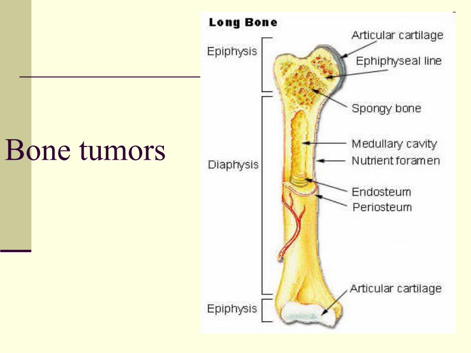
Bone tumors
| Date post: | 15-Jul-2015 |
| Category: |
Health & Medicine |
| Upload: | gopisankar-mg |
| View: | 200 times |
| Download: | 6 times |
Bone tumors
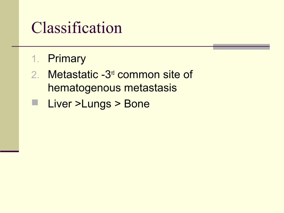
Classification
1. Primary
2. Metastatic -3rd common site of hematogenous metastasis
Liver >Lungs > Bone
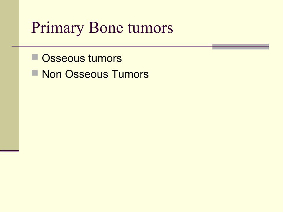
Primary Bone tumors
Osseous tumors Non Osseous Tumors
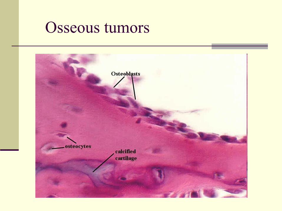
Osseous tumors

Benign Malignant
Bone forming/osteoblastic
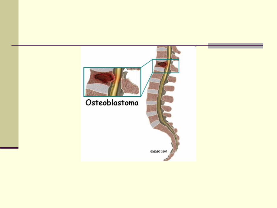
Osteoma Osteod osteomaOsteoblastoima
Osteosarcoma
Cartilage forming Enchondroma OsteochondromaChondroblastoma
Chondrosarcoma
Hematopoietic Myeloma Lymphoma
Unknown Giant Cell tumor Malignant GCTEwing’s sarcoma
Notochordal Chordoma

Non Osseous Tumors
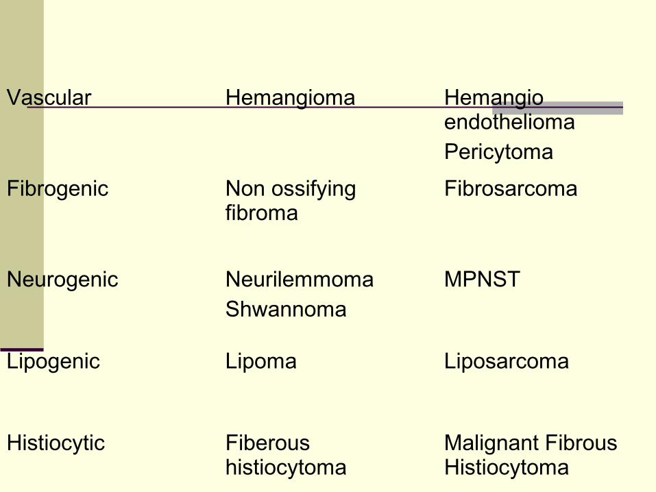
Vascular Hemangioma Hemangio endothelioma Pericytoma
Fibrogenic Non ossifying fibroma
Fibrosarcoma
Neurogenic NeurilemmomaShwannoma
MPNST
Lipogenic Lipoma Liposarcoma
Histiocytic Fiberous histiocytoma
Malignant Fibrous Histiocytoma


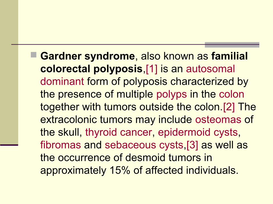
Gardner syndrome, also known as familial colorectal polyposis,[1] is an autosomal dominant form of polyposis characterized by the presence of multiple polyps in the colon together with tumors outside the colon.[2] The extracolonic tumors may include osteomas of the skull, thyroid cancer, epidermoid cysts, fibromas and sebaceous cysts,[3] as well as the occurrence of desmoid tumors in approximately 15% of affected individuals.


Osteosarcoma
MC primary tumor of bone Tumor cells can produce osteiod – tumor ostiod /
tumor bone Malignancy of mesenchyme Two types
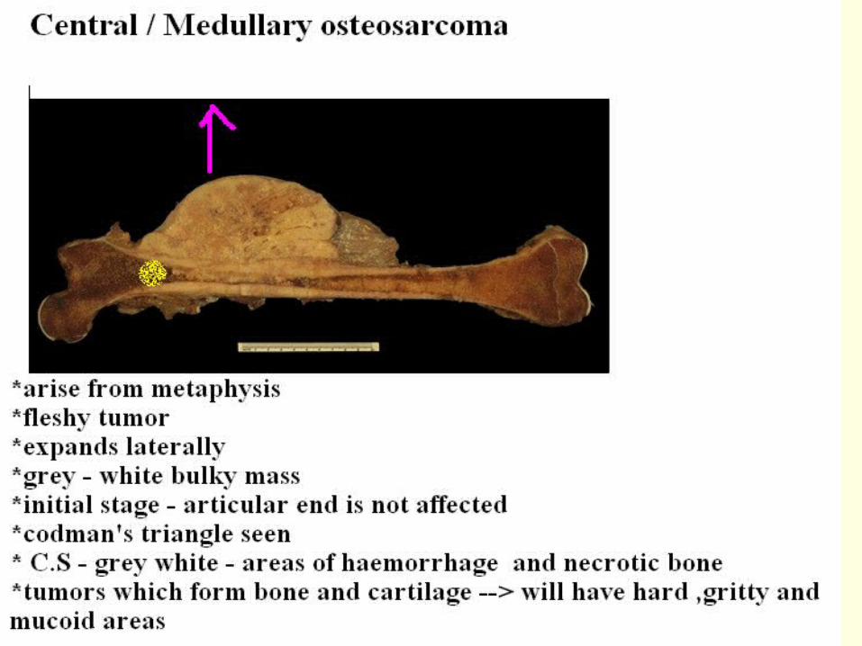
1. Central / medullary
2. Surface / parosteal / Perosteal Pathogenesis
1. Primary
2. Secondary
Pathogenesis
Genetic Constitutional factors Environmental Factors
Genetic
RB gene mutation – 1000 times more risk 13q14 Homozygous mutation in RB locus Mutation in p53 gene Over expression of MDM2

Constitutional factors
10- 20 yrs – maximum Bone growth Males more risk – inc osteoblastic activity Inc risk in paget’s disease Metaphysis - osteogenesis

Pathogenesis
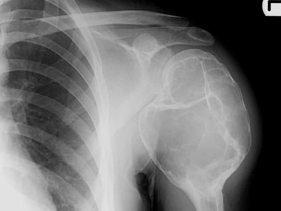
Metaphysis Medullary cavity Perisoteal eleation – codmann’s triangle Xray – metaphyseal radio dense lesion with
codmann’s triangle Sunburst / sunray appearance

Gross
Fleshy tumor Expands laterally


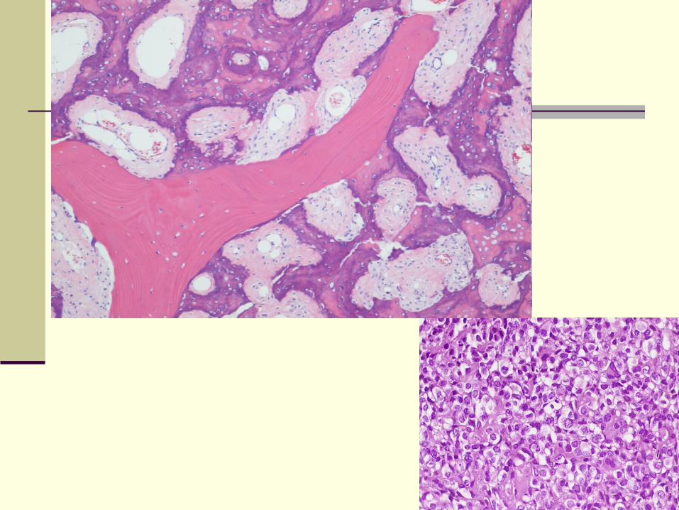

Microscopy
Tumor cells– very in shape –giant cells- spindle ,oval polgonal cells
Osteiod – not rimmed by osteoblast But surrounded by tumor cells Varients –1. Chondroblastic2. Fibroblastic3. Small cell4. telangectactic
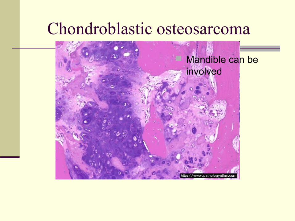
Chondroblastic osteosarcoma
Mandible can be involved
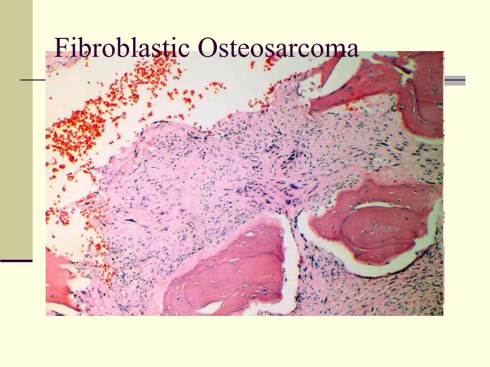
Fibroblastic Osteosarcoma
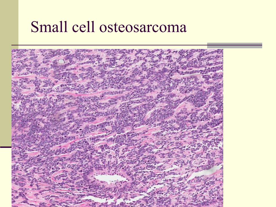
Small cell osteosarcoma
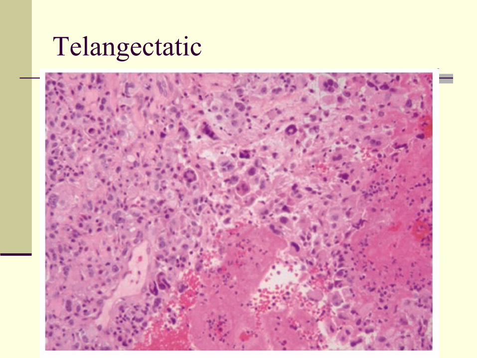
Telangectatic

T/t
Limb salvaged surgery
Extra skeletal osteosarcoma
Mediastinum Breast retroperitonim
Surface Osteosarcoma
1. Parosteal / Juxta cortical Osteosarcoma
2. Periosteal osteosarcoma

Ewing’s sarcoma
Highly malignant small round cell tumor More in females 5 – 20 yrs Cell of origin – Primitive Neurectodermal cells

Variants of ES
1. Classic / Skeletal ES
2. Soft tissue Ewing’s sarcoma
3. PNET


