Page 1
REVIEW ON BOVINE CYSTICERCOSIS AND ITS PUBLIC HEALTH IMPORTANCE
IN ETHIOPIA
By
KASSAHUN SEMIE
A paper presented for the course Seminar on animal health (VST-566)
UNIVERSITY OF GONDAR
FACULTY OF VETERINARY MEDICINE
April, 2014
GONDAR
Page 2
I
ACKNOWLEDGEMENT
First and for most Praise to God, the most Gracious, the most Merciful, the self sufficient master,
whom all creatures need.
I wish to sincerely express my profound thanks to my advisor Mr. Aschalew Assefa for his
motivations, valuable suggestions, unreserved and steady guidance, encouragement, and
meticulous correction to have the paper its form.
I am also grateful to the course coordinator, Dr. Seleshe Negatu for his overall guidance in
preparation of seminar paper.
I will be failing in my duty if I do not mention here the inspiring appreciation and lavish love of
my family.
Finally, I want to thank my batch students and other persons who has come into my life and
inspired, touched and illuminated me through their presence.
Page 3
II
TABLE OF CONTENTS
Contents Pages
ACKNOWLEDGEMENT ............................................................................................................. I
TABLE OF CONTENTS ............................................................................................................ II
LIST OF TABLES ...................................................................................................................... IV
LIST OF FIGURES .................................................................................................................... IV
LIST OF ABBREVIATIONS ..................................................................................................... V
SUMMARY ................................................................................................................................. VI
INTRODUCTION......................................................................................................................... 1
2. BOVINE CYSTICERCOSIS ................................................................................................... 4
2.1 Etiology ................................................................................................................................. 4
2.1.1 Taxonomic classification ................................................................................................ 4
2.1.2 Morphology .................................................................................................................... 4
2.2 Epidemiology ....................................................................................................................... 5
2.2.1 Host range ....................................................................................................................... 5
2.2.2 Geographic distribution: Status in Ethiopia.................................................................... 5
2.2.3 Source of infection and mode of transmission ............................................................... 9
2.3 Life cycle............................................................................................................................. 10
2.4 Pathogenesis and clinical manifestation .......................................................................... 13
2.4.1 Disease in human .......................................................................................................... 13
2.4.2 Disease in cattle ............................................................................................................ 13
2.5 Diagnosis ............................................................................................................................ 13
2.5.1 Diagnosis in human ...................................................................................................... 13
2.5.2 Diagnosis in cattle ........................................................................................................ 14
2.6 Differential diagnosis ........................................................................................................ 14
2.7 Treatment ........................................................................................................................... 15
2.8 Importance of the disease ................................................................................................. 16
2.8.1 Public health importance .............................................................................................. 16
2.8.2 Economic importance ................................................................................................... 18
Page 4
III
2.9 Control and prevention ..................................................................................................... 19
3. CONCLUSION AND RECOMMENDATIONS .................................................................. 21
4. REFERENCES ........................................................................................................................ 22
Page 5
IV
LIST OF TABLES
Table 1: Bovine cysticercosis in different parts of Ethiopia…………………....................6
Table 2: Traditional anticestodal drugs……………………………………………………16
LIST OF FIGURES
Figure 1: Cysticercus bovis cyst distribution in different organs……………………………9
Figure 2: Life cycle of T.saginata……………………………………………………………12
Page 6
V
LIST OF ABBREVIATIONS
µm micrometer
C. bovis Cysticercus bovis
e.g. Example
EARO Ethiopian Agricultural Research Organization
ELISA Enzyme Linked Immunosorbent Assay
FAO Food and Agricultural Organization
kg kilogram
mg milligram
mm millimeter
OIE Office International des Epizootics
PCR Polymerase Chain Reaction
T. saginata Taenia saginata
WHO World Health Organization
Page 7
VI
SUMMARY
Bovine cysticercosis is a zoonotic disease that affects the musculature of cattle and is caused by
the metacestode stage of human intestinal cestode, Taenia saginata. It is cosmopolitan in its
distribution and occurs in developing as well as in developed countries. Distribution is associated
with economic conditions, religious beliefs and close proximity of humans to cattle in utility
function. Its life cycle is indirect and entirely dependent on the link between man and cattle; so
that any break in this link can result in the total elimination of the parasite. Cysts of Cysticercus
bovis can be found anywhere in the carcass and viscera, but there seems to be special affinity
towards some parts which are described as sites of predilection (masseter, tongue, heart, triceps,
inter costal muscles and the diaphragm). Most of these organs except the heart are consumed raw
or under cooked. If man consumes these muscles containing viable cysticerci, a tapeworm may
develop and this is a potential public health hazard in contracting taeniasis. The custom of eating
raw or undercooked beef dishes such as kourt, lebleb, kitffo and the habit of defecating in open
fields coupled with the tradition of allowing cattle to graze in such fields made taeniasis of
human and cysticercosis of cattle common in Ethiopia. The prevalence of the disease both in
human and animals is high. T.saginata in small intestine of humans absorbs digested food and its
proglottids migrate to different organs causing different signs. Economic loss from cysticercosis
is determined by disease prevalence, grade of animals affected, potential market policy of cattle
and treatment cost for detained carcasses. In Ethiopia most Slaughtering practices are often
carried out in open air in the absence of abattoirs. This allows the parasite to continue its life
cycle till to the date and in the coming future.
Keywords: Bovine, Cysticercosis, Ethiopia, economic /public health importance.
Page 8
1
1. INTRODUCTION
Tapeworm infection has been recorded in history 1500 years ago and recognized as one of the
earliest human parasite. Taenia saginata is a worldwide zoonotic cestode whose epidemiology is
ethnically and culturally determined with estimation of 50-77 million cases of worldwide
annually. Both adult and larval forms hazardously affect health of their respective hosts, either
directly or indirectly accompanied with several secondary infections, particularly in human. The
occurrence of larvae (C.bovis) in cattle musculature causes bovine cysticercosis while the adult
worms in human small intestine causes taeniasis (WHO, 1996; Minozzo et al., 2002).
Bovine cysticercosis is a disease that affects the muscle of cattle and is caused by the
metacestode stage, of the human intestinal cestode, Taenia saginata. It is cosmopolitant in its
distribution and occurs in developing as well as in developed countries. Its life cycle is entirely
dependent on the link between man and cattle; so that any break in this link can result in the total
elimination of the parasite. Cysts of Cysticercus bovis can be found anywhere in the carcass and
viscera, but there seems to be special affinity towards some parts which are described as sites of
predilection (masseter, tongue, heart, triceps, intercostal muscles and the diaphragm). Most of
these organs except the heart are consumed raw or under cooked and could be a potential public
health hazard in contracting taeniasis (Gracey, 1981).
Distribution is associated with economic conditions, religious beliefs and close proximity of
humans to cattle in utility function. Slaughtering is often carried out in open air in the absence of
abattoirs. This allows the parasite to continue its life cycle till to the date and in the coming
future. Transmission of the parasite occurs most commonly in areas characterized by poor
hygiene, primitive livestock husbandry practice and inadequate meat inspection, management
and control policy (Mann, 1993). Bovine cysticercosis and taeniasis are common where hygienic
conditions are poor and the inhabitants traditionally eat raw or insufficiently cooked or sun-cured
meat (Minozzo et al., 2002).
In developing countries, taeniasis/bovine cysticercosis constitutes a serious but less recognized
public health problem (Minozzo et al., 2002). Bovine cysticercosis is very common in Africa and
is endemic in areas of Central and East African countries like Ethiopia, Kenya and Zaire
Page 9
2
(Harrison, 1991). The custom of eating raw or undercooked beef dishes such as kourt, lebleb,
kitffo and the habit of defecating in open fields coupled with the tradition of allowing cattle to
graze in such fields made taeniasis of human and cysticercosis of cattle common in Ethiopia
(Teka, 1997). A high prevalence of human infection in different agro-climatic zones of the
country has been reported (Tembo, 2001). Estimates made by different investigators on
prevalence of taeniasis in Ethiopia vary widely from 2% - 16% to over 70% (Mohammed and
Waqtola, 2006).
Among the prevalent livestock diseases, zoonotic represents major constraint to the development
of livestock productivity in Ethiopia. Of zoonotic diseases, bovine cysticercosis is the disease
that remains a major public health problem in lower income and some industrialized countries
(Utulas et al., 2007). T. saginata infection is usually asymptomatic. However, heavy infection
often results in weight loss, dizziness, abdominal pain, diarrhea, headaches, nausea, constipation
or chronic indigestion, and loss of appetite. There can be intestinal obstruction in humans and
this can be alleviated by surgery. The tapeworm can also expel antigens that can cause an
allergic reaction in the individual. It is also rare cause of pancreatitis, cholecystitis
and cholangitis (WHO, 2013). FAO (2004) states that the disease can also cause obstruction of
the bowel, stomach-ache and migrating proglottids cause inflammation of the appendix,
inflammation of the bile duct, unpleasant surprise when seen in the feces; whereas Teka (1997)
stated that taeniasis in humans causes anal pruritis due to emerging tapeworm segments but with
severe infection humans may experience increased appetite or loss of appetite, abdominal
discomfort and digestive upset.
Cysticercosis affects both the health of the consumer and more significantly the country’s
economy, which approaches 30% if allowance is made for the loss in the carcass weight and the
cost of freezing of the infected meat (Fufa, 2006). Generally loss from cysticercosis is
determined by disease prevalence, grade of animals affected, potential market policy of cattle
and treatment cost for detained carcasses. The average annual loss due to taenicidal drugs for
treatment in Ethiopia was estimated to be 4,937,583 Ethiopian birr (Ahmed, 1990; Dawit, 2004;
Fufa, 2006). Inadequate health education and low availability of taenicides are the major
obstacles for the control of such infections in Ethiopia (Pawlowski, 1996; Fufa, 2006).
Page 10
3
In foreign trade, although Ethiopia is ideally placed to export live animals to the big markets of
the Middle East and substantial markets of North and West Africa, export earning is relatively
low. This is mainly due to the presence of a number of unimproved animal health problems,
among which, Taenia saginata/Cysticercus bovis is one that remains a major public and animal
health problem (EARO, 2000)
Therefore the objective of this review is:
To highlight the status of bovine cysticercosis in Ethiopia,
To highlight control and prevention strategies, and public health and economic impacts
of the disease.
Page 11
4
2. BOVINE CYSTICERCOSIS
2.1. Etiology
Bovine cysticercosis is a disease that affects the musculature of cattle and is caused by the
metacestode stage of human intestinal cestode, Taenia saginata (Tylor et al., 2007).
2.1.1. Taxonomic classification
Taenia saginata and its metacestode, Cysticercus bovis, the unarmed beef tapeworm, is classified
under the kingdom of Animalia, phylum of Platyhelminthes, class of Cestoda, order of
Cyclophylidea, family of Taeniidae, genus of Taenia and species of Taenia saginata (Soulsby,
1982; Symth, 1994; Urquhart et al., 1996)
2.1.2. Morphology
The adult tapeworm, Taenia saginata, is a large ribbon shaped, multi segmented, white flat
worm usually 4-15 m long consisting of thousands of segments (proglottids) arranged in a chain
(Urquhart et al., 1996; Andrews et al., 2003). Its body divided in to three distinct parts consisting
of head (scolex), neck and strobilla (Gracey, 1981). The head or scolex bearing attachment
organs, a short unsegmented neck and chain of segments. The chain is known as strobilla and
each segment as proglottids. Unlike other taeniids, the head (scolex) has no rostellum or hooks.
The proglottids are continually budded from the neck region and become sexually mature as they
pass down the strobilla. Each proglottids is hermaphrodite with one or two sets of reproductive
organs. Gravid segments usually leave the host singly and often migrate spontaneously from the
anus (Blancou et al., 2010; Soulsby, 1982).
Taeniid eggs passed in the stool or discharged from ruptured segments are sub-spherical to
spherical in shape and very resistant, remaining viable for 6 months in pasture and vegetables, 5
weeks in water, 10 weeks in stool or hay and 12 weeks in silage sludge. Taeniid eggs measure
about 30-45 µm in diameter; contain an oncosphere (hexacanth embryo) bearing three pairs of
hook; have a thick, brown, radially striated embryophore or ‘shell’ composed of hooks; and there
is an outer, oval, membranous coat, the true egg shell, that is lost from fecal eggs (OIE, 2008).
The larval stage, or metacestode also referred to as “beef measles”, are found in all striated
Page 12
5
muscles of the intermediate host. Cysticercus bovis is a small, pea-sized oval in shape (OIE,
2000), translucent and contains a single white scolex that is morphologically similar to the scolex
of the future adult tapeworm. They are contained in a thin, host-produced fibrous capsule (OIE,
2008).
2.2. Epidemiology
2.2.1. Host range
Cattle are the preferred intermediate hosts and humans are the only final hosts of Taenia
saginata. Cattle of all ages are susceptible; however, young age groups are more susceptible.
Parasitism sometimes observed in other ruminants (like sheep, goats, antelopes, gazelles and
buffaloes) but Cysticercus development is unlikely (Pawlowski, 1972).
2.2.2. Geographic distribution: Status in Ethiopia
In developing countries, taeniasis/bovine cysticercosis constitutes a serious but less recognized
public health problem (Minozzo et al., 2002). Bovine cysticercosis has a cosmopolitant
distribution and is very common in Africa. It is highly endemic in areas of Central and East
African countries like Ethiopia, Kenya and Zaire (Harrison, 1991). The custom of eating raw or
undercooked beef dishes such as kourt, lebleb, kitffo and the habit of defecating in open fields
coupled with the tradition of allowing cattle to graze in such fields made taeniasis of human and
cysticercosis of cattle common in Ethiopia (Teka, 1997). A high prevalence of human infection
in different agro-climatic zones of the country has been reported (Tembo, 2001). Estimates made
by different investigators on prevalence of taeniasis in Ethiopia vary widely from 2% - 16% to
over 70% (Mohammed and Waqtola, 2006). Ethiopia is divided into nine ethnically-based
administrative regions and three chartered cities and bovine cysticercosis has been reported from
different parts of the country (Table 1).
Page 13
6
Table 1: Bovine cysticercosis in different parts of Ethiopia
Place Percent Prevalence Reference
Addis Ababa 13.3% Kebede et al. (2009)
DebreZeit 13.85% Getachew (1990)
Wolaita Soddo 11.3% Megersa et al. (2009)
Mekelle 7.23% Getachew (2008)
Southern Nations Nationalities
People’s Region
26.25%
Abunna et al. (2008)
Amhara National Regional
State
18.49% Kabede (2008)
Bahir Dar 19.4% Mulugeta (1997)
Nekemte 21.7% Ahemmed (1990)
Gondar
4.9%
Dawit (2004)
Risk factors of taeniasis:
The prevalence of taeniasis is associated with different risk factors. The potential risk factors of
taeniasis are: habit of raw meat consumption, age, sex, religion, educational level, and presence
and usage of sanitary facilities especially toilets. Different scholars have controversies regarding
to disease prevalence in association with such risk factors.
Hailu (2005) reported that there is highly significant variation among raw meat and cooked meat
eaters, in which prevalence is high in those eating raw meat. But no significant variations were
observed between age, sex and religion. In contrast, Megersa et al. (2009) reported in such a way
that taeniasis has significant association with ages of individuals, indicating higher prevalence of
infection in adult people. The possible suggestion for this case is that adults has habit of raw
meat consumption than younger, as youngs are not allowed to consume raw meat, and adults
have income that afford in consuming raw meat like kurt which may be expensive for young
individuals.
Page 14
7
In contrast to Hailu (2005), Abunna et al. (2008) reported taeniasis has significant association
with sex. Prevalence is higher in males than females. This could be due to economic reasons and
cultural practices in that males do not prepare their dish at home, rather consume at restaurants
and butcheries.
Most researchers underline that there is higher prevalence of taeniasis in those who consumes
raw meat than those having cooked meat dishes (Abunna et al., 2008; Megersa et al., 2009;
Tembo, 2001; Hailu, 2005).
Taeniasis is prevalent in those who do not use latrines (Abunna et al., 2008).
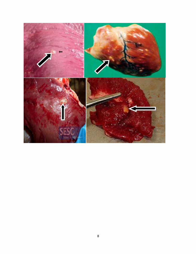
Distribution of metacestode in different organs:
The metcestodes were found throughout the edible parts of the carcass which includes masseter
muscles, cardiac muscles, triceps muscles, thigh muscles, shoulder muscles, diaphragm,
intercostal muscles, liver, heart, tongue, lung and kidney (Kebede et al., 2009; Megersa et al.,
2009; Abunna et al., 2008; Getachew, 2008; Kabede, 2008).
The tongue, masseter muscles, heart muscles, triceps muscles and thigh muscles were the main
predilection sites of the cysts (Kebede, 2008). Abunna et al. (2008) reported these cysts in heart
(29.2%), shoulder muscle (25.3%), masseter muscle (26.7%), tongue (10.4%), diaphragm
(5.4%), liver(1.4%), lung (0.9%) and kidney(0.5%) while Kebede (2008) reported cysts from
tongue (0.61%), masseter muscles (0.59%), shoulder muscles (0.26%), heart (0.26% ) and liver
(7.45%).
Page 16
9
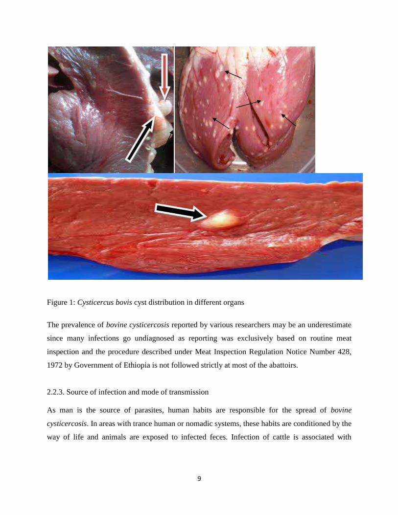
Figure 1: Cysticercus bovis cyst distribution in different organs
The prevalence of bovine cysticercosis reported by various researchers may be an underestimate
since many infections go undiagnosed as reporting was exclusively based on routine meat
inspection and the procedure described under Meat Inspection Regulation Notice Number 428,
1972 by Government of Ethiopia is not followed strictly at most of the abattoirs.
2.2.3. Source of infection and mode of transmission
As man is the source of parasites, human habits are responsible for the spread of bovine
cysticercosis. In areas with trance human or nomadic systems, these habits are conditioned by the
way of life and animals are exposed to infected feces. Infection of cattle is associated with
Page 17
10
directly to the non-hygienic disposal of stool by infected humans or indirectly by the use of
human sewage on pasture as fertilizer (Kassai, 1999).
Man’s customs and traditions of consuming raw, sun-cured, and inadequately cooked beef dishes
like kourt, lebleb and kitffo in Ethiopia, containing viable cysticerci perpetuate human infection
(Teka, 1997).
Man cannot spread taeniasis to his own species. Management of animals in their natural
environment predisposes them to infection. Cattle grazing communally have a higher risk of
picking up T.saginata eggs as they are frequently in contact with the human feces compared to
commercial herds. The risk of cattle coming into contact with T.saginata eggs is much higher
when cattle are at pasture (Pawlowski, 1996).
In developing countries like Ethiopia, cattle are reared on extensive scale, human sanitation is
poorly developed which makes the incidence of T.saginata infection in humans very high.
Calves are infected usually in early life, often with in the first few days after birth from infected
stockmen whose hands are contaminated with Taenia eggs (Maedia et al., 1996; Teka, 1997).
2.3. Life cycle
The life cycle of T.saginata is indirect where the definitive host is human and intermediate hosts
are cattle (Urquhart et al., 1996).
Typically, the tapeworm life cycle consists of an adult tapeworm in the final host (i.e. human).
The worms produce segments (proglottids) containing a considerable number of eggs which are
shed on defecation. Taenia eggs containing an embryo (or oncosphere) are spread into the
environment through sewage and may be orally ingested by the intermediate hosts (i.e. cattle). In
cattle the embryo move from the intestine to striated musculature. Here they develop into small
vesicles called cysticerci containing one protoscolex, head of the future adult tapeworm
(Lightowlers et al., 1996).
The metcestodes were found throughout the edible parts of the carcass which included masseter
muscles, cardiac muscles, triceps muscles, thigh muscles, shoulder muscles, diaphragm,
Page 18
11
intercostal muscles, liver, heart, tongue, lung and kidney, (Kebede et al., 2009; Megersa et
al.,2009; Abunna et al., 2008; Getachew , 2008; Kabede, 2008). The tongue, masseter muscles,
heart muscles, triceps muscles and thigh muscles were the main predilection sites of the cysts
(Kebede, 2008). If man consumes these muscles containing viable cysticerci, a tapeworm may
develop. One viable Cysticercus can be sufficient, although immunity of the host can alter that.
Prevention of human taeniasis and bovine cysticercosis is achieved by interrupting the life cycle
of the parasite (Teka, 1997).
Page 19
12
Figure 2: life cycle of T. saginata (source: slide share)
Page 20
13
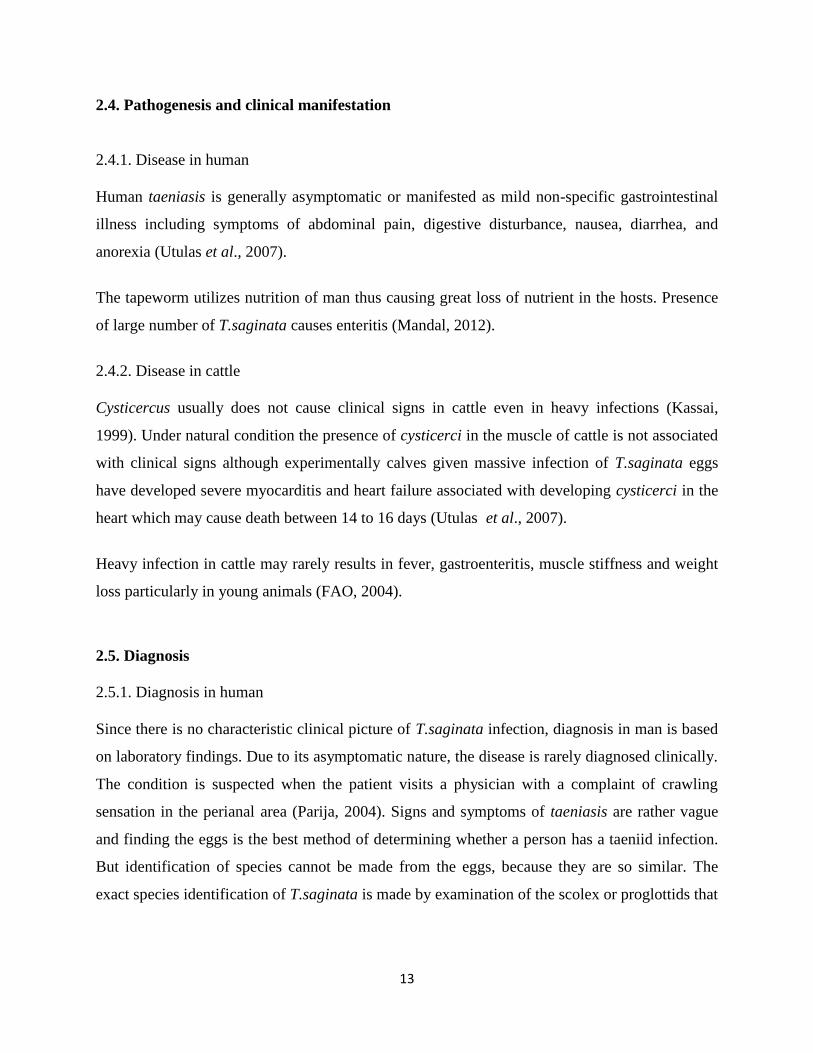
2.4. Pathogenesis and clinical manifestation
2.4.1. Disease in human
Human taeniasis is generally asymptomatic or manifested as mild non-specific gastrointestinal
illness including symptoms of abdominal pain, digestive disturbance, nausea, diarrhea, and
anorexia (Utulas et al., 2007).
The tapeworm utilizes nutrition of man thus causing great loss of nutrient in the hosts. Presence
of large number of T.saginata causes enteritis (Mandal, 2012).
2.4.2. Disease in cattle
Cysticercus usually does not cause clinical signs in cattle even in heavy infections (Kassai,
1999). Under natural condition the presence of cysticerci in the muscle of cattle is not associated
with clinical signs although experimentally calves given massive infection of T.saginata eggs
have developed severe myocarditis and heart failure associated with developing cysticerci in the
heart which may cause death between 14 to 16 days (Utulas et al., 2007).
Heavy infection in cattle may rarely results in fever, gastroenteritis, muscle stiffness and weight
loss particularly in young animals (FAO, 2004).
2.5. Diagnosis
2.5.1. Diagnosis in human
Since there is no characteristic clinical picture of T.saginata infection, diagnosis in man is based
on laboratory findings. Due to its asymptomatic nature, the disease is rarely diagnosed clinically.
The condition is suspected when the patient visits a physician with a complaint of crawling
sensation in the perianal area (Parija, 2004). Signs and symptoms of taeniasis are rather vague
and finding the eggs is the best method of determining whether a person has a taeniid infection.
But identification of species cannot be made from the eggs, because they are so similar. The
exact species identification of T.saginata is made by examination of the scolex or proglottids that
Page 21
14
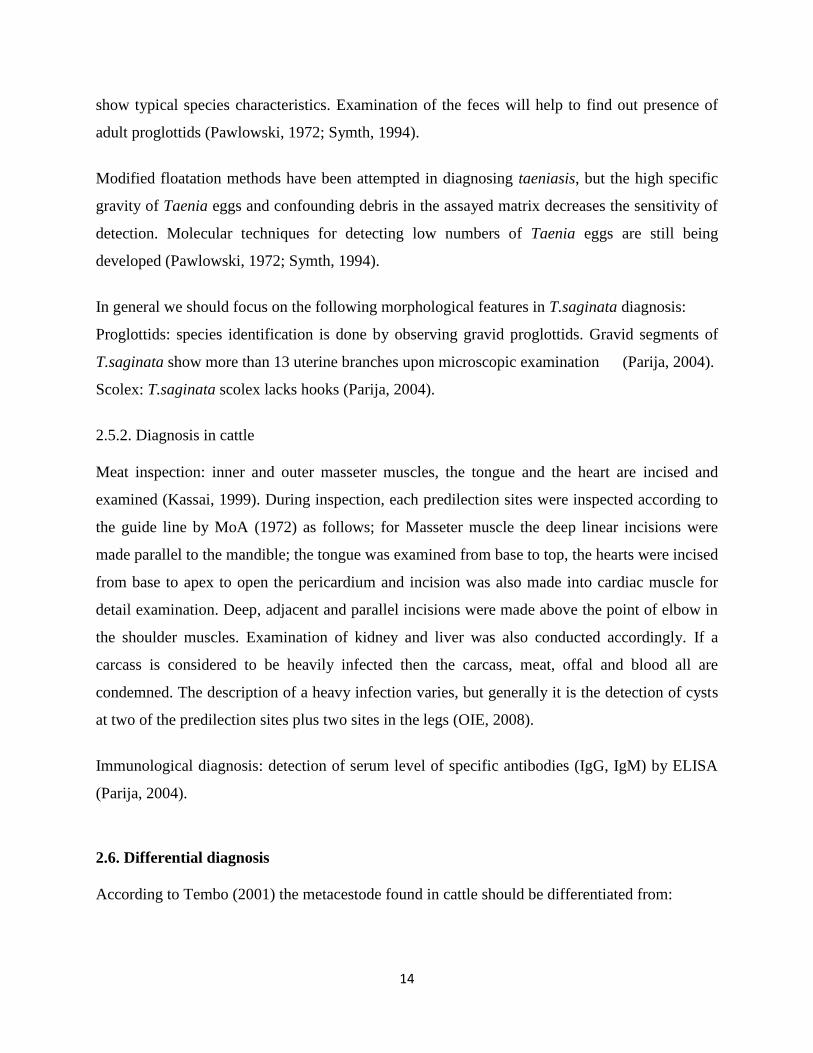
show typical species characteristics. Examination of the feces will help to find out presence of
adult proglottids (Pawlowski, 1972; Symth, 1994).
Modified floatation methods have been attempted in diagnosing taeniasis, but the high specific
gravity of Taenia eggs and confounding debris in the assayed matrix decreases the sensitivity of
detection. Molecular techniques for detecting low numbers of Taenia eggs are still being
developed (Pawlowski, 1972; Symth, 1994).
In general we should focus on the following morphological features in T.saginata diagnosis:
Proglottids: species identification is done by observing gravid proglottids. Gravid segments of
T.saginata show more than 13 uterine branches upon microscopic examination (Parija, 2004).
Scolex: T.saginata scolex lacks hooks (Parija, 2004).
2.5.2. Diagnosis in cattle
Meat inspection: inner and outer masseter muscles, the tongue and the heart are incised and
examined (Kassai, 1999). During inspection, each predilection sites were inspected according to
the guide line by MoA (1972) as follows; for Masseter muscle the deep linear incisions were
made parallel to the mandible; the tongue was examined from base to top, the hearts were incised
from base to apex to open the pericardium and incision was also made into cardiac muscle for
detail examination. Deep, adjacent and parallel incisions were made above the point of elbow in
the shoulder muscles. Examination of kidney and liver was also conducted accordingly. If a
carcass is considered to be heavily infected then the carcass, meat, offal and blood all are
condemned. The description of a heavy infection varies, but generally it is the detection of cysts
at two of the predilection sites plus two sites in the legs (OIE, 2008).
Immunological diagnosis: detection of serum level of specific antibodies (IgG, IgM) by ELISA
(Parija, 2004).
2.6. Differential diagnosis
According to Tembo (2001) the metacestode found in cattle should be differentiated from:
Page 22
15
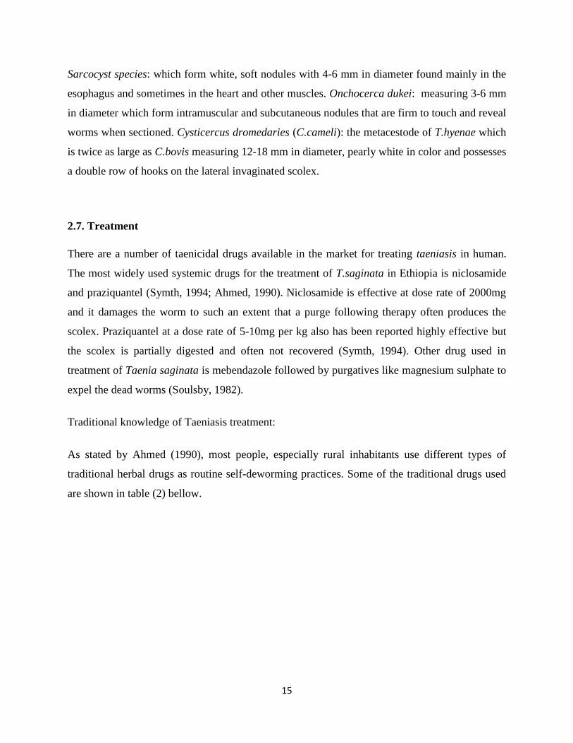
Sarcocyst species: which form white, soft nodules with 4-6 mm in diameter found mainly in the
esophagus and sometimes in the heart and other muscles. Onchocerca dukei: measuring 3-6 mm
in diameter which form intramuscular and subcutaneous nodules that are firm to touch and reveal
worms when sectioned. Cysticercus dromedaries (C.cameli): the metacestode of T.hyenae which
is twice as large as C.bovis measuring 12-18 mm in diameter, pearly white in color and possesses
a double row of hooks on the lateral invaginated scolex.
2.7. Treatment
There are a number of taenicidal drugs available in the market for treating taeniasis in human.
The most widely used systemic drugs for the treatment of T.saginata in Ethiopia is niclosamide
and praziquantel (Symth, 1994; Ahmed, 1990). Niclosamide is effective at dose rate of 2000mg
and it damages the worm to such an extent that a purge following therapy often produces the
scolex. Praziquantel at a dose rate of 5-10mg per kg also has been reported highly effective but
the scolex is partially digested and often not recovered (Symth, 1994). Other drug used in
treatment of Taenia saginata is mebendazole followed by purgatives like magnesium sulphate to
expel the dead worms (Soulsby, 1982).
Traditional knowledge of Taeniasis treatment:
As stated by Ahmed (1990), most people, especially rural inhabitants use different types of
traditional herbal drugs as routine self-deworming practices. Some of the traditional drugs used
are shown in table (2) bellow.
Page 23
16
Table 2: Traditional anticestodal drugs
No. Local name Scientific name Parts of plants
used
1 Bisana Corton macrustachys Bark(hard outer
cover)
2 Duba firie Cucurbita pepo: the pump kin Seed
3 Enkoko Embelia schimperi Fruit
4 Kosso Hygenia abyssinica Flower
5 Metre Glinus lotoides Seed
6 Wogert Silen macrosclen Root
Source: Ahmed (1990)
In cattle treatment with compounds such as albendazole (50mg per kg), praziquantel (50mg per
kg), mebendazole (50mg per kg) can be given but they are not fully effective (Kassai, 1999).
2.8. Importance of the disease
2.8.1. Public health importance
Taenia saginata is a very long (3-15 meters in length) tapeworm parasite, whose adult form is
found attached to the small intestinal tracts of human beings. In man it has been known to live
for 20 years within a single individual. It is an intestinal parasite of cattle and humans,
causing taeniasis in humans. It is found globally and most prevalent where cattle are raised,
and beef is consumed. It is relatively common in Africa, some parts of Eastern
Europe, Southeast Asia, and Latin America. Humans are generally infected as a result of
poor hygiene (WHO, 1996).
The effect on human health is generally slight and symptoms may vague or absent. Taeniasis has
debilitating effect on people who already have live of protein deficiency diets suffer from iron
deficiency and infested by hook worm (FAO, 2004). T.saginata in small intestine of humans
Page 24
17
absorbs digested food and its proglottids migrate to different organs causing different signs
(Urquhart et al., 1996).
T. saginata infection is usually asymptomatic. However, heavy infection often results in weight
loss, dizziness, abdominal pain, diarrhea, headaches, nausea, constipation or chronic indigestion,
and loss of appetite. There can be intestinal obstruction in humans and this can be alleviated by
surgery. The tapeworm can also expel antigens that can cause an allergic reaction in the
individual. It is also rare cause of pancreatitis, cholecystitis and cholangitis (WHO, 2013); FAO
(2004) states that the disease can also cause obstruction of the bowel, stomach-ache and
migrating proglottids cause inflammation of the appendix, inflammation of the bile duct,
unpleasant surprise when seen in the feces; whereas Teka (1997) stated that taeniasis in humans
causes anal pruritis due to emerging tapeworm segments but with severe infection humans may
experience increased appetite or loss of appetite, abdominal discomfort and digestive upset.
Generally, according to WHO (2013), adult Taenia parasites located in the intestinal tracts of
people can pose a variety of problems including:
Non-specific intestinal disturbances - tapeworms can produce some non-specific signs of
intestinal discomfort and pain (e.g. colic signs) in humans. Vomiting may also result.
Non-specific appetite changes - tapeworms can cause some people to go off their food or to
become fussy or picky about their eating habits (this appetite loss is possibly the result of such
factors as abdominal pain and nausea). In contrast, certain other individuals develop a
ravenous appetite in the face of heavy tapeworm infestations because they are competing with
the parasite/s for nutrients (they need to physically eat more to provide enough nutrition for
both themselves and the worms).
Body weakness, headaches, dizziness, irritability and delirium.
Malnutrition - very large numbers of adult Taenia tapeworms present in the intestinal tracts of
man can result in the malabsorption of nutrients. This can cause the tapeworm-parasitized
individual to not receive the nutrition it needs (i.e. to not absorb its food properly), resulting in
malnourishment, weight loss, ill-thrift and poor growth.
Poor hair quality - severe malnutrition and malabsorption of vitamins, minerals and proteins
can result in reduced quality of the hair.
Page 25
18
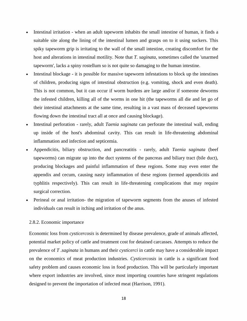
Intestinal irritation - when an adult tapeworm inhabits the small intestine of human, it finds a
suitable site along the lining of the intestinal lumen and grasps on to it using suckers. This
spiky tapeworm grip is irritating to the wall of the small intestine, creating discomfort for the
host and alterations in intestinal motility. Note that T. saginata, sometimes called the 'unarmed
tapeworm', lacks a spiny rostellum so is not quite so damaging to the human intestine.
Intestinal blockage - it is possible for massive tapeworm infestations to block up the intestines
of children, producing signs of intestinal obstruction (e.g. vomiting, shock and even death).
This is not common, but it can occur if worm burdens are large and/or if someone deworms
the infested children, killing all of the worms in one hit (the tapeworms all die and let go of
their intestinal attachments at the same time, resulting in a vast mass of deceased tapeworms
flowing down the intestinal tract all at once and causing blockage).
Intestinal perforation - rarely, adult Taenia saginata can perforate the intestinal wall, ending
up inside of the host's abdominal cavity. This can result in life-threatening abdominal
inflammation and infection and septicemia.
Appendicitis, biliary obstruction, and pancreatitis - rarely, adult Taenia saginata (beef
tapeworms) can migrate up into the duct systems of the pancreas and biliary tract (bile duct),
producing blockages and painful inflammation of these regions. Some may even enter the
appendix and cecum, causing nasty inflammation of these regions (termed appendicitis and
typhlitis respectively). This can result in life-threatening complications that may require
surgical correction.
Perineal or anal irritation- the migration of tapeworm segments from the anuses of infested
individuals can result in itching and irritation of the anus.
2.8.2. Economic importance
Economic loss from cysticercosis is determined by disease prevalence, grade of animals affected,
potential market policy of cattle and treatment cost for detained carcasses. Attempts to reduce the
prevalence of T .saginata in humans and their cysticerci in cattle may have a considerable impact
on the economics of meat production industries. Cysticercosis in cattle is a significant food
safety problem and causes economic loss in food production. This will be particularly important
where export industries are involved, since most importing countries have stringent regulations
designed to prevent the importation of infected meat (Harrison, 1991).
Page 26
19
The cost implication can be broken down into those involved in treating human taeniasis and
cattle carcasses (cost of freezing, boiling) or condemned, as well as the cost involved in the
inspection procedures. The average annual loss due to taenicidal drugs for treatment in Ethiopia
was estimated to be 4,937,583 Ethiopian birr (Ahmed, 1990; Dawit, 2004; Fufa, 2006).
2.9. Control and prevention
Control of taeniasis or cysticercosis aimed at breaking the epidemiological cycle of T. saginata
infection. This involves cattle and humans, the intermediate host and final host respectively
(Pawlowski, 1972).
In cattle:
Sanitary measures are important to ensure:
Improvement of livestock farming techniques, for example, the establishment of cattle
farms with controlled hygienic conditions in which the animals do not have access to
pasture contaminated by human feces (Lightowlers et al., 1996).
Reinforcement of veterinary inspections during slaughter in abattoirs and more meat
inspection both in municipal slaughter houses and slaughter establishments at markets
(Blancou et al., 2010).
Vaccination of cattle would the most cost-effective control strategy. It has been shown
that the T. saginata oncosphere extracts and oncosphere secretions produce a high level
of protective immunity to challenge infections with T .saginata eggs (Lightowlers et al.,
1996).
More recently, an 18kDa T. saginata oncosphere secreted and surface expressed adhesion
molecule HP6 was used to successfully vaccinate calves against oral challenge with T.
saginata eggs. However, no vaccine is currently marketed (Blancou et al., 2010).
In human:
As stated by Blancou et al (2010), control of infection in human is based on the following:
Page 27
20
Diagnosis of carriers and treatment with a taenicides (e.g. Niclosamide or praziquantel) to
eliminate the parasite that is the source of contamination for the environment and cattle.
Improvement of personal hygiene and installation of good sanitary accommodation for
family use.
Enhance environmental hygiene and suitable drainage of waste water.
Continuous public health education of the population, stressing the danger of
consumption of uncooked or partially cooked beef.
Mass education to use latrines, and avoid eating of raw meat.
Page 28
21
3. CONCLUSION AND RECOMMENDATIONS
Cysticercosis is an important zoonotic disease that affects both human and animals in Ethiopia.
The prevalence of the disease both in human and animals is high and economically significant.
Nowadays, since there are accustoms of eating raw meat, lack of knowledge about ways of
disease transmission, backyard slaughtering of animals especially during holydays, ignorance
incision of meat by meat inspectors and lack of sanitation can give a great favor for continual
existence of the parasite within the human and animal population.
Based on the above conclusion the following recommendations are forwarded:
There should be public awareness about the health and economic importance of the
disease through social and public media.
Avoid eating of raw meat (Kurt, lebleb and kitffo) that is not inspected by well
experienced meat inspector.
Infected meat and meat products must be undergoing the processes of freezing and
boiling.
There should be strong and close collaboration between medical and veterinary
professionals to reduce impact of the disease both in humans and animals.
The community should use latrines to improve personal as well as environmental
hygiene.
Untreated human feces should not be used as fertilizers.
Strict routine meat inspection of slaughtered animals should be carried out.
Further researches should be conducted on the epidemiology and control strategies of
cestodes in Ethiopia.
Page 29
22
4. REFERENCES
Abunna, F., G. Tilahun, B. Megersa, A. Regassa and B. Kumsa (2008): Bovine Cysticercosis in
Cattle Slaughtered at Hawassa Municipal Abattoir, Ethiopia: Prevalence, Cyst
Viability, Distribution and Its Public Health Implication. Zoonosis and Public Health,
55(2): 82-88.
Ahmed, I. (1990): Bovine Cysticercosis in animals slaughtered at Nekemte abattoir. DVM
Thesis. Addis Ababa University, Faculty of Veterinary Medicine, Debre Zeit,
Ethiopia.
Andrews, A.H., Blowey, R.W., Boyd, H. and Eddy, R.G. (2003): Bovine Medicine: Disease and
Husbandry of cattle. 2nd ed. Singapore, Blackwell Science. Pp. 360-363.
Blancou, J., Uilenberg, G., Lefevre, P-C, and Charmette, R. (2010): Infectious and Parasitic
Diseases of Livestock. Lavoisier. vol. 2, P. 1646.
Dawit, S. (2004): Epidemiology of Taenia saginata taeniasis and cysticercoids in north Gondar
zone, north western Ethiopia. DVM Thesis. Faculty of Veterinary Medicine, Addis
Ababa University, Debre zeit, Ethiopia.
EARO (2000): Beef Research strategy, Animal Science Directorate.
FAO (2004): Veterinary Public Health Disease Fact Sheet: Cysticercosis.
Fertig, D.L. and Daron, C.R. (1985): Taenia saginata Cysticercosis in an Ohio cattle feeding
operation. Journal of American veterinary Medicine Association, 186, Pp. 1280-1284.
Fufa, A. (2006): Study on the prevalence of Bovine Cysticercosis at Awassa town and its
surroundings, southern Ethiopia. Addis Ababa University, Faculty of Veterinary
Medicine, Debre zeit, Ethiopia.
Getachew, B. (1990): Prevalence and significance of Cysticercus bovis among cattle slaughtered
at Debre zeit abattoir. Addis Ababa University, Faculty of Veterinary Medicine,
Debre zeit, Ethiopia.
Gracey, J.L. (1981): Thornton’s Meat Hygiene. 9th ed. London, Ballier Tindal. 24-28 Oval Road,
London NW17DX.
Hailu, D. (2005): Prevalence and risk factors for T. saginatsa Cysticercosis in three selected
areas of eastern Shoa. MSc Thesis. Addis Ababa University, Faculty of Veterinary
Medicine, Debre zeit, Ethiopia.
Page 30
23
Harrison, L.J.S., and Swell, M.M.H. (1991): The zoonotic Taenia of Africa. In: Parasitic
Helminthes and Zoonosis in Africa. London, Unwin Hyman, Pp. 50-53.
Kassai, T. (1999): Veterinary Helminthology. 1st ed. New Delhi, Butter Worth. Heinemann, Pp.
37 - 42.
Kebede, N., G., Tilahun and A. Hailu (2009): Current Status of Bovine Cysticercosis of
Slaughtered Cattle in Addis Ababa Abattoir, Ethiopia. Tropical Animal Health and
Production, 41: 291-294.
Lightowlers, M.W., Rolfe, R., and Gauci, C.G. (1996): Taenia saginata: Vaccination against
Cysticercosis in cattle with recombinant oncosphere antigens. Experim.parasitol.
84: 330-338.
Maedia, G.E., Kyvsgaard, N.P., Nansen, C., and Bogh, H.O. (1996): Disease Transmitted from
animal to man. 6th ed. Pp. 88-96.
Mandal, S.C. (2012): Veterinary Parasitology at a Glance. 2nded. New Delhi, Ibdc, India. Pp. 194
- 203.
Megersa, B., Tesfaye, E., Regassa, A., Abebe, R., and Abunna, F. (2009): Bovine Cysticercosis
in cattle slaughtered at Jimma Municipal Abattoir, Southern Ethiopia.
Minozzo, J.C., Gusso, R.L.F., De Castro, E.A., Lago, O. and Soccoi, V.T. (2002): Experimental
Bovine Infection with Taenia saginata Eggs: Recovery Rates and Cysticerci Location.
Braz.arch.biol.tfchnol. 45: 4.
Ministry of Agriculture (1972): Meat Inspection Regulations. Legal notice no. 428 Negarit
Gazeta. Addis Ababa, Ethiopia.
Mohammed, A. A. and Waqtola, C. (2006): Medical Parasitology for Medical laboratory
Technology students. Jimma University, Faculty of Medical Science, Jimma. Pp.
339-341.
Mulugeta, A. (1997): Bovine Cysticercosis: prevalence, economic and public health importance,
DVM Thesis. Addis Ababa University, Faculty of Veterinary Medicine, Debre zeit,
Ethiopia.
OIE (2000): Manual of Standards for Diagnostic Tests and Vaccines. Cysticercosis, Pp. 420-422.
OIE (2008): Terrestrial Manual. Cysticercosis, Chapter 2.9.5
Parija, J., (2004): Text book of Medical Parasitology. 2nd ed. New Delhi, All Indian publishers.
Pp. 207 – 212.
Page 31
24
Pawlowski, Z. and Schultz, M.G. (1972): Taeniasis and Cysticercosis (T.saginata).
Adv.Parasitol. 10: 269-343.
Pawlowski, Z.S. (1996): Helminthic Zoonosis affecting human in Africa. Lindberg, Pp. 55-60.
Soulsby, E.J.W. (1982): Helminthes, Arthropods an d Protozoa of Domestic Animals. 7th ed.
London, Balliere Tindal.
Symth, S.D. (1994): Introduction to Animal Parasitology. 3rd ed. London, Hodder and Stoughton,
Pp. 250-263.
Teka, G. (1997): Food Hygiene Principles and Food Born Disease Control with special
Reference to Ethiopia. Addis Ababa University, Faculty of Medicine, Department of
Community Health, Addis Ababa, Ethiopia.
Tembo, A. (2001): Epidemiology of Taenia saginata, Taeniasis/Cysticercosis in three selected
agro climatic zones. MSc Thesis. Addis Ababa University, Faculty of Veterinary
Medicine, Debre zeit, Ethiopia.
Tylor, M.A., Coop, R.S., wall.R.L. (2007): Veterinary Parasitology. 3rd ed. USA: Blackwell. Pp.
121 - 123.
Urquhart, G.M., Armour, J., Duncan, J.L., Dunn, A.M. and Jennings, F.W. (1996): Veterinary
Parasitology. 2nd ed. London, Blackwell Science, Pp. 121-124.
Utulas, M., Esatgil, and Tuzer (2007): Prevalence of hydatidosis in slaughtered animals in
Thrace, Turkey Parasitology Dergisit, 31(1), Pp. 41 – 45.
WHO (1996): Investigating in health research and development. Report of the committee on
health research relating to future intervention options. Geneva, Switzerland: WHO,
Pp. 270 – 275.
WHO (2013): Taeniasis/cysticercosis. WHO Fact sheet N°376.