Analysis of Glycosaminoglycans in Stem Cell Glycomics
Boyangzi Li, Haiying Liu, Zhenqing Zhang, Hope E. Stansfield, Jonathan S. Dordick, and Robert J. Linhardt
Abstract
Glycosaminoglycans (GAGs) play a critical role in the binding and activation of growth factors in cell signal transduction required for biological development. A glycomics approach can be used to examine GAG content, composition, and structure in stem cells in order to characterize their general differentia-tion. Specifically, this method may be used to evaluate chondrogenic differentiations by profiling for the GAG content of the differentiated cells. Here, embryonic-like teratocarcinoma cells, NCCIT, a develop-mentally pluripotent cell line, were used as a model for establishing GAG glycomic methods, but will be easily transferrable to embryonic stem cell cultures.
Glycomics is the study of the structure and function of glycans, glycoconjugates including glycosphingolipids, glycoproteins, such as proteoglycans (PGs), and glycan-binding proteins. An under-standing of the cellular glycome should explain some mysteries associated with these frequent and important posttranslational modifications (1). The structural and functional glycomics of the glycosaminoglycan (GAG) chains (GAGome) of PGs from different tissues and cells are under intensive study in our laboratory (2–5). GAGs are linear, sulfated, heterogeneous polysaccharides consisting of various repeating disaccharide and are mainly located on both the external membrane of eukaryotic cells and within the extracellular matrix (6, 7). There are four distinct families of GAGs: chondroi-tin/dermatan sulfate (CS/DS), heparin/heparan sulfate (HS), ker-atan sulfate (KS), and hyaluronan (HA). GAGs are involved in
numerous biological activities and are important as molecular coreceptors, in cell–cell interactions, cell adhesion, cell migration, cell signaling, cell growth, and cell differentiation (8–10).
Embryonic stem cells (ESCs) have enormous potential as a source of cells for cell replacement therapy and have been used as in vitro models to study specific aspects of early embryonic development (11, 12). GAGs, particularly HS and CS, within stem cells play key roles in maintaining cell proliferation and differentiation (5). Understanding the glycomics of ESCs should shed light on devel-opment, including the differentiation of chondrocytes from mes-enchymal cells (13–15). In our lab, we are using teratocarcinoma cells (NCCIT), a developmentally pluripotent cells, to offer a convenient model for ESCs. Methods for the elucidation of the GAGome within NCCIT cells, generally applicable for ESCs and their derivatives, such as chondrocytes, are described here for use in better understanding cell pluripotency and differentiation.
ESCs are able to differentiate into the mesenchymal cells that ultimately give rise to chondrocytes and endochondral ossification (13–15). Cartilage is a specialized connective tissue that provides support for other tissues or prevents friction of the joints. The cartilage is comprised of chondrocytes that sparsely distribute in extracellular matrix filled with collagen fibrils and PGs. The fibrous structure of collagen provides support and maintains tis-sue shape, while PGs form gels and act as filler to facilitate com-pressibility and prevent friction as well as perform other critical signaling functions. The PGs in cartilage, such as decorin, bigly-can, and aggrecan, are glycosylated with one or multiple GAG chains. In cartilage, the GAG components are mainly CS and KS and HA as well as smaller amounts of DS and HS. CS, the most abundant GAG in the cartilage, is composed of 4-O-sulfo, 6-O-sulfo, and 4,6-di-O-sulfo sequences (16, 17). Research has shown that both the relative amounts of these sequences within CS and the length of CS chains change in aging and in diseases such as osteoarthritis (18–21). The GAG profile of differentiating ESCs may therefore help to elucidate whether or not chondro-cytes have been formed, to what extent and may help to charac-terize the quality of the generated ESC-derived chondrocytes.
1. 70% (v/v) ethanol. 2. NCCIT cells (ATCC: CRL-2073), frozen and preserved in
95% culture medium and 5% DMSO, were from American
1.2. GAGs in Embryonic Stem Cells
1.3. GAGs in the Extracellular Matrix of Chondrocytes
2. Materials
2.1. Cell Culture
287Analysis of Glycosaminoglycans in Stem Cell Glycomics
Type Culture Collection. Cells are stored in liquid nitrogen until immediately prior to use (see Note 1).
3. NCCIT medium: 500 mL RPMI-1640 medium with L-glutamine, 50 mL fetal bovine serum (FBS, Invitrogen) and 5 mL 10,000 U/mL penicillin/streptomycin stock solution. FBS and penicillin/streptomycin stock solution are stored at −20°C before use and after mixing the culture medium is stored at 4°C before use.
4. Cell detachment process solution: 0.25% trypsin (Invitrogen) and 1 mM ethylenediamine tetraacetic acid (EDTA). Store at −20°C.
5. Trypan blue stain from Invitrogen for cell viability measurements.
6. Falcon® sterile polystyrene disposable aspirating pipettes (1, 5, 10, and 25 mL), sterile centrifuge tubes (15 and 50 mL) and sterile tissue culture flasks with vented cap (canted-neck; growth area: 25 cm2; total volume of the flask: 50 mL), for example from BD Biosciences.
1. Defatting solution prepared from HPLC purity chloroform and HPLC purity methanol.
2. Proteolysis enzyme solution, actinase E (5 mg/mL in water) (see Note 2). Store at −20°C.
3. Protein and peptide denaturing solution: 8 M urea with 2% 3-[(3-cholamidopropyl)dimethylammonio]-1-propanesul-fonate (CHAPS) adjusted to pH 8.3 using 1 M HCl.
4. Prewash solution: 200 mM sodium chloride (NaCl). 5. GAG collection solution: 16% (w/w) NaCl. 6. Methanol used as GAG precipitation solvent. 7. Millex™ 0.22- m syringe driven filter unit from Millipore to
remove particulates. 8. Vivapure MINI QH columns (Viva science) for GAG recovery. 9. Microcon® Centrifugal Filter Units-Microcon Ultracel YM-3
(3,000 MWCO), i.e. from Millipore for desalting. 10. 0.1 M sodium hydroxide (NaOH). 11. ColorpHast® pH strips (universal, pH ranging from 0 to 14),
EMD Chemicals or similar.
1. Resolving gel buffer and lower chamber buffer: 100 mM boric acid, 100 mM Tris, and 1 mM disodium ethylenedi-aminetetraacetic acid (EDTA) at pH of 8.3. Store at room temperature.
2.2. Recovery and Purification of GAGs
2.3. Molecular Weight Analysis of GAGs by PAGE
288 Li et al.
2. Upper buffer: 1.24 M glycine and 200 mM Tris as written. Store at room temperature.
3. Front gel unpolymerized solution: 20.02% (w/v) acrylamide, 2% (w/v) N,N-methylenebisacrylamide, and 15% (w/v) sucrose in resolving gel buffer. Store at 4°C.
4. Stacking gel unpolymerized solution: 4.75% (w/v) acrylam-ide, 0.25% (w/v) N,N-methylenebisacrylamide in resolving gel buffer at pH 6.3 using 1 M HCl. Store at 4°C.
5. Polymerization reagents: N,N,N,N -tetramethyl-ethylenediamine (TEMED), and 10% (w/v) aqueous ammonium persulfate (APS). Store separately at 4°C.
6. 50% (w/v) sucrose in water for density increase in GAGs. Store at 4°C.
7. Heparin (e.g. from Celsus Laboratory). Heparin oligosaccha-rides mixture, as the standard heparin ladder for molecular weight calculation can be obtained from mixing several oligo-saccharides (e.g. tetrasaccharide, octasaccharide, decasaccha-ride, and dodecasaccharide) available from Iduron. Alternatively, heparin can be partially digested by heparinase I (Seikagaku) and used as substitute set of standards. In this protocol, we used partially digested heparin and a pure heparin-derived octasac-charide standard prepared in our laboratory (22). Phenol red solution can be added to aid in real-time visualization during electrophoresis.
8. Gel staining reagent: 0.5% (w/v) Alcian blue in 2% (v/v) aqueous acetic acid.
9. Mini-gel electrophoresis system PowerPac 1000 from Bio-Rad.
10. Gel-loading pipette tips (200 L). 11. UN-SCAN-IT™ digitizing software, i.e. Silk Scientific or
similar.
1. 20 mM Tris(hydroxymethyl)-aminomethane (Tris)–HCl buffer, pH 7.2.
2. Chondroitin sulfate depolymerization enzymes: 10 mU of chondroitinase ABC and 5 mU of chondroitinase ACII (Seikagaku) prepared in 20 mM, pH 7.2 Tris–HCl buffer containing 0.1% BSA. Store at −20°C.
3. Heparin/heparan sulfate depolymerization enzymes: Heparinase I, II, and III (Seikagaku) prepared as a mixture 5 mU each in 20 mM pH 7.2 PBS buffer. Store at −20°C.
di-NS6S, UA-GlcNS6S; di-UA2S6S, UA2S-GlcNAc6S; and di-triS, UA2S-GlcNS6S (Iduron).
3. Disaccharide detection system: LC-MS system (Agilent, LC/MSD trap MS).
4. HPLC solution A for CS/DS disaccharide analysis: 0% (v/v) HPLC grade acetonitrile in HPLC grade water, 15 mM hexy-lamine (HXA) and 100 mM 1,1,1,3,3,3,-hexafluoro-2-pro-panol (HFIP).
5. HPLC solution B for CS/DS disaccharide analysis: 75% (v/v) HPLC grade acetonitrile in HPLC grade water, 15 mM HXA, and 100 mM HFIP.
6. HPLC solution C for heparin/HS disaccharide analysis: 15% (v/v) HPLC grade acetonitrile in HPLC grade water, 37.5 mM NH4CH3COO, and 11.25 mM tributylamine (TBA), pH 6.5 adjusted with glacial acetic acid.
7. HPLC solution D for heparin/HS disaccharide analysis: 65% (v/v) HPLC grade acetonitrile in HPLC grade water, 37.5 mM NH4CH3COO, and 11.25 mM TBA, pH 6.5 adjusted with glacial acetic acid.
8. ACQUITY UPLC™ BEH C18 column (Waters, 2.1 × 150 mm, 1.7 m) for CS/DS disaccharide analysis and Zorbax SB-C18 column (Agilent, 0.5 × 250 mm, 5 m) for heparin/HS disac-charide analysis.
Disaccharide analysis is useful for assessing the structure of the GAGome in pluripotent cells such as teratocarcinoma cells and embryonic stem cells. Changes in the GAGome can then be cor-related to alteration in the transcription levels for enzymes involved in GAG biosynthesis, PG core proteins, GAG-binding proteins such as growth factors, growth factor receptors, chemok-ines, and adhesion proteins. An improved knowledge of struc-tural glycomics of GAGs should result in a better understanding
2.5. Disaccharide Analysis by LC-MS
3. Methods
290 Li et al.
of the relationship of the GAGome to the functional glycomics associated with stem cell differentiation.
The following protocol will explain how to characterize the GAGome of embryonic stem cells and their differentiated progeny using teratocarcinoma cells as a model. In brief, terato-carcinoma cells are grown to confluency and 107 cells are col-lected. The washed cell pellet is defatted, proteolyzed and GAGs are extracted into CHAPS/Urea. Spin column-based ion chro-matography is then used to recover the CS and HS GAGs that are washed and then released in salt. After membrane-based desalt-ing, the molecular weights of purified CS and HS GAGs are ana-lyzed by PAGE. The CS and HS GAGs are then individually depolymerized to disaccharide mixtures using either chondroi-tinases or heparinases. The resulting disaccharide mixtures are analyzed by reversed-phase ion-pairing high performance liquid chromatography and detected by UV and MS.
Before performing the following steps, media should be taken out of the refrigerator and warmed to 37°C using a water bath. Make certain that the temperature never rises above 40°C. All steps per-formed in Subheading 3.1 were done in a laminar flow hood in a Biosafety Level 2 laboratory. The hood was sterilized with 70% (v/v) ethanol and exposed to UV light before use. All items taken into the hood were swabbed with 70% (v/v) ethanol.
1. Remove a vial of cells from the liquid nitrogen tank and thaw quickly by swirling in the 37°C water bath. Make sure not to submerge the vial. Transfer the contents of the vial to a 15-mL centrifuge tube. With swirling add in a dropwise fashion 9 mL of warmed (37°C) NCCIT medium.
2. Centrifuge the cells at 250 × g for 5 min. Discard the superna-tant and resuspend the pellet in 10 mL of prewarmed (37°C) NCCIT medium. Transfer the cells to a T25-cm2 flask and place in the 37°C incubator.
3. Change culture medium when the color indicator in the medium changes from rosy pink to yellow (approximately every 2 days, change the media at least three times per week).
4. Take the cell culture flask out of the 37°C incubator and remove the medium carefully using sterile glass pipette. Avoid touching the side where cells are growing and add 10 mL of fresh medium to the flask before returning the cell culture flask to the incubator.
5. NCCIT cells should be passaged when they reach 80% of the confluency on the wall of cell culture flask (in our experience about 4 days) (Fig. 1). For passaging, take the cell culture flask out of the 37°C incubator. Using a microscope (20-fold magnification), estimate the confluency of cells growth on the flask wall.
3.1. Preparation of Cells
3.1.1. NCCIT Cell Culture Inoculation and Maintenance
291Analysis of Glycosaminoglycans in Stem Cell Glycomics
6. Take the cell culture flask out of the incubator and remove the medium carefully using sterile glass pipette. Avoid touching the side where cells are growing and add 1 mL of the cell detachment solution.
7. Lay the flask down to let the solution completely contact with the layer of cells. Wait for about 5 min.
8. Agitate flask to make sure the cells are completely detached. Add 9 mL of NCCIT medium. Repeatedly rinse the flask wall with the medium in the flask to detach as many cells as possible from the wall of the flask. Make sure most of the cells are at the bottom of the flask suspended in the medium. Transfer the entire suspension into a 15-mL centrifuge tube and cetrifuge at 1,000 x g for 3 min. Remove the supernatant and resuspend the pellet in 10 mL of fresh NCCIT medium. Transfer an equal volume of 2.5 mL cell suspension into four new culture flasks. Add 7.5 mL of fresh and warmed (37°C) culture medium to each of those new cell culture flasks. Return the cell culture flasks to the 37°C incubator.
1. Take the cell culture flask out of the incubator and remove the medium carefully using sterile glass pipette. Avoid touch-ing the side where cells are growing and add 1 mL of cell detachment solution.
2. Lay the flask down to let the solution completely contact with the layer of cells and wait for about 5 min. Add 9 mL of NCCIT medium.
3.1.2. NCCIT Cell Harvest
Fig. 1. View of cultured teratocarcinoma cells using phase-contrast microscopy.
292 Li et al.
3. Repeatedly rinse the wall cells grow on for several times with the medium in the flask, detaching as many cells as possible from the wall of the flask. Ensure that most of the cells are at the bottom of the flask suspended in the medium.
4. Transfer the entire suspension into a 15-mL centrifuge tube. 5. Centrifuge the cultured cells at 1,000 × g for 3 min and
remove the supernatant. 6. Resuspend the pellet in 5 mL of fresh NCCIT medium. 7. Always count cell before harvesting and only harvest cells
when they reach 106 cells/mL. 8. Before cell counting, make sure the medium and the cells are
mixed well. Combine 10 L cell suspension and 10 L trypan blue stain in one well of a 96-well plate and mix well.
9. Inject 10 L mixture into the cleft of the hemocytometer and count the number of both living cells (transparent) and dead cells (blue).
10. Determine the percentile of cell viability and calculate the approximate total cell number in the medium. If viability is >50%, replate at higher density.
11. Repeat step 5. To the pellet, add 10 mL of PBS buffer. 12. Gently mix the cells and buffer using a pipette. Centrifuge at
1,000 × g for 3 min and rinse with PBS another two times, then collect the cell pellet after the last centrifugation mini-mum amount of PBS buffer (see Note 3).
Start with approximately 107 NCCIT cells prepared and counted using a hemocytometer as described in Subheading 3.1. Total GAG extraction as described previously (23) requires a multistep procedure (see Note 4).
1. Lyophilize cells by freezing the cell pellet from Subheading 3.1.2.12 at −60°C for 30 min. Place the frozen cell pellet into a tube in a freeze dryer bottle (Fisher Scientific) and attach to a lyophilizer. The sample is freeze-dried over-night under pressure of <1.3 × 10⁻⁸ bar at a collector tempera-ture of −40°C.
2. Defatting involves the three-step washing of the cell pellet with 3 mL each of 2:1, 1:1, 1:2 (v/v) chloroform/methanol. Samples are placed on a shaker at a speed of 200 rpm at room temperature. Each step takes about 8–10 h.
3. Between the steps, leave the mixture to sediment. Remove the supernatant portion with a glass pipette before adding the new wash.
4. Perform a proteolysis step by incubating defatted cell pellets with actinase E proteolysis solution at 55°C overnight.
3.2. Recovery and Purification of GAGs
293Analysis of Glycosaminoglycans in Stem Cell Glycomics
5. For GAGs extraction, add dry urea and dry CHAPS to the proteolyzed aqueous sample to obtain a final concentration of 2% (wt%) in CHAPS and 8 M in urea. Remove particles from the resulting solutions by either centrifuging at 4,000 × g or by passing the samples through a 0.22- m syringe filter.
6. To recover and purify GAG use a Vivapure Mini Q H spin column (see Note 5). Wash and preequilibrate spin columns with 200 L denaturing solution by centrifuging at 2,000 × g. Load sample (approximately 0.5 mL) onto the wet spin col-umn and run through the spin columns under 2,000 × g. Wash the spin column once with 200 L denaturing solution at 2,000 × g.
7. Next, wash the spin column five times at 2,000 × g with 200 L prewash solution to remove nonspecific binding materials.
8. Elute HS and CS GAGs from column by washing three times at 2,000 × g with 50 L of collection solution.
9. Desalt GAGs with a Microcon® Centrifugal Filter Units YM-3 (3,000 MWCO) spin column (see Note 6). To do so, load 100 L of NaOH to prewash the spin column and cen-trifuge at 12,000 × g.
10. Rinse the column five times with 400 L of water to remove all the NaOH, centrifuging after each wash at 12,000 × g. Make sure the eluate is at pH 7 before proceeding further using pH strips.
11. Load GAG samples and centrifuge at 12,000 × g. 12. Wash the membrane five times with 400 L of water to com-
pletely remove salts and other small molecules, centrifuging at 12,000 × g after each wash.
13. The GAGs are recovered from the top layer of the filtration membrane by inverting the membrane and centrifuging at 1,000 × g.
14. Then rinse the surface of membrane three times, each time with 100 L of water centrifuging at 1,000 × g to obtain residual GAGs on membrane. Store the GAG-containing wash (approximately 350 L) at 4°C or lyophilize for future use.
1. Preparing the gel in a mini-gel electrophoresis system begins by washing all equipment and glass plates thoroughly with detergent before and rinsed extensively with distilled water before and after each use.
2. Prepare a 0.75 mm thick, 22% gel by mixing 6 mL of front gel buffer, 36 L of 10% (w/w) aqueous ammonium persulfate solution, and 6 L of TEMED, and mix rapidly with a needle.
3.3. Analysis of Intact GAGs
3.3.1. Preparing a Gel
294 Li et al.
3. Inject the gel into the sandwich glass plates by syringe, leaving some space for a stacking gel.
4. Cover the upper layer of the gel with water. The gel should polymerize within about 30 min depending on the room temperature. After the polymerization is set, carefully remove the water.
5. Prepare the stacking gel by mixing 2 mL of stacking gel buf-fer with 60 L of 10% (w/v) aqueous ammonium persulfate solution, and 2 L of TEMED, mixing rapidly with a needle. Using a syringe, inject the stacking gel to the top of separat-ing gel. Insert a comb, carefully avoiding any air bubbles. The stacking gel should polymerize within about 30 min at room temperature.
6. Assemble the inner core of gel system. Once the stacking gel polymerization is done, carefully remove the comb and pour in upper buffer. Make certain there is no leakage on the assembled inner core. Add the resolving buffer to the lower chambers of the gel system.
1. Mix 5 L of sample and 5 L of 50% (w/v) sucrose. 2. Load 10 L of each sample into a well. One well should contain
a mixture of heparin oligosaccharide standards, such as oligo-saccharides prepared by the partial enzymatic depolymerization of heparin, and one well should contain a purified heparin oli-gosaccharide, such as an octasaccharide. Phenol Red (0.5 L) can be added to this well if a visible indicator if needed.
3. Complete the assembly of the gel system and connect to a power supply. Gel electrophoresis is performed at 200 V for 90 min or until the phenol red reaches the bottom of the plate.
1. After electrophoresis is complete, carefully separate the gel from the glass plates.
2. Stain the gel with Alcian blue dye reagent for 30–60 min. 3. Completely destain the gel by shaking overnight in water. 4. Wrap the gel in clear plastic wrap and scan the gel with on a
standard computer scanner (Fig. 2). The scan can then be digitized using UN-Scan-it software.
5. A standard curve, the log of the molecular weight of each heparin oligosaccharide band (disaccharide 665, tetrasaccha-ride 1330, hexasaccharide 1995, etc.) as a function of migra-tion distance, is plotted.
6. The pure oligosaccharide is used to provide a counting frame by identifying the size of the oligosaccharides in the heparin oligosaccharide mixture.
7. The average molecular weights of the GAGs isolated from the cells are calculated based on this standard curve (24).
3.3.2. Loading and Running the Gel
3.3.3. Staining, Destaining, and Quantifying the Data
295Analysis of Glycosaminoglycans in Stem Cell Glycomics
1. Incubate intact GAGs recovered in Subheading 3.2 with the chondroitinase ABC and ACII enzymes at 37°C for 10 h.
2. Recover the products of the chondroitinase treatment using the Microcon® Centrifugal Filter Units YM-3 (3,000 MWCO). Refer to Subheading 3.2, steps 9–14 for detailed procedures.
3. In the current case, collect both the portion, which passes through the membrane, and the portion remaining above the membrane (the retentate). The CS/DS disaccharides have a molecular weight <3,000 and passing through the membrane should be collected in three washes, combined and lyophilized and used for further LC-MS analysis (Subheading 3.5.1). Continue with the retentate with the following step.
4. GAGs in the retentate, remaining on the top of the filtration membrane, are next incubated with the heparinase I, II, and III enzyme mixture at 37°C for 10 h. The HS disaccharides have a molecular weight <3,000 and passing through the Microcon® Centrifugal Filter Units YM-3 (3,000 MWCO) membrane should be collected in three washes, combined,
3.4. GAG Depolymerization
Fig. 2. PAGE analysis on GAGs isolated from NCCIT cells. Lane 1 shows heparin oligosaccharide with degree of polymer-ization of eight (dp8). Lane 2 shows heparin oligosaccharide standard where the degree of polymerization (dp) from four (tetrasaccharide) to 14 tetradecasaccharide is labeled. Lane 3 shows the isolated intact GAG mixture. PAGE analysis with Alcian blue staining confirmed that GAGs were present by a broad band of expected polydispersity. After digitizing the gels using UN-Scan-it software, the average MW of GAGs were calculated based on the heparin oligosaccharide stan-dards. The average molecular weight of GAGs from NCCIT cells is 15.53 kDa.
296 Li et al.
and lyophilized and used for further LC-MS analysis (see Subheading 3.5.2).
This method has been optimized by our laboratory and has been found to work well (2, 25, 26). Two different eluent systems are required for the optimum resolution of the CS/DS and heparin/HS disaccharides.
1. Inject 8 L of disaccharide standards containing 10 ng of each disaccharide or 8 L of CS/DS disaccharide sample from Subheading 3.4) onto an ACQUITY UPLC™ BEH C18 column.
2. Use HPLC solution A and HPLC solution B to elute CS/DS disaccharides at 100 L/min.
3. The elution conditions at 45°C are solution A for 10 min, followed by a linear gradient from 10 to 40 min of 0–50% solution B.
4. The column effluent enters the UV detector followed by the source of the electrospray ionization mass spectrometer for continuous detection. Set the electrospray interface in posi-tive ionization mode with the skimmer potential 40.0 V, cap-illary exit 40.0 V and a source of temperature of 350°C to obtain maximum abundance of the ions in a full scan spectra (350–2,000 Da, ten full scans/s).
5. Use nitrogen as a drying (8 L/min) and nebulizing gas (40 p.s.i.). 6. Use UV detection at 232 nm with simultaneous extracted ion
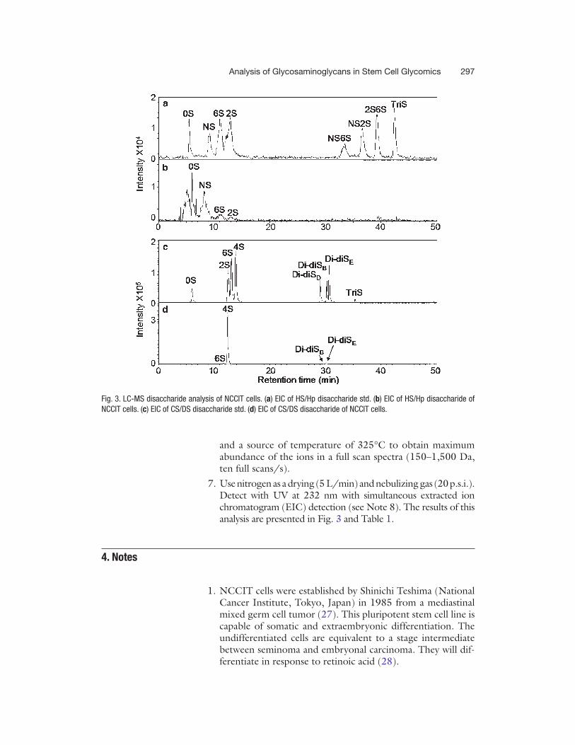
chromatogram (EIC) detection (see Note 7). The results of this analysis are presented in Fig. 3 and Table 1.
1. Use a Zorbax SB-C18 column (Agilent, 0.5 × 250 mm, 5 m) and inject 8 L of disaccharide standards containing 10 ng of each disaccharide or 8 L of heparin/HS disaccharide sample from Subheading 3.4.
2. Use HPLC solution C and HPLC solution D to elute hepa-rin/HS disaccharides at 10 L/min.
3. The elution conditions at 20°C are solution C for 20 min, followed by a linear gradient from 20 to 45 min of 0–50% solution D.
4. The column effluent should enter the UV detector followed by the source of the electrospray ionization mass spectrome-ter for continuous detection.
5. Add another 5 L/min of acetonitrile just after column and before MS to make the solvent and TrBA easy spray, and easy evaporate in the ion-source.
6. Set the electrospray interface in negative ionization mode with the skimmer potential −40.0 V, capillary exit −40.0 V
3.5. Disaccharide Analysis
3.5.1. CS/DS Disaccharide Analysis
3.5.2. Heparin/HS Disaccharide Analysis
297Analysis of Glycosaminoglycans in Stem Cell Glycomics
and a source of temperature of 325°C to obtain maximum abundance of the ions in a full scan spectra (150–1,500 Da, ten full scans/s).
7. Use nitrogen as a drying (5 L/min) and nebulizing gas (20 p.s.i.). Detect with UV at 232 nm with simultaneous extracted ion chromatogram (EIC) detection (see Note 8). The results of this analysis are presented in Fig. 3 and Table 1.
1. NCCIT cells were established by Shinichi Teshima (National Cancer Institute, Tokyo, Japan) in 1985 from a mediastinal mixed germ cell tumor (27). This pluripotent stem cell line is capable of somatic and extraembryonic differentiation. The undifferentiated cells are equivalent to a stage intermediate between seminoma and embryonal carcinoma. They will dif-ferentiate in response to retinoic acid (28).
4. Notes
Fig. 3. LC-MS disaccharide analysis of NCCIT cells. (a) EIC of HS/Hp disaccharide std. (b) EIC of HS/Hp disaccharide of NCCIT cells. (c) EIC of CS/DS disaccharide std. (d) EIC of CS/DS disaccharide of NCCIT cells.
298 Li et al.
Tabl
e 1
Disa
ccha
ride
com
posi
tion
of N
CCIT
cel
ls
HS/H
p0S
NS6S
2SNS
6SNS
2S2S
6STr
iS
23.8
%47
.6%
18.1
%10
.5%
n.d.
n.d.
n.d.
n.d.
CS/D
S0S
2S6S
4SDi
-diS
DDi
-diS
BDi
-diS
ETr
iS
n.d.
n.d.
1.2%
94.3
%n.
d.0.
3%4.
2%n.
d.
HS/
Hp:
0S,
U
A-G
lcN
Ac;
NS,
U
A-G
lcN
S; 6
S,
UA
-Glc
NA
c6S;
2S,
U
A2S
-Glc
NA
c; N
S2S,
U
A2S
-Glc
NS;
NS6
S,
UA
-Glc
NS6
S; 2
S6S,
U
A2S
-Glc
NA
c6S;
TriS
, U
A2S
-Glc
NS6
S. C
S/D
S: 0
S,
UA
-Gal
NA
c; 2
S,
UA
2S-G
alN
Ac;
6S,
U
A-G
alN
Ac6
S; 4
S,
UA
-Gal
NA
c4S;
Di-d
iSB,
UA
2S-G
alN
Ac4
S; D
i-diS
D,
UA
2S-G
alN
Ac6
S;
Di-d
iSE,
UA
-Gal
NA
c4S6
S; T
riS,
UA
2S-G
alN
Ac4
S6S;
n.d
., no
t det
ecte
d.
299Analysis of Glycosaminoglycans in Stem Cell Glycomics
2. It is best to freshly prepare actinase E proteolysis enzyme solution from dry protein immediately before use.
3. We tested series of different number of cells for GAGs extrac-tion, recovery and subsequent disaccharide analysis, we found that 1 × 10⁶ is currently the minimum number of cells required for disaccharide quantification.
4. This method for total GAGs extraction and recovery was devel-oped in our lab, involving in the use of a simple recovery and purification that relies on protease digestion and strong anion-exchange chromatography on a spin column followed by salt. Urea, a nonionic denaturant, is known to solubilize most proteins, and Chaps, a zwitterionic surfactant, is commonly used to solubi-lize hydrophobic molecules such as triglycerides. Approximately 90% of GAGs can be recovered using this method.
5. When Vivapure spin filters are used, make sure the centrifugal force is not >2,000 × g. For each centrifugation, adjust time carefully so that there is always residual liquid above on the top of the membrane to avoid dryness and membrane cracking.
6. When Microcon® Centrifugal Filter Units YM-3 (3,000 MWCO) spin columns are used, make sure the centrifugal force is not >14,000 × g. For each centrifugation, adjust time carefully so that there is always residual liquid above on the top of the membrane to avoid dryness and membrane cracking.
7. The sensitivity in this method improved to 0.2 ng/disaccha-ride of CS/DS.
8. The sensitivity in this method improved to 2 ng/disaccharide of HS/Hp.
Acknowledgment
Our laboratory acknowledges generous support from the New York State Department of Health and the Empire State Stem Cell Board in the form of grant number N08G-264.
References
1. Raman, R., Raguram, S., Venkataraman, G., Paulson, J. C., and Sasisekharan, R. (2005) Glycomics: an integrated systems approach to structure-function relationships of glycans. Nat. Methods 2, 817–824.
2. Zhang, F., Zhang, Z., Thistle, R., McKeen, L., Hosoyama, S., Toida, T., et al. (2009) Structural characterization of glycosaminogly-cans from zebrafish in different ages. Glycoconj. J. 26, 211–218.
3. Park, Y., Yu, G., Gunay, N. S., and Linhardt, R. J. (1999) Purification and characterization of heparan sulphate proteoglycan from bovine brain. Biochem. J. 344(Pt 3), 723–730.
4. Warda, M., Toida, T., Zhang, F., Sun, P., Munoz, E., Xie, J., et al. (2006) Isolation and characterization of heparan sulfate from vari-ous murine tissues. Glycoconj. J. 23, 555–563.
5. Nairn, A. V., Kinoshita-Toyoda, A., Toyoda, H., Xie, J., Harris, K., Dalton, S., et al. (2007)
300 Li et al.
Glycomics of proteoglycan biosynthesis in murine embryonic stem cell differentiation. J. Proteome Res. 6, 4374–4387.
6. Linhardt, R. J., and Toida, T. (2004) Role of glycosaminoglycans in cellular communica-tion. Acc. Chem. Res. 37, 431–438.
7. Linhardt, R. J. (2003) 2003 Claude S. Hudson Award address in carbohydrate chem-istry. Heparin: structure and activity. J. Med. Chem. 46, 2551–2564.
8. Johnson, Z., Proudfoot, A. E., and Handel, T. M. (2005) Interaction of chemokines and gly-cosaminoglycans: a new twist in the regulation of chemokine function with opportunities for thera-peutic intervention. Cytokine Growth Factor Rev. 16, 625–636.
9. Capila, I., and Linhardt, R. J. (2002) Heparin-protein interactions. Angew. Chem. Int. Ed. Engl. 41, 390–412.
10. Beenken, A., and Mohammadi, M. (2009) The FGF family: biology, pathophysiology and therapy. Nat. Rev. Drug Discov. 8, 235–253.
11. Hoffman, L. M., and Carpenter, M. K. (2005) Characterization and culture of human embry-onic stem cells. Nat. Biotechnol. 23, 699–708.
12. Giacomini, M., Baylis, F., and Robert, J. (2007) Banking on it: public policy and the ethics of stem cell research and development. Soc. Sci. Med. 65, 1490–1500.
13. Uygun, B. E., Stojsih, S., and Matthew, H. (2009) Effects of immobilized glycosamino-glycans influence proliferation and differentia-tion of mesenchymal stem cells. Tissue Eng. Part A 15(11), 3499–3512.
14. Kumarasuriyar, A., Murali, S., Nurcombe, V., and Cool, S. M. (2009) Glycosaminoglycan composition changes with MG-63 osteosar-coma osteogenesis in vitro and induces human mesenchymal stem cell aggregation. J. Cell Physiol. 218, 501–511.
15. Dombrowski, C., Song, S. J., Chuan, P., Lim, X., Susanto, E., Sawyer, A. A., et al. (2009) Heparan sulfate mediates the proliferation and differentia-tion of rat mesenchymal stem cells. Stem Cells Dev. 18, 661–670.
16. Carney, S. L., and Muir, H. (1988) The struc-ture and function of cartilage proteoglycans. Physiol. Rev. 68, 858–909.
17. Poole, A. R. (1986) Proteoglycans in health and disease: structures and functions. Biochem. J. 236, 1–14.
18. Bayliss, M. T., Osborne, D., Woodhouse, S., and Davidson, C. (1999) Sulfation of chon-droitin sulfate in human articular cartilage. The effect of age, topographical position, and
zone of cartilage on tissue composition. J. Biol. Chem. 274(22), 15892–15900.
19. Rizkalla, G., Reigner, A., Bogoch, E., and Poole, A. R. (1992) Studies of the articular cartilage proteoglycan aggrecan in health and osteoarthritis. Evidence for molecular hetero-geneity and extensive molecular changes in disease. J. Clin. Invest. 90, 2268–2277.
20. Plaas, A. H., Wong-Palms, S., Roughley, P. J. Midura, R. J., and Hascall, V. C. (1997) Chemical and immunological assay of the nonreducing terminal residues of chondroitin sulfate from human aggrecan. J. Biol. Chem. 272, 20604–20610.
21. Hitchcock, A. M., Yates, K. E., Shortkroff, S., Costello, C. E., and Zaia, J. (2007) Optimized extraction of glycosaminoglycans from nor-mal and osteoarthritic cartilage for glycomics profiling. Glycobiology 17(1), 25–35.
22. Pervin, A., Gallo, C., Jandik, K. A., Han, X.-J., and Linhardt, R. J. (1995) Preparation and structural characterization of large heparin-derived oligosaccharides. Glycobiology 5, 83–95.
23. Zhang, F., Sun, P., Munoz, E., Chi, L., Sakai, S., Toida, T., et al. (2006) Microscale isola-tion and analysis of heparin from plasma using an anion-exchange spin column. Anal. Biochem. 353, 284–286.
24. Edens, R. E., Al-Hakim, A., Weiler, J. M., Rethwisch, D. G., Fareed, J., and Linhardt, R. J. (1992) Gradient polyacrylamide gel electropho-resis for determination of the molecular weights of heparin preparations and low-molecular-weight heparin derivatives, J. Pharm. Sci. 81, 823–827.
25. Zhang, Z., Park, Y., Kemp, M. M., Zhao, W., Im, A. R., Shaya, D., et al. (2009) Liquid chromatography-mass spectrometry to study chondroitin lyase action pattern. Anal. Biochem. 385, 57–64.
26. Zhang, Z., Xie, J., Liu, H., Liu, J., and Linhardt, R. J. (2009) Quantification of hepa-ran sulfate disaccharides using ion-pairing reversed-phase microflow high-performance liquid chromatography with electrospray ion-ization trap mass spectrometry. Anal. Chem. 81, 4349–4355.
27. Teshima, S., Shimosato, Y., Hirohashi, S., Tome, Y., Hayashi, I., Kanazawa, H., Kakizoe, T. (1988) Four new human germ cell tumor cell lines. Lab Invest. 59, 328–336.
28. Damjanov, I., Horvat, B., Gibas, Z. (1993) Retinoic acid-induced differentiation of the developmentally pluripotent human germ cell tumor-derived cell line, NCCIT. Lab Invest. 68, 220–232.