I N V ITRO Issue 7 • 2010 Interview with Dr. Adel M. H. Azab Deputy Director for Production at the Veterinary Serum and Vaccines Research Institute of Cairo, Egypt BioFiles Staining Protocol Series: ACCUSTAIN® Gomori’s and Masson’s Trichrome Stain Prestige Antibodies® as tools in detection studies and Biomarker discovery Comparative genomic hybridisation of DNA ampli- fied with GenomePlex®
Transcript
INVITROIssue 7 • 2010
Interview with Dr. Adel M. H. AzabDeputy Director for Production at the Veterinary Serum and
Vaccines Research Institute of Cairo, Egypt
BioFiles
Staining Protocol Series:
ACCUSTAIN® Gomori’s and
Masson’s Trichrome Stain
Prestige Antibodies® as tools
in detection studies and
Biomarker discovery
Comparative genomic
hybridisation of DNA ampli-
fied with GenomePlex®
Our Innovation, Your Research – Shaping the Future of Life Science
Our Innovation, Your Research – Shaping the Future of Life Science
Dear Researcher,
Table of contents
Interview with Dr. AdeI M. H. Azab 4
Ni-NTA-Atto conjugates for sensitive, specific detection of polyhistidine-tagged proteins 7
ACCUSTAIN® Gomori’s Trichrome Stain 10
ACCUSTAIN® Masson’s Trichrome Stain 11
Comparative genomic hybridisation of DNA amplified with GenomePlex® 15
Vibrio detection 18
Dr. Adel Azab is an internationally renowned expert for veterinary viral
vaccine research and production. The Egyptian scientist lives and works
in Cairo, where he played a major role in developing The Veterinary Serum
& Vaccine Research Institute to its high standards today. As a Deputy
Director he helps the Institute to cover the vaccine production for his
country, and to be prepared for future challenges arising from new
epidemics in Africa.
Accurate and reliable detection in molecular biology and diagnostics is in
the focus of this In Vitro issue. Constant improvement in chemistry has led
to the development of Atto dyes with unprecedented brightness and
photostability, which can be used in time saving in gel-staining protocols.
One highlight of this issue is a scientific article describing how whole
genome amplification was combined with comparative genome hybrid-
isation to identify chromosomal abnormalities from limited amounts of
DNA. Moreover, an update on Prestige Antibodies® and our regular Bio-
Files are included.
This BioFiles In Vitro series on classical histological staining protocols con-
tains two protocols for Trichrome staining provided in accordance with
the IVD product range, which can be collected in your lab work stain
protocols.
Our Prestige Antibody® range, to which more than 2,500 antibodies are
added per year, now covers more than 6,100 antibodies, with 5,200
human protein targets. The benefit of the validation of these antibodies
by the human protein atlas (HPA) programme is impressively demonstrat-
ed by detection of a highly specific tumour marker in a diagnostic
application.
In this issue an article describes how the use of the GenomePlex® WGA kit
for whole genome amplification (WGA) can be combined with compara-
tive genome hybridisation to identify chromosomal abnormalities. The
GenomePlex® WGA kit allows highly accurate conducting of the CGH
analysis from a limited amount of DNA, providing a valuable tool for
genetic research.
Wrapping up, In Vitro introduces a solution for selective detection of
Vibrio cholerae, using the chromogenic medium HiCrome Vibrio Agar.
The use of this medium allows the for differentiation of Vibrio cholerae in
the presence of co-cultured contaminating bacteria.
We hope you enjoy our BioFiles In Vitro journal.
Kind regards,
Walter Gmelin, PhD
European Marketing Manager Life Science
Sigma-Aldrich Chemie GmbH
Walter Gmelin, PhD
It is our great pleasure to present an interview with Dr. Adel M. H. Azab,
Deputy Director of The Veterinary Serum & Vaccine Research Institute (VSRI).
4
Our Innovation, Your Research – Shaping the Future of Life Science
Interview with Dr. AdeI M. H. AzabDeputy Director for Production at the Veterinary Serum Vaccines Research Institute (VSVRl),Agriculture Research Center of Cairo, Egypt
CURRICULUM VITAE
Personal Information
Name: Dr. AdeI M. H. Azab
Birth date: 10 December, 1949
Place of birth: Cairo, Egypt
Gender: Male
Social status: Married
Nationality: Egyptian
Work address: Vet-Ser. and Vacc. Res. Instit. (VSVRI)
P. O. Box No.: 131, Postal Code: 1 1381
Abbassia, Cairo, Egypt.
Scientific Degrees
1982 Ph.D. in Microbiology, Faculty of
Veterinary Medicine, Cairo University.
1979 Master of Veterinary Sciences,
Microbiology, Faculty of Veterinary
Medicine, Cairo University.
1973 Bachelor of Veterinary Sciences,
Faculty of Veterinary Medicine,
Cairo University.
Career History
2005– today Deputy Director for Production at the
Veterinary Serum and Vaccine Research
Institute (VSVRI), Agriculture Research
Center.
1973– 2003 Different positions at the VSVRI.
1991– 2000 Expert in viral vaccine production at
the Ministry of Agriculture and Water,
Kingdom of Saudi Arabia. For
production of: rinderpest, sheep pox,
new castel rabies.
1983–1990 Associate Professor at the faculty of
veterinary medicine, EI-Mousel
University, Iraq, teaching: virology,
poultry viral disease.
The Veterinary Serum and Vaccine Research Institute
The Veterinary Serum and Vaccine Research Institute (VSVRI) was established in
1903. It consists of 13 departments. Each one is responsible for the production of
and research into specific vaccines. The departments of VSVRI include: FMD, Rift
Valley fever, pet animals, new castel, sheep pox, rinderpest, African horse sickness,
rinderpest like disease, aerobic, anaerobic, parasitic, antigens and sera, genetic
engineering. The institute products satisfy Egyptian market needs in some vaccines,
e.g. FMD, Rift Valley fever, sheep pox, some poultry vaccines, rabies etc.
The institute also includes a maintenance workshop, library, administration office,
meeting hall and other supportive services. The institute employs about 1,200
people, 174 of them with M.Sc. and Ph.D.
I have a training course in Sandia National Laboratories Albuquerque NM-USA in:
• The transportation of infectious substances and diagnostic specimens.
This training course meets or exceeds all IATA/ICAO requirements.
Biebrich scarlet, 0.9%, acid fuchsin 0.1%, in acetic acid,
1.0%.
Phosphotungstic Acid Solution
Cat. no. HT15-2, phosphotungstic acid, 10%.
Phosphomolybdic Acid Solution
Cat. no. HT15-3, phosphomolybdic acid, 10%.
Aniline Blue Solution
Cat. no. HT15-4, aniline blue, 2.4% and acetic acid, 2%.
Reagent preparation
Prepare working Weigert’s iron hematoxylin solution
(Cat. no. HT10-79) by adding equal amounts of Part A and
Part B. Prepare working phosphotungstic-phosphomolyb-
dic acid solution by mixing 1 volume of phosphotungstic
acid solution with 1 volume of phosphomolybdic acid solu-
tion with two volumes of deionised water. Use once and
discard.
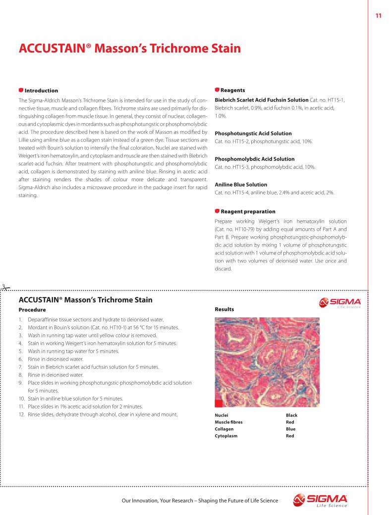
Results
Nuclei Black
Muscle fibres Red
Collagen Blue
Cytoplasm Red
Our Innovation, Your Research – Shaping the Future of Life Science
Turn your research into a masterpiece with Prestige Antibodies®
The most highly characterized antibodies in the industry — powered by Atlas Antibodies.
■ Over 3,800 antibodies, covering 3,600 protein targets
■ Validated by the Human Proteome Resource (HPR)
■ Standardized in universal protocols
■ Over 500 IF, IHC and WB images per antibody
■ All data conveniently searchable online
Visit sigma.com/prestige for more information.
Our Innovation, Your Research — Shaping the Future of Life SciencePrestige Antibodies is a registered trademark of Sigma-Aldrich Biotechnology L.P. and Sigma-Aldrich Co.
5,3006,100
The most highly characterised antibodies in the industry – powered by Atlas Antibodies.
■ Over 6,100 antibodies, covering 5,300 human protein targets
■ Validated by the Human Protein Atlas (HPA) Programme
■ Standardised in universal protocols
■ Over 700 IHC, IF and WB images per antibody
■ All data conveniently searchable online
13
Our Innovation, Your Research – Shaping the Future of Life Science
Prestige Antibodies® powered by Atlas Antibodies – highly
characterised for use in several applications
With all characterisation data published on the Human Protein Atlas
(HPA) portal (www.proteinatlas.org), Prestige Antibodies® are the most
highly characterised antibodies in the industry today. At the moment,
there are over 6,100 Prestige Antibodies® available, covering 5,300
human protein targets. This corresponds to 25% of the human pro-
teome. Each year, more than 2,500 new Prestige Antibodies® are made
available to the scientific community. The antibodies are directed
against both new and previously unknown proteins as well as estab-
lished protein families.
Each Prestige Antibody has been analysed and annotated in a large vari-
ety of normal and diseased human tissues, and the expression profiles
are conveniently searchable online. Immunohistochemical (IHC) staining
has been performed in 48 normal human tissues, the 20 most common
cancer types, 47 cell lines and 12 primary cell types. In addition, all anti-
bodies have been tested for performance in Immunofluorescence (IF)
and Western Blot (WB) applications. In total, more than 700 IHC, as well
as IF and WB images per antibody, are shown online.
The Human Protein Atlas Program systematically generates antibodies
against the human proteome,1,2 and the resulting Human Protein Atlas
provides a useful platform for biomarker discovery efforts.3
Malignant melanoma
Malignant melanoma is a common skin tumour with a rapidly increasing
incidence rate. Survival rates are high, but melanomas tend to meta-
stasise relatively early, and for patients with metastatic melanoma prog-
nosis is poor, with a five-year survival rate of less than 10%. Today, there
are no validated biomarkers for use in a clinical setting that are able to
give prognostic information and estimate the risk for metastatic
disease.
Syntaxin- 7
Syntaxin-7 (STX7) was identified on the Human Protein Atlas as a protein
showing a selective expression profile in cancer tissue as shown in
Figure 1.
Syntaxin-7 is a protein that belongs to the SNARE family, proposed to be
mediators of all intracellular fusion events. Syntaxin-7 is believed to be a
regulator of membrane vesicular trafficking between late endosomes
and lysosomes.
The Prestige Antibody HPA001467, directed against Syntaxin-7 was used
to evaluate the potential of Syntaxin-7 as a marker for malignant melan-
oma prognosis.4 The antibody was successfully used in IHC, WB and IF
applications. IHC staining was performed on tissue microarrays as well as
on two extended melanoma cohorts. As can be seen in Figure 2, there
was a clear differentiation in expression in different melanomas. This dif-
ferential expression could be correlated to tumour aggressiveness. It was
shown that high expression of STX7 indicates a good prognosis.
Prestige Antibodies® as tools in detection
studies and biomarker discovery
1
Figure 1: Detection of STX7 in HPA screening tissue micro arrays (TMAs). This
schematic overview shows the staining results in cancer tissue with the colour
codes representing different levels of immunoreactivity (red = strong staining,
orange = moderate staining, yellow = weak staining, white = negative staining and
black = missing or non-representative tissues). Duplicate samples are displayed
from each individual.
Figure 2: Immunohistochemical staining of malignant melanoma using the Anti-
STX7 antibody. Strong staining is illustrated in A and negative/weak staining in B.
2A B
14
Our Innovation, Your Research – Shaping the Future of Life Science
Summary
• The Human Protein Atlas was used to identify STX7 as a potential
prognostic marker for malignant melanoma.
• It was shown that reduced levels of STX7 are associated with more
aggressive tumours.
• The Prestige Antibody HPA001467 has been validated as an excellent
tool for biomarker detection.
Use of Prestige Antibodies® in detection studies
Prestige Antibodies® are excellent tools for detection studies. These are
highly characterised antibodies evaluated in several applications, such
as IHC, IF and WB, and tested on a large variety of normal and diseased
human tissues. Standardised universal protocols can be used
and all characterisation data are conveniently searchable online
(www.proteinatlas.org)
Each year the Human Protein Atlas portal is updated with characterisa-
tion data from 2,500 new human antibodies. During 2010, The Human
Protein Atlas portal will contain expression profiles and localisation data
for more than 50% of the human proteome.
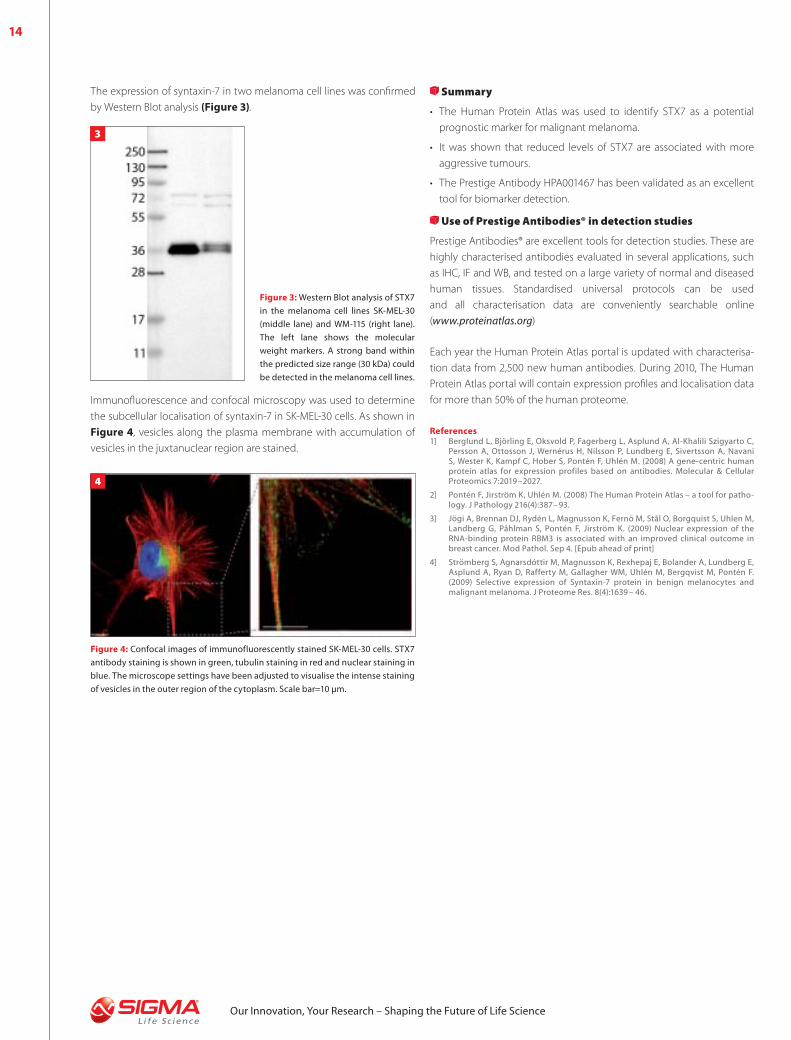
The expression of syntaxin-7 in two melanoma cell lines was confirmed
by Western Blot analysis (Figure 3).
Figure 3: Western Blot analysis of STX7
in the melanoma cell lines SK-MEL-30
(middle lane) and WM-115 (right lane).
The left lane shows the molecular
weight markers. A strong band within
the predicted size range (30 kDa) could
be detected in the melanoma cell lines.
3
Figure 4: Confocal images of immunofluorescently stained SK-MEL-30 cells. STX7
antibody staining is shown in green, tubulin staining in red and nuclear staining in
blue. The microscope settings have been adjusted to visualise the intense staining
of vesicles in the outer region of the cytoplasm. Scale bar=10 μm.
4
Immunofluorescence and confocal microscopy was used to determine
the subcellular localisation of syntaxin-7 in SK-MEL-30 cells. As shown in
Figure 4, vesicles along the plasma membrane with accumulation of
vesicles in the juxtanuclear region are stained.
References1] Berglund L, Björling E, Oksvold P, Fagerberg L, Asplund A, Al-Khalili Szigyarto C,
Persson A, Ottosson J, Wernérus H, Nilsson P, Lundberg E, Sivertsson A, Navani S, Wester K, Kampf C, Hober S, Pontén F, Uhlén M. (2008) A gene-centric human protein atlas for expression profiles based on antibodies. Molecular & Cellular Proteomics 7:2019–2027.
2] Pontén F, Jirström K, Uhlén M. (2008) The Human Protein Atlas – a tool for patho-logy. J Pathology 216(4):387– 93.
3] Jögi A, Brennan DJ, Rydén L, Magnusson K, Fernö M, Stål O, Borgquist S, Uhlen M, Landberg G, Påhlman S, Pontén F, Jirström K. (2009) Nuclear expression of the RNA-binding protein RBM3 is associated with an improved clinical outcome in breast cancer. Mod Pathol. Sep 4. [Epub ahead of print]
4] Strömberg S, Agnarsdóttir M, Magnusson K, Rexhepaj E, Bolander A, Lundberg E, Asplund A, Ryan D, Rafferty M, Gallagher WM, Uhlén M, Bergqvist M, Pontén F. (2009) Selective expression of Syntaxin-7 protein in benign melanocytes and malignant melanoma. J Proteome Res. 8(4):1639 – 46.
15
Our Innovation, Your Research – Shaping the Future of Life Science
Cut off log2 ratios > 0.2 Cut off log2 ratios > 0.2
GainChr.21
GainChr.18
Normalised NormalisedCombined Combined
The data (Figure 2) shows that CGH microarray analysis of the Down's
syndrome patient is consistent with a genomic gain as detected by 15
target clones in the array for unamplified gDNA and the whole genome
amplified DNA, reflecting the presence of a third chromosome corre-
sponding to region 21q22. Furthermore, the array for Edward’s syndrome
(Figure 3) showed a genomic gain in 30 of 33 possible test clones for
the unamplified genomic DNA sample and a gain of 27 target clones
from 33 in the whole genome amplified DNA sample in 18p11.2
p11.3q11.2q22.2q23.3q23 regions. This represents strong indication of
trisomy 18. The CGH array analysis was accurate in detecting clones dis-
playing a gain for each patient tested and confirms that the GenomePlex®
Whole Genome amplification method provides a sufficient amount of
DNA without any bias.
17
Our Innovation, Your Research – Shaping the Future of Life Science
Clone Location Genomic combined WGA combined
RP11-625C23 21:q11.2 0.251 0.278
RP11-840D8 21:q21.1 0.26 0.367
RP11-143A3 21:q21 0.276 0.258
RP11-108H5 21:q21 0.384 0.312
RP11-17O20 21:q22.1 0.403 0.393
RP11-166F15 21:q22.1 0.36 0.352
RP11-401I23 21:q22.1 0.497 0.356
RP11-35C4 21:q22.3 0.364 0.287
RP11-92D3 21:q22.3 0.259 0.29
RP11-88N2 21:q22.3 0.352 0.34
RP11-190A24 21:q22.3 0.353 0.36
RP11-40L10 21:q22.3 0.31 0.189
RP11-16B19 21:q22.3 0.369 0.276
RP11-640F21 21:q22.3 0.358 0.273
GS-63H24 21:qter 0.31 0.229
Table 1: Shows quantified data for trisomy 21, where a copy number gain was
detected in all possible clones for the unamplified genomic DNA and 14 of 15
clones were detected for the WGA product. All clones were detected in the expect-
ed 21q22 region, ranging from 0.251 to 0.497.
Clone Location Genomic combined WGA combined
RP11-70501 18:p11.3 0.191 0.197
RP11-14P20 18:p11.3 0.224 0.243
RP11-607C2 18:p11.3 0.29 0.213
RP11-78H1 18:p11.3 0.294 0.243
RP11-55N14 18:p11.3 0.193 0.174
RP11-193E15 18:p11.3 0.239 0.217
RP11-838N2 18:p11.3 0.208 0.188
RP11-874J12 18:p11.3 0.271 0.15
RP11-183C12 18:p11.3 0.33 0.153
RP11-105C15 18:p11.3 0.339 0.356
RP11-781P6 18:p11.3 0.308 0.257
RP11-931H21 18:p11.2 0.244 0.231
RP11-772F18 18:p11.2 0.213 0.21
RP11-752I5 18:p11.2 0.38 0.272
RP11-807E13 18:p11.2 0.305 0.235
RP11-411B10 18:p11.2 0.203 0.201
RP11-380C8 18:q11.1 0.168 0.169
RP11-459H24 18:p11.2 0.219 0.163
RP11-758N17 18:p11.2 0.336 0.245
RP11-540M4 18:p11.2 0.326 0.256
RP11-90G7 18:p11.2 0.33 0.221
RP11-704G7 18:q22.2 0.246 0.191
RP11-47G4 18:q22.3 0.306 0.204
RP11-669I1 18:q22.3 0.299 0.214
RP11-27C7 18:q22.3 0.373 0.26
RP11-357H3 18:q23 0.402 0.345
RP11-162A12 18:q23 0.2 0.199
RP11-90L3 18:q23 0.311 0.31
RP11-451L19 18:q23 0.354 0.342
RP11-91C19 18:q23 0.322 0.219
RP11-154H12 18:q23 0.325 0.427
GS-964M9 18:qter 0.23 0.241
RP11-89N1 18:q23 0.298 0.34
Table 2: Shows quantified data for trisomy 18, where a copy number gain was
detected in 30 of 33 possible clones for the unamplified genomic DNA, while whole
genome amplified DNA showed a gain in 27 of 36 possible clones. Clones were
detected in expected regions, ranging from 0.2 to 0.427 compared to the selected
gain threshold of 0.2.
Conclusions
The data presented proves that whole genome amplification using the
GenomePlex® Whole Genome Amplification Kit in conjunction with
Comparative Genomic Hybridisation array allows for the identification of
genome-wide copy number and chromosomal abnormalities ampli-
fying limited DNA without bias.
The results verify that the GenomePlex® WGA Kit is able to amplify DNA
across >800 loci without detectable bias. When using DNA amplified
with the GenomePlex® technology, four to five CGH arrays were run
using a total of 10 ng genomic DNA isolated from the patients. Typical
CGH arrays require 500 ng of genomic DNA for a single array. Using the
GenomePlex® WGA Kit eliminates the need for obtaining a significant
quantity of patient samples for CGH and other assay methodologies.
Acknowledgements
The authors would like to thank Dr. Sau W. Cheung for providing Trisomy
21 and Trisomy 18 DNA samples along with normal control DNA for
whole genome amplification; Dr. Xinyan Lu for performing chromo-
somal microarray analysis; and Steve D. Bland for his illustrations. We also
would like to thank Dr. Arthur Beaudet for facilitating communication
with the microarray facilities in the Department of Molecular and Human
Genetics at Baylor College of Medicine, Houston, TX.
References1] Yu, W.; Ballif, B. C.; Kashork, C. D.; Heilstedt, H. A.; Howard, L. A.; Cai, W. W.; White,
L.D.; Liu, W.; Beaudet, A. L.; Bejjani, B. A.; Shaw, C. A.; Shaffer, L. G. Development of a comparative genomic hybridiation microarray and demonstration of its utility with 25 wellcharacterized 1p36 deletions. Hum. Mol. Gen. 2003, 12, 2145.
2] Shaw, C. J.; Shaw, C. A.; Yu, W.; Stankiewicz, P.; White, L. D.; Beaudet, A. L.; Lup-ski, J. R. Comparative genomic hybridization using a proximal 17p BAC/PAC array detects rearrangements responsible for four genomic disorders. J. Med. Genet. 2004, 41, 113.
3] Shaw, C. J.; Lupski, J. R. Implications of human genome architecture for rearrange-ment-based disorders: the genomic basis of disease. Hum. Mol. Gen. 2004, 13, 57.
4] Cheung, S. W.; Shaw, C. A.; Yu, W.; Li, J.; Ou, Z.; Patel, A.; Yatsenko, S. A.; Cooper, M. L.; Furman, P.; Stankiewicz, P.; Lupski, J. R.; Chinault, A. C.; Beaudet, A. L. Develop-ment and validation of a CGH microarray for clinical cytogenetic diagnosis. Gen. Med. 2005, 7, 422.
TrademarksAll products supplied by Sigma-Aldrich unless otherwise indicated. GenomePlex® is a registered trademark of Rubicon Genomics, Inc. GenomePlex® WGA technology patent pending. GenElute is a trademark of Sigma-Aldrich Biotechnology, L.P. SYBR is a registered trademark of Molecular Probes, Inc. PureGene is a registered trademark of Gentra Systems, Inc.
Tables of results listing signal quantities for loci showing major differences
Signal values for critical diagnostic clones are listed in the tables below. The threshold for designation of over-representation in these experiments
was 0.2.
18
Our Innovation, Your Research – Shaping the Future of Life Science
Vibrio cholerae causes cholera in humans, Vibrio parahaemo-
lyticus and Vibrio vulnificus are the leading cause of seafood-
associated gastroenteridis.
Vibrios are motile, curved or comma-shaped bacilli and have a single
polar flagella with sheet proteins. They are often found in open water,
freshwater and saltwater. Vibrios are facultative aerobe and Gram-
negative bacterium and do not form spores. The metabolism can be
oxidative and fermentative. Most species are oxidase-positive, except
V. metschnikovii. Some vibrios such as Vibrio fischeri exhibit biolumines-
cence (Quorum sensing) under certain conditions. In most ways, vibrios
are close to Enterobacteriaceae, but also share some properties with
pseudomonads. They can be differentiated from enteric bacteria by
oxidase-positive reaction and motility. Differentiation from Pseudomonas
can be made based on the ability of vibrios to undergo oxidative and
fermentative metabolism.
Most vibrios are not fastidious and a simple C-source like glucose serves
as an energy source. As it is a typically marine organism, most species
require 2–4% NaCl or other salts and trace elements present in sea water
for optimal growth. Some species are like Pseudomonas and can use
diverse energy sources, and show great versatility in their metabolism.
The widely used media for Vibrio isolation are TCBS Agar and Alkaline
Peptone Water. However, accompanying sucrose-fermenting bacteria
may pose a problem in the identification of Vibrio species on TCBS Agar.3
The TCBS Agar contains a mixed indicator of bromothymol blue and
thymol. This system reacts upon acid production from sucrose fermen-
tation. On a chromogenic medium like HiCrome™ Vibrio Agar (see
Table 2), the colour development by Vibrio species is not affected by the
presence of colonies of other bacteria. This is because the amount of
colour developed depends on the reaction of the bacterial β-galac-
tosidase with the substrate contained in the media. The TCBS Agar also
contains a sodium thiosulphate and ferric citrate indicator system which
detects the production of hydrogen sulphide.
Pepton from animal origin provides carbonaceous, nitrogeneous and
essential nutrients to the Vibrio species to promote growth. High con-
centrations of sodium chloride in the medium are used to get an inhibi-
tory effect on the accompanying microflora. Sodium thiosulphate,
sodium citrate and sodium cholate are used as well to inhibit the growth
of gram-positive and some gram-negative bacteria, but not members of
Enterobacteriaceae. The strongly alkaline pH of the medium is also an
important tool to get selectivity for Vibrio species.
V. cholerae is a non-invasive bacteria, affecting the small intestine by pro-
ducing the cholera enterotoxins. The result is a life-threatening watery
diarrhea because of activation of the adenylate cyclase in the intestinal
cells. This reaction causes water and electrolytes from blood and tissues
to be pumped into the intestinal tract. The rapid loss of fluids leads to
dehydration, anuria, acidosis and shock. An additional loss of potassium
ions may result in cardiac complications and circulatory failure. The mor-
tality rate is very high (50–60%) if the disease is not treated. Infection
source is the water or food contaminated with human faeces.
V. parahaemolyticus causes gastroenteritis. It is an invasive organism
affecting primarily the colon tissue, and excretes a presently unidentified
toxin. The origin of an infection leads in most cases back to contamin-
ated raw and improper refrigerated seafood or a faecal contamination of
water and food.
V. vulnificus lives in warm seawater and is halophile, meaning it requires
salt for growth. Contaminated seafood which is eaten raw or is under-
cooked is in most cases the source of infections and causes gastroenteri-
tis, or a syndrome known as “primary septicemia”. Also, open wounds
that are exposed to seawater can lead to a wound infection.
Figure 1: Vibrio vulnificus is the cause of seafood-related mortality. Scientists from
Northwest Fisheries Science Center have isolated and characterised a key surface
protein involved in the ability of Vibrio vulnificus to attach to shellfish, such as oys-
ters (microscopic image from Northwest Fisheries Science Center, Seattle, USA).
1
19
Our Innovation, Your Research – Shaping the Future of Life Science
V. cholerae V. parahaemolyticus V. vulnificus
Growth in nutrient broth
without NaCl
with 1% NaCl
+
+
-
+
-
+
Oxidase + + +
Nitrate reduction + + +
myo-Inositol fermentation - - -
Arginine dihydrolase - - -
Lysine decarboxylase + + +
Ornithine decarboxylase + + V
ONPG + + +
Table 1: Typical biochemical reactions
Brand Cat. no. Name ISO
Nonselective
media
Fluka 43856 Alkaline Peptone Water (ISO) 8914
Fluka 77185 Peptone Water
Fluka T2117 Thiol broth
Nonselective
media
w/differential
system
Fluka 62895 Lysine Decarboxylase Salt Broth 8914
Fluka 75315 OF Test Nutrient Agar
Fluka 22091 Tryptic Soy Agar with supplement:
TTC solution
Selective media Fluka 49281 Glucose Salt Teepol Broth
Selective media
w/differential
system
Fluka 17134 CPC-Agar (Base) 8914
Fluka 70135 DCLS Agar
Fluka 90035 DCLS Agar No. 2
Fluka 92323 HiCrome™ Vibrio Agar
Fluka 86348 TCBS Agar
Table 2: Media for enrichment, detection and differentiation of Vibrio species
References1] Thompson et. al (ed.), The Biology of Vibrios, ASM Press, chapter1, pg 3 (2006).
2] E.I. Alcamo, Fundamentals of Microbiology, 6th ed, Jones and Bartlett Publishers, Inc. pg 254, 244 (2001).
3] Clesceri, Greenberg and Eaton (ed.), Standard Method for the Examination of Water and Waste Water, 20th ed.
4] American Public Health Association, Washington, D. C. (1998) H.Y. Kudo et. al, Improved Method for Detection of Vibrio parahaemolyticus in Seafood. ASM. Vol 67, No. 12, pg 5819 – 5823 (2001).
5] ISO 8914:1990, Microbiology – General Guidance for the Detection of Vibrio parahaemolyticus.
6] Color Atlas and Textbook of Diagnostic Microbiology, 5th edition, Lippincott Williams &Wilkins (1997).
Brand Cat. no. Name
Fluka 1850 Oxidase Reagent acc. Gordon-McLeod
Fluka 40560 Oxidase Strips
Fluka 70439 Oxidase Test
Fluka 07345 Oxidase Reagent acc. Gaby-Hadley A
Fluka 07817 Oxidase Reagent acc. Gaby-Hadley B
Fluka 49940 ONPG Disks
Fluka 51138 Nitrate Reagent Disks Kit
Fluka 38497 Nitrate Reagent A
Fluka 39441 Nitrate Reagent B
Fluka 77730 Gram Staining Kit
Table 4: Biochemical products and kits for Vibrio identification and differentiation
Brand Cat. no. Name
Fluka P9602 Polymyxin B Selective Supplement
Fluka 17779 TTC Solution
Table 3: Supplements for Vibrio media
Figure 2: HiCrome™ Vibrio Agar, a selective Agar with chromogenic system as


![Monitoring multiple myeloma by idiotype-specific peptide ... › track...Aldrich], and analyzed by an ELISA reader at 405 nm [Labsystems multiscan MS]. In vitro cell binding Cultured](https://static.documents.pub/doc/80x56/60d60bc658c9b43e297a2fd8/monitoring-multiple-myeloma-by-idiotype-specific-peptide-a-track-aldrich.jpg)