Introduction It has long been suspected that generalized brain dysfunc- tion may predispose to violent behavior. Studies using electroencephalographic (EEG), neurological, neuropsy- chological, and cognitive test techniques have repeatedly shown that violent offenders have poorer brain functioning than normal controls (Eichelman 1993; Eysenck and Gudjonsson 1989; Elliott 1987; Lewis et al 1988; Moffitt 1988; Raine 1993), but until recently it has not been
From the Department of Psychology, University of Southern California, Los Angeles, California (AR, LL); and Department of Psychiatry, Mount Sinai School of Medicine, New York, New York (MB).
Address reprint requests to Adrian Raine, Department of Psychology, S.G.M. Building, University of Southern California, Los Angeles, CA 90089-1061.
Received November 2, 1995; revised July 22, 1996.
possible to localize which brain areas in particular may be dysfunctional in violent offenders.
Clues do however exist with respect to the source of brain dysfunction predisposing to violence. It has long been thought that dysfunction of the prefrontal cortex may disrupt the regulation of aggression, and this notion has been supported by neurological studies of patients with damage to the prefrontal cortex (Damasio et al 1990; Weiger and Bear 1988). Some neuropsychological and psychophysiological studies on violent and forensic pop- ulations have shown abnormalities in hemispheric asym- metries of function (Convit et al 1991; Hare and McPher- son 1984; Raine et al 1990a) and reduced EEG interhemispheric coherence (Flor-Henry et al 1991), which may be linked to dysfunction of the corpus callosum (Nachshon 1983; Yeudall 1977), but this hypothesis has
496 BIOL PSYCHIATRY A. Raine et al 1997;42:495-508
not been tested using direct measures of callosal function- ing. Recent event-related potential mapping techniques have implicated dysfunction in the left angular gyrus in violent offenders as indicated by reduced slow-wave amplitudes (Barratt et al in press). Experimental animal research together with neurological studies of patients have further implicated limbic structures such as the amygdala and hippocampus in modulating aggression (Bear 1991; Elliott 1992; Gorenstein and Newman 1980; Mirsky and Siegel 1994; Watson et al 1983a), while the thalamus also provides an important afferent source of the hypothalamic-induced attack in cats (Mirsky and Siegel 1994). Nevertheless, such research on animals and humans who have suffered brain insults, although of key impor- tance, is one step removed from the question of whether severely violent offenders have brain dysfunction local- ized to specific brain areas.
The advent of brain imaging research has recently made it possible for the first time to directly assess brain functioning in violent individuals. Initial research in this area has again implicated frontal brain regions in addition to the temporal cortex (Goyer et al 1994; Volkow and Tancredi 1987). These important initial studies support the notion of localized brain dysfunction in aggressive pa- tients, although inevitable limitations of such initial re- search include small sample sizes in hospitalized patients, and a focus on aggressive personality as opposed to seriously violent behavior.
One particularly important group of violent offenders in forensic psychiatry consists of those who commit murder and plead not guilty by reason of insanity (NGRI). Although it is thought that such individuals have localized brain impairments, there has been no previous brain imaging research on this important population to support or refute this notion. In a prelim- inary report on a pilot sample of 22 such offenders compared to 22 normals, we provided some initial support for the notion of prefrontal dysfunction in this group (Raine et al 1994). In the present study the sample size is extended to 41 murderers and 41 con- trois, and analysis of subcortical structures is now undertaken. To our knowledge, this is the largest sample of violent offenders assessed on functional brain imaging. It is hypothesized that these seriously violent individuals have relatively localized brain dysfunction in the prefrontal cortex, angular gyrus, amygdala, hip- pocampus, thalamus, and the corpus callosum, brain areas previously linked empirically or conceptually to violence. Conversely, no dysfunction is expected in other brain areas (caudate, putamen, globus pallidus, midbrain, cerebellum), which have been implicated in other psychiatric conditions but which have not been related to violence.
Methods
Subjects MURDERERS. The experimental group consisted of 41
subjects tried in the state of California (39 men, 2 women) with a mean age of 34.3 years (SD = 10.1) who had been charged with either murder or manslaughter (labeled below as "murderers" for ease of reference). Subjects were referred to the University of California, h'vine (UCI) imaging center to obtain evidence relating to a NGRI defense or to capability of understanding the judicial process (incompetence to stand trial), while some who had been found guilty were referred to obtain information for diminished capacity as an ameliorating circumstance in the sentencing phase of the trial. Reasons for referral were very diverse and included schizophrenia (6 cases), history of head injury or organic brain damage (23), history of psychoactive substance abuse (3), affective disorder (2), epilepsy (2), history of hyperactivity and learning disabil- ity (3), and passive-aggressive or paranoid personality disorder (2). In 7 of the above cases, there were also unusual circumstances surrounding the crime that addi- tionally lead to the suspicion of some mental impairment. Offenders were not receiving regulated psychoactive med- ication at the time of positron emission tomography (PET) scans, and were instructed to be medication-free for the 2-week period preceding brain scanning. All subjects were in custody during this period, and penal authorities agreed to refrain from administering medication. Urine screens at the time of PET scanning were negative for every mur- derer referred for study.
CONTROLS. A control group was formed by matching each murderer with a normal subject of the sanae sex and age who had been tested using identical PET imaging procedures in the same laboratory. Six murderers (all men) had been diagnosed as schizophrenic by psychiatrists. These 6 were individually matched on age and sex with 6 schizophrenics from a larger psychiatric sample tested under identical procedures at the Brain Imaging Center at the University of California, Irvine (Buchsbaum et al 1990). The resulting 41 controls (39 men, 2 women) had a mean age of 31.7 years (SD = 10.3), which did not differ from murderers (p > .26). Normal controls had been screened for health by physical exam, medical history, and a psychiatric interview. No subject was taking any medi- cation, had a history of psychiatric illness, in self or first-degree relatives, or had current significant medical illness. Subjects with a history of seizure disorder, head trauma, or substance abuse were excluded. Subjects par- ticipated under protocols and consent forms approved by the Human Subjects Committee of University of Califor- nia, Irvine.
Brain Abnormalities in Murderers BIOL PSYCHIATRY 497 1997;42:495-508
P E T Task Procedure
Full details of general PET scanning procedures and quantification may be found in Buchsbaum et al (1990). Briefly, the fluorodeoxyglucose (FDG) tracer was injected into the subject in the test room and taken up by the brain as a tracer of brain metabolic rate for a 32-min period during which the subject completed the continuous per- formance task (CPT; Nuechterlein et al 1983). A degraded stimulus version of the CPT was employed as the frontal challenge task because it has been shown to produce increases in relative glucose metabolic rates in the frontal lobes in normal controls, in addition to increases in fight temporal and parietal lobes (Buchsbaum et al 1990). The key signal detection performance measure of d' reflects target recognition accuracy across the 32-min period (Davies and Parasuraman 1982; Nuechterlein 1991). Split- half reliability for the task is high (r = .843, p < .001). Full procedural details are reported in Buchsbaum et al (1990).
Ten minutes before the FDG injection, subjects were given practice trials on the CPT. Thirty seconds before injection, the task was started so that initial task novelty would not be FDG labeled. After 32 min of FDG uptake, the subject was transferred to the adjacent PET scanner room. An individually molded, thermosetting plastic head holder was used to hold the head still during the scan. Ten slices at 10-mm intervals parallel to the canthomeatal line were obtained. Scans started at the level of 80% of head height above the canthomeatal line (vertex to can- thomeatal line, usually 12-14 cm) and step downward at 10-mm intervals.
Brain regions were identified using two techniques as follows:
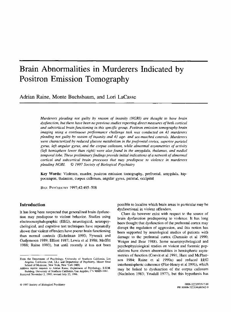
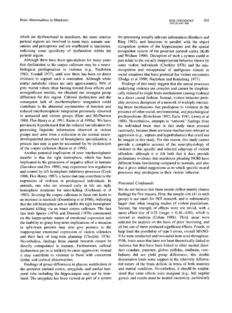
Cortical Peel Technique (lateral areas). Surface cortical regions of interest were measured using a modifica- tion of the original cortical peel technique (Buchs- baum et al 1990) with the four lobes and four anatomical subdivisions of each identified stereotac- tically (Buchsbaum et al 1989). This technique has been used by at least nine different PET groups, and a review of its advantages for facilitating intrasubject and intersubject differences may be found in Harris et al (1991). Absolute glucose values for each region of interest were expressed as a measure relative to all other regions contained in that slice. Relative rather than absolute metabolic rates were used because relative rates are more widely reported, have the advantages of removing whole brain metabolic rate, are more likely to be related to function in specific neuroanatomical systems (Fox and Mintum 1989), and show greater reliability within subjects over time (Bartlett et al 1991). The following three prefrontal
Precentrol I Postcentral
Middle f ro~t~, ,~.~
Superior ~ i i i i i i ~ i ~ frontal \ ~ i i i : : i i : : : : ~
i~ F..iiiii~i~g,7//A:,.~! I I:::::::::::::::::::::::::
i i i !~. . . r~/ / /~ kl[i::iiiiw/////~r
Inferior frontal
amarginal erior parietal Iobule
r / :::::::::::::::::::::: ~.ngular gyrus I lii!!::::~!~ii.::'~Jf/lll//-----"Jl~l lateral occipital I ================================= ~J----18
Figure 1. Lateral view of 10 stacked slices showing surface superior, middle, and inferior cortical prefrontal areas, precentral frontal cortex, and temporal, parietal, and occipital areas from cortical peel analysis. The top slice corresponds to slice #2, or 80% of head height in the brain atlas of Matsui and Hirano (1978).
values (averaged across slices) for each hemisphere were extracted: superior frontal gyrus, middle frontal gyms, and inferior frontal gyms (see Figure 1). Bilateral temporal (superior, middle, inferior, and posterior), parietal (postcentral, supramarginal, supe- rior parietal lobule, and angular gyrus), and occipital (area 19, area 17 superior, area 17 inferior, and area 18) measures averaged across slices were also taken (see Figure 1).
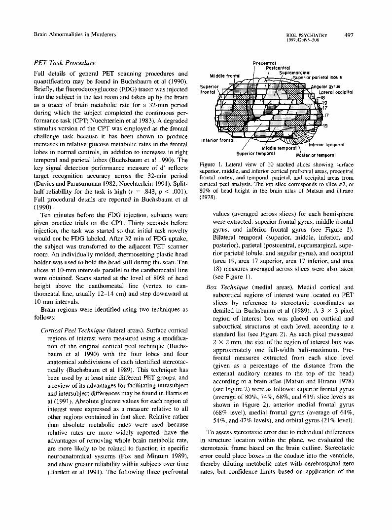
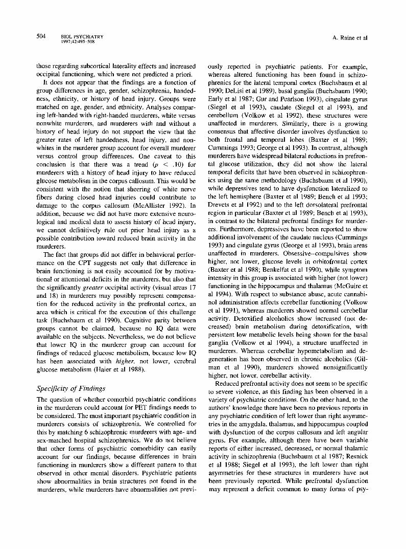
Box Technique (medial areas). Medial cortical and subcortical regions of interest were located on PET slices by reference to stereotaxic coordinates as detailed in Buchsbaum et al (1989). A 3 × 3 pixel region of interest box was placed on cortical and subcortical structures at each level, according to a standard list (see Figure 2). As each pixel measured 2 × 2 ram, the size of the region of interest box was approximately one full-width half-maximum. Pre- frontal measures extracted from each slice level (given as a percentage of the distance from the external auditory meatus to the top of the head) according to a brain atlas (Matsui and Hirano 1978) (see Figure 2) were as follows: superior frontal gyrus (average of 80%, 74%, 68%, and 61% slice levels as shown in Figure 2), anterior medial frontal gyrus (68% level), medial frontal gyms (average of 61%, 54%, and 47% levels), and orbital gyrus (21% level).
To assess stereotaxic error due to individual differences in structure location within the plane, we evaluated the stereotaxic frame based on the brain outline. Stereotaxic error could place boxes in the caudate into the ventricle, thereby diluting metabolic rates with cerebrospinal zero rates, but confidence limits based on application of the
498 BIOL PSYCHIATRY A. Raine et al 1997;42:495-508
MFG F . W .
PreC. " ~ ¢'~¢J74"/. "4t~=r,7 ~ 68% "V,.J v 61% ~ v 54%
" ~ ¢ ~ ) ~ FLW~ O ~ ~ d ~ FLWM-~. ~ [] "~'~ FLWM ~ T u . ~ , ~ ,RG
\% ~Zll ~ ,~=~ A. Calc'~"-- :~ ~[[ ~ Fus. G ~ 4 ~ ~, .~ / "~"~2~%Y AM"
Figure 2. Transverse view of the 10 slices showing medial cortical prefrontal structures used in box analysis. Percentages refer to percent of head height above the canthomeatal line. Key to abbreviations: A. Calc = anterior calcarine gyms, A. Cer = anterior cerebellum, ACG = anterior cingulate gyms, A. Corp = anterior corpus callosum, AMFG = anterior medial frontal gyrus, Amyg = amygdala, ARG = anterior rectal gyms, A. Thai = anterior thalamus, CG = cingulate gyms, CN = caudate nucleus, FLWM = frontal lobe white matter, Fus. G = fuisform gyms, GP = globus pallidus, Hipp = hippocampus, I. Coil = inferior colliculus, L. Thal= lateral thalamus, MCG = middle cingulate gyms, M. Corp = middle corpus callosum, MFG = medial frontal gyms, Midb -- midbrain, MTG = medial temporal gyms, M. Thai = medial thalmus, OG = orbital gyms, OR = optic radiation, Para = paracentral lobule, P. Calc = posterior calcarine gyms, P. Cer = posterior cerebellum, PCG = posterior cingulate gyms, P. Corp = posterior corpus callosum, PMFG = posterior medial frontal gyms, P. Put = posterior putamen, P. Thai = posterior thalamus, Prec = precuneus, PRG = posterior rectal gyms, Put = putamen, S. Co l l= superior colliculus, SFG = superior frontal gyms, Unc = uncus.
current system to magnetic resonance images confirm 2-SD limits within the caudate (Buchsbaum et al 1992).
Subcortical regions theorized to relate to violence were as follows: corpus callosum (47% level, see Figure 2); medial temporal lobe, including the hippocampus (average of 34%, 28%, and 21%), amygdala (21%); thalamus (41%); and cingulate (61%, 54%, 41%, and 34%). Sub- cortical regions theorized not to be related to violence were extracted as follows: caudate (average of 41% and 34%), putamen (average of 41%, 34%, and 28%), globus pallidus (34%), midbrain (21%), and cerebellum (21%).
Results
For both cortical and subcortical analyses, values were averaged across slices and two-way group (murderers and controls) x hemisphere (left and right) repeated-measures multivariate analyses of variance using the MANOVA approach (Vasey and Thayer 1987) were conducted. For some brain areas gyrus was added as a third factor in a three-way MANOVA. All tests of significance for planned
comparisons (t tests) are two tailed. Means and SDs for all
brain areas are shown in Table 1.
Cortical Regions
PREFRONTAL. AS anticipated on the basis of the pre- vious pilot data, the expanded group of 41 murderers had
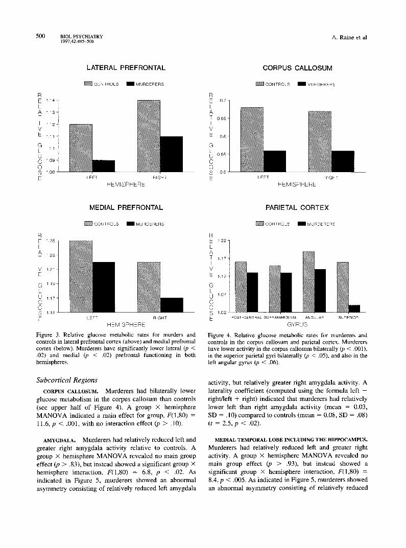
lower glucose metabolism relative to controls in both lateral and medial prefrontal cortical areas (,;ee Table 1
and Figure 3). We repeated exactly the same analyses that
we had previously conducted on a smaller sample (Raine
et al 1994) and found from two separate group x hemi- sphere MANOVAs a main effect for both lateral IF(l ,80)
= 5.6, p < .02] and medial IF(I,80) = 6.2, p < .02] prefrontal areas, with no interactions for hemisphere (p >
.75). A more detailed breakdown of prefrontal subregions
indicated that murderers had significantly lower glucose
metabolism for left and right medial superior frontal
cortex (t = 2.6, p < .02), left anterior medial cortex (t =
3.1, p < .003), right orbitofrontal cortex (t = 2.1, p < .04),
Brain Abnormalities in Murderers BIOL PSYCHIATRY 499 1997 ;42:495-508
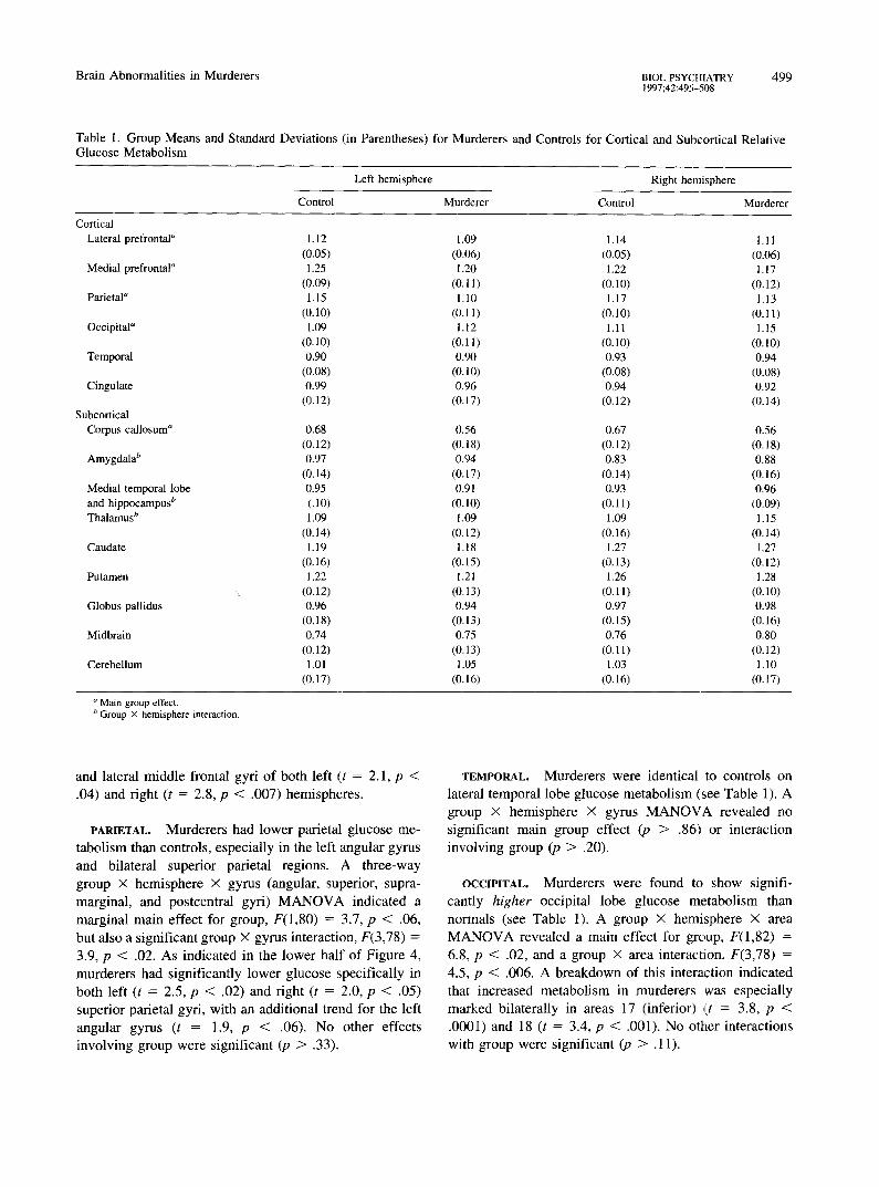
Table 1. Group Means and Standard Deviations (in Parentheses) for Murderers and Controls for Cortical and Subcortical Relative Glucose Metabolism
a Main group effect. b Group X hemisphere interaction.
and lateral middle frontal gyri of both left (t = 2.1, p <
.04) and fight (t = 2.8, p < .007) hemispheres.
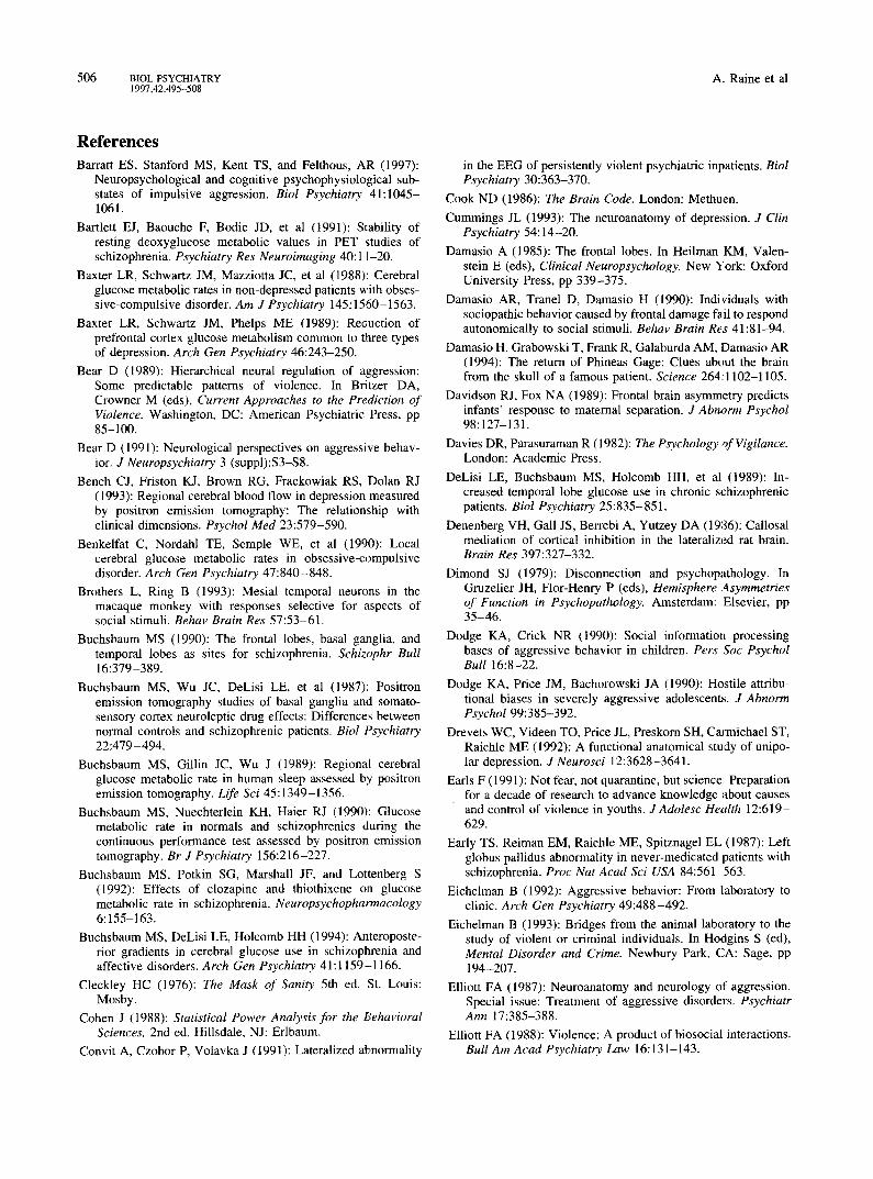
PARIETAL. Murderers had lower parietal glucose me-
tabol ism than controls, especial ly in the left angular gyrus
and bilateral superior parietal regions. A three-way
group X hemisphere x gyrus (angular, superior, supra-
marginal , and postcentral gyri) M A N O V A indicated a
margina l ma in effect for group, F(1,80) = 3.7, p < .06, but also a s ignif icant group X gyrus interaction, F(3,78) =
3.9, p < .02. As indicated in the lower half o f Figure 4,
murderers had signif icantly lower glucose specifically in
both left (t = 2.5, p < .02) and right (t = 2.0, p < .05) superior parietal gyri, with an addit ional t rend for the left
angular gyrus (t = 1.9, p < .06). No other effects
invo lv ing group were s ignif icant (p > .33).
TEMPORAL. Murderers were identical to controls on
lateral temporal lobe glucose metabol i sm (see Table 1). A
group X hemisphere x gyrus M A N O V A revealed no signif icant ma in group effect (p > .86) or interact ion
involv ing group (p > .20).
OCCIPITAL. Murderers were found to show signifi-
cantly higher occipital lobe glucose metabol i sm than normals (see Table 1). A group x hemisphere × area
M A N O V A revealed a main effect for group, F(1,82) =
6.8, p < .02, and a group × area interaction, F(3,78) =
4.5, p < .006. A breakdown of this interact ion indicated that increased metabol i sm in murderers was especial ly
marked bilaterally in areas 17 (inferior) (t = 3.8, p < .0001) and 18 (t = 3.4, p < .001). No other interactions
with group were s ignif icant (p > . 11).
500 BIOL PSYCHIATRY ,at. Raine et al 1997;42:495-508
R E 114 L A 1.18 T I 112
V E 111
G L 11 U C 1.o9 O S 1.De E
L A T E R A L P R E F R O N T A L
CONTROLS 1 MURDERERS
LEFT
HEMISPHERE RIGHT
R E o7- L A T 065 I
V E 06
G L U o.e5 C 0 S o6 E
C O R P U S C A L L O S U M
~ C O N T R O L S 1MURDERERS
LEFT RIGFT
HEMISPHERE
M E D I A L P R E F R O N T A L
R E 1.25 L A 1.23 T I
V 1.21 E
G 1.19 L U 1.17 C O S 116 E
~ C O N T R O L S 1MURDERERS
LEFT RIGHT
HEMISPHERE
Figure 3. Relative glucose metabolic rates for murders and controls in lateral prefrontal cortex (above) and medial prefrontal cortex (below). Murderers have significantly lower lateral (p < .02) and medial (p < .02) prefrontal functioning in both hemispheres.
Subcortical Regions
CORPUS CALLOSUM. Murderers had bilaterally lower glucose metabolism in the corpus callosum than controls (see upper half of Figure 4). A group X hemisphere MANOVA indicated a main effect for group, F(1,80) = 11.6, p < .001, with no interaction effect (p > .10).
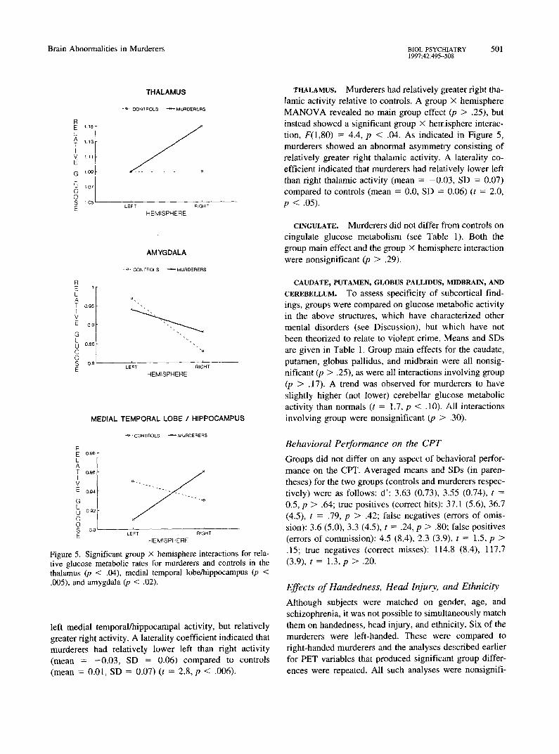
AMYGDALA. Murderers had relatively reduced left and greater right amygdala activity relative to controls. A group X hemisphere MANOVA revealed no main group effect (p > .83), but instead showed a significant group x hemisphere interaction, F(1,80) = 6.8, p < .02. As indicated in Figure 5, murderers showed an abnormal asymmetry consisting of relatively reduced left amygdala
PARIETAL C O R T E X
~ C O N T R O L S 1MURDERER8
R E 1.22- L A T 1.17i I V E 1.12"
G L U 1.o7
C O S 1.02- E POST-CENTRAL SUPRAMARGPNAL ANGULAR
GYRUS
SUPERIOR
Figure 4. Relative glucose metabolic rates for murderers and controls in the corpus callosum and parietal cortex. Murderers have lower activity in the corpus callosum bilaterally (p < .001), in the superior parietal gyri bilaterally (p < .05), and also in the left angular gyrus (p < .06).
activity, but relatively greater fight amygdala activity. A laterality coefficient (computed using the formula left - fight/left + right) indicated that murderers had relatively lower left than right amygdala activity (mean = 0.03, SD = .10) compared to controls (mean = 0.08, SD = .08) (t = 2.5, p < .02).
MEDIAL TEMPORAL LOBE INCLUDING THE HEPPOCAMPUS. Murderers had relatively reduced left and greater fight activity. A group X hemisphere M A N O V A revealed no main group effect (/7 > .93), but insteadi showed a significant group X hemisphere interaction, F(1,80) = 8.4, p < .005. As indicated in Figure 5, murderers showed an abnormal asymmetry consisting of relatively reduced
Brain Abnormalities in Murderers BIOL PSYCHIATRY 501 1997;42:495--508
R E 1,15
L A ~.~3 T I
V 111 E
G loe L U "i.o7 C O S lOe E
THALAMUS
- ~- OOHTROLS ~ MURDERER,~
I I
LEFT RIGHT
HEMISPHERE
1
o g 5
0 0
0,85
0,8
AMYGDALA
- 6 - CONTROLS ~ MURDERERS
cl
"u
i i
LEFT RIGHT
HEMISPHERE
MEDIAL TEMPORAL LOBE / HIPPOCAMPU8
- ~ ' C O N T R O L $ ~ M U R D E R E R S
R E o.ee L A T o, oe I v E o.04
G L U 0 9 2
c O s 0~ E
, i
LEFT RIGHT
HEMISPHERE
Figure 5. Significant group X hemisphere interactions for rela- tive glucose metabolic rates for murderers and controls in the thalamus (p < .04), medial temporal lobe/hippocampus (p < .005), and amygdala (p < .02).
left medial temporal/hippocampal activity, but relatively greater right activity. A laterality coefficient indicated that murderers had relatively lower left than right activity (mean = -0 .03 , SD = 0.06) compared to controls (mean = 0.01, SD = 0.07) (t = 2.8, p < .006).
THALAMUS. Murderers had relatively greater right tha- lamic activity relative to controls. A group X hemisphere M A N O V A revealed no main group effect (p > .25), but instead showed a significant group x hemisphere interac- tion, F(1,80) = 4.4, p < .04. As indicated in Figure 5, murderers showed an abnormal asymmetry consisting of relatively greater right thalamic activity. A laterality co- efficient indicated that murderers had relatively lower left than right thalamic activity (mean = -0 .03 , SD = 0.07) compared to controls (mean = 0.0, SD = 0.06) (t = 2.0, p < .05).
CINGULATE. Murderers did not differ fi'om controls on cingulate glucose metabolism (see Table 1). Both the group main effect and the group X hemisphere interaction were nonsignificant (p > .29).
CAUDATE, PUTAMEN, GLOBUS PALLIDUS, MIDBRAIN, AND
CEREBELLUM. TO assess specificity of subcortical find- ings, groups were compared on glucose metabolic activity in the above structures, which have characterized other mental disorders (see Discussion), but which have not been theorized to relate to violent crime. Means and SDs are given in Table 1. Group main effects for the caudate, putamen, globus pallidus, and midbrain were all nonsig- nificant (p > .25), as were all interactions involving group (p > .17). A trend was observed for murderers to have slightly higher (not lower) cerebellar glucose metabolic activity than normals (t = 1.7, p < .10). All interactions involving group were nonsignificant (p > .30).
Behavioral Performance on the CPT
Groups did not differ on any aspect of behavioral perfor- mance on the CPT. Averaged means and SDs (in paren- theses) for the two groups (controls and murderers respec- tively) were as follows: d ' : 3.63 (0.73), 3.:55 (0.74), t = 0.5, p > .64; true positives (correct hits): 37.1 (5.6), 36.7 (4.5), t = .79, p > .42; false negatives (eJrrors of omis- sion): 3.6 (5.0), 3.3 (4.5), t = .24, p > .80; false positives (errors of commission): 4.5 (8.4), 2.3 (3.9), t = 1.5, p > .15; true negatives (correct misses): 114.8 (8.4), 117.7 (3.9), t = 1.3, p > .20.
Effects of Handedness, Head Injury, and Ethnicity
Although subjects were matched on gender, age, and schizophrenia, it was not possible to simultaneously match them on handedness, head injury, and ethnicity. Six of the murderers were left-handed. These were compared to right-handed murderers and the analyses described earlier for PET variables that produced significant group differ- ences were repeated. All such analyses were nonsignifi-
502 BIOL PSYCHIATRY A. Raine et al 1997;42:495-508
cant, with the exception that left-handed murderers tended to have higher (not lower) medial prefrontal activity (p < .08), and had a significant less abnormal amygdala asym- metry (p < .002) than right-handed murderers. Results indicate therefore that greater rates of left-handedness in the murderer group relative to controls cannot account for reduced prefrontal activity and the abnormal amygdala asymmetry.
Fourteen of the murderers were nonwhite. Analyses comparing them to white murderers on PET measures were nonsignificant (p > . 14) in all cases, indicating that ethnic status did not influence findings.
Twenty-three murderers had a history of head injury. They did not differ from murderers without a history of head injury on PET measures (p > .27), with the one exception of a trend for head-injured murderers to have lower activity in the corpus callosum (p < .08) than non-head-injured murderers. Although analyses suggest that history of head injury cannot account for most findings, the possibility that they account for group dif- ferences in the corpus callosum cannot be ruled out.
Discussion
Key Findings
The key findings from this preliminary study are that murderers pleading NGRI are characterized by a) reduced glucose metabolism in bilateral prefrontal cortex, the posterior parietal cortex (bilateral superior gyrus and left angular gyrus), and the corpus callosum, and b) abnormal asymmetries of activity (left hemisphere lower than right) in the amygdala, thalamus, and medial temporal gyrus including the hippocampus. These data both confirm deficits in the prefrontal cortex from our earlier pilot study, and also yield new findings. These in turn provide both some general support for preexisting biological the- ories of violence, and also suggest new perspectives for understanding the type of brain dysfunction that may predispose to violence in this specific group of offenders.
Biosocial Pathways from Brain Deficits to Violence
A key question concerns how these multisite deficits can translate into violence via neuropsychological, psycholog- ical, cognitive, social, and situational pathways. Regarding prefrontal deficits, damage to this brain region can result in impulsivity, loss of self-control, immaturity, altered emotionality, and the inability to modify behavior, which can all in turn facilitate aggressive acts (Damasio 1985; Damasio et al 1994; Moffitt and Henry 1991; Stuss and Benson 1986; Weiger and Bear 1988). Regarding limbic deficits, the amygdala has been repeatedly associated with aggressive behavior in both animals and humans (Bear
1989; Mirsky and Siegel 1994; Weiger and Bear 1988). The amygdala, hippocampus, and prefrontal cortex make up part of the limbic system governing the expression of emotion, while the thalamus relays inputs from subcortical limbic structures to the prefrontal cortex (Fuster 1989; Mirsky and Siegel 1994). The hippocampal formation is thought to modulate aggression in cats through its action on the lateral hypothalamus via the lateral septal area (Mirsky and Siegel 1994; Siegel and Flynn 1968), and together with the septal area and prefrontal cortex forms the neurobiological basis of the behavioral inhibition system of Gray (1982), which is theorized to be dysfunc- tional in violent and psychopathic individuals (Gorenstein and Newman 1980). The amygdala is believed to act on the medial hypothalamus through at least two pathways in the modulation of aggression in animals (Watson et al 1983b). The hippocampus, amygdala, and thalamus are also of critical importance to learning, memory, and attention; abnormalities in their functioning may relate to deficits in forming conditioned emotional responses and the failure to learn from experience displayed by criminal and violent offenders (Cleckley 1976; Raine 1993). The amygdala additionally plays a role in the recognition of affective and socially significant stimuli (Nishijo et al 1988), with destruction of the amygdala in animals result- ing in a lack of fear (Bear 1991) and in man in a reduction in autonomic arousal (Lee et al 1988); thus, abnormalities in the amygdala could be relevant to a fearles.sness theory of violence based on psychophysiological findings of reduced autonomic arousal in offenders (Raine et al 1990b; Raine 1993).
The posterior parietal cortex (including superior and angular gyri) is centrally involved in the integration of sensory input and the formation of abstract concepts (Kolb and Wishaw 1990), and in conjunction with its reciprocal connections with the dorsolateral prefrontal cortex (Gold- man-Rakic et al 1983) may contribute to the cognitive and social information processing deficits observed in violent offenders (Dodge and Crick 1990; Moffitt and Silva 1988). Reductions in glucose metabolism in the left angular gyrus have been correlated with reduced verbal ability (Gur et al 1994), while damage to the left angular gyrus has been linked to deficits in reading anti arithmetic. Such cognitive dysfunction could predispose to educa- tional and occupational failure, which in turn predispose to crime and violence. Learning deficits have been found to be common in violent offenders who also haw~ low verbal IQs (Quay 1987; Raine 1993). One caveat here is that the finding for the left angular gyrus was a trend using a two-tailed test (p < .06), although the effect is significant on a one-tailed test (p < .03) and was predicted on the basis of event-related brain potential data from Barratt et al in press. In contrast to these posterior parietal areas,
Brain Abnormalities in Murderers BIOL PSYCHIATRY 503 1997;42:495--508
which are dysfunctional in murderers, the more anterior parietal regions are involved in more basic somatic sen- sations and perceptions and are unaffected in murderers, indicating some specificity of dysfunction within the parietal region.
Although there have been speculations for many years that dysfunction to the corpus callosum may be a neuro- biological predisposition to violence (e.g., Nachshon 1983; Yeudall 1977), until now there has been no direct evidence to support such a contention. Although white matter metabolic values are only approximately 50% of grey matter values (thus biasing toward floor effects and nonsignificant results), we obtained our strongest group difference for this region. Callosal dysfunction and the consequent lack of interhemispheric integration could contribute to the abnormal asymmetries of function and reduced interhemispheric integration previously observed in antisocial and violent groups (Hare and McPherson 1984; Flor-Henry et al 1991; Raine et al 1990a). We have previously hypothesized that the reduced lateralization for processing linguistic information observed in violent groups may arise from a reduction in the normal neuro- developmental processes of hemispheric specialization, a process that may in part be accounted for by dysfunction of the corpus callosum (Raine et al 1995).
Another potential implication of poor interhemispheric transfer is that the right hemisphere, which has been implicated in the generation of negative affect in humans (Davidson and Fox 1989), may experience less regulation and control by left hemisphere inhibitory processes (Cook 1986; Flor-Henry 1987), a factor that may contribute to the expression of violence in predisposed individuals. In animals, rats who are stressed early in life are right hemisphere dominant for mice-killing (Garbanati et al 1983). Severing the corpus callosum in these rats leads to an increase in muricide (Denenberg et al 1986), indicating that the left hemisphere acts to inhibit the right hemisphere mediated killing via an intact corpus callosum. The fact that both Sperry (1974) and Dimond (1979) commented on the inappropriate nature of emotional expression and the inability to grasp long-term implications of a situation in split-brain patients may also give pointers to the inappropriate emotional expression of violent offenders and their lack of long-term planning (Cleckley 1976). Nevertheless, findings from animal research cannot be directly extrapolated to humans. Furthermore, callosal dysfunction per se is unlikely to cause aggression; instead it may contribute to violence in those with concurrent limbic and cortical abnormalities.
Findings of group differences in glucose metabolism in the posterior parietal cortex, amygdala, and medial tem- poral lobe including the hippocampus may not be unre- lated. The amygdala has been viewed as part of a system
for processing socially relevant information (Brothers and Ring 1993), and functions in parallel with the object recognition system of the hippocampus and the spatial recognition system of the posterior parielLal cortex (Kolb and Wishaw 1990). Disruption of such a system could in part relate to the socially inappropriate behavior shown by some violent individuals (Cleckley 1976) and the mis- recognition and misappraisal of ambiguous stimuli in social situations that have potential for violent encounters (Dodge et al 1990; Nachshon and Rotenberg 1977).
Findings of this study suggest that the neural processes underlying violence are complex and cannot be simplisti- cally reduced to single brain mechanisms causing violence in a direct causal fashion. Instead, violent behavior prob- ably involves disruption of a network of multiply interact- ing brain mechanisms that predispose to violence in the presence of other social, environmental, and psychological predispositions (Eichelman 1992; Earls 1991; Lewis et al 1988). Nevertheless, attempts to "network" findings from the individual brain sites in this study must proceed cautiously, because there are brain mechanisms relevant to aggression (e.g., septum and hypothalamus) that could not be imaged in this study. For this reason, this study cannot provide a complete account of the neurophysiology of violence in this specific and selected subgroup of violent offenders, although it is felt both that it does provide preliminary evidence that murderers pleading NGRI have different brain functioning compared to normals, and also that it gives initial suggestions as to which specific neural processes may predispose to their violent behavior.
Potential Confounds
We do not believe that these results reflect merely chance findings for five reasons. First, the sample ,;ize (41 in each group) is not small for PET research, and is substantially larger than other imaging studies of violent populations. Second, the strength of effects were not trivial, with a mean effect size of 0.55 (range = 0.36-0.80), which is viewed as medium (Cohen 1988). Third, areas were selected for analysis on the basis of prior l:heorizing, and all but one of these produced significant effects. Fourth, to help limit the possibility of type I errors, overall MANO- VAs were conducted and two-tailed tests used throughout. Fifth, brain areas that have not been theoretically linked to violence but that have been linked to other mental disor- ders (caudate, putamen, globus pallidus, raidbrain, cere- bellum) did not yield group differences; this double dissociation lends some support to the relalively differen- tial nature of the brain deficits in terms of both anatomy and mental condition. Nevertheless, it shoald be empha- sized that some effects were marginal (e.g., left angular gyrus), and results must be treated cautiously, particularly
504 BIOL PSYCHIATRY A. Raine et al 1997;42:495-508
those regarding subcortical laterality effects and increased occipital functioning, which were not predicted a priori.
It does not appear that the findings are a function of group differences in age, gender, schizophrenia, handed- ness, ethnicity, or history of head injury. Groups were matched on age, gender, and ethnicity. Analyses compar- ing left-handed with right-handed murderers, white versus nonwhite murderers, and murderers with and without a history of head injury do not support the view that the greater rates of left handedness, head injury, and non- whites in the murderer group account for overall murderer versus control group differences. One caveat to this conclusion is that there was a trend (p < .10) for murderers with a history of head injury to have reduced glucose metabolism in the corpus callosum. This would be consistent with the notion that sheering of white nerve fibers during closed head injuries could contribute to damage to the corpus callosum (McAllister 1992). In addition, because we did not have more extensive neuro- logical and medical data to assess history of head injury, we cannot definitively rule out prior head injury as a possible contribution toward reduced brain activity in the murderers.
The fact that groups did not differ in behavioral perfor- mance on the CPT suggests not only that difference in brain functioning is not easily accounted for by motiva- tional or attentional deficits in the murderers, but also that the significantly greater occipital activity (visual areas 17 and 18) in murderers may possibly represent compensa- tion for the reduced activity in the prefrontal cortex, an area which is critical for the execution of this challenge task (Buchsbaum et al 1990). Cognitive parity between groups cannot be claimed, because no IQ data were available on the subjects. Nevertheless, we do not believe that lower IQ in the murderer group can account for findings of reduced glucose metabolism, because low IQ has been associated with higher, not lower, cerebral glucose metabolism (Haier et al 1988).
Specificity of Findings The question of whether comorbid psychiatric conditions in the murderers could account for PET findings needs to be considered. The most important psychiatric condition in murderers consists of schizophrenia. We controlled for this by matching 6 schizophrenic murderers with age- and sex-matched hospital schizophrenics. We do not believe that other forms of psychiatric comorbidity can easily account for our findings, because differences in brain functioning in murderers show a different pattern to that observed in other mental disorders. Psychiatric patients show abnormalities in brain structures not found in the murderers, while murderers have abnormalities not previ-
ously reported in psychiatric patients. For example, whereas altered functioning has been found in schizo- phrenics for the lateral temporal cortex (Buchsbaum et al 1990; DeLisi et al 1989), basal ganglia (Buchsbaum 1990; Early et al 1987; Gur and Pearlson 1993), cingulate gyrus (Siegel et al 1993), caudate (Siegel et al 1993), and cerebellum (Volkow et al 1992), these structures were unaffected in murderers. Similarly, there i,; a growing consensus that affective disorder involves dysfunction to both frontal and temporal lobes (Baxter et al 1989; Cummings 1993; George et al 1993). In contrast, although murderers have widespread bilateral reductions in prefron- tal glucose utilization, they did not show the lateral temporal deficits that have been observed in schizophren- ics using the same methodology (Buchsbaum et al 1990), while depressives tend to have dysfunction lateralized to the left hemisphere (Baxter et al 1989; Bench et al 1993; Drevets et al 1992) and to the left dorsolateral prefrontal region in particular (Baxter et al 1989; Bench et al 1993), in contrast to the bilateral prefrontal findings for murder- ers. Furthermore, depressives have been reported to show additional involvement of the caudate nucleus (Cummings 1993) and cingulate gyrus (George et al 1993), brain areas unaffected in murderers. Obsessive-compulsives show higher, not lower, glucose levels in orbitofrontal cortex (Baxter et al 1988; Benkelfat et al 1990), while symptom intensity in this group is associated with higher (not lower) functioning in the hippocampus and thalamus (McGuire et al 1994). With respect to substance abuse, acute cannabi- nol administration affects cerebellar functioning (Volkow et al 1991), whereas murderers showed normal cerebellar activity. Detoxified alcoholics show increased (not de- creased) brain metabolism during detoxification, with persistent low metabolic levels being shown for the basal ganglia (Volkow et al 1994), a structure unaffected in murderers. Whereas cerebellar hypometabolism and de- generation has been observed in chronic alcoholics (Gil- man et al 1990), murderers showed nonsignificantly higher, not lower, cerebellar activity.
Reduced prefrontal activity does not seem to be specific to severe violence, as this finding has been observed in a variety of psychiatric conditions. On the other hand, to the authors' knowledge there have been no previous reports in any psychiatric condition of left lower than right asymme- tries in the amygdala, thalamus, and hippocampus coupled with dysfunction of the corpus callosum and left angular gyrus. For example, although there have been variable reports of either increased, decreased, or normal thalamic activity in schizophrenia (Buchsbaum et al 1987; Resnick et al 1988; Siegel et al 1993), the left lower than right asymmetries for these structures in murderers have not been previously reported. While prefrontal dysfunction may represent a deficit common to many fo~mas of psy-
Brain Abnormalities in Murderers BIOL PsYCmATRY 505 1997 ;42:495-508
chopathology, additional dysfunction to these other brain structures may lead to a pathway toward violence as opposed to other conditions. In drawing comparisons across imaging studies, it must be borne in mind that some studies have used exactly the same imaging methodology employed in the present study (e.g., Buchsbaum et al 1990; Siegel et al 1993; DeLisi et al 1989), whereas others have employed different methodologies (e.g., Baxter et al 1989; Volkow et al 1992). As such, strict comparisons across studies are not possible.
Although coexisting psychopathology may contribute to violence and should not be discounted as unimportant, it does not seem that such pathology per se can account for the specific network of brain dysfunction observed in this violent group. Nevertheless, although subjects constituted a relatively specific subgroup of violent offenders (all had committed homicide and were pleading NGRI), it must be acknowledged that they do not constitute a homogenous clinical group. Specifically, heterogeneity would contrib- ute to type II error and the failure to observe significant group differences in some brain regions of interest. As such, it must be emphasized that these initial findings must be viewed with caution and due circumspection.
Strengths, Limitations, and Conclusions
As with all initial findings, the current study has limita- tions, including relatively modest spatial resolution rela- tive to the most advanced present-day PET techniques, the lack of standardized diagnostic and neuropsychological assessments, and the use of the canthomeatal line for slice placement, which has a variable orientation to brain landmarks, and which can lead to significant variability across subjects in the anatomical localization of regions of interest. Limitations such as the absence of psychiatric control groups have also characterized the first brain imaging studies of other conditions such as schizophrenia as well as some current studies. In addition, it must be reiterated that findings apply only to a select subgroup of severely violent offenders and cannot be generalized at this stage to violence per se. Furthermore, findings for subcortical asymmetries and the occipital cortex were not predicted a priori and need to be replicated in an indepen- dent study.
Balancing these limitations, it is felt that the study also has a number of strengths. These include by far the largest sample of seriously violent offenders ever imaged, match- ing for age, sex, and schizophrenia, ruling out confounds of handedness, ethnicity, and head injury, and establishing group equivalence on behavioral performance of the chal- lenge task and psychopharmacologic control over medica- tion and illegal drug use in weeks prior to scanning. Such strengths are not common in this field, which is hampered
by multiple obstacles to research. Despite some limitations in the research, we nevertheless feel it appropriate to report these findings, because they constitute the first to document multiple but selective brain deficits assessed using PET in a group of severely violent offenders who are of particular importance in forensic psychiatry, and be- cause they provide both theoretical directions and a critical empirical base upon which future brain imaging studies of violent offenders may build. At the same time, the need for caution in interpreting findings due to the preliminary nature of the findings and the need for independent replication must be reemphasized.
Although the study has strengths, it is critically impor- tant to document what this study does and does not indicate. First, these findings cannot be taken to demon- strate that violence is determined by biology alone; clearly, social, psychological, cultural, and situational factors also play important roles in predisposing to vio- lence (Eichelman 1992; Elliott 1987, 1988). Second, these data do not demonstrate that murderers pleading NGRI are not responsible for their actions, nor do they demonstrate that PET can be used as a diagnostic technique. Third, our findings cannot speak to the issue of the cause (genetic or environmental) of the brain dysfunction, nor do they establish causal direction. Fourth, findings cannot be generalized at the present date from NGRI murder cases to other types of violent offenders. Fifth, specificity to violence as opposed to crime per se has not been estab- lished, as this requires the inclusion of a nonviolent criminal control group, which was not available. What these initial findings do document, however, is that as a group, murderers pleading NGRI have statistically signif- icant differences in glucose metabolism in selected brain regions compared to normals. They also suggest, but do not conclusively demonstrate, that reduced activity in the prefrontal, parietal, and callosal regions of the brain, together with abnormal asymmetries of activity in the amygdala, thalamus, and medial temporal lobe including the hippocampus, may be one of many predispositions toward violence in this specific group. As with all initial findings in the field, future independent replication, refine- ment, and extension to less select populations of violent offenders are greatly needed.
This research was supported in part by a Research Scientist Development Award from NIH (1 KO2 MH01114-01) to the first author, and a grant to the first author from the Southern California ]injury Prevention Research Center (Center for Disease Control, grant R49/CCR903622).
We thank Jill Stanley for research assistance, Steven Lottenberg and Leonard Abel for help in data collection, Joseph Woo for discussions on alternative methods of data analysis, and Keith Nuechterlein for helpful advice on the continuous performance test.
506 BIOL PSYCHIATRY A. Raine et al 1997;42:495-508
References Barratt ES, Stanford MS, Kent TS, and Felthous, AR (1997):
Neuropsychological and cognitive psychophysiological sub- states of impulsive aggression. Biol Psychiatry 41:1045- 1061.
Bartlett EJ, Baouche F, Bodie JD, et al (1991): Stability of resting deoxyglucose metabolic values in PET studies of schizophrenia. Psychiatry Res Neuroimaging 40:111-20.
Baxter LR, Schwartz JM, Mazziotta JC, et al (1988): Cerebral glucose metabolic rates in non-depressed patients with obses- sive-compulsive disorder. Am J Psychiatry 145:1560-1563.
Baxter LR, Schwartz JM, Phelps ME (1989): Reduction of prefrontal cortex glucose metabolism common to three types of depression. Arch Gen Psychiatry 46:243-250.
Bear D (1989): Hierarchical neural regulation of aggression: Some predictable patterns of violence. In Britzer DA, Crowner M (eds), Current Approaches to the Prediction of Violence. Washington, DC: American Psychiatric Press, pp 85-100.
Bear D (1991): Neurological perspectives on aggressive behav- ior. J Neuropsychiatry 3 (suppl):S3-S8.
Bench CJ, Friston KJ, Brown RG, Frackowiak RS, Dolan RJ (1993): Regional cerebral blood flow in depression measured by positron emission tomography: The relationship with clinical dimensions. Psychol Med 23:579-590.
Benkelfat C, Nordahl TE, Semple WE, et al (1990): Local cerebral glucose metabolic rates in obsessive-compulsive disorder. Arch Gen Psychiatry 47:840-848.
Brothers L, Ring B (1993): Mesial temporal neurons in the macaque monkey with responses selective for aspects of social stimuli. Behav Brain Res 57:53-61.
Buchsbaum MS (1990): The frontal lobes, basal ganglia, and temporal lobes as sites for schizophrenia. Schizophr Bull 16:379-389.
Buchsbaum MS, Wu JC, DeLisi LE, et al (1987): Positron emission tomography studies of basal ganglia and somato- sensory cortex neuroleptic drug effects: Differences between normal controls and schizophrenic patients. Biol Psychiatry 22:479-494.
Buchsbaum MS, Gillin JC, Wu J (1989): Regional cerebral glucose metabolic rate in human sleep assessed by positron emission tomography. Life Sci 45:1349-1356.
Buchsbaum MS, Nuechterlein KH, Haier RJ (1990): Glucose metabolic rate in normals and schizophrenics during the continuous performance test assessed by positron emission tomography. Br J Psychiatry 156:216-227.
Buchsbaum MS, Potkin SG, Marshall JF, and Lottenberg S (1992): Effects of clozapine and thiothixene on glucose metabolic rate in schizophrenia. Neuropsychophatrnacology 6:155-163.
Buchsbaum MS, DeLisi LE, Holcomb HH (1994): Anteroposte- rior gradients in cerebral glucose use in schizophrenia and affective disorders. Arch Gen Psychiatry 41:1159--1166.
Cleckley HC (1976): The Mask of Sanity 5th ed. St. Louis: Mosby.
Cohen J (1988): Statistical Power Analysis for the Behavioral Sciences, 2nd ed. Hillsdale, NJ: Erlbaum.
Convit A, Czobor P, Volavka J (1991): Lateralized abnormality
in the EEG of persistently violent psychiatric inpatients. Biol Psychiatry 30:363-370.
Cook ND (1986): The Brain Code. London: Methuen.
Cummings JL (1993): The neuroanatomy of depression. J Clin Psychiatry 54:14-20.
Damasio A (1985): The frontal lobes. In Heilman KM, Valen- stein E (eds), Clinical Neuropsychology. New York: Oxford University Press, pp 339-375.
Damasio AR, Tranel D, Damasio H (1990): Individuals with sociopathic behavior caused by frontal damage fail to respond autonomically to social stimuli. Behav Brain Res 41:81-94.
Damasio H, Grabowski T, Frank R, Galaburda AM, Damasio AR (1994): The return of Phineas Gage: Clues about the brain from the skull of a famous patient. Science 264:1102-1105.
Davidson RJ, Fox NA (1989): Frontal brain asymmetry predicts infants' response to maternal separation. J Abnorm Psychol 98:127-131.
Davies DR, Parasuraman R (1982): The Psychology of Vigilance. London: Academic Press.
DeLisi LE, Buchsbaum MS, Holcomb HH, et al (1989): In- creased temporal lobe glucose use in chronic schizophrenic patients. Biol Psychiatry 25:835- 851.
Denenberg VH, Gall JS, Berrebi A, Yutzey DA (1986): Callosal mediation of cortical inhibition in the lateralized rat brain. Brain Res 397:327-332.
Dimond SJ (1979): Disconnection and psychopathology. In Gruzelier JH, Flor-Henry P (eds), Hemisphere Asymmetries of Function in Psychopathology. Amsterdam: Elsevier, pp 35-46.
Dodge KA, Crick NR (1990): Social information processing bases of aggressive behavior in children. Pers Soc Psychol Bull 16:8-22.
Dodge KA, Price JM, Bachorowski JA (1990): Hostile attribu- tional biases in severely aggressive adolescent,;. J Abnorm Psychol 99:385-392.
Drevets WC, Videen TO, Price JL, Preskorn SH, Carmichael ST, Raichle ME (1992): A functional anatomical study of unipo- lar depression. J Neurosci 12:3628-3641.
Earls F (1991): Not fear, not quarantine, but science: Preparation for a decade of research to advance knowledge about causes and control of violence in youths. J Adolesc Health 12:619- 629.
Early TS, Reiman EM, Raichle ME, Spitznagel EL (1987): Left globus pallidus abnormality in never-medicated patients with schizophrenia. Proc Nat Acad Sci USA 84:561-.563.
Eichelman B (1992): Aggressive behavior: From laboratory to clinic. Arch Gen Psychiatry 49:488-492.
Eichelman B (1993): Bridges from the animal laboratory to the study of violent or criminal individuals. In Hodgins S (ed), Mental Disorder and Crime. Newbury Park, CA: Sage, pp 194-207.
Elliott FA (1987): Neuroanatomy and neurology of aggression. Special issue: Treatment of aggressive disorders. Psychiatr Ann 17:385-388.
Elliott FA (1988): Violence: A product of biosocial interactions. Bull Am Acad Psychiatry Law 16:131-143.
Brain Abnormalities in Murderers BIOL PSYCHIATRY 507 1997;42:495-508
Elliott FA (1992): Violence: The neurologic contribution; An overview. Arch Neurol 49:595-603.
Eysenck HJ, Gudjonsson GH (1989): The Causes and Cures of Criminality. New York: Plenum.
Flor-Henry P (1987): Cerebral aspects of sexual deviation. In Wilson GD (ed), Variant Sexuality: Research and Theory. London: Croom Helm, pp 49-83.
Flor-Henry P, Lang RA, Koles ZJ, Frenzel RR (1991): Quanti- tative EEG studies of pedophilia, lnt J Psychophysiol 10:253- 258.
Fox PT, Mintum MA (1989): Noninvasive functional brain mapping by change-distribution analysis of averaged PET images of H2-150 tissue activity. J Nucl Med 30:141-149.
Fuster JM (1989): The Prefrontal Cortex: Anatomy, Physiology, and Neuropsychology of the Frontal Lobe, 2nd ed. New York: Raven Press.
Garbanati JA, Sherman GF, Rosen GD, Hofmann M J, Yutzey DA, Denenberg VH (1983): Handling in infancy, brain laterality and muricide in rats. Behav Brain Res 7:351-359.
George MS, Ketter TA, Post RM (1993): SPECT and PET imaging in mood disorders. J Clin Psychiat~ 54:6-13.
Gilman S, Adams K, Koeppe RA, et al (1990): Cerebellar and frontal hypometabolism in alcoholic cerebellar degeneration studied with positron emission tomography. Ann Neurol 28:775-785.
Goldman-Rakic PS, Isseroff A, Schwartz ML, Bugbee NM (1983): The neurobiology of cognitive development. In Mus- sen P (ed), Handbook of Child Psychology: Biology and Infancy Development. New York: Wiley, pp 281-344.
Gorenstein EE, Newman JP (1980): Disinhibitory psychopathol- ogy: A new perspective and a model for research. Psychol Rev 87:301-315.
Goyer PF, Andreason PJ, Semple WE, et al (1994): Positron- emission tomography and personality disorders. Neuropsy- chopharmacology 10:21-28.
Gray JA (1982): The Neuropsychology of Anxie~: An Enquiry into the Functions of the Septo-Hippocampal 5~stem. Oxford: Oxford University Press.
Gur RE, Pearlson GD (1993): Neuroimaging in schizophrenia research. Schizophr Bull 19:337-353.
Gur RC, Ragland JD, Resnick SM, et al (1994): Lateralized increases in cerebral blood flow during performance of verbal and spatial tasks: Relationship with performance level. Brain Cogn 24:244-258.
Haier RJ, Siegel BV, Nuechterlein KH, et al (1988): Cortical glucose metabolic rate correlations of abstract reasoning and attention studied with positron emission tomography. Intelli- gence 12:199-217.
Hare RD, McPherson LM (1984): Psychopathy and perceptual asymmetry during verbal dichotic listening. J Abnorm Psy- chol 93:141-149.
Harris GJ, Links JM, Pearlson GD, Camargo EE (1991): Cortical circumferential profile of SPECT cerebral perfusion in Alz- heimer's disease. Psychiatry Res Neuroimaging 40:167-180.
Kolb B, Wishaw IQ (1990): Fundamentals of Neuropsychology, 3rd ed. New York: WH Freeman.
Lee GP, Arena JG, Meador KJ, Smith JR, Loring DW, Flanigan
HF (1988): Changes in autonomic responsiveness following bilateral amygdalectomy in humans. Neuropsychiatry Neuro- psychol Behav Neurol 1:119-129.
Lewis DO, Pincus JH, Bard B, et al (1988): Neuropsychiatric, psycho-educational, and family characteristics of 14 juveniles condemned to death in the United States. Am J Psychiatry 145:584-589.
Matsui T, Hirano A (1978): An Atlas of the Human Brain for Computerized Tomography. Tokyo: Igaku-Shoin.
McAllister TW (1992): Neuropsychological sequelae of head injuries. Psychiatr Clin North Am 15:395-413.
McGuire PK, Bench C J, Frith CD, Marks IM. Frackowiak RS, Dolan RJ (1994): Functional anatomy of obsessive-compul- sive phenomenon. Br J Psychiatry 164:459-468.
Mirsky AF, Siegel A (1994): The neurobiology of violence and aggression. In Reiss AJ, Miczek KA, Roth JA (eds), Under- standing and Preventing Violence. Vol. 2. Biobehavioral Influences. Washington, DC: National Academy Press, pp 59-172.
Moffitt TE (1988): Neuropsychology and self-reported early delinquency in an unselected birth cohort. In Moffitt TE, Mednick SA (eds), Biological Contributions to Crime Cau- sation. New York: Martinus Nijhoff, pp 93-120.
Moffitt TE, Henry B (1991): Neuropsychological studies of juvenile delinquency and juvenile violence. ]in Milner JS (ed), Neuropsychology of Aggression. Boston: Kluwer Academic Publishers.
Moffitt TE, Silva PA (1988): IQ and delinquency: A direct test of the differential detection hypothesis. J Abnorm Psychol 97:227-240.
Nachshon I (1983): Hemisphere dysfunction in psychopathic and behavior disorders. In Myslobodsky MS (ed), Hemisyn- dromes: Psychobiology, Neurology, Psychiatry. New York: Academic Press, pp 389-414.
Nachshon I, Rotenberg M (1977): Perception of violence by institutionalized offenders. J Crim Law Criminol 68:454- 457.
Nishijo H, Ono T, Nishino H (1988): Single neuron responses in amygdala of alert monkey during complex sensory stimula- tion with affective significance. J Neurosci 8:3570-3583.
Nuechterlein KH (199l): Vigilance in schizophrenia and related disorders. In Steinhauer SR, Gruzelier JH. Zubin J (eds), Handbook of Schizophrenia, Vol. 5: Neuropsychology, Psy- chophysiology, and Information Processing. New York: Elsevier Science Publishers.
Quay HC (1987): Intelligence. In Quay HC (ed), Handbook of Juvenile Delinquency. New York: Wiley, pp 106-117.
Raine A (1993): The Psychopathology of (?rime: Criminal Behavior as a Clinical Disorder. San Diego: Academic Press.
Raine A, O'Brien M, Smiley N, Scerbo A, Chan CJ (1990a): Reduced lateralization in verbal dichotic listening in adoles- cent psychopaths. J Abnorm Psychol 99:272-277.
Raine A, Venables PH, Williams M (1990b): Relationships between central and autonomic measures of arousal at age 15 years and criminality at age 24 years. Arch Gen Psychiatry 47:1003-1007.
508 BIOL PSYCHIATRY A. Raine et al 1997;42:495-508
Raine A, Buchsbaum MS, Stanley J, Lottenberg S, Abel L, Stoddard S (1994): Selective reductions in prefrontal glucose metabolism in murderers. Biol Psychiatry 36:365-373.
Raine A, Lencz T, Scerbo A (1995): Antisocial behavior: Neuroimaging, neuropsychology, neurochemistry, and psy- chophysiology. In Ratey JJ (ed), Neuropsychiatry of Person- ality Disorders. Cambridge: Blackwell Science, pp 50-78.
Resnick SM, Gut RE, Gur RC, Reivich M (1988): Positron emission tomography and subcortical glucose metabolism in schizophrenia. Psychiatry Res 24:1-11.
Siegel A, Flynn JP (1968): Differential effects of electrical stimulation and lesions of the hippocampus and adjacent regions upon attack behavior in the cat. Brain Res '7:252-267.
Siegel BV, Buchsbaum MS, Bunney WE, et al (1993): Cortical- striatal-thalamic circuits and brain glucose metabolic activity in 70 unmedicated male schizophrenic patients. Am J Psychi- atry 150:1325-1336.
Sperry RW (1974): Lateral specialization in the surgically separated hemispheres. In Schmitt FO, Worden FG (eds), The Neurosciences: Third Study Program. Cambridge, MA: MIT Press.
Stuss DT, Benson DF (1986): The Frontal Lobes. New York: Raven Press.
Vasey MW, Thayer JF (1987): The continuing problem of false positives in repeated measures ANOVA in psychophysiol- ogy: A multivariate solution. Psychophysiology 24:479-486.
Volkow ND, Tancredi L (1987): Neural substralEes of violent behavior: A preliminary study with positron emission tomog- raphy. Br J Psychiatry 151:668-673.
Volkow ND, Gillespie H, Mullani N, et al (1991): Cerebellar metabolic activation by delta-9-tetra hydrocannabinol in the human brain: A study with positron emission tomography and 18F-2-fluro-2-deoxyglucose. Psychiatry Res Neuroimaging 40:69 -78.
Volkow ND, Levy A, Brodie JD, et al (1992): Low cerebellar metabolism in medicated patients with chronic schizophrenia. Am J Psychiatry 149:686-688.
Volkow ND, Wang GJ, Hitzemann R, et al (1994): Recovery of brain glucose metabolism in detoxified alcoholics. Am J Psychiatry 151:178-183.
Watson REJ, Edinger HM, Siegel A (1983a): An analysis of the mechanisms underlying hippocampal control of hypothalami- cally-elicited aggression in the cat. Brain Pes 269:327- 345.
Watson EEJ, Troiano R, Poulakos J, Weiner S, Block CH, Siegel A (1983b): A 14C-2-deoxyglucose analysis of the functional neural pathways of the limbic forebrain in the rat. 1. The amygdala. Brain Res Rev 5:1-44.
Weiger WA, Bear DM (1988): An approach to the neurology of aggression. J Psychiatry Res 22:85-98.
Yeudall LT (1977): Neuropsychological assessment of forensic disorders. Can Ment Health 25:7-16.


![Lestas Ephaske [Greek]: Oedipus and Laius' Many Murderers ...](https://static.documents.pub/doc/80x56/6255c59327ff0331260d6c86/lestas-ephaske-greek-oedipus-and-laius-many-murderers-.jpg)


