Mem. Fac. Integrated Arts and Sci., Hiroshima Univ., Ser. IV, Vol. 29 1-25, Dec. 2003 Received October 1 2003; Accepted November 1 2003 * Corresponding author: Tel. +81-824-24-6569; Fax +81-824-24-0759. E-mail: [email protected]Brain Atlas of the Japanese Eel: Comparison to Other Fishes T. Mukuda 1, 2 and M. Ando 1 * 1 Laboratory of Integrative Physiology, Faculty of Integrated Arts and Sciences, Hiroshima University, Higashi-Hiroshima 739-8521, Japan. 2 Division of Morphological Analysis, Department of Functional, Morphological and Regulatory Sciences, Faculty of Medicine, Tottori University, Yonago, Tottori 683-8503, Japan. ABSTRACT The whole brain atlas of the eel was constructed in the first place by Klüver-Barréra’s staining. Eighty one nuclei and thirty fiber tracts were identified in the present study. Basically, the brain topology of the eel was similar to that of the rainbow trout, the goldfish, the zebrafish, and the catfish. However, some details differed from those of other teleosts. The parvocellular preoptic nucleus (PP) was not subdivided, whereas the anterior PP is distinguished from the posterior part in the zebrafish and the rainbow trout. The intermediate thalamic nucleus was not distinguished, whereas it is identified in the zebrafish, the goldfish, and the rainbow trout. The paraventricular organ (PVO) was single, while paired PVOs are observed in the zebrafish. The torus semicircularis (TS) was smaller than that in the goldfish and rainbow trout. The cell size of the nucleus of medial longitudinal fascicle (NMLF) in the tegmentum was larger than that in the glass knifefish and the zebrafish. The protrusion of the nucleus lateralis valvulae (NLV) into the mesencephalic ventricle (VMes) was larger than that in the zebrafish and the rainbow trout. The valvula cerebelli was smaller than those in the goldfish and the zebrafish. The facial lobes (LVII) ran through the medulla oblongata (MO), whereas the two lobes fuse at the caudal cerebellum in the goldfish, the catfish, and the zebrafish. The expansion of the vagal lobe (LX) in the caudal MO was smaller than that in the goldfish and the zebrafish. The glossopharyngeal motor nucleus (MNIX) and the vagal motor nucleus (MNX) were fused to make a columnar structure named glossopharyngeal-vagal motor complex (GVC). Such a columnar complex seems to be common in fishes, since similar columns are observed in the lamprey, the elasmobranch and other teleost fishes. The facial motor nucleus (MNVII) was separated from the GVC, whereas it is fused with the GVC in the sturgeon, the reedfish and the tarpon. Key words: brain atlas; external morphology; Klüver-Barréra’s staining; Japanese eel; glossopharyngeal-vagal motor complex INTRODUCTION Maintenance of body fluid homeostasis is essential to life for vertebrates. Especially, drinking behavior is most important for terrestrial vertebrates and marine teleosts to compensate for water loss. However, the neu-
Transcript
Mem. Fac. Integrated Arts and Sci., Hiroshima Univ., Ser. IV, Vol. 29 1-25, Dec. 2003
Received October 1 2003; Accepted November 1 2003* Corresponding author: Tel. +81-824-24-6569; Fax +81-824-24-0759. E-mail: [email protected]
Brain Atlas of the Japanese Eel:Comparison to Other Fishes
T. Mukuda1, 2 and M. Ando1*
1 Laboratory of Integrative Physiology, Faculty of Integrated Arts and Sciences,
Hiroshima University, Higashi-Hiroshima 739-8521, Japan.2 Division of Morphological Analysis, Department of Functional, Morphological and
Maintenance of body fluid homeostasis is essential to life for vertebrates. Especially, drinking behavior is
most important for terrestrial vertebrates and marine teleosts to compensate for water loss. However, the neu-
T. Mukuda and M. Ando2
ronal control of drinking behavior is not clarified even in mammals (Bourque et al., 1994; Fitzsimons, 1998;
Takei, 2000). In mammals, after perception of thirst they must first seek for water, which is then ingested and
finally swallowed. Furthermore, the neuronal networks of thermo- and osmo-regulation seem to be overlapped
in mammals (Takahashi et al., 2001). In contrast, fish can swallow immediately following thirst perception,
since they live in water and water is constantly held in the mouth for respiration. Therefore, the neuronal circuit
for controlling drinking behavior in fish may be less complex, and fish can be expected as a suitable model
system to analyze regulatory mechanisms in drinking behavior.
Until now, drinking behavior in fish has been analyzed only in eels (Hirano, 1974; Takei et al., 1979, 1998;
Ando and Nagashima, 1996; Ando et al., 200a, b; Kozaka et al., 2003). However, few morphological studies are
performed in the eel, whereas a partial description of brain morphology has been reported in the European eel in
relation to audition (Meredith and Roberts, 1986, 1987; Meredith et al., 1987) or to vision (Wullimann et al.,
1991). Immunohistochemical studies show the configuration of dopaminergic and cholinergic neurons in the
European eel (Roberts et al., 1989; Molist et al., 1993). In relation to the drinking behavior, anyway, no mor-
phological studies are performed.
Because brain atlas is indispensable to analyze for any behaviors, the present study aims to construct a
comprehensive whole brain atlas in the Japanese eel. By constructing the whole brain atlas, we identified eighty
one nuclei and thirty fiber tracts following the previous reports in various teleosts: the European eel (Meredith
and Roberts, 1986, 1987; Meredith et al., 1987; Roberts et al., 1989; Wullimann et al., 1991; Molist et al.,
1993), the rainbow trout (Meek and Nieuwenhuys, 1998), the gray mullet (Díaz-Regueira and Anadón, 1992),
the goldfish (Peter and Gill, 1975; Morita and Finger, 1987a, b; Goehler and Finger, 1992), the zebrafish
(Wullimann et al., 1996), the catfish (Kanwal and Caprio, 1987), or other teleosts (Meek and Nieuwenhuys,
1998). The nomenclature of the nuclei and the fiber tracts corresponded to Wullimann et al. (1996) and Meek
and Nieuwenhuys (1998).
Although the basic topology of the eel brain was similar to those of rainbow trout (Meek and Nieuwenhuys,
1998), the goldfish (Peter and Gill, 1975; Morita and Finger, 1987a, b; Goehler and Finger, 1992; Meek and
Nieuwenhuys, 1998), the zebrafish (Wullimann et al., 1996), and the catfish (Kanwal and Caprio, 1987), some
details differed from those in other teleosts.
MATERIALS AND METHODS
KLÜVER-BARRÉRA ’S STAININGCultured Japanese eels Anguilla japonica, weighting approximately 200 g, obtained from a commercial
source, were acclimated to artificial seawater for a week at 20˚C. After decapitation, the muscular tissues
surrounding the skull were removed. The skull was fenestrated bilaterally to allow the fixative to infiltrate
effectively into the brain, and immediately immersed into 4 % paraformaldehyde (PFA; Kanto Chemical, To-
kyo, Japan) in 0·1 M phosphate buffer (PB; pH 7·4) for 12 h at 4˚C. After fixation, the brain was isolated from
the skull. The fixed brain was dehydrated with an ethanol series, cleared with xylene, and embedded in paraffin.
Both transverse and sagittal sections were made at 7 µm thickness with a microtome. The sections were stained
with the modified method of Klüver and Barréra (1953). Briefly, after removing paraffin, the sections were
rinsed with distilled water (DW), and immersed in acetic acid solution (approximately 20 drops of 10 % acetic
acid in 100 ml DW) for 5 min, and in 95 % ethanol. The sections were, then, incubated in Luxol Fast Blue MSB
Brain Atlas of the Japanese Eel: Comparison to Other Fishes 3
Fig. 1. External morphology of the eel brain (left lateral view). The length from the olfactory bulb (OB) to thearea postrema (AP) was approximately 10 mm. All names are abbreviated as shown in Table 1.
(LFB; Chroma-Gesellschaft, Könger, Germany) solution (1·0 g LFB in 1000 ml of 95 % ethanol) for 24 h at
58˚C. After rinsing in 95 % ethanol, followed by DW, they were differentiated in 0·05 % lithium carbonate for
a few seconds at room temperature (RT), and immersed in 70 % ethanol 5 times. The sections were subse-
quently incubated in Cresyl Violet (CV; Katayamakagaku, Tokyo, Japan) solution (0·1 g CV and a few drops of
10 % acetic acid in 10 ml DW, then filtered prior to incubation) for approximately 30 min at RT. After rinsing
in DW, they were dehydrated with ethanol series, cleared with xylene, and coverslipped. The stained slides
were examined with an optical microscope (BH-2, Olympus, Tokyo, Japan) equipped with color digital camera
(Dimage EX, Minolta, Tokyo, Japan).
RESULTS
EXTERNAL MORPHOLOGY
The external brain morphology of the Japanese eel is shown in Fig. 1. The eel brain extended rostrocaudally
with approximately 10 mm long. In the lateral view, four dorsal expansions were distinguished rostrocaudally;
the olfactory bulb (OB), the telencephalon (Tel), the optic tectum (TeO) and the cerebellum (Ce). The size of
these expansions was nearly equal except for the OB being relatively small. The OB of the eel was close to the
Tel, which was rostrocaudally long and ellipsoidal shape. At the level of the TeO, the brain stem was protuberated
ventrolaterally, forming the inferior lobe (IL) of the hypothalamus. The eel medulla oblongata (MO) shifted
gradually to the spinal cord, whereas the boundary between the MO and the spinal cord was obscure. Ten
cranial nerves (I, olfactory; II, optic; III, oculomotor; IV, trochlear; V, trigeminal; VI, abducens; VII, facial; VIII,
octaval; IX, glossopharyngeal; X, vagal) and a few spino-occipital nerves (SO) were distinguished.
In a dorsal view, the OB, the Tel and the TeO consisted of paired hemispheres, while the caudal margin of
the Ce was subdivided into 3 lobes by two sulci. Caudally to the Ce, the fourth ventricle (V4) existed as a deep
and long excavation. More caudally, the area postrema (AP) was identified as a shallow excavation elongated
from the MO to the spinal cord. The lateral walls of the AP were reddish in intact brain. In a ventral view, the
saccus vasculosus (SV) appeared as a single red disk-like structure (1 mm) situated caudally to the IL.
T. Mukuda and M. Ando4
Table 1. Abbreviation of nuclei and nerve fibers used in the present studyAbbrevi-
ation
A
ALL
AON
AP
Cans
Cant
CC
Ccer
Ce
Chor
CM
CO
CP
CPN
Cpop
Cpost
Ctec
Cven
D
Dc
Dd
Die
DIL
Dl
Dld
Dlv
Dm
DON
DOT
DP
DV
E
ECL
EG
EW
FR
GL
GVC
Ha
Had
Hav
ICL
IL
INF
LC
LFB
LLF
LOT
Nomenclature
anterior thalamic nucleus
anterior lateral line nerve
anterior octaval nucleus
area postrema
ansulate commissure
anterior commissure
crista cerebellaris
cerebellar commissure
cerebellum
horizontal commissure
corpus mamillare
optic chiasm
dorsal central thalamic nucleus
central pretectal nucleus
postoptic commissure
posterior commissure
commissura tecti
commissura ventralis
rhombencephali
dorsal telencephalic area
central zone of the D
dorsal zone of the D
diencephalon
diffuse nucleus of the inferior lobe
lateral zone of the D
dorsal part of the Dl
ventral part of the Dl
medial zone of the D
descending octaval nucleus
dorsomedial optic tract
dorsal posterior thalamic nucleus
descending trigeminal root
epiphysis
external cellular layer
granular eminence
Edinger-Westphal nucleus
fasciculus retroflexus
glomerular layer
glossopharyngeal-vagal motor complex
habenular nucleus
dorsal habenular nucleus
ventral habenular nucleus
internal cellular layer
inferior lobe of the hypothalamus
infundibulum
locus coeruleus
lateral forebrain bundle
lateral longitudinal fascicle
lateral olfactory tract
Appearance in Fig. 1-2
Area
Die
Rho
Rho
Rho
Mes
Tel
Rho
Rho
Rho
Die,Mes
Die
Die
Die
Die
Die
Die
Mes
Rho
Tel
Tel
Tel
Die
Tel
Tel
Tel
Tel
Rho
Die
Die
Rho
Die
OB
Rho
Mes
Die,Mes
OB
Rho
Die
Die
Die
OB
Die
Die
Rho
Tel,Die
Mes,Rho
OB,Tel
Plane
6
17-19
17
27
11,12
4
17-25
15-18
6-9
11
5
7-9
8
6
7-9
8,9
14-27
2-5
3,4
3,4
7-12
3
4
4
3,4
20-23
6,7
7-9
15-27
5,6
1
15-22
12
7-12
1
24-27
5,6
6
5,6
1
8-10
13,14
4-6
10-16
2
Abbrevi-
ation
LPM
LVII
LX
MaON
MC
Mes
MFB
MLF
MNIII
MNIV
MNV
MNVII
MNX
MO
MON
MOT
NAT
NCC
NDV
NFM
NI
NIn
NLV
NMLF
NPT
NR
NRL
NSO
NTL
OB
OEN
OS
OT
OX
OVLT
PCN
PG
PGZ
Pit
PLL
PM
PP
PPd
PPv
Nomenclature
nucleus lateralis profundus
facial lobe
vagal lobe
magnocellular octaval nucleus
Mauthner cell
mesencephalon
median forebrain bundle
medial longitudinal fascicle
oculomotor nucleus
trochlear nucleus
trigeminal motor nucleus
facial motor nucleus
vagal motor nucleus
medulla oblongata
medial octavolateral nucleus
medial olfactory tract
anterior tuberal nucleus
commissural nucleus of the Cajal
nucleus of the descending
trigeminal root
medial funicular nucleus
nucleus isthmi
interpeduncular nucleus
nucleus lateralis valvula cerebelli
nucleus of the MLF
postreior tuberal nucleus
nucleus ruber
nucleus recessi lateralis
spinooccipital motor nucleus
nucleus tori lateralis
olfactory bulb
octavolateral efferent nucleus
superior olive
optic tract
obex
vascular organ of the lamina
terminalis
paracommissural nucleus
preglomerular complex
periventricular gray zone of the
optic tectum
pituitary
posterior lateral line nerve
magnocellular preoptic nucleus
parvocellular preoptic nucleus
dorsal part of the periventricular
pretectal nucleus
ventral part of the periventricular
pretectal nucleus
Appearance in Fig. 1-2
Area
Mes
Rho
Rho
Rho
Rho
Tel,Die
Mes,Rho
Mes
Mes
Rho
Rho
Rho
Rho
Rho
OB,Tel
Die
Rho
Rho
Rho
Mes
Mes
Mes
Die
Mes
Die
Rho
Die
Tel
Rho
Rho
Die
Rho
Die
Die
Die
Mes
Rho
Die
Die
Die
Die
Plane
12
20-23
24-26
19
17
4-6
10-27
11,12
13
15-17
21-23
17-23
2-3
7-10
27
18,19
27
13,14
13
11-14
10
11
9,10
8-12
26,27
7-9
20-23
17
5
4
8
7-10
7-15
20-23
5
4-6
8,9
8,9
Brain Atlas of the Japanese Eel: Comparison to Other Fishes 5
ContinuedAbbrevi-
ation
PSp
PVO
RF
Rho
RInf
RInt
RL
RS
RT
SC
SCO
SD
SGa
SGr
SGT
SMo
SO
SV
SY
Tel
TeO
TL
TPp
TS
TSc
TSvl
TTB
Nomenclature
parvocellular superficial
pretectal nucleus
paraventricular organ
reticular formation
rhombencephalon
inferior raphe nucleus
intermediate raphe nucleus
recess lateralis
superior raphe nucleus
rostral tegmental nucleus
suprachiasmatic nucleus
subcommissural organ
saccus dorsalis
intermediate ganglionic layer
granular layer
secondary gustatory tract
outer molecular layer
spinooccipital nerve
saccus vasculossus
sulcus ypsiloniformis
telencephalon
optic tectum
torus longitudinalis
periventricular nucleus of the
posterior tuberculum
torus semicircularis
central nucleus of the TS
ventrolateral nucleus of the TS
tractus tectobulbaris
Appearance in Fig. 1-2
Area
Die
Die
Mes,Rho
Rho
Rho
Die
Rho
Mes
Die
Die
Die
Rho
Rho
Rho
Rho
Die
Tel
Mes
Mes
Die
Mes
Mes
Mes
Mes
Plane
7
8,9
13-27
21-26
19-20
9-11
14
10
5,6
7-9
3-6
13-21
13-21
16-27
13-22
11-14
3,4
6-15
7-15
7-10
10-14
11-14
11-14
11-14
Abbrevi-
ation
TTBc
TVS
V
V3
V4
Vas
Vd
Vl
VL
VM
VMes
VOT
Vp
Vs
VT
Vv
I
II
III
IV
V
VI
VII
VIIs
VIII
IX
X
Nomenclature
tractus tectobulbaris cruciatus
vestibulo-spinal tract
ventral telencephalic area
third ventricle
fourth ventricle
vascular lacuna of area postrema
dorsal nucleus of the V
lateral nucleus of the V
ventrolateral thalamic nucleus
ventromedial thalamic nucleus
mesencephalic ventricle
ventrolateral optic tract
postcommissural nucleus of the V
supracommissural nucleus of
the V
telencephalic ventricle
ventral nucleus of the V
olfactory nerve
optic nerve
oculomotor nerve
trochlear nerve
trigeminal nerve
abducens nerve
facial nerve
sensory root of the facial nerve
octaval nerve
glossopharyngeal nerve
vagal nerve
Appearance in Fig. 1-2
Area
Rho
Rho
Tel
Die
Rho
Mes
Tel
Die
Die
Mes
Die
Tel
Tel
Tel
Tel
Rho
Plane
14-22
15-27
2-4
4-9
13-26
10,11
2,3
2
6
6
9-14
6-11
4
3
2-4
2,3
17-23
BRAIN ATLAS
The brain atlas of the eel is shown in Fig. 2. All descriptions refer the previous morphological reports from
various fishes: the European eel (Meredith and Roberts, 1986, 1987; Meredith et al., 1987; Roberts et al., 1989;
Wullimann et al., 1991; Molist et al., 1993), the rainbow trout (Meek & Nieuwenhuys, 1998), the gray mullet
Finger, 1992), the zebrafish (Wullimann et al., 1996), the catfish (Kanwal & Caprio, 1987), or other teleosts
(Meek & Nieuwenhuys, 1998). Basically, nomenclature of the nuclei and the fiber tracts corresponds to Wullimann
et al. (1996) and Meek and Nieuwenhuys (1998), and is abbreviated as shown in Table 1.
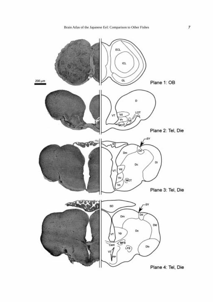
ForebrainThe eel forebrain consisted of the olfactory bulb (OB) rostrally, the telencephalon (Tel) caudally, and the
diencephalon (Die) ventrally. In the OB, three ring-like layers were observed (Plane 1), corresponding respec-
tively to the internal cellular layer (ICL), the external cellular layer (ECL), and the glomerular layer (GL) in the
zebrafish (Wullimann et al., 1996). Every layer consisted of small neurons (ca. 5 µm), and the density of the
somata was highest in the GL.
T. Mukuda and M. Ando6
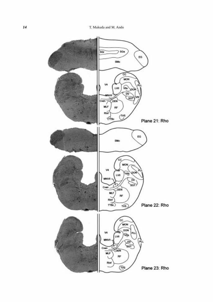
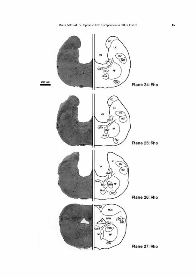
Fig. 2. Brain atlas of the Japanese eel (cross section). A: Lateral view of the eel brain indicating the levels(planes) for cross sectioning. Plane numbers (1-27) were given orderly from rostral to caudal. B: Crosssection of the eel brain. Klüver-Barréra’s staining is shown on the left and a schematic illustration of thefigure is drawn on the right. Somata are colored violet and fibers blue. Blood vessels are omitted in theillustrations. Scale bar (200 µm) is common in all planes 1-27. The nuclei and the fiber trunks are allabbreviated as shown in Table 1.
Telencephalon. The telencephalon (Tel) was divided into the left and right telencephalic hemispheres by
a central cavity, the telencephalic ventricle (VT). The telencephalic hemisphere consisted of the dorsal and
ventral telencephalic areas. The dorsal telencephalic area (D) was composed of small granular somata (Planes
2, 3). At the middle part of the Tel, the D was subdivided into the central (Dc), the medial (Dm), the dorsal (Dd),
and the lateral (Dl) zones (Plane 3). At the caudal part, the Dl was further subdivided into the dorsal (Dld) and
the ventral (Dlv) parts by a hollow (Plane 4). A distinct concavity between the Dm and the Dd was identified
as the sulcus ypsiloniformis (SY) (Planes 3, 4). The Dd was characterized as condensed somata (Plane 4). The
ventral telencephalic area (V) was subdivided into five nuclei; the dorsal (Vd), the ventral (Vv), the lateral (Vl),
the supracommissural (Vs) and the postcommissural (Vp) nuclei (Planes 2-4).
Fiber tracts. The medial olfactory tract (MOT) ran through the middle part of the Tel (Planes 2, 3), while
the lateral olfactory tract (LOT) disappeared at the anterior part of the telencephalon (Planes 2, 3). The telen-
cephalic hemispheres were connected with the anterior commissure (Cant) at the caudal part (Plane 4). In the
caudal Tel, the median forebrain (MFB) and lateral forebrain (LFB) bundles were morphologically distinguished
(Plane 4). Both bundles extended to the diencephalon (Die) (Planes 5, 6).
Diencephalon. The diencephalon (Die), located caudally to the Tel, was divided into six divisions; the
preoptic area, the epithalamus, the thalamus, the hypothalamus, the posterior tuberculum and the pretectum. In
the rostral part of the Die, the third ventricle (V3) appeared on the median line (Planes 4-9). At the caudal part,
the V3 was expanded transversely and divided into the infundibulum (INF) (Planes 8-10) and the recess lateralis
(RL) (Planes 9-11).
Preoptic area. The preoptic area surrounded the rostral part of the V3. The parvocellular preoptic nucleus
(PP, < 5 µm) and the magnocellular preoptic nucleus (PM, ca. 20 µm) were arrayed along the rostral V3, the PP
ventrally and the PM dorsally (Plane 5). The ventral margin of the V3 was surrounded by the suprachiasmatic
nucleus (SC) (Planes 5, 6).
Epithalamus. The epithalamus consisted of the habenular nucleus (Ha), the saccus dorsalis (SD) and the
epiphysis (E; pineal gland). The Ha surrounded the dorsal periventricular region of the V3, and was divided into
Brain Atlas of the Japanese Eel: Comparison to Other Fishes 7
T. Mukuda and M. Ando8
Brain Atlas of the Japanese Eel: Comparison to Other Fishes 9
T. Mukuda and M. Ando10
Brain Atlas of the Japanese Eel: Comparison to Other Fishes 11
T. Mukuda and M. Ando12
Brain Atlas of the Japanese Eel: Comparison to Other Fishes 13
T. Mukuda and M. Ando14
Brain Atlas of the Japanese Eel: Comparison to Other Fishes 15
T. Mukuda and M. Ando16
two parts by a staining pattern; the dorsal (Had) and the ventral (Hav) habenular nuclei (Planes 5, 6). The SD
projected rostrally and covered the telencephalic ventricle (VT) and the V3 (Planes 2-6).
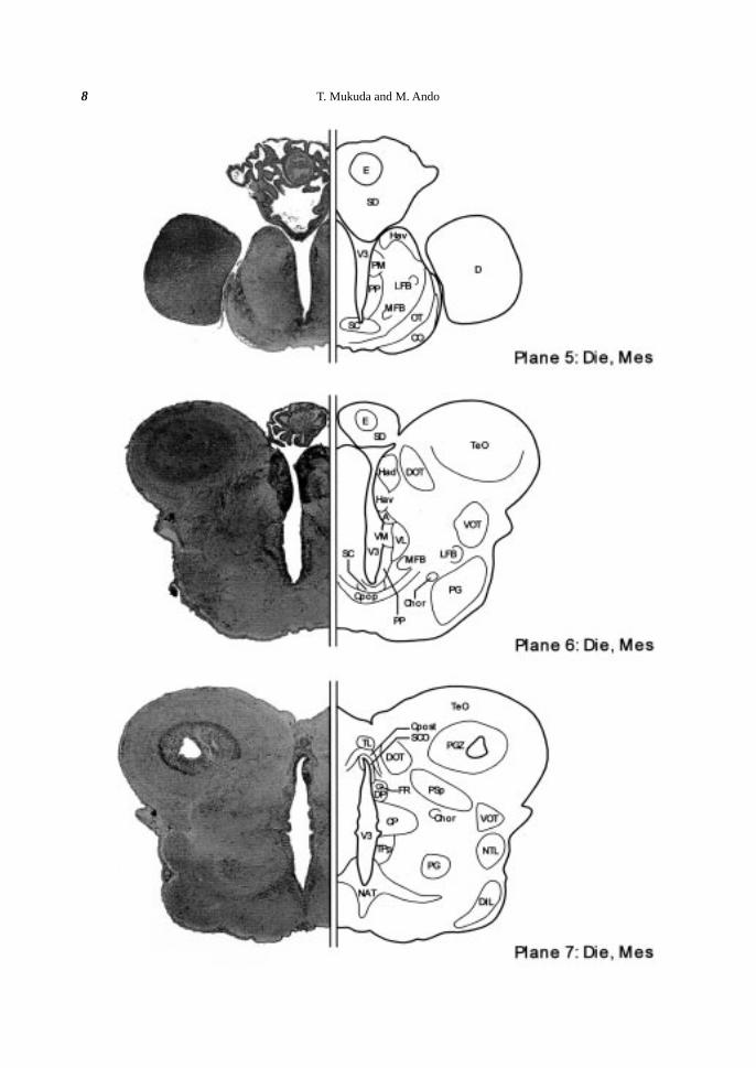
Thalamus. The thalamus was located caudally to the epithalamus, and characterized by five nuclei; the
anterior, the dorsal posterior, the central posterior, the ventromedial, and the ventrolateral thalamic nuclei. The
anterior thalamic nucleus (A) was located beneath the ventral habenular nucleus (Hav) (Plane 6). Further
ventrally, the ventromedial (VM) and the ventrolateral (VL) thalamic nuclei were situated (Plane 6). Caudally
to the A, the dorsal posterior thalamic nucleus (DP) and the central posterior thalamic nucleus (CP) appeared
along the V3 (Planes 7-9).
Posterior tuberculum. The posterior tuberculum (TP) was located caudally to the ventral thalamus, and
elongated caudally along the V3. The rostralmost part of the TP was the periventricular nucleus of the posterior
tuberculum (TPp) (Planes 7-10). Ventrally to the TPp, the paraventricular organ (PVO) appeared abut on the
infundibulum (INF) (Planes 8, 9). The PVO was characterized by well-developed vasalia. Laterally to the TPp,
there were the preglomerular nucleus (PG) centrally (Planes 7-10), and the nucleus tori lateralis (NTL) more
peripherally (Planes 7-9). At the caudalmost part of the TP, there was the corpus mamillare (CM) centrally
(Plane 11).
Hypothalamus. In the hypothalamus, the infundibulum (INF) appeared ventrally to the V3 (plane 8), and
the INF was further expanded laterally to form the recess lateralis (RL) at more caudal part (Plane 9-11). Most
hypothalamic nuclei were situated along these ventricles. At the rostralmost part, the anterior tuberal nucleus
(NAT) appeared beneath the V3 (Plane 7) and the NAT surrounded the INF (Planes 8-10), which was sur-
rounded by the posterior tuberal nucleus (NPT) at the more caudal part (Plane 11). Around the RL, the nucleus
recessi lateralis (NRL) existed periventricularly (Planes 8-12). Peripherally in the inferior lobe (IL), the diffuse
nucleus of the inferior lobe (DIL) was observed (Planes 7-12). The saccus vasculosus (SV) appeared at the
ventromedian area of the hypothalamus, caudally to the pituitary (Planes 11-14).
Pretectum. The pretectal region between the diencepharon (Die) and the mesencephalon (Mes) was char-
acterized by the relatively thick posterior commissure (Cpost) situated above the V3 (Planes 7-9). Beneath the
Cpost, the subcommissural organ (SCO), consisting of well-developed ependymal cells, contacted with the V3
(Planes 7-9). Laterally to the SCO, the ventral and the dorsal parts of the periventricular pretectal nuclei (PPv
and PPd) were distinguished (Planes 8, 9). Dorsally to the PPd, the paracommissural nucleus (PCN) was de-
tected and the central pretectal nucleus (CPN) was distingished laterally to the PCN (Plane 8). At more rostral
part, the parvocellular superficial pretectal nucleus (PSp) was observed (Plane 7).
Fiber tracts. In the ventrolateral side of the Die, the optic tract (OT) and the optic chiasm (CO) extended
ventrolaterally (Plane 5). The postoptic commissure (Cpop) appeared caudally to the optic chiasm (CO) (Plane
6). The dorsomedial optic tract (DOT) extended dorsally toward the optic tectum (TeO) (Planes 6, 7), while the
ventrolateral optic tract (VOT) was elongated more caudally (Planes 6-11). The fasciculus retroflexus (FR)
emerged caudally to the habenular nucleus (Ha) in the epithalamus (Plane 7) and extended to a region of the
mesencephalon (Mes), where the interpeduncular nucleus (NIn) appeared (Plane 13). The horizontal commis-
sure (Chor) emerged ventrally in the rostral Die (Plane 6), went up to the dorsal side, and then ran longitudinally
to the mesencephalic region (Planes 7-9).
Midbrain (Mesencephalon)The mesencephalon (Mes) was characterized by the dorsally expanded optic tectum (TeO) and the wide
Brain Atlas of the Japanese Eel: Comparison to Other Fishes 17
mesencephalic ventricle (VMes) (Planes 7-15), and was divided into three major divisions from dorsal to ven-
tral; the optic tectum, the torus semicircularis and the tegmentum.
Optic tectum. The somata of the TeO were condensed as a layer along the VMes (Planes 6-15), forming the
periventricular gray zone (PGZ) of the TeO. The torus longitudinalis (TL) hung over the median VMes and
extended rostrocaudally (Planes 7-15). Above the TL, the commissura tecti (Ctec) connected both lobes of the
TeO (Planes 8, 9).
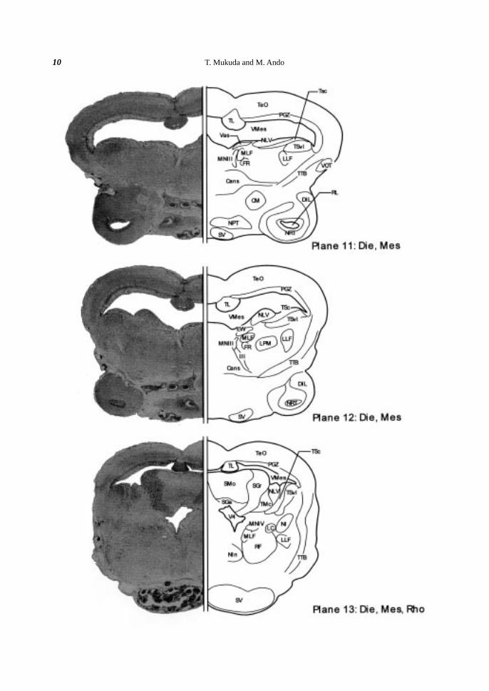
Torus semicircularis. The torus semicircularis (TS), situated ventrolaterally to the VMes and consisted of
many condensed somata, was expanded into the VMes (Planes 10-14). The TS was subdivided into the ventro-
lateral nucleus of the torus semicircularis (TSvl) and the central nucleus of the torus semicircularis (TSc) (Planes
11-14).
Tegmentum. The tegmentum, ventromedian region to the TS, possessed large somata (> 20 µm). The
somata of the nucleus ruber (NR), located in the rostralmost part of the tegmentum, were oval in shape (ca. 25
µm) (Planes 9, 10). In the more median side of the NR, there was the nucleus of the medial longitudinal fascicle
(NMLF) (Plane 10). The cell bodies of the NMLF were oval or round in shape with huge size (ca. 40 µm). In
the rostral tegmentum, the vascular lacuna of area postrema (Vas) occurred as a well-developed vasalium in the
median corner facing to the VMes (Planes 10, 11). Laterally to the NMLF, the rostral tegmental nucleus (RT)
was identified as a dorsoventrally diffused cluster (Plane 10). Caudally to the NMLF, many medium-sized (ca.
10 µm) and oval perikarya were conspicuously distinguished around the median line (Planes 11-13). These
somata were classified into three groups: the oculomotor nucleus (MNIII) (Planes 11, 12), the Edinger-Westphal
nucleus (EW) (Plane 12) and the trochlear nucleus (MNIV) (Plane 13). The nucleus lateralis valvulae (NLV)
was protruded to the VMes (Planes 11, 12). The protruded region of the NLV extended caudally, and then fused
with the valvula cerebelli in the rhombencephalon (Rho) (Planes 13, 14). Ventrally to the NLV, the nucleus
lateralis profundus mesencephali (LPM) was distinguished (Plane 12). In the caudal part of the tegmentum, the
interpeduncular nucleus (NIn) was present at the median portion (Plane 13).
Fiber tracts. Two major longitudinal bundles were distinguished in the midbrain; the medial longitudinal
fascicle (MLF) (Planes 10-27) and the lateral longitudinal fascicle (LLF) (Planes 10-16). The MLF started near
the NMLF, while the LLF started beneath the torus semicircularis (TS) (Plane 10). Both fascicles extended
caudally toward the rhombencephalon (Rho). The tractus tectobulbaris (TTB) traversed from the TeO to the
ventro-median part of the tegmentum (Planes 11-14). Many fibers of the TTB crossed the median line through
the ansulate commissure (Cans) (Planes 11, 12), which was located rostrally to the NIn (Plane 13). Beneath the
MNIII, the oculomotor nerve (III) extended to the ventral region (Plane 12).
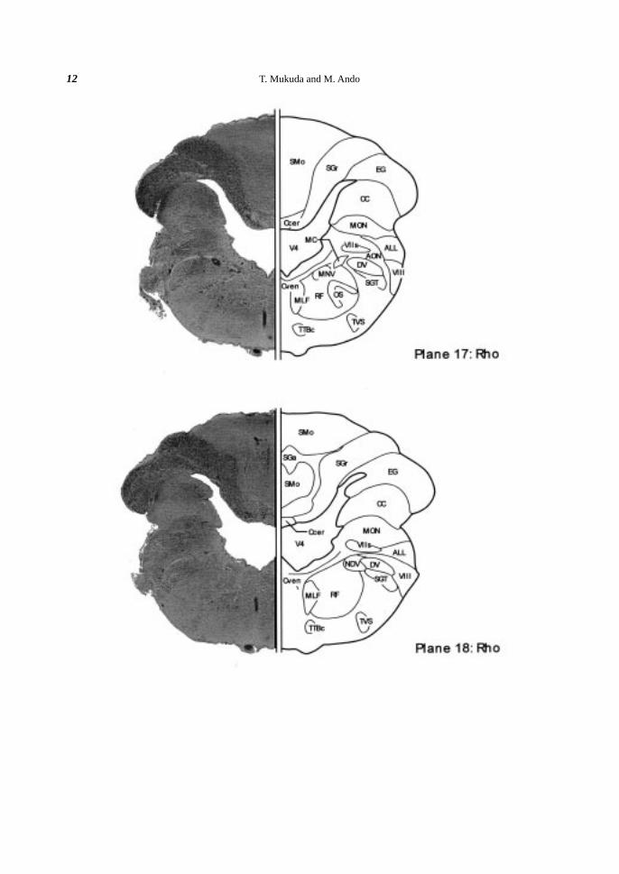
Hindbrain (Rhombencephalon)The rhombencephalon (Rho), the most caudal brain (hindbrain), consisted of the metencephalon and the
myelencephalon. Since fish lack the pons, the metencephalon means the cerebellum (Ce), and the myelen-
cephalon is the medulla oblongata (MO).
Cerebellum. The cerebellum (Ce) was divided into three main regions longitudinally; the valvula cerebelli,
the corpus cerebelli, and the caudal cerebellar region. The valvula cerebelli represented a median bulge into the
VMes, and consisted of three layers; the outer molecular layer (SMo), the intermediate ganglionic layer (SGa),
and the deep granular layer (SGr) (Planes 13-15). The corpus cerebelli was also composed of three major layers
of the SMo, the SGa and the SGr (Planes 16-22). The size (ca. 5 µm) and distribution of the SGa neurons in the
T. Mukuda and M. Ando18
eel was relative to those of Purkinje cells in the rainbow trout (Meek & Nieuwenhuys, 1998). The granular
eminence (EG) emerged at the junction between the Ce and the MO (Plane 15) and ran caudally (Planes 15-22).
The caudal cerebellar region was turned rostrally and connected to the MO by the crista cerebellaris (CC)
(Planes 17-25). At the ventral part of the Ce, the cerebellar commissure (Ccer) was distinguished above the
fourth ventricle (V4) (Planes 15-18).
Fourth ventricle. Between the cerebellum (Ce) and the medulla oblongata (MO), there existed the huge
ventricle, fourth ventricle (V4). However, the V4 was extremely narrow at the rostral part (Plane 13, 14). It
became wider caudally (Plane 15-18) with greatest width at the middle part of the MO (Plane 17, 18), and then
the width was reduced gradually (Plane 19-26).
Medulla oblongata. The medulla oblongata (MO) was connected with the mesencephalon at the
rhombencephalic isthmal region rostrally (Plane 13) and with the spinal cord at the obex (OX) level caudally
(Plane 27).
Rostral area (planes 13-17). In the rostralmost part of the MO, the nucleus isthmi (NI) was located mid-
laterally (Planes 13, 14). At more median site of the NI, the locus coeruleus (LC) with several large-sized
perikarya (ca. 20µm) and the superior raphe nucleus (RS), the most anterior part of the raphe nuclei on the
medial line, were identified (Planes 13, 14). Caudally to the LC, the trigeminal motor nucleus (MNV) appeared
(Planes 15-18). At more caudal part, the superior olive (OS) was located ventrally to the MNV (Plane 17). At
the ventral part of the anterior octaval nucleus (AON), paired extremely huge perikarya were present (Plane 17).
These perikarya were dorsolaterally elongated and ellipsoidal in shape (ca. 90 µm longitudinally and 50 µm
transversely), and identified as the Mauthner cells (MC) following the observations in the European eel (Meredith
and Roberts, 1987; Meredith et al., 1987), the goldfish (Nieuwenhuys et al., 1998), and various teleosts (Zottoli,
1978).
Middle area (Planes 17-23). At the middle part of the MO, the medial octavolateral nucleus (MON)
emerged from the crista cerebellaris (CC) (Plane 17). The cell mass situated ventrally to the MON was divided
rostrocaudally into three groups; the anterior octaval nucleus (AON) (Plane 17), the magnocellular octaval
nucleus (MaON) (Plane 19) and the descending octaval nucleus (DON) (Planes 20-23). More caudally, the
intermediate raphe nucleus (RInt) (Planes 19-20), the octavolateral efferent nucleus (OEN) (Planes 20-23), the
facial motor nucleus (MNVII) (Planes 21-23), and the inferior raphe nucleus (RInf) (Planes 21-26) appeared.
The lateral wall of the V4 was divided into the crista cerebellaris (CC) and the facial lobe (LVII) by a sulcus
(Plane 20-23).
Caudal area (Planes 24-27). The glossopharyngeal (MNIX) and the vagal (MNX) motor nuclei were
arrayed along the V4 as a continuous column (Planes 24-27), therefore the column was called glossopharyn-
geal-vagal motor complex (GVC). Ventrally to the GVC, the spinooccipital motor nucleus (NSO) consisting of
multipolar or ellipsoidal medium-sized perikarya (ca. 20 µm) extended caudally beyond the obex (Planes 26,
27). The vagal lobe (LX) appeared caudally to the facial lobe (LVII) (Plane 24-26). At the most caudal area of
the brain, the area postrema (AP) was recognized as a median structure with many capillaries in its dorsal region
(Plane 27). Ventrolaterally to the AP, the commissural nucleus of the Cajal (NCC) and medial funicular nucleus
(NFM) were distinguished (Plane 27). Both nuclei consisted of small diffuse somata.
Fiber tracts. The medial longitudinal fascicle (MLF) and the lateral longitudinal fascicle (LLF) were
major longitudinal fascicles in the MO. The MLF extended caudally through the obex (OX) level (Planes 10-
27), while the LLF disappeared at the anterior MO (Plane 17). In the octavolateral area, three fiber tracts were
Brain Atlas of the Japanese Eel: Comparison to Other Fishes 19
Fig. 3. Longitudinal distribution of various nuclei in the medulla oblongata (MO) (dorsal view). The figure isconstructed schematically from the data of planes 13-27 in Fig. 2B. The left half nuclei situate dorsallyto the right half nuclei. All abbreviations are illustrated in Table 1.
distinguished; the anterior lateral line nerve (ALL) (Planes 17-19), the posterior lateral line nerve (PLL) (Planes
20-23) and the octaval nerve (VIII) (Planes 17-21). The tractus tectobulbaris cruciatus (TTBc) ran longitudi-
nally in planes 15-23. The descending trigeminal root (DV) and the secondary gustatory tract (SGT) emerged at
the Mes (Plane 15) and extended caudally through the OX (Plane 27). The vestibulo-spinal tract (TVS) emerged
at the Mes (Plane 15) and elongated caudally through the OX (Plane 27) in parallel to the reticular formation
(RF). The commissure ventralis rhombencephali (Cven) ran beneath the V4 or the central canal (C) (Planes 14-
27).
Longitudinal distribution of medullary nucleiMany nuclei in the MO were distributed longitudinally along the V4, mostly with a column structure, and
some were already described above. The raphe nuclei were located in the median region of the MO, and
consisted of three parts; the superior raphe nucleus (RS), the intermediate raphe nucleus (RInt), and the inferior
raphe nucleus (RInf). The RS emerged immediately caudal to the NIn (Plane 14). Another two nuclei appeared
below the medial longitudinal fascicle (MLF) (Planes 19, 20). The most caudal raphe nucleus was the RInf
(Planes 21-26) (Fig. 3). Lateral to the raphe nuclei and the MLF, the reticular formation (RF) ran through the
MO. Within the RF, there were large-sized somata (> 25 µm) with multipolar or oval shapes (Planes 13-27).
Figure 3 shows a schematic distribution of some major nuclei in the MO. The length of the column of the
MNV, the MNVII and the GVC were approximately 450 µm, 500 µm, and 1700 µm, respectively. The gaps
T. Mukuda and M. Ando20
between the MNV and the MNVII and between the MNVII and the GVC were approximately 300 µm and 50
µm, respectively (left half in Fig. 3). The raphe nuclei (the RS, the RInt and the RInf) were arrayed on the
median line, while the superior olive (OS), the octavolateral efferent nucleus (OEN) and the spinooccipital
motor nucleus (NSO) were situated bilaterally (right half in Fig. 3). Therefore, the OEN and the NSO were
positioned closely to the MNVII and the GVC (Planes 21-23) (Planes 26, 27), and the formers (left half in Fig.
3) were ventral to the latter (right half in Fig. 3).
Cranial ganglia. Apart from the brain, three ganglia of the trigeminal nerve (V), the facial nerve (VII),
and the glossopharyngeal (IX) or the vagal (X) nerve were observed at the level of planes 13-15, of planes 15-
16, and of planes 19-26, respectively (data not shown). The somata in these ganglia are all round-shaped (> 20
µm). These ganglia may correspond to the semilunar, geniculate, and jugular/nodose ganglia in human, respec-
tively (Martini et al., 2000).
DISCUSSION
The external morphology of the eel brain was characterized by a close apposition between the olfactory
bulb (OB) and the telencephalon (Tel), a relatively small expansion of the optic tectum (TeO), a smooth bound-
ary between the medulla oblongata (MO) and the spinal cord, and three lobes of the cerebellum (Ce). A similar
juxtaposition of the OB and Tel is also observed in the zebrafish (Wullimann et al., 1996) and the rainbow trout
(Nieuwenhuys et al., 1998), while the OB is separated from the Tel in the goldfish (Morita and Finger, 1987a;
Nieuwenhuys et al., 1998). The TeO covers the midbrain dorsolaterally in the eel, while ventrolaterally in the
rainbow trout (Nieuwenhuys et al., 1998), the zebrafish (Wullimann et al., 1996), and the goldfish (Morita and
Finger, 1987a; Nieuwenhuys et al., 1998). The eel medulla oblongata (MO) shifts gradually to the spinal cord
as in the rainbow trout (Nieuwenhuys et al., 1998), while the caudal part of the MO expands dorsally to form the
facial (LVII) and vagal (LX) lobes in the zebrafish (Wullimann et al., 1996) and the goldfish (Nieuwenhuys et
al., 1998). The fourth ventricle (V4) of the eel seems to be longer than those of the zebrafish (Wullimann et al.,
1996) and the goldfish (Nieuwenhuys et al., 1998), whose V4 are covered with the LVII. In the rainbow trout,
the V4 is completely covered with the Ce, and thus not visible from the outside.
The present study is the first report of the whole brain atlas of the eel, whereas partial brain morphology has
been described in the European eel (Meredith and Roberts, 1986, 1987; Meredith et al., 1987; Roberts et al.,
1989; Wullimann et al., 1991; Molist et al., 1993), and all results obtained in this study are consistent with the
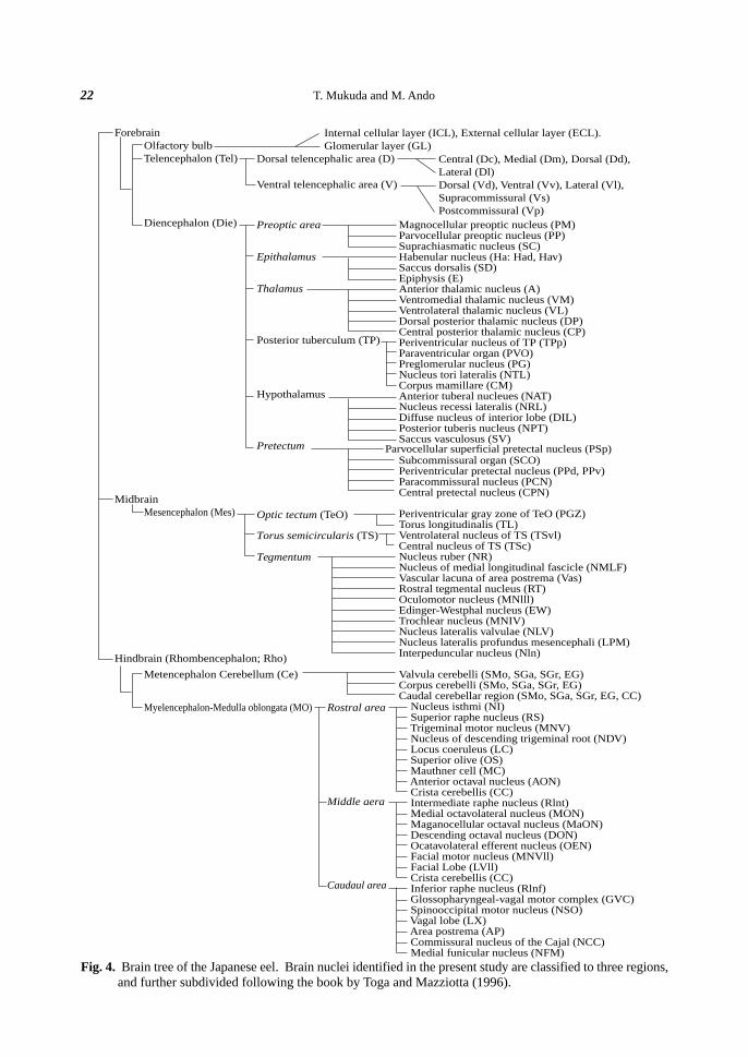
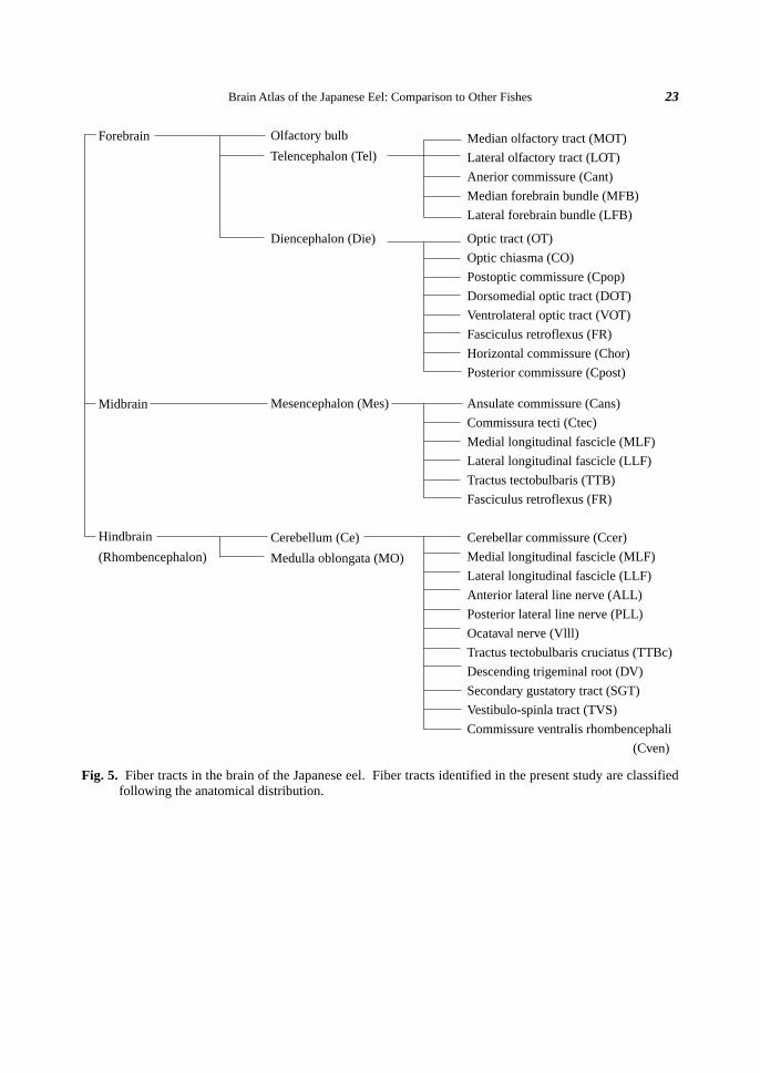
restricted descriptions in the European eel. Figures 4 and 5 summarize the brain nuclei and the fiber tracts
identified in the present study, respectively. A hierarchical representation of the anatomical nomenclature serves
to establish the relationship among structures and provides a map of the organization of the nervous system.
Many nuclei in Fig. 4 appear to be adjacent to the ventricles in general.
Basically, the brain topology of the eel is similar to those of the rainbow trout (Meek and Nieuwenhuys,
1998), the goldfish (Peter and Gill, 1975; Morita and Finger, 1987a, b; Goehler and Finger, 1992; Meek and
Nieuwenhuys, 1998), the zebrafish (Wullimann et al., 1996), and the catfish (Kanwal and Caprio, 1987). How-
ever, some fine structures differ from those of other teleosts. 1) The parvocellular preoptic nucleus (PP) could
not be subdivided in the eel, whereas the anterior and posterior parts of the PP are distinguished in the zebrafish
(Wullimann et al., 1996) and the rainbow trout (Meek and Nieuwenhuys, 1998). 2) The intermediate thalamic
nucleus was not distinguished in the eel thalamus, although it is located dorsally to the ventromedial and vent-
Brain Atlas of the Japanese Eel: Comparison to Other Fishes 21
rolateral thalamic nuclei (VM and VL) in the zebrafish (Wullimann et al., 1996), the goldfish (Nieuwenhuys et
al., 1998), and the rainbow trout (Nieuwenhuys et al., 1998). The ventral thalamic nuclei are known to have a
different distribution among teleosts (Wullimann et al., 1991). 3) The paraventricular organ (PVO) was single
in the eel, while paired PVOs are observed in the zebrafish (Wullimann et al., 1996). 4) The torus semicircularis
(TS) in the eel was smaller than that in the goldfish (Peter and Gill, 1975) and rainbow trout (Nieuwenhuys et
al., 1998). 5) The cell size (40 µm) of the nucleus of medial longitudinal fascicle (NMLF) in the tegmentum was
similar to that in the elephantfish (Hlavacek et al., 1984), but larger than that in the glass knifefish (Behred and
Donicht, 1990), or the zebrafish (Wullimann et al., 1996). 6) The protrusion of the nucleus lateralis valvulae
(NLV) into the mesencephalic ventricle (VMes) in the eel was larger than that in the zebrafish (Wullimann et al.,
1996) and the rainbow trout (Nieuwenhuys et al., 1998). 7) The valvula cerebelli in the eel was similar to that
in the rainbow trout (Nieuwenhuys et al., 1998), but smaller than those in the goldfish (Peter and Gill, 1975) and
the zebrafish (Wullimann et al., 1996). 8) The facial lobes (LVII) ran through the medulla oblongata (MO) in
the eel similarly as in the rainbow trout (Nieuwenhuys et al., 1998), while two lobes fuse at the caudal cerebel-
lum in the goldfish (Morita and Finger, 1987a; Nieuwenhuys et al., 1998), the catfish (Kanwal and Caprio,
1987; Nieuwenhuys et al., 1998), and the zebrafish (Wullimann et al., 1996). 9) The expansion of the vagal lobe
(LX) in the caudal MO of the eel was similar to that in the gray mullet (Diaz-Regueira and Anadón, 1992) and
the rainbow trout (Nieuwenhuys et al., 1998), but smaller than that in the goldfish (Morita and Finger, 1987a, b;
Goehler and Finger, 1992) and the zebrafish (Wullimann et al., 1996). 10) The glossopharyngeal motor nucleus
(MNIX) and the vagal motor nucleus (MNX) were fused to make a columnar structure named glossopharyn-
geal-vagal motor complex (GVC). Similar complex is observed in the lamprey (Aríëns-Kappers et al., 1936),
the elasmobranch (Aríëns-Kappers et al., 1936; Anadón et al., 2000) and other teleost fishes (Aríëns-Kappers et
al., 1936; Kanwal and Caprio, 1987; Morita and Finger, 1987b; Goehler and Finger, 1992; Wullimann et al.,
1996; Nieuwenhuys et al., 1998; Pérez et al., 2000). Therefore, such a columnar complex seems to be common
in fishes. 11) The facial motor nucleus (MNVII) was separated from the GVC in the eel similarly as in the
bowfin (Nieuwenhuys et al., 1998), the catfish (Kanwal and Caprio, 1987), the rainbow trout (Pérez et al.,
2000), the anglerfish (Aríëns-Kappers et al., 1936), the puffer fish (Aríëns-Kappers et al., 1936), the sunfish
(Aríëns-Kappers et al., 1936), and the goldfish (Morita and Finger, 1987b; Goehler and Finger, 1992; Nieuwenhuys
et al., 1998), whereas it is fused with the GVC in the sturgeon (Adrio et al., 2000), the reedfish (Aríëns-Kappers
et al., 1936) and the tarpon (Aríëns-Kappers et al., 1936).
Among the nuclei identified in the present study, the magnocellular preoptic nucleus (PM), anterior tuberal
nucleus (NAT), area postrema (AP), and glossopharyngeal- vagal motor complex (GVC) might be involved in
drinking behavior in the Japanese eel. The PM, the NAT and the AP seem to be the circumventricular organs
which lack the blood-brain barrier (BBB) and thus accept dipsogens and antidipsogens produced in the systemic
circulation, because these nuclei are stained by intraperitoneal Evans blue which can not pass the BBB (T.
Mukuda, Y. Matsunaga, K. Kawamoto, K. Yamaguchi and M. Ando, unpublished observation). The GVC
innervates the upper esophageal sphincter muscle cholinergically (Mukuda and Ando, 2003; Kozaka and Ando,
Periventricular gray zone of TeO (PGZ)Torus longitudinalis (TL)Ventrolateral nucleus of TS (TSvl)Central nucleus of TS (TSc)Nucleus ruber (NR)Nucleus of medial longitudinal fascicle (NMLF)Vascular lacuna of area postrema (Vas)Rostral tegmental nucleus (RT)Oculomotor nucleus (MNlll)Edinger-Westphal nucleus (EW)Trochlear nucleus (MNIV)Nucleus lateralis valvulae (NLV)Nucleus lateralis profundus mesencephali (LPM)Interpeduncular nucleus (Nln)
Valvula cerebelli (SMo, SGa, SGr, EG)Corpus cerebelli (SMo, SGa, SGr, EG)Caudal cerebellar region (SMo, SGa, SGr, EG, CC) Nucleus isthmi (NI) Superior raphe nucleus (RS) Trigeminal motor nucleus (MNV) Nucleus of descending trigeminal root (NDV) Locus coeruleus (LC) Superior olive (OS) Mauthner cell (MC) Anterior octaval nucleus (AON) Crista cerebellis (CC) Intermediate raphe nucleus (Rlnt) Medial octavolateral nucleus (MON) Maganocellular octaval nucleus (MaON) Descending octaval nucleus (DON) Ocatavolateral efferent nucleus (OEN) Facial motor nucleus (MNVll) Facial Lobe (LVll) Crista cerebellis (CC) Inferior raphe nucleus (Rlnf) Glossopharyngeal-vagal motor complex (GVC) Spinooccipital motor nucleus (NSO) Vagal lobe (LX) Area postrema (AP) Commissural nucleus of the Cajal (NCC) Medial funicular nucleus (NFM)
Parvocellular superficial pretectal nucleus (PSp)
Fig. 4. Brain tree of the Japanese eel. Brain nuclei identified in the present study are classified to three regions,and further subdivided following the book by Toga and Mazziotta (1996).
Brain Atlas of the Japanese Eel: Comparison to Other Fishes 23
Fig. 5. Fiber tracts in the brain of the Japanese eel. Fiber tracts identified in the present study are classifiedfollowing the anatomical distribution.
Forebrain
Midbrain
Hindbrain
(Rhombencephalon)
Olfactory bulb
Telencephalon (Tel)
Diencephalon (Die)
Mesencephalon (Mes)
Cerebellum (Ce)
Medulla oblongata (MO)
Median olfactory tract (MOT)
Lateral olfactory tract (LOT)
Anerior commissure (Cant)
Median forebrain bundle (MFB)
Lateral forebrain bundle (LFB)
Optic tract (OT)
Optic chiasma (CO)
Postoptic commissure (Cpop)
Dorsomedial optic tract (DOT)
Ventrolateral optic tract (VOT)
Fasciculus retroflexus (FR)
Horizontal commissure (Chor)
Posterior commissure (Cpost)
Ansulate commissure (Cans)
Commissura tecti (Ctec)
Medial longitudinal fascicle (MLF)
Lateral longitudinal fascicle (LLF)
Tractus tectobulbaris (TTB)
Fasciculus retroflexus (FR)
Cerebellar commissure (Ccer)
Medial longitudinal fascicle (MLF)
Lateral longitudinal fascicle (LLF)
Anterior lateral line nerve (ALL)
Posterior lateral line nerve (PLL)
Ocataval nerve (Vlll)
Tractus tectobulbaris cruciatus (TTBc)
Descending trigeminal root (DV)
Secondary gustatory tract (SGT)
Vestibulo-spinla tract (TVS)
Commissure ventralis rhombencephali
(Cven)
T. Mukuda and M. Ando24
ACKNOWLEDGMENTS
This research was supported in part by Grants-in-Aid for Scientific Research (C) nos.13640681 and 15570064
from the Ministry of Education, Culture, Sports, Science and Technology, Japan, and also by the Fisheries
Agency of Japan.
References
Adrio F, Anadón R Rodriguez-Moldes I (2000) Distribution of choline acetyltransferase (ChAT) immunoreac-
tivity in the central nervous system of a chondrostean, the Siberian sturgeon (Acipenser baeri). J Comp
Neurol 426: 602-621
Anadón R, Molist R, Rodriguez-Moldes I, Lopez JM, Quintela I, Cervino MC, Barja P, Gonzalez A (2000)
Distribution of choline acetyltransferase immunoreactivity in the brain of an elasmobranch, the lesser