1 Brain Concentrations of Methylone and its Metabolites After Systemic Methylone Administration: Relationship to Pharmacodynamic Effects Nicole Centazzo 1 , Michael R. Chojnacki 2 , Joshua S. Elmore 2 , Raider Rodriguez 1 , Teeshavi Acosta 1 , Masaki Suzuki 3 , Kenner C. Rice 3 , Michael H. Baumann 2, * , Marta Concheiro 1, * 1 Department of Sciences, John Jay College of Criminal Justice, City University of New York, New York, NY, USA 2 Designer Drug Research Unit, Intramural Research Program (IRP), National Institute on Drug Abuse (NIDA), National Institutes of Health (NIH), Baltimore, MD, USA 3 Drug Design and Synthesis Section, IRP, NIDA, NIH, Rockville, MD, USA * Corresponding authors This article has not been copyedited and formatted. The final version may differ from this version. JPET Fast Forward. Published on March 30, 2021 as DOI: 10.1124/jpet.121.000531 at ASPET Journals on November 26, 2021 jpet.aspetjournals.org Downloaded from
Transcript
1
Brain Concentrations of Methylone and its Metabolites After Systemic Methylone
Administration: Relationship to Pharmacodynamic Effects
Nicole Centazzo1, Michael R. Chojnacki
2, Joshua S. Elmore
2, Raider Rodriguez
1, Teeshavi
Acosta1, Masaki Suzuki
3, Kenner C. Rice
3, Michael H. Baumann
2, *, Marta Concheiro
1, *
1Department of Sciences, John Jay College of Criminal Justice, City University of New York,
New York, NY, USA
2Designer Drug Research Unit, Intramural Research Program (IRP), National Institute on Drug
Abuse (NIDA), National Institutes of Health (NIH), Baltimore, MD, USA
3Drug Design and Synthesis Section, IRP, NIDA, NIH, Rockville, MD, USA
* Corresponding authors
This article has not been copyedited and formatted. The final version may differ from this version.JPET Fast Forward. Published on March 30, 2021 as DOI: 10.1124/jpet.121.000531
This article has not been copyedited and formatted. The final version may differ from this version.JPET Fast Forward. Published on March 30, 2021 as DOI: 10.1124/jpet.121.000531
HPLC-ECD: high-performance liquid chromatography with electrochemical detection
HVA: homovanillic acid
IRP: Intramural Research Program
LC-MS: liquid chromatography–mass spectrometry
LC-MSMS: liquid chromatography-tandem mass spectrometry
LOQ: limits of quantification
MDMA: 3,4-methylenedioxy-N-methylamphetamine
MDC: 3,4-methylenedioxycathinone
MDMC: methylone
MDPV: 3,4-methylenedioxypyrovalerone
MRM: multiple reaction monitoring
NPS: new psychoactive substances
SMBS: sodium metabisulfite
This article has not been copyedited and formatted. The final version may differ from this version.JPET Fast Forward. Published on March 30, 2021 as DOI: 10.1124/jpet.121.000531
3,4-Methylenedioxy-N-methylcathinone (methylone) is a new psychoactive substance with
stimulant properties and potential for abuse. Despite its popularity, limited studies have
examined relationships between brain concentrations of methylone, its metabolites, and
pharmacodynamic effects. The goal of the present study was two-fold: 1) to determine
pharmacokinetics of methylone and its major metabolites, 4-hydroxy-3-methoxy-N-
methylcathinone (HMMC), 3,4-dihydroxy-N-methylcathinone (HHMC), and 3,4-
methylenedioxycathinone (MDC) in rat brain and plasma; 2) to relate brain pharmacokinetic
parameters to pharmacodynamic effects including locomotor behavior and post-mortem
neurochemistry. Male Sprague-Dawley rats received s.c. methylone (6, 12, or 24 mg/kg) or
saline vehicle (n=16/dose), and subgroups were decapitated after 40 or 120 min. Plasma and
prefrontal cortex were analyzed for concentrations of methylone and its metabolites by liquid
chromatography-tandem mass spectrometry. Frontal cortex and dorsal striatum were analyzed
for dopamine, 5-HT, and their metabolites by high-performance liquid chromatography-
electrochemical detection. Brain and plasma concentrations of methylone and its metabolites
rose with increasing methylone dose, but brain methylone and MDC concentrations were greater
than dose-proportional. Brain-to-plasma ratios for methylone and MDC were >3 (range 3-12),
whereas those for HHMC and HMMC were <0.2 (range 0.01-0.2). Locomotor activity score was
positively correlated with brain methylone and MDC, whereas cortical 5-HT was negatively
correlated with these analytes at 120 min. Our findings show that brain concentrations of
methylone and MDC display non-linear accumulation. Behavioral and neurochemical effects of
systemically administered methylone are related to brain concentrations of methylone and MDC,
but not its hydroxylated metabolites, which do not effectively penetrate into the brain.
This article has not been copyedited and formatted. The final version may differ from this version.JPET Fast Forward. Published on March 30, 2021 as DOI: 10.1124/jpet.121.000531
Behavioral and neurochemical effects of methylone are related to brain concentrations of
methylone and its metabolite MDC, but not its hydroxylated metabolites, HMMC and HHMC,
which do not effectively penetrate into the brain. Methylone and MDC display non-linear
accumulation in the brain, which could cause untoward effects on 5-HT neurons in vulnerable
brain regions, including the frontal cortex.
This article has not been copyedited and formatted. The final version may differ from this version.JPET Fast Forward. Published on March 30, 2021 as DOI: 10.1124/jpet.121.000531
In the past decade, non-medical (i.e., recreational) drug markets worldwide have seen an
increase in the availability of stimulant-like new psychoactive substances (NPS), including
synthetic cathinones. These substances are chemically similar to amphetamines, and they have
been sold as “bath salts” or “research chemicals” to evade drug control legislation in the United
States (US) and elsewhere (Baumann et al., 2013; Madras, 2016). In 2011, the most prevalent
synthetic cathinones - methylone, mephedrone, and 3,4-methylenedioxypyrovalerone (MDPV) -
were placed into emergency Schedule I control by the US Drug Enforcement Administration
(DEA), and the substances were permanently scheduled in 2013 (Drug Enforcement
Administration (DEA), 2013). The first forensic identifications of methylone occurred in 2009
with 4 case reports, but serious drug exposures increased markedly in the following years,
reaching a peak of 3,976 case reports in 2013 (Drug Enforcement Administration (DEA), 2019).
In more recent times, methylone and its analogs (e.g., pentylone) are found as adulterants in
counterfeit Ecstasy pills sold as the club drug 3,4-methylenedioxy-N-methylamphetamine
(MDMA), and therefore, many drug users consume these compounds unknowingly (Oliver et al.,
2019).
Methylone is the -keto analog of MDMA, and not surprisingly, it produces similar
pharmacological effects to MDMA (De Felice et al., 2014; Baumann et al., 2018). More
specifically, methylone acts as a substrate-type releasing agent at high-affinity transporters for
dopamine, norepinephrine, and 5-HT in rat brain tissue and in cells transfected with human
transporters (Baumann et al., 2012; Eshleman et al., 2013; Simmler et al., 2013). The
monoamine-releasing effects of methylone produce elevations in extracellular dopamine and 5-
HT in brain reward pathways, as measured by in vivo microdialysis (Schindler et al., 2016;
This article has not been copyedited and formatted. The final version may differ from this version.JPET Fast Forward. Published on March 30, 2021 as DOI: 10.1124/jpet.121.000531
Elmore et al., 2017). Drug self-administration studies in rats demonstrate that methylone exhibits
reinforcing properties, which suggests the drug has abuse potential (Watterson et al., 2012;
Vanderwater et al., 2015).
In humans, methylone is metabolized by cytochrome P450 2D6 (CYP2D6), with minor
contributions from CYP1A2, CYP2B6, and CYP2C19 (Pedersen et al., 2013), whereas in rats,
the precise cytochrome(s) responsible are not well established but might involve CYP2D1, the
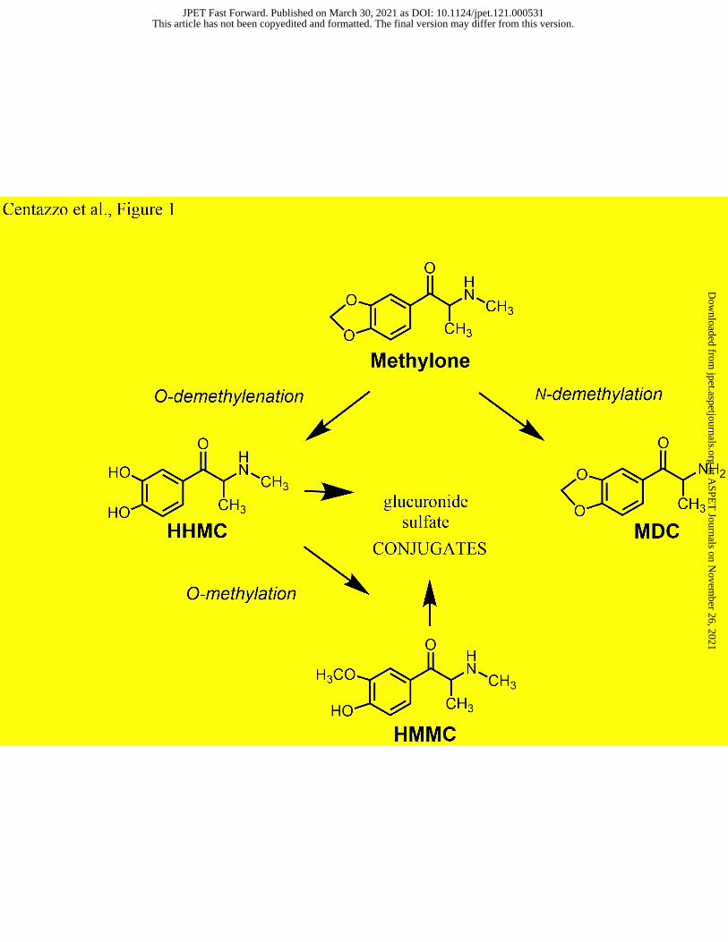
rat isoform of CYP2D6 (Malpass et al., 1999). Similar to MDMA, methylone is metabolized via
two distinct pathways in the liver (see Figure 1): 1) O-demethylenation to form 3,4-dihydroxy-N-
methylcathinone (HHMC), which is rapidly converted to 4-hydroxy-3-methoxy-N-
methylcathinone (HMMC), and 2) N-demethylation to form 3,4-methylenedioxycathinone
(MDC) or normethylone. It is noteworthy that HMMC is the predominant metabolite of
methylone in blood and plasma from both rats and humans. Phase II metabolism includes the
formation of glucuronide and sulfate conjugates of the hydroxylated metabolites HHMC and
HMMC (Kamata et al., 2006; Meyer et al., 2010). Our previous studies show that certain phase I
methylone metabolites are bioactive (Elmore et al., 2017; Luethi et al., 2019). MDC and HHMC
are substrate-type releasers at monoamine transporters in vitro, but only MDC produces
significant elevations in brain extracellular dopamine and 5-HT in vivo. The reason why HHMC
lacks bioactivity in vivo is not known but could be related to poor penetration across the blood-
brain barrier, due to its rapid conjugation in the bloodstream or higher polarity when compared to
methylone.
- Insert Figure 1 here -
This article has not been copyedited and formatted. The final version may differ from this version.JPET Fast Forward. Published on March 30, 2021 as DOI: 10.1124/jpet.121.000531
Severe clinical side effects have been reported from the misuse of methylone, such as
aggressive behavior, psychosis, hyperthermia, seizures, and even death (Cawrse et al., 2012;
Ellefsen et al., 2015), but limited studies have examined relationships between methylone
pharmacokinetics, metabolism, and its pharmacodynamic effects. Controlled administration
studies with methylone and other NPS in humans are limited due to ethical constraints, so
pharmacokinetic studies in animal models fill a critical void. Several research groups have
investigated the pharmacokinetics and pharmacodynamics of methylone in rodent models
(López-Arnau et al., 2013; Elmore et al., 2017; Grecco et al., 2017; Štefková et al., 2017). In
particular, Lopez-Arnau and colleagues (López-Arnau et al., 2013) examined pharmacokinetics
of methylone in rats, and their findings suggest that metabolites may contribute to
pharmacodynamic effects of the drug in vivo. Given the aforementioned information, the goal of
the present study was two-fold: 1) to determine the pharmacokinetics of methylone and its three
major metabolites - HMMC, HHMC, MDC - in rat brain and plasma; 2) to relate brain
pharmacokinetic parameters to acute pharmacodynamic effects including locomotor behavior
and post-mortem neurochemistry.
Materials and Methods
Drugs, Chemicals, and Reagents
(±)-3,4-Methylenedioxy-N-methylcathinone (methylone) for animal studies was acquired
from the National Institute on Drug Abuse (NIDA), Drug Supply Program (Rockville, MD,
USA). For analytical procedures, methylone (1 mg/mL in methanol) and its deuterated standard
methylone-d3 (100 g/mL in methanol) were obtained from Cerilliant (Round Rock, TX, USA).
This article has not been copyedited and formatted. The final version may differ from this version.JPET Fast Forward. Published on March 30, 2021 as DOI: 10.1124/jpet.121.000531
ethylenediaminetetraacetic acid (EDTA), formic acid, isopropanol, methanol, and sodium
metabisulfite (SMBS) were obtained from Thermo Fisher Scientific (Fair Lawn, NJ, USA).
BG100® liquid β-glucuronidase from Red Abalone Haliotis rufescens (>100 KU/mL) was
purchased from Kura Biotec (Inglewood, CA, USA) and 4-methylcatechol from Sigma-Aldrich
(Milwaukee, WI, USA). Hydrochloric acid (HCl) 36.5-38% was obtained from J.T. Baker
Chemical Company (Phillipsburg, NJ, USA). Brains from drug-naïve male Sprague Dawley rats
were acquired from BioIVT (Hicksville, NY, USA) and used for development of the method to
quantify methylone in brain tissue. Two-mL 1.4 mm ceramic beads were obtained from Thermo
Fisher Scientific (Fair Lawn, NJ, USA) and 10-mL 100 x 16 mm polypropylene tubes were
purchased from Sarstedt Inc. (Newton, NC, USA).
Animals, Dosing Regimen, and Tissue Collection
Male Sprague-Dawley rats (300-400 g), purchased from Envigo (Frederick, MD, USA),
were double-housed under conditions of controlled temperature (22 ± 2ºC) and humidity (45 ±
5%), with ad libitum access to food and water. Lights were on between 7:00 AM and 7:00 PM.
The Institutional Animal Care and Use Committee of the NIDA IRP approved the animal
experiments, and all procedures were carried out in accordance with the National Institutes of
This article has not been copyedited and formatted. The final version may differ from this version.JPET Fast Forward. Published on March 30, 2021 as DOI: 10.1124/jpet.121.000531
Health Guide for the Care and Use of Laboratory Animals. Vivarium facilities were fully
accredited by the Association for Assessment and Accreditation of Laboratory Animal Care.
Experiments were designed to minimize the number of animals included in the study.
One week prior to drug treatments, rats were single-housed. On the day of the
experiment, groups of rats received s.c. methylone (6, 12, or 24 mg/kg) or its saline vehicle
(n=16/dose) and were returned to their home cages; subgroups were subsequently killed by
decapitation at 40 min or 120 min post-injection. These particular time points were chosen for
tissue collection based on our previous study which showed plasma MDC concentrations peak at
30-40 min post-injection, while plasma HMMC concentrations peak much later at 90-120 min
(Elmore et al., 2017). Injections were carried out in the vivarium, whereas decapitation was
carried out in a separate necropsy room. Trunk blood was collected, brains were rapidly removed
from the skull, and tissue from prefrontal cortex, frontal cortex, and dorsal striatum was
dissected on ice. Plasma and brain tissue were stored frozen at -80°C until the time of analysis.
Assessment of Locomotor Behavior and Body Temperature
Just prior to decapitation, each rat was observed for 1 min in its home cage to discern
locomotor behavior, and core body temperature was measured. Behavior was scored using a
numerical scale: 1 = asleep or still; 2 = in-place activities; 3 = locomotion, rearing, or sniffing; 4
= any two (locomotion, rearing, or sniffing); 5 = 10 s of continuous sniffing without locomotion
or rearing; 6 = 10 s of continuous sniffing with locomotion or rearing; 7 = 5 s of patterned
sniffing; 8 = 10 s of patterned sniffing. Patterned sniffing was defined as any repeated head
motion (e.g., up and down ‘head bobbing’) that occurred simultaneously with sniffing behavior.
This behavioral scale is sensitive to dose-related changes in motor activation caused by
This article has not been copyedited and formatted. The final version may differ from this version.JPET Fast Forward. Published on March 30, 2021 as DOI: 10.1124/jpet.121.000531
methanol (pH = 2.75) was recirculated at 0.9 mL/min. Known monoamine standards, ranging in
concentration from 10 to 1,000 pg/µL, were assayed along with each set of samples. Data were
acquired by a Waters Empower software system (Waters Millipore), where peak heights of
unknowns were compared with those of standards. The lower limit of assay sensitivity (3×
baseline noise) was 30 pg/20 μL sample.
Methylone and Metabolites Analysis in Rat Plasma
This article has not been copyedited and formatted. The final version may differ from this version.JPET Fast Forward. Published on March 30, 2021 as DOI: 10.1124/jpet.121.000531
Plasma samples were analyzed by liquid chromatography-tandem mass spectrometry
(LC-MSMS) as previously described by Ellefsen et al. (Ellefsen et al., 2015). Specifically, 20 µL
of 250 mM SMBS, 10 µL of 250 mM EDTA, 50 µL of internal standard (methylone-d3) at 100
ng/mL, and 100 µL rat plasma were mixed in 1.5-mL microcentrifuge tubes and gently vortexed.
After enzymatic hydrolysis (10 µL of β-glucuronidase, incubation at 50°C, one hour), 20 µL of
4-methylcathechol and 10 µL of perchloric acid were mixed with each sample. The samples were
extracted using mixed-mode cation exchange solid phase extraction. The eluent was acidified
with 100 µL of 1% HCl in methanol and evaporated to dryness in a Turbovap® (Biotage,
Charlotte, NC, USA). Two hundred µL of 0.1% formic acid in water was utilized for
reconstitution, and the solution was transferred to injection vials. A LC-MSMS system, with a
Nexera UHPLC system coupled to a triple quadrupole LCMS-8050 from Shimadzu (Columbia,
MD, USA), was employed for the instrumental analysis. The chromatographic separation was
performed using a Synergi Polar-RP LC column (100 x 2 mm, 2.5µm particles, 100Å pore size)
(Phenomenex, Torrance, CA, USA), and the mobile phase in gradient mode was a combination
of 0.1% formic acid in water (mobile phase A) and 0.1% formic acid in acetonitrile (mobile
phase B). Mass spectrometer data were collected in positive electrospray ionization mode with
two multiple reaction monitoring (MRM) transitions per analyte (Supplemental Table S1). The
method was linear from 0.5 (methylone, HMMC and MDC) or 10 (HHMC) to 1,000 ng/mL.
Validation details are described in Ellefsen et al. (Ellefsen et al., 2015). If a plasma sample was
quantified above the upper limit of quantification (1,000 ng/mL), the sample was diluted 1:10
with blank rat plasma, and reanalyzed. Once the diluted sample was quantified within the
calibration range, the final concentration was obtained by multiplying the measured
concentration of the diluted sample by the dilution factor.
This article has not been copyedited and formatted. The final version may differ from this version.JPET Fast Forward. Published on March 30, 2021 as DOI: 10.1124/jpet.121.000531
Prefrontal cortical tissue from each rat was weighed and transferred into a bead mill tube
containing ceramic beads and 500 µL of 7.5 mM SMBS, 7.5 mM EDTA in 10 mM formic acid
(SMBS-EDTA-FA mixture). The samples were homogenized on a Bead Ruptor Elite bead mill
homogenizer by OMNI International (Kennesaw, GA, USA). The homogenization program
consisted of one cycle lasting 20 s at a speed of 4.85 m/s. After centrifugation (6,200 x g, 5 min),
25 µL internal standard (methylone-d3) at 100 ng/mL was added and the enzymatic hydrolysis
was performed (10 µL of β-glucuronidase, incubation at 50℃, one hour). After adding 20 µL of
4-methylcatechol to each sample, 800 µL of cold acetonitrile was used for protein precipitation.
One hundred µL of 1% HCl in methanol was added to the supernatant and samples were
evaporated to dryness. Reconstitution was performed by adding 200 µL of MP-A, and the
sample was transferred into a nanoFilter Vial® 0.2 µm PVDF with red screw cap (Thomson
Instrument Company, Oceanside, CA, USA), before being analyzed by LC-MSMS as described
for the plasma samples (injection volume 20 µL). The method was linear from 5 to 1,000 ng/g
for all compounds. Validation parameters are summarized in Supplemental Tables S2-S4. If a
brain sample quantified above the upper limit of quantification (1,000 ng/g), the brain
homogenate was diluted at 1:10 or 1:100 with the SMBS-EDTA-FA mixture, and reanalyzed.
Once the diluted sample quantified within the calibration range, the final concentration was
obtained by multiplying the measured concentration of the diluted sample by the dilution factor.
Data Analysis and Statistics
This article has not been copyedited and formatted. The final version may differ from this version.JPET Fast Forward. Published on March 30, 2021 as DOI: 10.1124/jpet.121.000531
Data collected from the analysis of drug and metabolite concentrations, locomotor
activity scores, body temperature, and neurotransmitter levels were tabulated, analyzed and
graphed with GraphPad Prism (version 7, GraphPad Software, La Jolla, CA, USA). For the
pharmacokinetic findings, two-way ANOVA (dose x matrix) followed by Sidak’s multiple
comparison test was performed to compare plasma versus brain concentrations of analytes. As a
means to assess the potential for non-linear accumulation of analytes, two-way ANOVA (dose x
condition) followed by Sidak’s test was used to compare predicted versus observed brain
concentrations of methylone and MDC at each time point. Predicted brain concentrations at the
12 and 24 mg/kg doses were calculated by multiplying measured analyte concentrations after 6
mg/kg methylone by a factor of 2 and 4, respectively. Pharmacodynamic findings were examined
by one-way ANOVA (dose), followed by Bonferroni’s post-hoc test. A correlation matrix, which
included brain concentrations of methylone and MDC, neurotransmitter levels from the frontal
cortex and dorsal striatum, locomotor activity scores, and core temperature, was created and
subsequently analyzed by Pearson’s tests and linear regression analyses. In all the statistical
analyses, p<0.05 was considered significant.
Results
Pharmacokinetics of Methylone and its Metabolites
A total of 48 brain and plasma samples from rats receiving s.c. methylone (3 doses),
collected at 40 min or 120 min post-injection (2 time points), were analyzed with the described
LC-MSMS procedure (n=8 rats/dose at each time point). The limits of quantification (LOQ) in
brain and plasma were 5 ng/g and 0.5 ng/mL, with the exception of HHMC in plasma, which
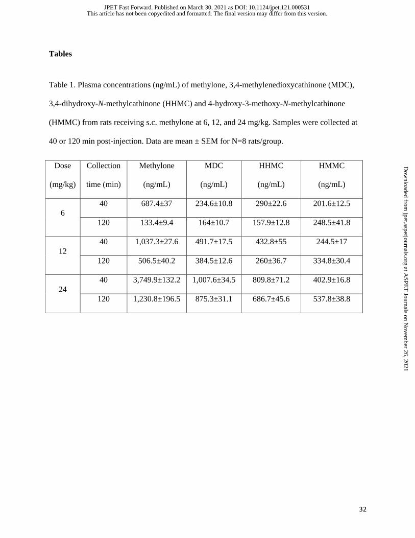
displayed an LOQ of 10 ng/mL. The plasma concentrations of analytes are summarized in Table
This article has not been copyedited and formatted. The final version may differ from this version.JPET Fast Forward. Published on March 30, 2021 as DOI: 10.1124/jpet.121.000531
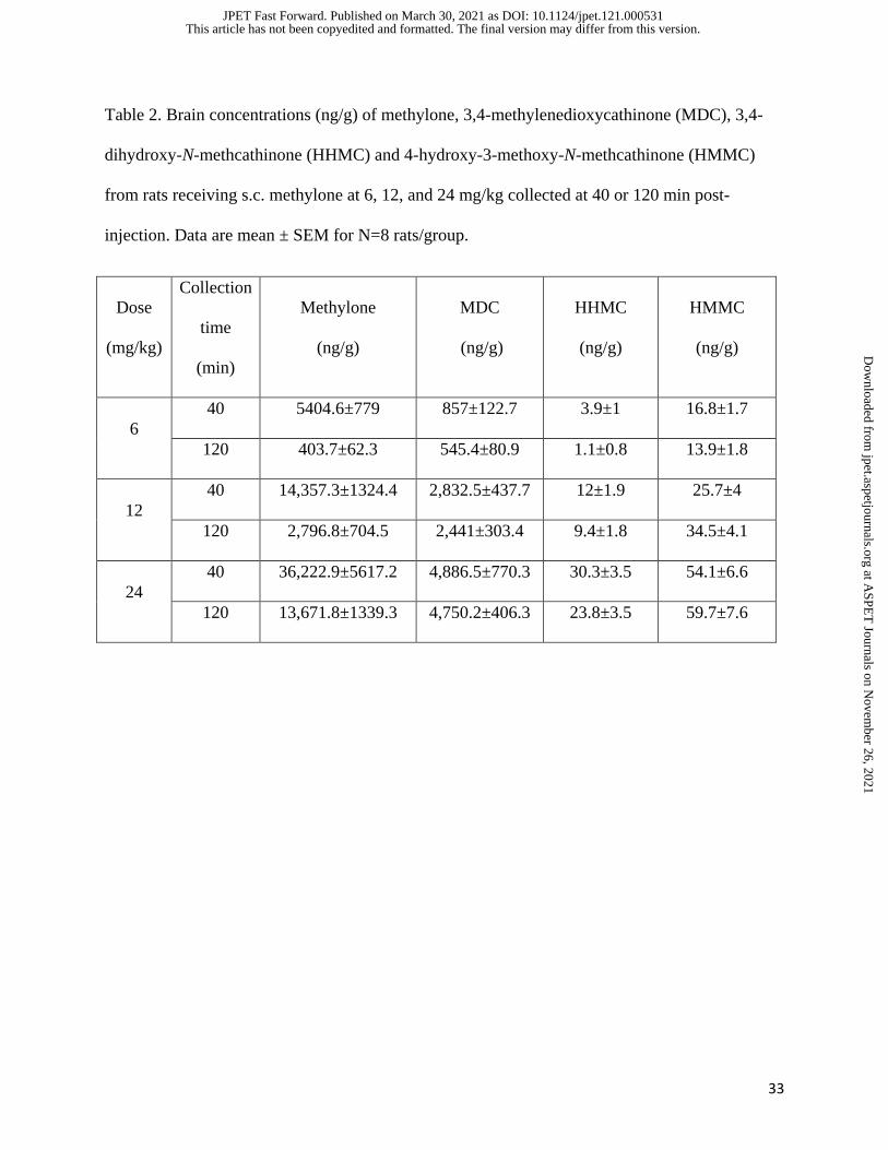
1, whereas brain concentrations are summarized in Table 2. In general, concentrations of
methylone and its metabolites increased in both matrices as the dose administered was increased.
- Insert Figure 2 here –
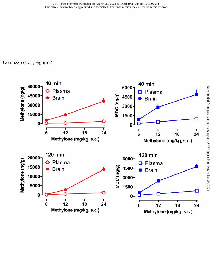
Figure 2 depicts the brain and plasma concentrations of methylone and MDC at the 40-
min and 120-min time points. A two-way ANOVA (dose x matrix) comparing brain and plasma
concentrations of methylone at 40 min revealed significant main effects of dose (F2,42=27.09,
p<0.0001) and matrix (F1,42=75.19, p<0.0001), with a significant dose x matrix interaction
(F2,42=17.85, p<0.0001). Similar results were found for methylone measures at 120 min. At all
doses and time points, brain concentrations of methylone were far greater than plasma
concentrations. A two-way ANOVA comparing brain and plasma concentration of MDC at 40
min revealed significant effects of dose (F2,42=21.62, p<0.0001) and matrix (F1,42=58.40,
p<0.001), with a significant dose x matrix interaction (F2,42=9.932, p<0.003). Similar results
were found for MDC measures at 120 min. At all doses and time points, brain concentrations of
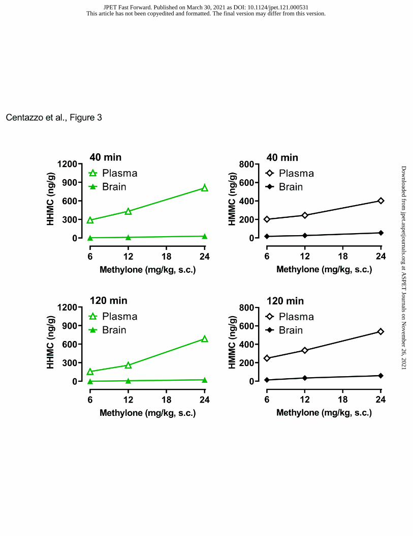
MDC exceeded those measured in plasma. Figure 3 illustrates the brain and plasma
concentrations of the hydroxylated metabolites HHMC and HMMC. In contrast to the findings
for methylone and MDC, brain concentrations of HHMC and HMMC were extremely low in all
tissue samples. A two-way ANOVA comparing the brain and plasma concentration of HHMC at
40 min revealed significant effects of dose (F2,42=25.90, p<0.0001) and matrix (F=1,42=244.6),
with a significant dose x matrix interaction (F2,42=21.18, P<0.0001). Similar results were found
for HHMC at 120 min. At all doses and time points, plasma concentrations of HHMC were
significantly greater than brain concentrations. A two-way ANOVA comparing brain and plasma
This article has not been copyedited and formatted. The final version may differ from this version.JPET Fast Forward. Published on March 30, 2021 as DOI: 10.1124/jpet.121.000531
concentration of HMMC at 40 min revealed significant effects of dose (F2,41=56.73, p<0.0001)
and matrix (F1,41=694, p<0.0001), with a significant dose x matrix interaction (F2,41=26.98,
p<0.0001). Similar results were found for HMMC at 120 min. At all doses and time points,
plasma concentrations of HMMC far exceeded those measured in brain.
- Insert Figure 3 here -
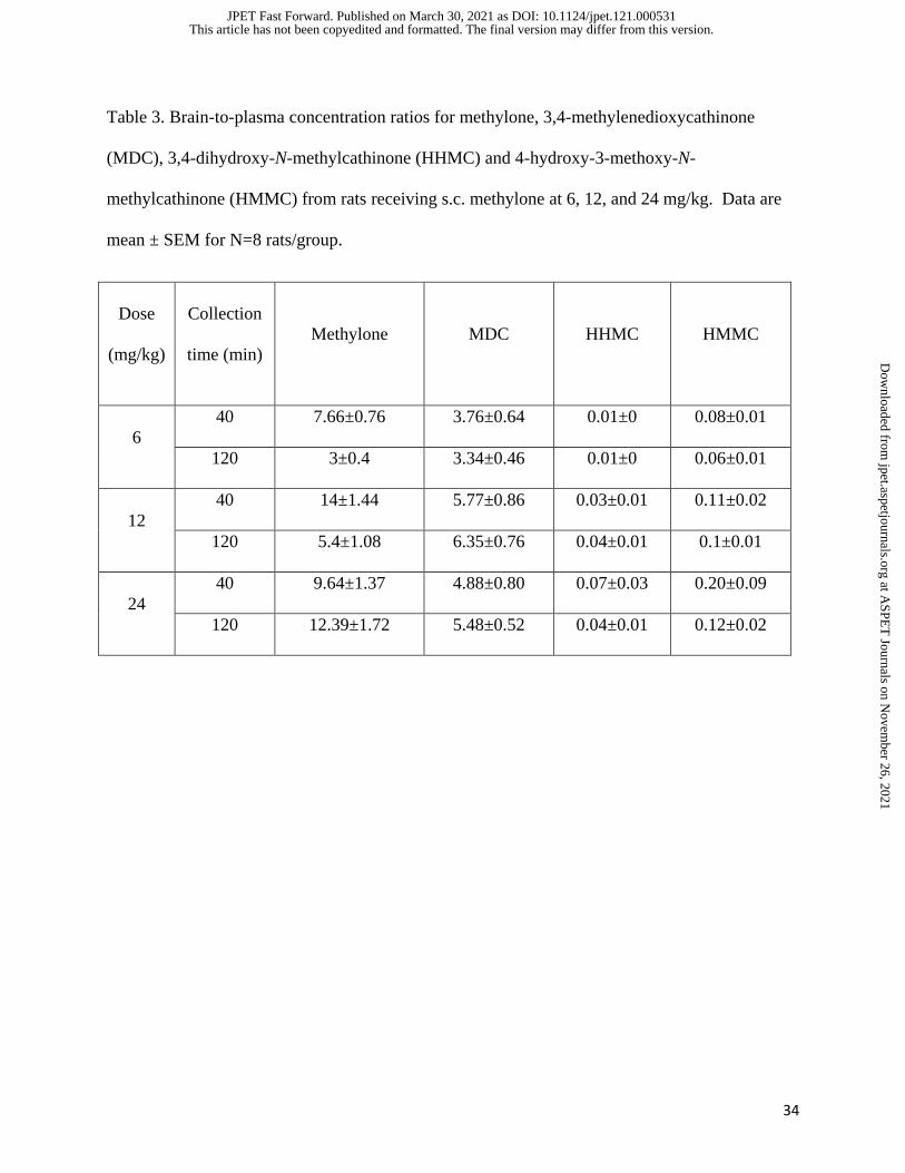
The data in Table 3 summarize brain-to-plasma ratios for all analytes. Methylone and
MDC displayed brain-to-plasma ratios >3 (range 3-14), whereas HHMC and HMMC had ratios
<0.2 (range of 0.01-0.2). These results confirm that methylone and its N-demethylated metabolite
MDC freely cross the blood-brain-barrier to reach the brain, whereas HHMC and HMMC do not.
To investigate the possible reasons underlying the lack of hydroxylated metabolites reaching the
brain, we explored the presence of glucuronide or sulfate conjugates in both plasma and brain.
Briefly, we compared analyte concentrations in plasma and brain samples which were subjected
to 2 separate analytical procedures, one that involved sample hydrolysis to cleave conjugated
metabolites and another that did not. In the brain, no phase II metabolites were detected for any
of the metabolites. In plasma, HMMC and HHMC were mainly present as conjugates. The
percentage of HMMC in conjugated form ranged from 47.6 to 95.7%, median 84.6%, and the
percentage of HHMC as conjugated metabolite ranged from 49.2 to 99.8%, median 87.6%.
These results show that HHMC and HMMC are predominantly present as conjugates in plasma,
and these conjugates do not cross the blood-brain barrier.
Data from our previous study suggested that methylone concentrations in plasma may
exhibit non-linear accumulation, where circulating drug concentrations are greater than dose-
This article has not been copyedited and formatted. The final version may differ from this version.JPET Fast Forward. Published on March 30, 2021 as DOI: 10.1124/jpet.121.000531
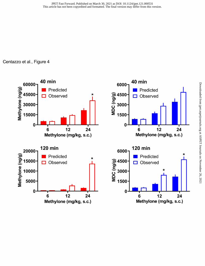
proportional (Elmore et al., 2017). Therefore, we compared the predicted concentrations of
methylone and MDC in brain tissue to their actual observed concentrations. Data in Figure 4
show the predicted versus observed concentrations for methylone and MDC in brain. A two-way
ANOVA (dose x condition) comparing predicted versus observed brain concentrations of
methylone at the 40-min time point revealed significant main effects of dose (F2,42=32.49,
p<0.0001) and condition (F1,42=6.05, p<0.01), where observed methylone concentrations were
significantly greater than predicted at the 24 mg/kg methylone dose (p<0.05 Sidak’s test).
Similar results were found for the 120-min time point, where the observed concentration of
methylone was significantly greater than the predicted concentration at 24 mg/kg dose. A two-
way ANOVA comparing the predicted versus observed brain concentrations of MDC at the 40-
min time point revealed significant main effects of dose (F2,42=29.45, p<0.001) and condition
(F1,42=5.93, p<0.01), but the predicted and observed concentrations did not differ significantly at
any dose. A similar analysis of MDC concentrations at 120 min found significant main effects of
dose (F2,42=64.36, p<0.0001) and condition (F1,42=38.34, p<0.0001), where the observed
concentrations were significantly greater than predicted at the 12 and 24 mg/kg doses. The
findings with MDC suggest that there is a delayed accumulation of this analyte in the brain.
- Insert Figure 4 here –
- Insert Figure 5 here -
Pharmacodynamic Effects of Methylone
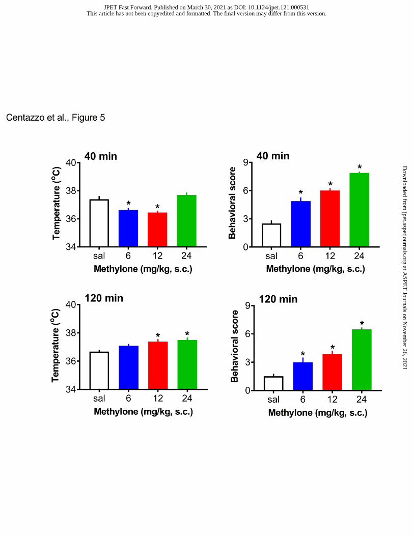
The effects of methylone on core body temperature and locomotor behavioral score are
shown in Figure 5. Methylone administration affected temperature in a dose- and time-dependent
This article has not been copyedited and formatted. The final version may differ from this version.JPET Fast Forward. Published on March 30, 2021 as DOI: 10.1124/jpet.121.000531
manner, with initial hypothermia followed by delayed hyperthermia. A one-way ANOVA (dose)
for temperature data demonstrated that methylone significantly affected body temperature at the
40-min time point (F3,28=11.90, p<0.0001), with modest hypothermia occurring after the 6 and
12 mg/kg doses. At the 120-min time point, methylone significantly affected temperature
(F3,28=5.734, p<0.003), with a modest but significant hyperthermia of about 0.5oC above normal,
observed at the 12 and 24 mg/kg doses. Methylone administration significantly altered locomotor
score at both 40 min (F3,28=56.36, p<0.0001) and 120 min (F3,28=36.55, p<0.0001). At both time
points, Bonferroni’s post hoc test revealed significant increases in behavioral score after the 6,
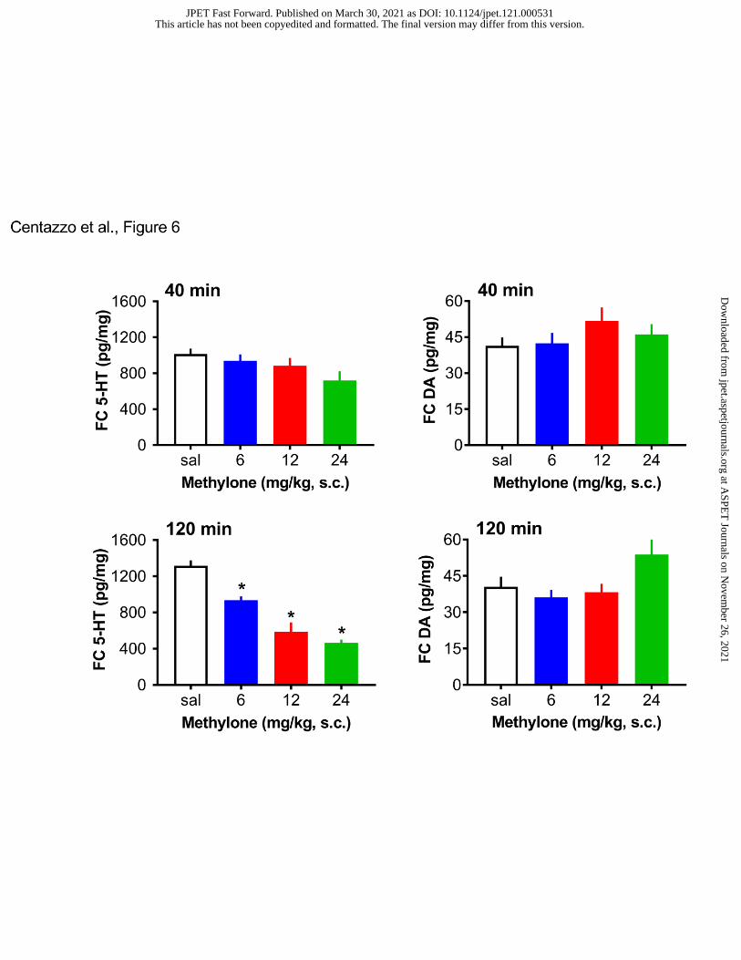
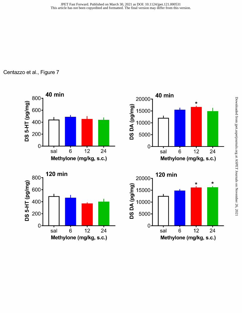
12, and 24 mg/kg doses when compared to saline control. Figure 6 depicts the effects of
methylone on post-mortem concentrations of 5-HT and dopamine in the frontal cortex.
Methylone did not affect 5-HT at 40 min, but significantly influenced 5-HT at 120 min
(F3,28=33.88, p<0.0001), with substantial dose-related decreases in 5-HT, which reached 60%
reduction at the 24 mg/kg dose. Methylone failed to alter dopamine concentrations in the frontal
cortex. Figure 7 shows the effects of methylone on post-mortem tissue 5-HT and dopamine in the
dorsal striatum. Methylone had no effect on striatal 5-HT at either time point. By contrast,
methylone slightly, albeit significantly, elevated striatal dopamine at both the 40-min (F3,28=3.73,
p<0.02) and 120-min (F3,28=7,29, p<0.001) time points.
- Insert Figure 6 here -
- Insert Figure 7 here -
Correlative Relationships
This article has not been copyedited and formatted. The final version may differ from this version.JPET Fast Forward. Published on March 30, 2021 as DOI: 10.1124/jpet.121.000531
We obtained pharmacokinetic and pharmacodynamic data from the same experimental
subjects, which allowed us to examine potential correlative relationships among various
endpoints. We were particularly interested in the relationship between brain analyte
concentrations and pharmacodynamic effects. The present pharmacokinetic findings revealed the
absence of hydroxylated metabolites in the brain, so all correlation analyses were confined to
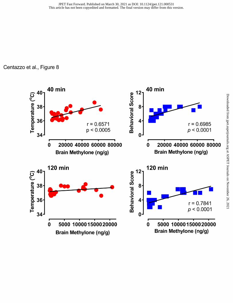
brain concentrations of methylone and MDC. Figure 8 depicts the correlations between brain
concentrations of methylone and body temperature or behavioral score. At the 40-min time point,
brain methylone was positively correlated with both temperature (Pearson’s r=0.6751, p<0.0005)
and behavioral score (r=0.6985, p<0.0001). At 120 min, methylone was not correlated with
temperature (r=0.3567, NS) but did correlate with behavioral score (r=0.7841, p<0.0001). Figure
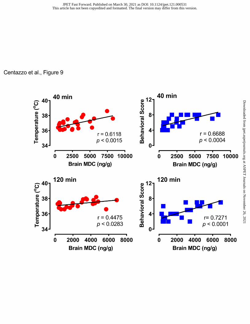
9 shows that brain MDC concentrations were positively correlated with body temperature
(r=0.6118, p<0.0015) and behavioral score (r=0.6688, p<0.0004) at 40 min post-injection, and
similar positive correlations were observed at the 120-min time point.
- Insert Figure 8 here -
- Insert Figure 9 here -
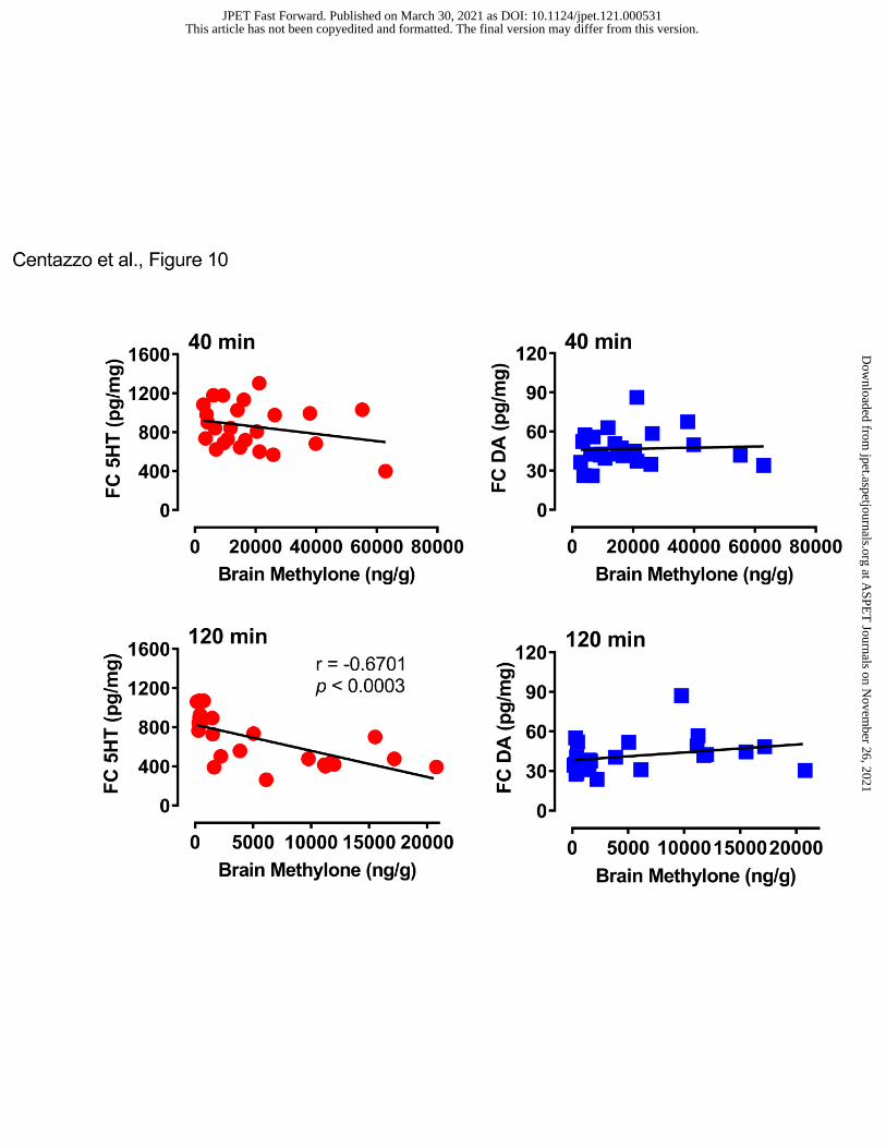
Figure 10 illustrates the correlations between brain methylone concentration and cortical
5-HT or dopamine. Brain concentrations of methylone did not correlate with either cortical
neurotransmitter at 40 min. However, at the 120 min time point, methylone was negatively
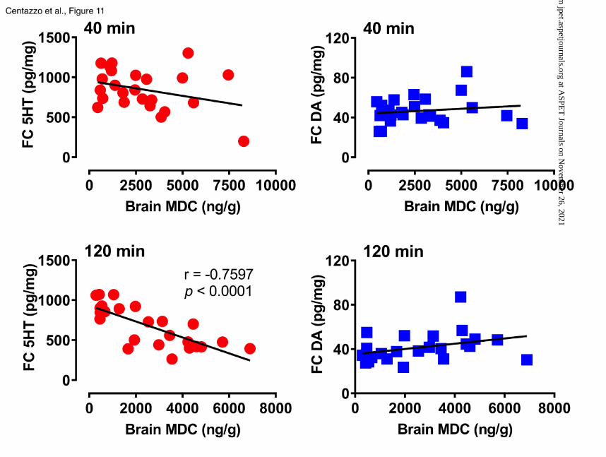
correlated with cortical 5-HT (r=-0.6701, p<0.0003). Figure 11 shows the correlations between
MDC concentrations and cortical 5-HT or dopamine. Brain concentrations of MDC did not
correlate with either neurotransmitter at the 40 min time point, but at 120 min, there was a
This article has not been copyedited and formatted. The final version may differ from this version.JPET Fast Forward. Published on March 30, 2021 as DOI: 10.1124/jpet.121.000531
significant negative correlation between MDC and 5-HT (r=-0.7597, p<0.0001). No correlations
were found when examining relationships among methylone, MDC, and striatal
neurotransmitters.
- Insert Figure 10 here -
- Insert Figure 11 here -
Discussion
A primary aim of the present study was to quantify brain and plasma concentrations of
methylone and its metabolites after systemic methylone administration to male rats. In general,
methylone and metabolite concentrations rose in parallel with increasing dose of methylone
administered, but brain methylone and MDC concentrations were greater than dose-proportional
at the highest dose administered. Methylone and MDC displayed brain-to-plasma ratios > 3
(range 3-12) whereas HHMC and HMMC had brain-to-plasma ratios < 0.2 (range 0.01-0.2).
These findings demonstrate that methylone and MDC freely penetrate into the central nervous
system, but hydroxylated metabolites do not. A secondary aim of the study was to relate brain
analyte concentrations with acute pharmacodynamic effects of methylone. In this regard,
locomotor activity score was positively correlated with brain concentrations of methylone and
MDC, while post-mortem 5-HT levels in the cortex were negatively correlated with these same
analytes. Overall, the findings show that acute pharmacodynamic effects of methylone are likely
related to brain concentrations of the parent compound and its N-demethylated metabolite.
It is notable that we found evidence for non-linear accumulation of methylone and MDC
in the brain. Elmore et al. (Elmore et al., 2017) reported non-linear kinetics for methylone in
This article has not been copyedited and formatted. The final version may differ from this version.JPET Fast Forward. Published on March 30, 2021 as DOI: 10.1124/jpet.121.000531
plasma, while López-Arnau et al. (López-Arnau et al., 2013) found no evidence for the
phenomenon. Here, we report the first dose-effect investigation of methylone and metabolite
concentrations in the brain. The doses of methylone we employed were chosen based on our
previous study (Elmore et al., 2017), where we observed higher than predicted concentrations of
methylone in plasma after s.c. administration of 12 mg/kg. The current findings show that non-
linear accumulation of methylone and MDC occurs in the brain at higher drug doses, and this
effect is more robust after 120 min compared to 40 min. Our data are consistent with the notion
that methylone induces a dose- and time-dependent inhibition of CYP2D1 (the rat isoform of
CYP2D6 in humans), the chief enzyme responsible for biotransformation of methylone. Indeed,
Pedersen et al. (Pedersen et al., 2013) found that methylone inhibits CYP2D6 with a Ki of 15
µM, which translates to ~3,000 ng/g, a concentration that is achieved in rat brain tissue after the
12 and 24 mg/kg doses of methylone (see Table 2). We hypothesize that methylone is capable of
inactivating CYP2D1 in rats, in a manner analogous to the effect of MDMA on CYP2D6 in
humans (de la Torre et al., 2004), and subsequent studies should address this hypothesis. From a
clinical perspective, the non-linear kinetics of methylone might be a contributing factor to the
adverse effects of the drug reported after high-dose exposure in humans (Cawrse et al., 2012;
Ellefsen et al., 2015). Similar toxicities have been reported in rats self-administering large doses
of methylone (Gannon et al.; 2018; Gannon et al.; 2019).
The collection of brain tissue and plasma from the same rats made it possible to
determine brain-to-plasma ratios for methylone and its metabolites. Our results reveal that
methylone and MDC are readily able to cross the blood-brain-barrier (brain-to-plasma ratios
from 3-14), while HHMC and HMMC do not (brain-to-plasma ratio from 0.01 to 0.2). In fact,
the small amounts of HHMC and HMMC detected in brain were likely related to analyte
This article has not been copyedited and formatted. The final version may differ from this version.JPET Fast Forward. Published on March 30, 2021 as DOI: 10.1124/jpet.121.000531
concentrations in residual blood found in post-mortem brain tissue samples. Limited information
is available about the distribution of methylone and its metabolites in the brain or other organs
(Štefková et al., 2017). Lopez-Arnau et al. (López-Arnau et al., 2013) reported a methylone
brain-to-plasma ratio of 1.42 after an oral dose of 30 mg/kg methylone. In that study, the oral
route of administration might explain lower concentrations of drug reaching the brain, secondary
to extensive gut and hepatic metabolism of the parent compound. Stefkova et al. (Štefková et al.,
2017) reported that s.c. methylone administration yields a brain-to-serum ratio of 7.97 whereas
Grecco et al. (Grecco et al., 2017) found that s.c. methylone yields a brain-to-plasma ratio of
39.5. It is noteworthy that the latter finding was based on area-under-curve estimates rather than
single time points. Regardless of the details, all of the available data from rats agree that
methylone and MDC freely cross the blood-brain-barrier. In contrast to methylone and MDC, we
show that HHMC and HMMC are found at particularly low concentrations in the brain. The
inability of the hydroxylated metabolites to enter the brain is most likely due to the high
percentage of HHMC and HMMC conjugates in plasma, which are too polar to penetrate into the
brain. Elmore et al. (Elmore et al., 2017) showed that methylone, MDC, and HHMC were
substrate-type releasers at monoamine transporters in vitro, but only methylone and MDC
produced significant elevations in brain extracellular dopamine and 5-HT when administered in
vivo. Thus, HHMC, in its unconjugated form, is able to serve as a monoamine transporter
substrate, but this metabolite does not normally reach the brain after systemic methylone
administration.
A secondary aim of the present study was to relate pharmacodynamic effects of
methylone to brain concentrations of the drug and its metabolites, especially MDC, since this is
the main metabolite reaching the brain. The behavioral scoring method that was used to assess
This article has not been copyedited and formatted. The final version may differ from this version.JPET Fast Forward. Published on March 30, 2021 as DOI: 10.1124/jpet.121.000531
locomotor activation is sensitive to dose-dependent changes in behavior induced by stimulant
drugs in rats (Elmore et al., 2017). Methylone produced dose-dependent increases in forward
locomotion, rearing, and patterned sniffing, consistent with previous reports of its stimulant
effects in rats (López-Arnau et al., 2013; Elmore et al., 2017; Štefková et al., 2017; Javadi-
Paydar et al., 2018). We found that behavioral scores were positively correlated with brain
methylone and MDC concentrations at both time points examined, suggesting these analytes
could contribute to motor stimulation. Effects of methylone on core temperature were more
complex, characterized by acute hypothermia followed by a delayed, albeit modest,
hyperthermia. The effects of methylone administration on body temperature were positively
correlated with brain methylone and MDC at 40 min, but less so at 120 min. In a previous study,
Elmore et al. (Elmore et al., 2017) failed to find any correlation between core temperature and
methylone or metabolite concentrations in plasma after 3, 6, or 12 mg/kg s.c. injections. Stefkova
et al. (Štefková et al., 2017) observed that methylone significantly increases colonic temperature
in individually-housed and group-housed rats after s.c. doses of 10 and 20 mg/kg, and the effects
are maintained for more than an hour. Javadi-Paydar et al. (Javadi-Paydar et al., 2018) observed
a modest but sustained hyperthermia (0.4-0.8ºC) for 4 hours after 10 mg/kg methylone in male
rats. Overall, methylone appears to induce modest and sustained hyperthermia in rats, but this
effect is influenced by dose and specific experimental conditions.
Perhaps the most important finding in the present report is the acute depletion of brain 5-
HT produced by methylone administration. The effects of methylone on tissue 5-HT were dose-
and time-dependent, such that the drug produced a delayed decrease in post-mortem tissue 5-HT
in the frontal cortex but not striatum. The acute effects of methylone on tissue 5-HT reported
here are similar to the effects reported for MDMA (Baumann et al., 2007). Previous research has
This article has not been copyedited and formatted. The final version may differ from this version.JPET Fast Forward. Published on March 30, 2021 as DOI: 10.1124/jpet.121.000531
demonstrated that methylone is a non-selective substrate-type releaser at the transporters for
dopamine, norepinephrine, and 5-HT (Baumann et al., 2013; Eshleman et al., 2013; Simmler et
al., 2013). Like other synthetic cathinones, methylone releases monoamine transmitters from
intracellular stores via reversal of normal transporter flux (i.e., reverse transport). The results
provided here show that the releasing actions of methylone may lead to acute depletion of
intracellular stores of transmitter, but this effect is selective for 5-HT since post-mortem
dopamine concentrations are actually increased and not depleted. The 5-HT depleting action of
methylone seems to be exacerbated as time passes, since the most robust effects are observed
after 2 hours. We have no explanation for why methylone produces selective decreases in
cortical 5-HT, but such reductions could have functional consequences. In animal models, low
cerebrospinal fluid concentrations of the 5-HT metabolite, 5-HIAA, and reduced 5-HT levels or
turnover in the brain are associated with increased aggressive behavior (Nelson and Chiavegatto,
2001). In human case studies, methylone overdose is sometimes associated with aggressive and
psychotic behaviors, and our preclinical findings suggest that decreased cortical 5-HT might
contribute to such adverse effects (Diestelmann et al., 2018).
In summary, we report the pharmacokinetics of methylone and its three major
metabolites in brain and plasma of male rats. Methylone and MDC freely penetrate the blood-
brain barrier, whereas HHMC and HMMC do not. Thus, hydroxylated metabolites of methylone
do not contribute to centrally mediated pharmacodynamic effects. Methylone and MDC exhibit
non-linear kinetics in the brain when assessed 120 min after methylone administration,
suggesting delayed accumulation into neurons. Locomotor activity score is positively correlated
with brain concentrations of methylone and MDC, while post-mortem levels of 5-HT in the
frontal cortex are negatively correlated with these analytes. Taken together, our findings indicate
This article has not been copyedited and formatted. The final version may differ from this version.JPET Fast Forward. Published on March 30, 2021 as DOI: 10.1124/jpet.121.000531
that non-linear accumulation of methylone and MDC in the brain could cause untoward effects
on 5-HT neurons in vulnerable brain regions, including the frontal cortex.
This article has not been copyedited and formatted. The final version may differ from this version.JPET Fast Forward. Published on March 30, 2021 as DOI: 10.1124/jpet.121.000531
Contributed new reagents or analytical tools: Suzuki, Rice, Concheiro
Performed data analysis: Centazzo, Baumann, Concheiro
Wrote or contributed to the writing of the manuscript: Centazzo, Baumann, Concheiro
This article has not been copyedited and formatted. The final version may differ from this version.JPET Fast Forward. Published on March 30, 2021 as DOI: 10.1124/jpet.121.000531
and Caffeine in Rats. Neuropsychopharmacology 43:761-769.
Gannon BM, Mesmin MP, Sulima A, Rice KC and Collins GT (2019) Behavioral economic
analysis of the reinforcing effects of “bath salts” mixtures: studies with MDPV, methylone,
and caffeine in male Sprague-Dawley rats (2019) Psychopharmacology 236:1031-1041.
This article has not been copyedited and formatted. The final version may differ from this version.JPET Fast Forward. Published on March 30, 2021 as DOI: 10.1124/jpet.121.000531
Pedersen AJ, Petersen TH, and Linnet K (2013) In vitro metabolism and pharmacokinetic studies
on methylone. Drug Metab Dispos 41:1247–1255.
Schindler CW, Thorndike EB, Goldberg SR, Lehner KR, Cozzi N V., Brandt SD, and Baumann
MH (2016) Reinforcing and neurochemical effects of the “bath salts” constituents 3,4-
methylenedioxypyrovalerone (MDPV) and 3,4- methylenedioxy-N-methylcathinone
(methylone) in male rats. Psychopharmacology (Berl) 233:1981–1990.
Simmler LD, Buser TA, Donzelli M, Schramm Y, Dieu LH, Huwyler J, Chaboz S, Hoener MC,
and Liechti ME (2013) Pharmacological characterization of designer cathinones in vitro. Br
J Pharmacol 168:458–470.
Štefková K, Židková M, Horsley RR, Pinterová N, Šíchová K, Uttl L, Balíková M, Danda H,
Kuchar M, and Pálenícek T (2017) Pharmacokinetic, ambulatory, and hyperthermic effects
of 3,4-methylenedioxy-n-methylcathinone (Methylone) in rats. Front Psychiatry 8:1–11.
Vanderwater SA, Creehan KM, and Taffe MA (2015) Intravenous self-administration of
entactogen-class stimulants in male rats. Neuropharmacology 99:538–545.
This article has not been copyedited and formatted. The final version may differ from this version.JPET Fast Forward. Published on March 30, 2021 as DOI: 10.1124/jpet.121.000531
Watterson LR, Hood L, Sewalia K, Tomek SE, Yahn S, Johnson CT, Wegner S, Blough BE,
Marusich JA, and Olive MF (2012) The Reinforcing and Rewarding Effects of Methylone,
a Synthetic Cathinone Commonly Found in “Bath Salts.” J Addict Res Ther Supple 9:002.
Footnotes
The research program of Dr. Baumann is generously supported by the Intramural Research
Program (IRP) of the National Institute on Drug Abuse (NIDA, National Institutes of Health
(NIH), grant# DA000523-13.
This article has not been copyedited and formatted. The final version may differ from this version.JPET Fast Forward. Published on March 30, 2021 as DOI: 10.1124/jpet.121.000531
methylcathinone (HMMC), and 3,4-methylenedioxycathinone (MDC).
Fig. 2. Plasma and brain concentrations of methylone and 3,4-methylenedioxycathinone (MDC)
at early and late time points (40 min and 120 min) after s.c. methylone injections (6, 12, and 24
mg/kg). Data are mean ± SEM for n=8 rats/group.
Fig. 3. Plasma and brain concentrations of 3,4-dihydroxy-N-methylcathinone (HHMC) and 4-
hydroxy-3-methoxy-N-methylcathinone (HMMC) at early and late time points (40 min and 120
min) after s.c. methylone injections (6, 12, and 24 mg/kg). Data are mean ± SEM for n=8
rats/group.
Fig. 4. Predicted versus observed brain concentrations of methylone and 3,4-
methylenedioxycathinone (MDC) at early and late time points (40 min and 120 min). Predicted
concentrations at the 12 and 24 mg/kg doses were calculated by multiplying the observed values
at 6 mg/kg by a factor of 2 and 4, respectively. Data are mean ± SEM for n=8 rats/group.
Asterisks represent significant difference compared to predicted group.
Fig. 5. Effects of s.c. methylone administration (6, 12, and 24 mg/kg dose) on body temperature
and behavioral score at 40 min and 120 min post-injection. Data are mean ± SEM for n=8
rats/group. Asterisks represent significant differences compared to saline-treated control group.
Fig. 6. Effect of s.c. methylone administration (6, 12, and 24 mg/kg) on post-mortem levels of
dopamine (DA) and serotonin (5-HT) from frontal cortex at 40 min and 120 min post-injection.
Data are mean ± SEM for n=8 rats/group. Asterisks represent significant differences compared to
saline-treated control group.
Fig. 7. Effect of s.c. methylone administration (6, 12, and 24 mg/kg) on post-mortem levels of
dopamine (DA) and serotonin (5-HT) from dorsal striatum at 40 min and 120 min post-injection.
Data are mean ± SEM for n=8 rats/group. Asterisks represent significant differences compared to
saline-treated control group.
Fig. 8. Correlations between brain methylone concentrations and body temperature or behavioral
score at 40 min and 120 min post-injection.
Fig. 9. Correlations between brain 3,4-methylenedioxycathinone (MDC) concentrations and
body temperature or behavioral score at 40 min and 120 min post-injection.
Fig. 10. Correlations between brain methylone concentrations and frontal cortical 5-HT and
dopamine (DA) at 40 min and 120 min post-injection.
This article has not been copyedited and formatted. The final version may differ from this version.JPET Fast Forward. Published on March 30, 2021 as DOI: 10.1124/jpet.121.000531
Fig. 11. Correlations between brain MDC concentrations and frontal cortical 5-HT and
dopamine (DA) at 40 min and 120 min post-injection.
This article has not been copyedited and formatted. The final version may differ from this version.JPET Fast Forward. Published on March 30, 2021 as DOI: 10.1124/jpet.121.000531
This article has not been copyedited and formatted. The final version may differ from this version.JPET Fast Forward. Published on March 30, 2021 as DOI: 10.1124/jpet.121.000531
This article has not been copyedited and formatted. The final version may differ from this version.JPET Fast Forward. Published on March 30, 2021 as DOI: 10.1124/jpet.121.000531
Table 3. Brain-to-plasma concentration ratios for methylone, 3,4-methylenedioxycathinone
(MDC), 3,4-dihydroxy-N-methylcathinone (HHMC) and 4-hydroxy-3-methoxy-N-
methylcathinone (HMMC) from rats receiving s.c. methylone at 6, 12, and 24 mg/kg. Data are
mean ± SEM for N=8 rats/group.
Dose
(mg/kg)
Collection
time (min)
Methylone
MDC HHMC HMMC
6
40 7.66±0.76 3.76±0.64 0.01±0 0.08±0.01
120 3±0.4 3.34±0.46 0.01±0 0.06±0.01
12
40 14±1.44 5.77±0.86 0.03±0.01 0.11±0.02
120 5.4±1.08 6.35±0.76 0.04±0.01 0.1±0.01
24
40 9.64±1.37 4.88±0.80 0.07±0.03 0.20±0.09
120 12.39±1.72 5.48±0.52 0.04±0.01 0.12±0.02
This article has not been copyedited and formatted. The final version may differ from this version.JPET Fast Forward. Published on March 30, 2021 as DOI: 10.1124/jpet.121.000531
This article has not been copyedited and formatted. The final version may differ from this version.JPET Fast Forward. Published on March 30, 2021 as DOI: 10.1124/jpet.121.000531
This article has not been copyedited and formatted. The final version may differ from this version.JPET Fast Forward. Published on March 30, 2021 as DOI: 10.1124/jpet.121.000531
This article has not been copyedited and formatted. The final version may differ from this version.JPET Fast Forward. Published on March 30, 2021 as DOI: 10.1124/jpet.121.000531
This article has not been copyedited and formatted. The final version may differ from this version.JPET Fast Forward. Published on March 30, 2021 as DOI: 10.1124/jpet.121.000531
This article has not been copyedited and formatted. The final version may differ from this version.JPET Fast Forward. Published on March 30, 2021 as DOI: 10.1124/jpet.121.000531
This article has not been copyedited and formatted. The final version may differ from this version.JPET Fast Forward. Published on March 30, 2021 as DOI: 10.1124/jpet.121.000531
This article has not been copyedited and formatted. The final version may differ from this version.JPET Fast Forward. Published on March 30, 2021 as DOI: 10.1124/jpet.121.000531
This article has not been copyedited and formatted. The final version may differ from this version.JPET Fast Forward. Published on March 30, 2021 as DOI: 10.1124/jpet.121.000531
This article has not been copyedited and formatted. The final version may differ from this version.JPET Fast Forward. Published on March 30, 2021 as DOI: 10.1124/jpet.121.000531
This article has not been copyedited and formatted. The final version may differ from this version.JPET Fast Forward. Published on March 30, 2021 as DOI: 10.1124/jpet.121.000531
This article has not been copyedited and formatted. The final version may differ from this version.JPET Fast Forward. Published on March 30, 2021 as DOI: 10.1124/jpet.121.000531