1 Brain Lesions associated Brain Lesions associated with Epilepsy with Epilepsy Daniel Seeburg, Harvard Medical School, Year IV Daniel Seeburg, Harvard Medical School, Year IV Gillian Lieberman, MD Gillian Lieberman, MD May 26 May 26 th th , 2009 , 2009
Transcript
1
Brain Lesions associated Brain Lesions associated with Epilepsywith Epilepsy
Daniel Seeburg, Harvard Medical School, Year IVDaniel Seeburg, Harvard Medical School, Year IVGillian Lieberman, MDGillian Lieberman, MD
May 26May 26thth, 2009, 2009
2
BackgroundBackground——EpilepsyEpilepsy
Second most common group of neurologic disorders Second most common group of neurologic disorders after stroke.after stroke.
~5~5--10% of the population will have at least one seizure in 10% of the population will have at least one seizure in their lifetime, with the highest incidence in early childhood their lifetime, with the highest incidence in early childhood and late adulthoodand late adulthoodPrevalence of epilepsy (two or more unprovoked seizures) is Prevalence of epilepsy (two or more unprovoked seizures) is ~5~5--10 persons per 100010 persons per 1000Seizures account for ~1% of ED visits (1 million annually)Seizures account for ~1% of ED visits (1 million annually)
•Lowenstein et al., Harrison's Principles of Internal Medicine, Ch 363 17th Edition•Pallin
Epilepsy has a high social, psychological and Epilepsy has a high social, psychological and economical impacteconomical impactAbout 30% of patients with epilepsy do not About 30% of patients with epilepsy do not respond to medical therapyrespond to medical therapyFor these patients, surgery may be beneficial, For these patients, surgery may be beneficial, especially if imaging reveals lesions potentially especially if imaging reveals lesions potentially related to the seizuresrelated to the seizures
•WHO fact sheet on epilepsy (http://www.who.int/mediacentre/factsheets/fs999/en/index.html)
4
Overview: Common Pathologies Overview: Common Pathologies Associated with Seizures Associated with Seizures
Hippocampal (Hippocampal (mesialmesial temporal) sclerosistemporal) sclerosisMalformations of cortical development (MCD)Malformations of cortical development (MCD)Dysplastic tumorsDysplastic tumorsVascular lesionsVascular lesionsOther mass lesionsOther mass lesionsInfectionsInfectionsInfarctionInfarctionTraumaTrauma
Pathologies more specifically associated with seizures
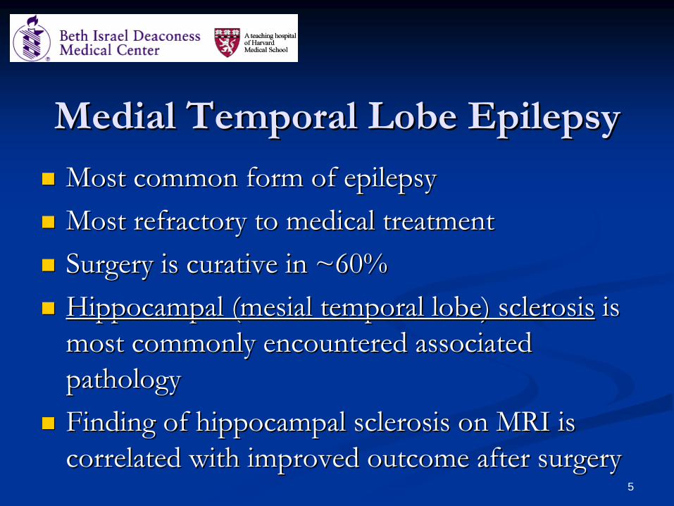
Most common form of epilepsyMost common form of epilepsyMost refractory to medical treatmentMost refractory to medical treatmentSurgery is curative in ~60%Surgery is curative in ~60%Hippocampal (Hippocampal (mesialmesial temporal lobe) sclerosistemporal lobe) sclerosis is is most commonly encountered associated most commonly encountered associated pathologypathologyFinding of hippocampal sclerosis on MRI is Finding of hippocampal sclerosis on MRI is correlated with improved outcome after surgerycorrelated with improved outcome after surgery
6
Hippocampal sclerosisHippocampal sclerosisExtensive cell lossExtensive cell lossDispersal of surviving Dispersal of surviving neuronsneuronsGliosisGliosisAxon sprouting Axon sprouting Formation of new Formation of new excitatory synaptic excitatory synaptic connections, forming connections, forming small small epileptogenicepileptogenicnetworksnetworks
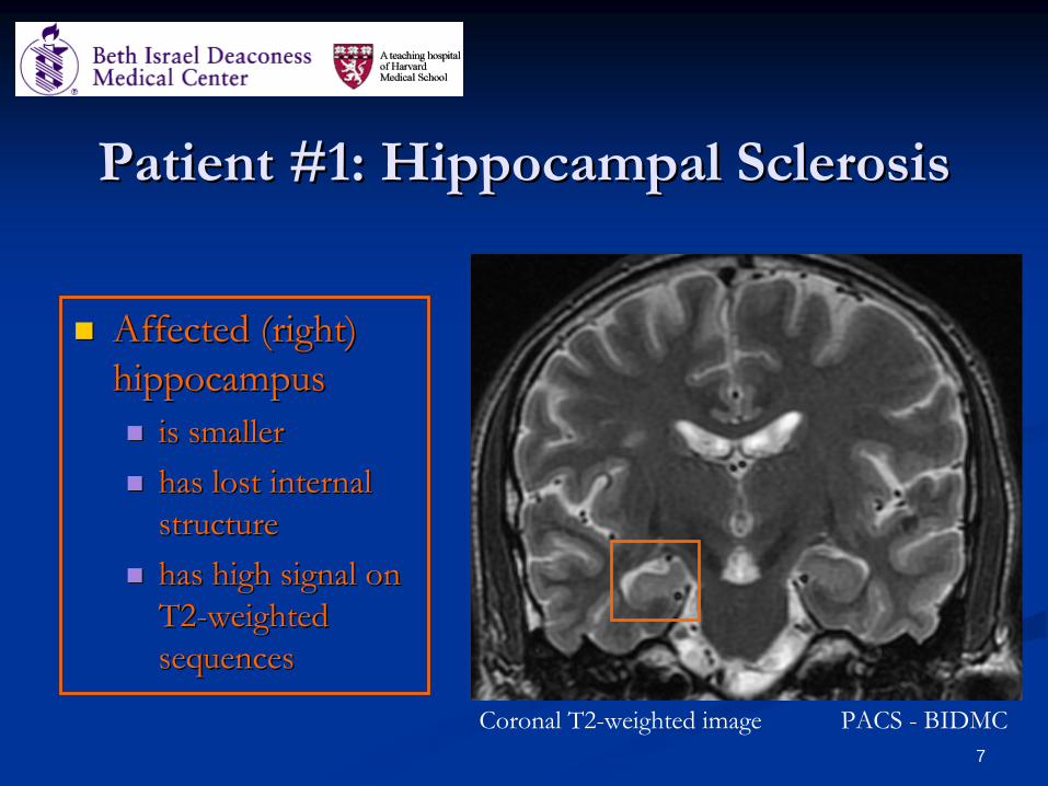
is smalleris smallerhas lost internal has lost internal structurestructurehas high signal on has high signal on T2T2--weighted weighted sequences sequences
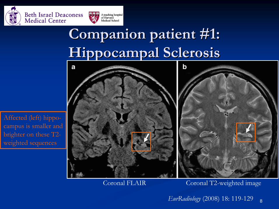
Affected (left) hippo-campus is smaller andbrighter on these T2-weighted sequences
9
Overview: Common Pathologies Overview: Common Pathologies Associated with Seizures cont’d Associated with Seizures cont’d
Hippocampal (Hippocampal (mesialmesial temporal) sclerosistemporal) sclerosisMalformations of cortical development (MCD)Malformations of cortical development (MCD)Dysplastic tumorsDysplastic tumorsVascular lesionsVascular lesionsOther mass lesionsOther mass lesionsInfectionsInfectionsInfarctionInfarctionTraumaTrauma
Pathologies more specifically associated with seizures
10
Malformations of Cortical Malformations of Cortical Development (MCD)Development (MCD)
Also commonly referred to as “Cortical Also commonly referred to as “Cortical DysplasiasDysplasias” or “Neuronal ” or “Neuronal MigrationalMigrational Disorders”Disorders”Several common types:Several common types:
HeterotopiaHeterotopia (clusters of normal gray matter in (clusters of normal gray matter in abnormal locations)abnormal locations)PolymicrogyriaPolymicrogyriaLissencephalyLissencephaly (“smooth brain”(“smooth brain”——absence of absence of sulcisulci and and gyrigyri))Focal Cortical Dysplasia Focal Cortical Dysplasia –– type II (of Taylor)type II (of Taylor)
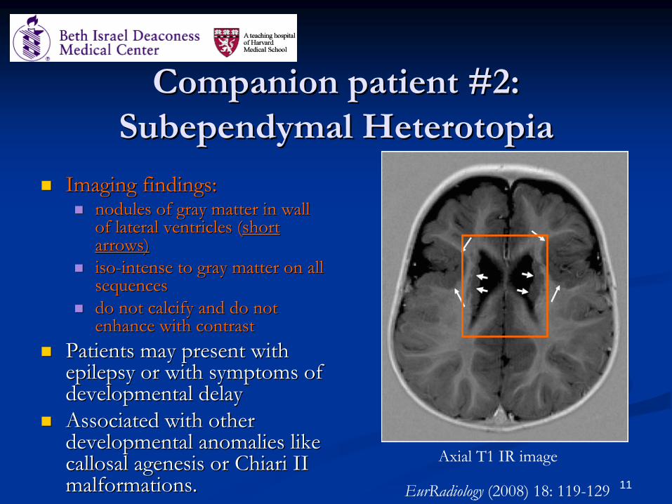
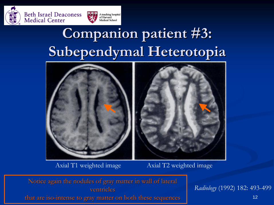
Imaging findings:Imaging findings:nodules of gray matter in wall nodules of gray matter in wall of lateral ventricles (of lateral ventricles (short short arrows)arrows)isoiso--intense to gray matter on all intense to gray matter on all sequencessequencesdo not calcify and do not do not calcify and do not enhance with contrastenhance with contrast
Patients may present with Patients may present with epilepsy or with symptoms of epilepsy or with symptoms of developmental delaydevelopmental delayAssociated with other Associated with other developmental anomalies like developmental anomalies like callosalcallosal agenesis or agenesis or ChiariChiari II II malformations.malformations. EurRadiology
Imaging findings:Imaging findings:loss of normal loss of normal sulcisulciirregular thickening of cortexirregular thickening of cortex
Most commonly encountered Most commonly encountered MCD in patients with MCD in patients with refractory epilepsyrefractory epilepsyBilateral Bilateral periperi--SylvianSylviandistribution is commondistribution is commonClinical presentation ranges Clinical presentation ranges from developmental delay to from developmental delay to epilepsy and can involve epilepsy and can involve focal or diffuse neurological focal or diffuse neurological deficitsdeficits
EurRadiology
(2008) 18: 119-129
Axial T1 IR
15
Malformations of Cortical Malformations of Cortical Development (MCD) cont’d Development (MCD) cont’d ----
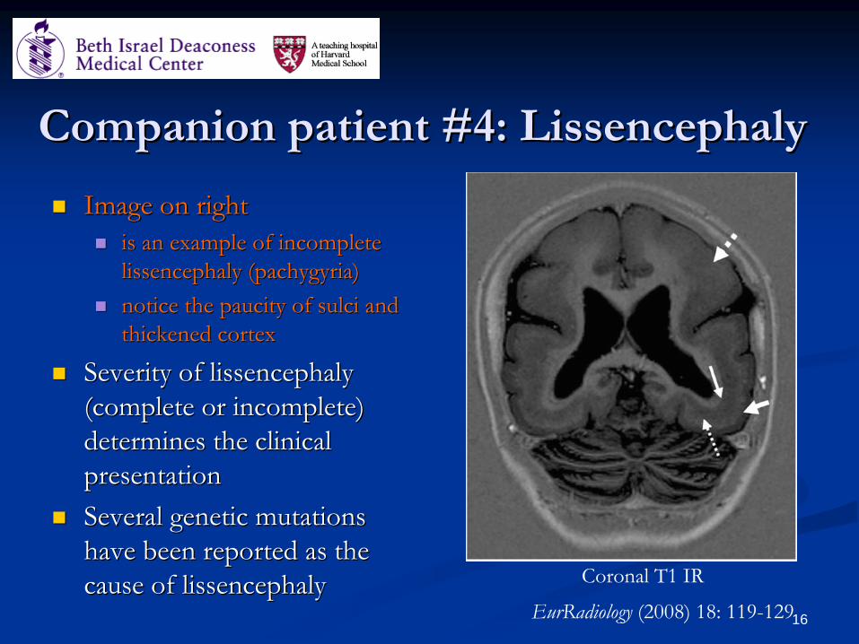
Image on right Image on right is an example of incomplete is an example of incomplete lissencephalylissencephaly ((pachygyriapachygyria))notice the paucity of notice the paucity of sulcisulci and and thickened cortexthickened cortex
Severity of Severity of lissencephalylissencephaly(complete or incomplete) (complete or incomplete) determines the clinical determines the clinical presentationpresentationSeveral genetic mutations Several genetic mutations have been reported as the have been reported as the cause of cause of lissencephalylissencephaly
EurRadiology
(2008) 18: 119-129
Coronal T1 IR
17
Malformations of Cortical Malformations of Cortical Development (MCD) cont’d Development (MCD) cont’d ––
Focal Focal
Cortical Dysplasia type II (Taylor)Cortical Dysplasia type II (Taylor)
18
Overview: FCD type II (Taylor)Overview: FCD type II (Taylor)
Most common type of focal cortical dysplasiaMost common type of focal cortical dysplasiaThought to have high degree of intrinsic Thought to have high degree of intrinsic epileptogenicityepileptogenicitySurgical treatment is often curativeSurgical treatment is often curative
19
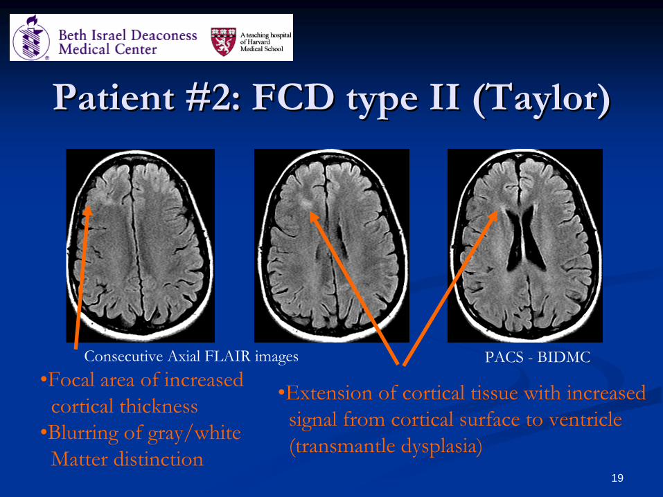
Patient #2: FCD type II (Taylor)Patient #2: FCD type II (Taylor)
•Focal area of increased cortical thickness
•Blurring of gray/white Matter distinction
•Extension of cortical tissue with increasedsignal from cortical surface to ventricle (transmantle
dysplasia)
PACS -
BIDMCConsecutive Axial FLAIR images
20
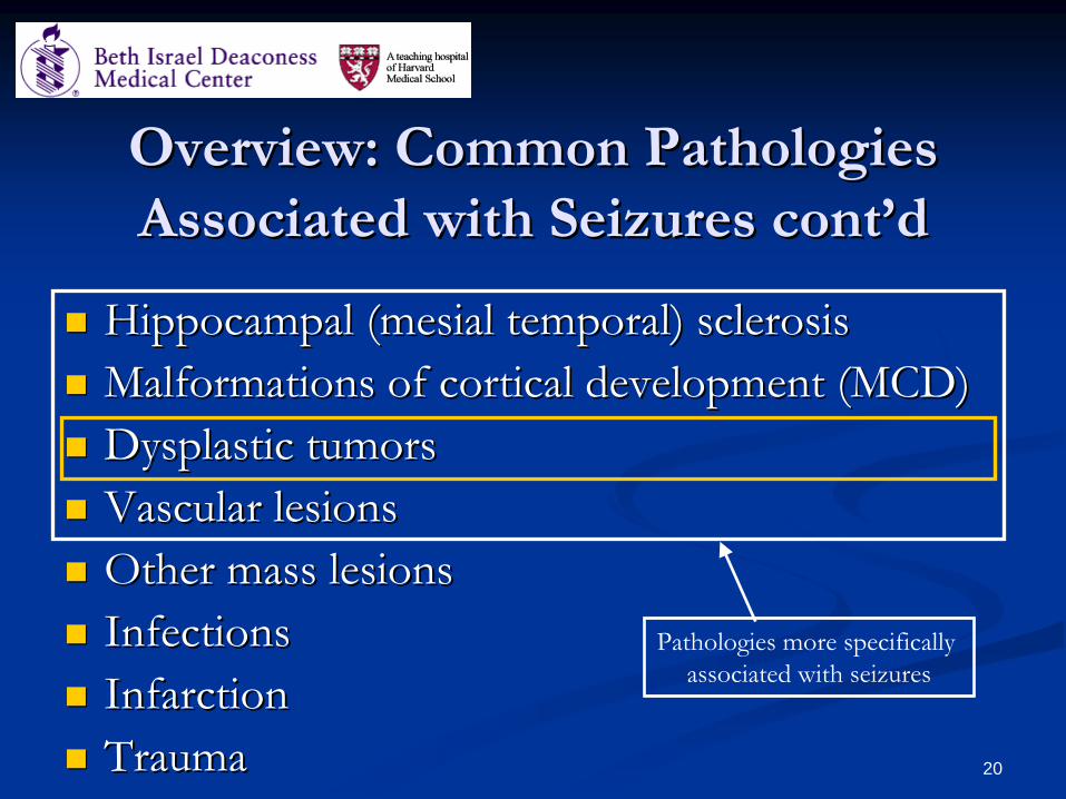
Overview: Common Pathologies Overview: Common Pathologies Associated with Seizures cont’dAssociated with Seizures cont’d
Hippocampal (Hippocampal (mesialmesial temporal) sclerosistemporal) sclerosisMalformations of cortical development (MCD)Malformations of cortical development (MCD)Dysplastic tumorsDysplastic tumorsVascular lesionsVascular lesionsOther mass lesionsOther mass lesionsInfectionsInfectionsInfarctionInfarctionTraumaTrauma
Pathologies more specifically associated with seizures
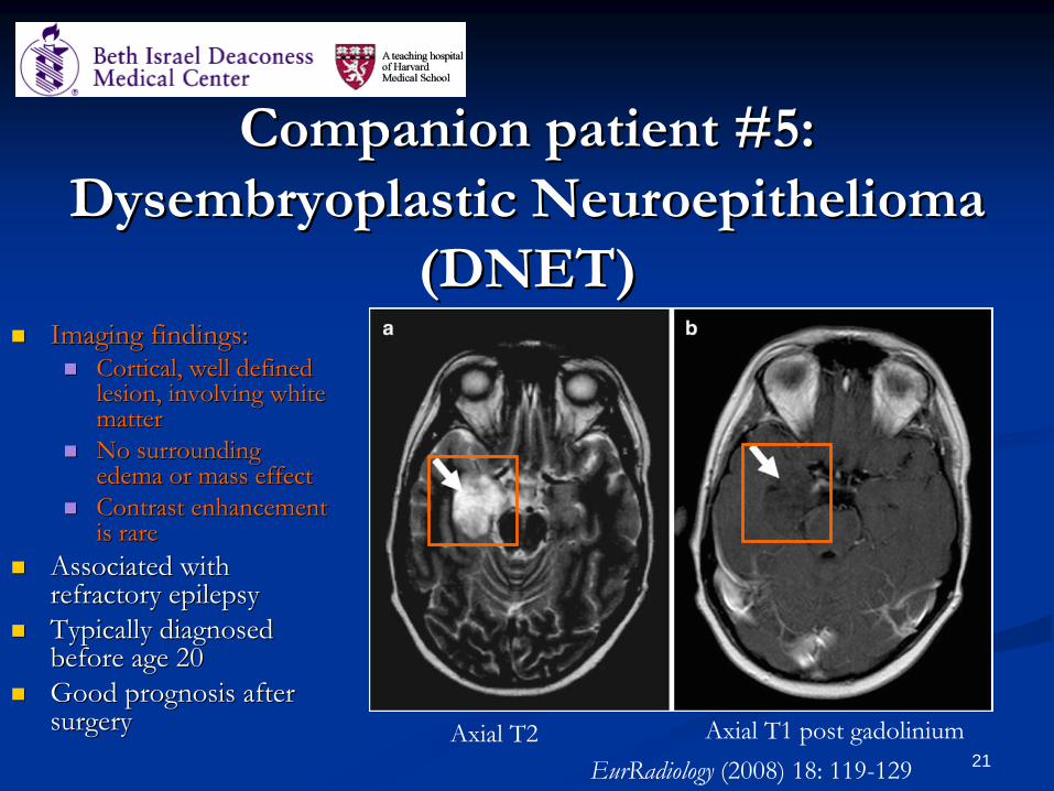
Cortical, well defined Cortical, well defined lesion, involving white lesion, involving white mattermatterNo surrounding No surrounding edema or mass effectedema or mass effectContrast enhancement Contrast enhancement is rare is rare
Associated with Associated with refractory epilepsyrefractory epilepsyTypically diagnosed Typically diagnosed before age 20before age 20Good prognosis after Good prognosis after surgerysurgery
EurRadiology
(2008) 18: 119-129Axial T2 Axial T1 post gadolinium
22
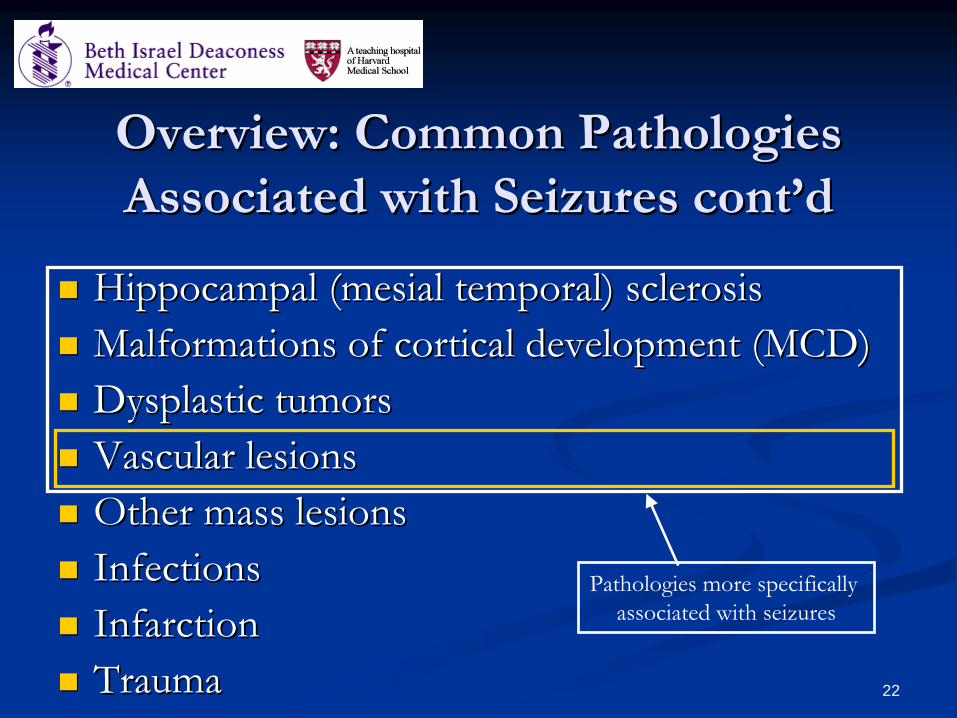
Overview: Common Pathologies Overview: Common Pathologies Associated with Seizures cont’dAssociated with Seizures cont’d
Hippocampal (Hippocampal (mesialmesial temporal) sclerosistemporal) sclerosisMalformations of cortical development (MCD)Malformations of cortical development (MCD)Dysplastic tumorsDysplastic tumorsVascular lesionsVascular lesionsOther mass lesionsOther mass lesionsInfectionsInfectionsInfarctionInfarctionTraumaTrauma
Pathologies more specifically associated with seizures
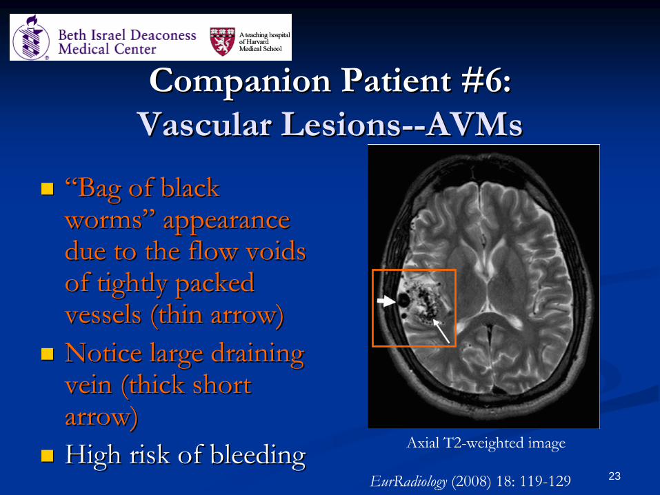
““Bag of black Bag of black worms” appearance worms” appearance due to the flow voids due to the flow voids of tightly packed of tightly packed vessels (thin arrow)vessels (thin arrow)Notice large draining Notice large draining vein (thick short vein (thick short arrow)arrow)High risk of bleedingHigh risk of bleeding
Imaging findings:Imaging findings:“popcorn“popcorn--like” like” appearanceappearancemixed signal core due to mixed signal core due to blood in different stages blood in different stages of degradationof degradation
Seizures and epilepsy are Seizures and epilepsy are most common most common symptomatic presentationsymptomatic presentationCarry risk of bleeding Carry risk of bleeding and development of focal and development of focal neurological deficitsneurological deficits
EurRadiology
(2008) 18: 119-129Coronal T2-weighted image
25
SummarySummary
Epilepsy is a common disorder that is associated Epilepsy is a common disorder that is associated with a number of different underlying lesions, with a number of different underlying lesions, the most common of which is hippocampal the most common of which is hippocampal sclerosis.sclerosis.MRI is useful in detecting these lesions. MRI is useful in detecting these lesions. MRI detection is a positive predictor of good MRI detection is a positive predictor of good outcome after surgeryoutcome after surgery
26
References References Lowenstein Daniel H, "Chapter 363. Seizures and Epilepsy" Lowenstein Daniel H, "Chapter 363. Seizures and Epilepsy" (Chapter). (Chapter). FauciFauci AS, AS, BraunwaldBraunwald E, Kasper DL, Hauser SL, E, Kasper DL, Hauser SL, Longo DL, Jameson JL, Longo DL, Jameson JL, LoscalzoLoscalzo J: Harrison's Principles of J: Harrison's Principles of Internal Medicine, 17th Edition: Internal Medicine, 17th Edition: http://www.accessmedicine.com.ezphttp://www.accessmedicine.com.ezp--prod1.hul.harvard.edu/content.aspx?aID=2901171. prod1.hul.harvard.edu/content.aspx?aID=2901171. DeblaereDeblaere and and AchtenAchten; ; EurEur RadiolRadiol (2008) 18: 119(2008) 18: 119--129129
•
Pallin
et al, Int
J Emerg
Med
(2008) 1(2):97-105BarkovichBarkovich and and KjosKjos; ; RadiologyRadiology (1992) 182: 493(1992) 182: 493--499499DichterDichter; ; Arch Arch NeurolNeurol (2009) 66(4): 443(2009) 66(4): 443--447447WHO fact sheet on epilepsy WHO fact sheet on epilepsy (http://www.who.int/mediacentre/factsheets/fs999/en/index.h(http://www.who.int/mediacentre/factsheets/fs999/en/index.html)tml)
27
Thanks!Thanks!Dr Gillian LiebermanDr Gillian LiebermanMaria Maria LevantakisLevantakisBIDMC BIDMC neuroradiologyneuroradiology faculty and radiology faculty and radiology residentsresidents