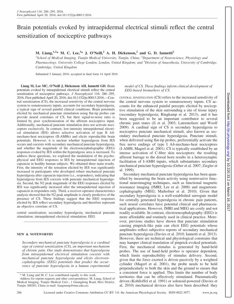
Brain potentials evoked by intraepidermal electrical stimuli reflect the central sensitization of nociceptive pathways M. Liang, 1,2 * M. C. Lee, 3 * J. O’Neill, 2 A. H. Dickenson, 2 and G. D. Iannetti 2 1 School of Medical Imaging, Tianjin Medical University, Tianjin, China; 2 Department of Neuroscience, Physiology and Pharmacology, University College London, London, United Kingdom; and 3 Division of Anaesthesia, University of Cambridge, Cambridge, United Kingdom Submitted 5 January 2016; accepted in final form 14 April 2016 Liang M, Lee MC, O’Neill J, Dickenson AH, Iannetti GD. Brain potentials evoked by intraepidermal electrical stimuli reflect the central sensitization of nociceptive pathways. J Neurophysiol 116: 286 –295, 2016. First published April 20, 2016; doi:10.1152/jn.00013.2016.—Cen- tral sensitization (CS), the increased sensitivity of the central nervous system to somatosensory inputs, accounts for secondary hyperalgesia, a typical sign of several painful clinical conditions. Brain potentials elicited by mechanical punctate stimulation using flat-tip probes can provide neural correlates of CS, but their signal-to-noise ratio is limited by poor synchronization of the afferent nociceptive input. Additionally, mechanical punctate stimulation does not activate noci- ceptors exclusively. In contrast, low-intensity intraepidermal electri- cal stimulation (IES) allows selective activation of type II A- mechano-heat nociceptors (II-AMHs) and elicits reproducible brain potentials. However, it is unclear whether hyperalgesia from IES occurs and coexists with secondary mechanical punctate hyperalgesia, and whether the magnitude of the electroencephalographic (EEG) responses evoked by IES within the hyperalgesic area is increased. To address these questions, we explored the modulation of the psycho- physical and EEG responses to IES by intraepidermal injection of capsaicin in healthy human subjects. We obtained three main results. First, the intensity of the sensation elicited by IES was significantly increased in participants who developed robust mechanical punctate hyperalgesia after capsaicin injection (i.e., responders), indicating that hyperalgesia from IES coexists with punctate mechanical hyperalge- sia. Second, the N2 peak magnitude of the EEG responses elicited by IES was significantly increased after the intraepidermal injection of capsaicin in responders only. Third, a receiver-operator characteristics analysis showed that the N2 peak amplitude is clearly predictive of the presence of CS. These findings suggest that the EEG responses elicited by IES reflect secondary hyperalgesia and therefore represent an objective correlate of CS. central sensitization; secondary hyperalgesia; mechanical punctate stimulation; intraepidermal electrical stimulation; EEG NEW & NOTEWORTHY Secondary mechanical punctate hyperalgesia is a cardinal sign of central sensitization (CS), an important mechanism of chronic pain. Our study demonstrates that hyperalgesia from intraepidermal electrical stimulation coexists with mechanical punctate hyperalgesia and elicits electroen- cephalographic (EEG) potentials that predict the occur- rence of punctate hyperalgesia in a human experimental model of CS. These findings inform clinical development of EEG-based biomarkers of CS. CENTRAL SENSITIZATION (CS) refers to the increased sensitivity of the central nervous system to somatosensory inputs. CS ac- counts for the enhanced painful percepts elicited by nocicep- tive stimulation of the skin surrounding a site of tissue injury (secondary hyperalgesia; Ringkamp et al. 2013), and it has been suggested to be an important contributor to several chronic pain states (Ji et al. 2003; Latremoliere and Woolf 2009). A cardinal sign of CS is secondary hyperalgesia to nociceptive punctate mechanical stimuli, also known as sec- ondary mechanical punctate hyperalgesia. Punctate stimuli, when delivered using flat-tip probes, preferentially activate the free nerve endings of type I A-mechano-heat nociceptors (I-AMH; Magerl et al. 2001). CS is typically established by an intense activation of C-fiber skin nociceptors: the resulting afferent barrage to the dorsal horn results in a heterosynaptic facilitation of I-AMH inputs, which substantiates secondary mechanical punctate hyperalgesia (Geber et al. 2007; Ziegler et al. 1999). Secondary mechanical punctate hyperalgesia has been quan- tified by measuring the brain activity using noninvasive func- tional neuroimaging techniques, such as functional magnetic resonance imaging (fMRI; Lee et al. 2008) and magnetoen- cephalography (MEG; Maihofner et al. 2010). Given that secondary hyperalgesia is a well-established surrogate model for centrally generated hyperalgesia in chronic pain patients, such neural correlates have potential clinical and pharmaceu- tical applications. However, fMRI and MEG are costly and not readily available. In contrast, electroencephalography (EEG) is more affordable and routinely used in clinical practice. More- over, previous studies have shown that punctate stimulation causing pinprick-like pain can elicit EEG potentials whose amplitudes reflect subjective reports of secondary mechanical punctate hyperalgesia (Davies et al. 2010; Iannetti et al. 2013). However, there are technical and physiological constrains that may hamper clinical translation of pinprick-evoked potentials. First, the mechanical stimulus is generated by hand-held probes. The use of hand-held probes is operator dependent, which limits reproducibility of stimulus delivery. Second, given that the force exerted is driven passively by a weighted cylinder (Magerl et al. 2001), the probe needs to be held perpendicularly to both the skin and the ground to ensure that a consistent force is applied. This limits the number of body territories that can be effectively stimulated. Pneumatically driven (Kohlloffel et al. 1991) or solenoid-powered (Davies et al. 2010) mechanical devices also have been described: they * M. Liang and M. C. Lee contributed equally to this work. Address for reprint requests and other correspondence: M. Liang, School of Medical Imaging, Tianjin Medical Univ., 1 Guangdong Road, Hexi District, Tianjin 300203, China (e-mail: [email protected]). J Neurophysiol 116: 286 –295, 2016. First published April 20, 2016; doi:10.1152/jn.00013.2016. 286 Licensed under Creative Commons Attribution CC-BY 3.0: the American Physiological Society. ISSN 0022-3077. www.jn.org by 10.220.33.2 on March 16, 2017 http://jn.physiology.org/ Downloaded from
Transcript
Brain potentials evoked by intraepidermal electrical stimuli reflect the centralsensitization of nociceptive pathways
M. Liang,1,2* M. C. Lee,3* J. O’Neill,2 A. H. Dickenson,2 and G. D. Iannetti21School of Medical Imaging, Tianjin Medical University, Tianjin, China; 2Department of Neuroscience, Physiology andPharmacology, University College London, London, United Kingdom; and 3Division of Anaesthesia, University of Cambridge,Cambridge, United Kingdom
Submitted 5 January 2016; accepted in final form 14 April 2016
Liang M, Lee MC, O’Neill J, Dickenson AH, Iannetti GD. Brainpotentials evoked by intraepidermal electrical stimuli reflect the centralsensitization of nociceptive pathways. J Neurophysiol 116: 286–295,2016. First published April 20, 2016; doi:10.1152/jn.00013.2016.—Cen-tral sensitization (CS), the increased sensitivity of the central nervoussystem to somatosensory inputs, accounts for secondary hyperalgesia,a typical sign of several painful clinical conditions. Brain potentialselicited by mechanical punctate stimulation using flat-tip probes canprovide neural correlates of CS, but their signal-to-noise ratio islimited by poor synchronization of the afferent nociceptive input.Additionally, mechanical punctate stimulation does not activate noci-ceptors exclusively. In contrast, low-intensity intraepidermal electri-cal stimulation (IES) allows selective activation of type II A�-mechano-heat nociceptors (II-AMHs) and elicits reproducible brainpotentials. However, it is unclear whether hyperalgesia from IESoccurs and coexists with secondary mechanical punctate hyperalgesia,and whether the magnitude of the electroencephalographic (EEG)responses evoked by IES within the hyperalgesic area is increased. Toaddress these questions, we explored the modulation of the psycho-physical and EEG responses to IES by intraepidermal injection ofcapsaicin in healthy human subjects. We obtained three main results.First, the intensity of the sensation elicited by IES was significantlyincreased in participants who developed robust mechanical punctatehyperalgesia after capsaicin injection (i.e., responders), indicating thathyperalgesia from IES coexists with punctate mechanical hyperalge-sia. Second, the N2 peak magnitude of the EEG responses elicited byIES was significantly increased after the intraepidermal injection ofcapsaicin in responders only. Third, a receiver-operator characteristicsanalysis showed that the N2 peak amplitude is clearly predictive of thepresence of CS. These findings suggest that the EEG responseselicited by IES reflect secondary hyperalgesia and therefore representan objective correlate of CS.
central sensitization; secondary hyperalgesia; mechanical punctatestimulation; intraepidermal electrical stimulation; EEG
NEW & NOTEWORTHY
Secondary mechanical punctate hyperalgesia is a cardinalsign of central sensitization (CS), an important mechanismof chronic pain. Our study demonstrates that hyperalgesiafrom intraepidermal electrical stimulation coexists withmechanical punctate hyperalgesia and elicits electroen-cephalographic (EEG) potentials that predict the occur-rence of punctate hyperalgesia in a human experimental
model of CS. These findings inform clinical development ofEEG-based biomarkers of CS.
CENTRAL SENSITIZATION (CS) refers to the increased sensitivity ofthe central nervous system to somatosensory inputs. CS ac-counts for the enhanced painful percepts elicited by nocicep-tive stimulation of the skin surrounding a site of tissue injury(secondary hyperalgesia; Ringkamp et al. 2013), and it hasbeen suggested to be an important contributor to severalchronic pain states (Ji et al. 2003; Latremoliere and Woolf2009). A cardinal sign of CS is secondary hyperalgesia tonociceptive punctate mechanical stimuli, also known as sec-ondary mechanical punctate hyperalgesia. Punctate stimuli,when delivered using flat-tip probes, preferentially activate thefree nerve endings of type I A�-mechano-heat nociceptors(I-AMH; Magerl et al. 2001). CS is typically established by anintense activation of C-fiber skin nociceptors: the resultingafferent barrage to the dorsal horn results in a heterosynapticfacilitation of I-AMH inputs, which substantiates secondarymechanical punctate hyperalgesia (Geber et al. 2007; Ziegler etal. 1999).
Secondary mechanical punctate hyperalgesia has been quan-tified by measuring the brain activity using noninvasive func-tional neuroimaging techniques, such as functional magneticresonance imaging (fMRI; Lee et al. 2008) and magnetoen-cephalography (MEG; Maihofner et al. 2010). Given thatsecondary hyperalgesia is a well-established surrogate modelfor centrally generated hyperalgesia in chronic pain patients,such neural correlates have potential clinical and pharmaceu-tical applications. However, fMRI and MEG are costly and notreadily available. In contrast, electroencephalography (EEG) ismore affordable and routinely used in clinical practice. More-over, previous studies have shown that punctate stimulationcausing pinprick-like pain can elicit EEG potentials whoseamplitudes reflect subjective reports of secondary mechanicalpunctate hyperalgesia (Davies et al. 2010; Iannetti et al. 2013).However, there are technical and physiological constrains thatmay hamper clinical translation of pinprick-evoked potentials.First, the mechanical stimulus is generated by hand-heldprobes. The use of hand-held probes is operator dependent,which limits reproducibility of stimulus delivery. Second,given that the force exerted is driven passively by a weightedcylinder (Magerl et al. 2001), the probe needs to be heldperpendicularly to both the skin and the ground to ensure thata consistent force is applied. This limits the number of bodyterritories that can be effectively stimulated. Pneumaticallydriven (Kohlloffel et al. 1991) or solenoid-powered (Davies etal. 2010) mechanical devices also have been described: they
* M. Liang and M. C. Lee contributed equally to this work.Address for reprint requests and other correspondence: M. Liang, School of
Medical Imaging, Tianjin Medical Univ., 1 Guangdong Road, Hexi District,Tianjin 300203, China (e-mail: [email protected]).
J Neurophysiol 116: 286–295, 2016.First published April 20, 2016; doi:10.1152/jn.00013.2016.
286 Licensed under Creative Commons Attribution CC-BY 3.0: the American Physiological Society. ISSN 0022-3077. www.jn.org
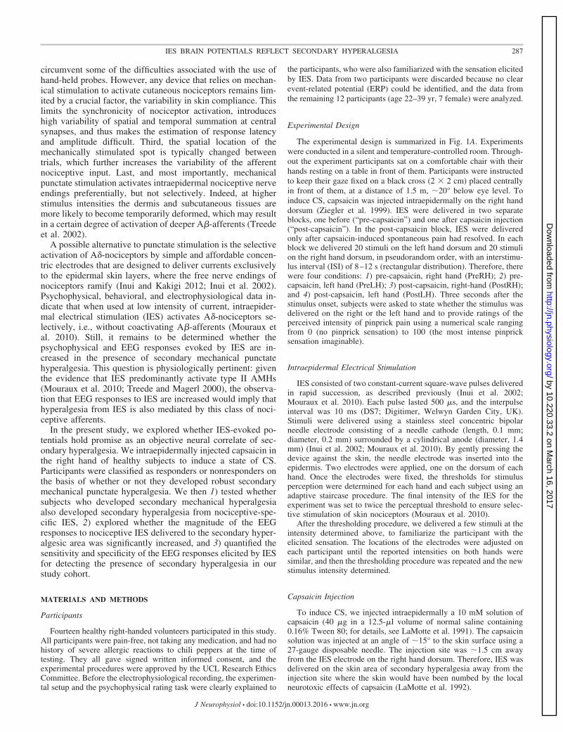
circumvent some of the difficulties associated with the use ofhand-held probes. However, any device that relies on mechan-ical stimulation to activate cutaneous nociceptors remains lim-ited by a crucial factor, the variability in skin compliance. Thislimits the synchronicity of nociceptor activation, introduceshigh variability of spatial and temporal summation at centralsynapses, and thus makes the estimation of response latencyand amplitude difficult. Third, the spatial location of themechanically stimulated spot is typically changed betweentrials, which further increases the variability of the afferentnociceptive input. Last, and most importantly, mechanicalpunctate stimulation activates intraepidermal nociceptive nerveendings preferentially, but not selectively. Indeed, at higherstimulus intensities the dermis and subcutaneous tissues aremore likely to become temporarily deformed, which may resultin a certain degree of activation of deeper A�-afferents (Treedeet al. 2002).
A possible alternative to punctate stimulation is the selectiveactivation of A�-nociceptors by simple and affordable concen-tric electrodes that are designed to deliver currents exclusivelyto the epidermal skin layers, where the free nerve endings ofnociceptors ramify (Inui and Kakigi 2012; Inui et al. 2002).Psychophysical, behavioral, and electrophysiological data in-dicate that when used at low intensity of current, intraepider-mal electrical stimulation (IES) activates A�-nociceptors se-lectively, i.e., without coactivating A�-afferents (Mouraux etal. 2010). Still, it remains to be determined whether thepsychophysical and EEG responses evoked by IES are in-creased in the presence of secondary mechanical punctatehyperalgesia. This question is physiologically pertinent: giventhe evidence that IES predominantly activate type II AMHs(Mouraux et al. 2010; Treede and Magerl 2000), the observa-tion that EEG responses to IES are increased would imply thathyperalgesia from IES is also mediated by this class of noci-ceptive afferents.
In the present study, we explored whether IES-evoked po-tentials hold promise as an objective neural correlate of sec-ondary hyperalgesia. We intraepidermally injected capsaicin inthe right hand of healthy subjects to induce a state of CS.Participants were classified as responders or nonresponders onthe basis of whether or not they developed robust secondarymechanical punctate hyperalgesia. We then 1) tested whethersubjects who developed secondary mechanical hyperalgesiaalso developed secondary hyperalgesia from nociceptive-spe-cific IES, 2) explored whether the magnitude of the EEGresponses to nociceptive IES delivered to the secondary hyper-algesic area was significantly increased, and 3) quantified thesensitivity and specificity of the EEG responses elicited by IESfor detecting the presence of secondary hyperalgesia in ourstudy cohort.
MATERIALS AND METHODS
Participants
Fourteen healthy right-handed volunteers participated in this study.All participants were pain-free, not taking any medication, and had nohistory of severe allergic reactions to chili peppers at the time oftesting. They all gave signed written informed consent, and theexperimental procedures were approved by the UCL Research EthicsCommittee. Before the electrophysiological recording, the experimen-tal setup and the psychophysical rating task were clearly explained to
the participants, who were also familiarized with the sensation elicitedby IES. Data from two participants were discarded because no clearevent-related potential (ERP) could be identified, and the data fromthe remaining 12 participants (age 22–39 yr, 7 female) were analyzed.
Experimental Design
The experimental design is summarized in Fig. 1A. Experimentswere conducted in a silent and temperature-controlled room. Through-out the experiment participants sat on a comfortable chair with theirhands resting on a table in front of them. Participants were instructedto keep their gaze fixed on a black cross (2 � 2 cm) placed centrallyin front of them, at a distance of 1.5 m, �20° below eye level. Toinduce CS, capsaicin was injected intraepidermally on the right handdorsum (Ziegler et al. 1999). IES were delivered in two separateblocks, one before (“pre-capsaicin”) and one after capsaicin injection(“post-capsaicin”). In the post-capsaicin block, IES were deliveredonly after capsaicin-induced spontaneous pain had resolved. In eachblock we delivered 20 stimuli on the left hand dorsum and 20 stimulion the right hand dorsum, in pseudorandom order, with an interstimu-lus interval (ISI) of 8–12 s (rectangular distribution). Therefore, therewere four conditions: 1) pre-capsaicin, right hand (PreRH); 2) pre-capsaicin, left hand (PreLH); 3) post-capsaicin, right-hand (PostRH);and 4) post-capsaicin, left hand (PostLH). Three seconds after thestimulus onset, subjects were asked to state whether the stimulus wasdelivered on the right or the left hand and to provide ratings of theperceived intensity of pinprick pain using a numerical scale rangingfrom 0 (no pinprick sensation) to 100 (the most intense pinpricksensation imaginable).
Intraepidermal Electrical Stimulation
IES consisted of two constant-current square-wave pulses deliveredin rapid succession, as described previously (Inui et al. 2002;Mouraux et al. 2010). Each pulse lasted 500 �s, and the interpulseinterval was 10 ms (DS7; Digitimer, Welwyn Garden City, UK).Stimuli were delivered using a stainless steel concentric bipolarneedle electrode consisting of a needle cathode (length, 0.1 mm;diameter, 0.2 mm) surrounded by a cylindrical anode (diameter, 1.4mm) (Inui et al. 2002; Mouraux et al. 2010). By gently pressing thedevice against the skin, the needle electrode was inserted into theepidermis. Two electrodes were applied, one on the dorsum of eachhand. Once the electrodes were fixed, the thresholds for stimulusperception were determined for each hand and each subject using anadaptive staircase procedure. The final intensity of the IES for theexperiment was set to twice the perceptual threshold to ensure selec-tive stimulation of skin nociceptors (Mouraux et al. 2010).
After the thresholding procedure, we delivered a few stimuli at theintensity determined above, to familiarize the participant with theelicited sensation. The locations of the electrodes were adjusted oneach participant until the reported intensities on both hands weresimilar, and then the thresholding procedure was repeated and the newstimulus intensity determined.
Capsaicin Injection
To induce CS, we injected intraepidermally a 10 mM solution ofcapsaicin (40 �g in a 12.5-�l volume of normal saline containing0.16% Tween 80; for details, see LaMotte et al. 1991). The capsaicinsolution was injected at an angle of �15° to the skin surface using a27-gauge disposable needle. The injection site was �1.5 cm awayfrom the IES electrode on the right hand dorsum. Therefore, IES wasdelivered on the skin area of secondary hyperalgesia away from theinjection site where the skin would have been numbed by the localneurotoxic effects of capsaicin (LaMotte et al. 1992).
Capsaicin-Induced Spontaneous Pain and Secondary HyperalgesiaAssessment
Spontaneous pain intensity after capsaicin injection was recordedusing a numerical rating scale ranging between 0 (no pain) and 100(worst pain imaginable). Participants were required to rate verballythe intensity of spontaneous pain every 10 s during the first 3 min andthen every 30 s until the pain intensity ratings were less than 5 out of100.
The development of mechanical hyperalgesia in the skin areasurrounding the injection site was confirmed by punctate mechanicalstimulation of the skin adjacent (within 1 cm) to the external circum-ference of the concentric IES electrode using a flat-tip punctate probe(256 mN). This probe comprises a stainless steel wire tip (diameter,0.25 mm) attached to a mounted weight (256 mN) that glides
smoothly within a hollow handheld cylindrical tube. When the probeis applied perpendicularly to the skin, its weight rests entirely on thewire tip, thus exerting a constant force of 256 mN. More details anda depiction of the punctate probe can be found in a previous report(Iannetti et al. 2013), as well as on the manufacturer website (MRCSystems; http://www.mrc-systems.de/en/products/pinprick). The samemechanical stimulus was applied to the corresponding position of the lefthand, to obtain a baseline for quantifying the effect of secondaryhyperalgesia, as follows. Participants were asked to report the inten-sity of punctate stimulation of the right hand (capsaicin injected) andof the left hand (control) using a numerical rating scale that rangedbetween 0 (no pinprick sensation) and 100 (the most intense pinpricksensation imaginable). For each hand, punctate stimuli were appliedthree times, with an ISI of �5 s, after the spontaneous pain induced
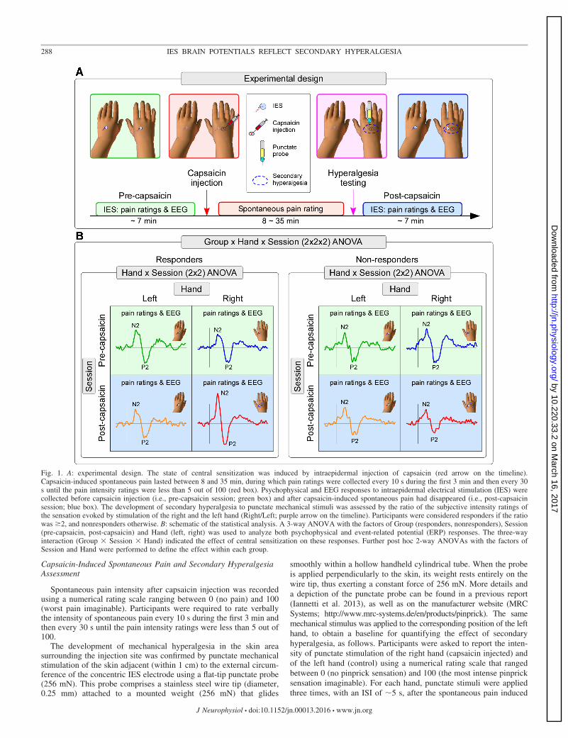
Fig. 1. A: experimental design. The state of central sensitization was induced by intraepidermal injection of capsaicin (red arrow on the timeline).Capsaicin-induced spontaneous pain lasted between 8 and 35 min, during which pain ratings were collected every 10 s during the first 3 min and then every 30s until the pain intensity ratings were less than 5 out of 100 (red box). Psychophysical and EEG responses to intraepidermal electrical stimulation (IES) werecollected before capsaicin injection (i.e., pre-capsaicin session; green box) and after capsaicin-induced spontaneous pain had disappeared (i.e., post-capsaicinsession; blue box). The development of secondary hyperalgesia to punctate mechanical stimuli was assessed by the ratio of the subjective intensity ratings ofthe sensation evoked by stimulation of the right and the left hand (Right/Left; purple arrow on the timeline). Participants were considered responders if the ratiowas �2, and nonresponders otherwise. B: schematic of the statistical analysis. A 3-way ANOVA with the factors of Group (responders, nonresponders), Session(pre-capsaicin, post-capsaicin) and Hand (left, right) was used to analyze both psychophysical and event-related potential (ERP) responses. The three-wayinteraction (Group � Session � Hand) indicated the effect of central sensitization on these responses. Further post hoc 2-way ANOVAs with the factors ofSession and Hand were performed to define the effect within each group.
by the capsaicin injection in the right hand had decreased to less than5 out of 100 (Fig. 2). For every individual, the mean ratings of thesensations elicited by the three stimuli was obtained for each hand andeach condition. The intensity of secondary hyperalgesia was quanti-fied as the ratio of the subjective ratings of the pinprick sensationelicited by mechanical stimulation of the right and the left hands(Right/Left). Participants were considered to have developed robustsecondary hyperalgesia from punctate stimuli if the ratio was �2 andwere thus classified as responders. All other participants were classi-fied as nonresponders. This ratio was chosen on the basis of a previousEEG study, which showed that an approximately twofold increase(�93%) in pinprick sensation elicited by punctate stimulation aftercapsaicin sensitization was associated with significant increases in theevoked EEG response (Iannetti et al. 2013).
EEG Recording
The EEG was recorded using 31 Ag-AgCl electrodes placed on thescalp according to the International 10-20 system and referenced tothe nose. Ocular movements and eye blinks were recorded using twosurface electrodes, one placed over the right lower eyelid and the otherplaced �1 cm lateral to the lateral corner of the right orbit. Signalswere amplified and digitized using a sampling rate of 1,024 Hz (SD32;Micromed, Mogliano Veneto, Italy).
Behavioral Data Analysis
Single-trial ratings of the sensation elicited by IES were firstnormalized between 0 and 100 for each participant (the minimumvalue was set to 0 and the maximum value was set to 100). Thisprocedure mitigates the differences in the range of values on thenumerical rating scale with which individuals reported the intensity ofpinprick pain elicited by IES (Huang et al. 2013). Normalized stim-
ulus intensity ratings were subsequently averaged across trials foreach condition, resulting in four average values for each participant(PreRH, PreLH, PostRH, and PostLH).
To test whether capsaicin injection had an effect on the perceivedIES intensity, we performed a three-way ANOVA with the followingexperimental factors: Group (2 levels: responders, nonresponders),Session (2 levels: pre-capsaicin, post-capsaicin), and Hand [2 levels:injected (right), control (left)]. Where effects were significant, posthoc analyses were performed to define their direction and possibleinteractions. Two-way repeated-measures ANOVA for the main andinteraction effects of Session and Hand were performed to define theeffects of capsaicin injection on the intensity of the sensation elicitedby IES within each group. The statistical threshold of the post hocanalyses was determined by Bonferroni correction accounting for thenumber of comparisons (P � 0.05/2 � 0.025).
EEG Data Analyses
EEG data analyses were performed using Letswave (www.nocions.org; Mouraux and Iannetti 2008) and MATLAB (The MathWorks,Natick, MA). Continuous EEG recordings were segmented into ep-ochs using a time window of 2 s (�0.5 to 1.5 s relative to the stimulusonset). Each epoch was baseline corrected (baseline interval rangingfrom �0.2 to 0 s) and bandpass filtered (1–30 Hz). Artifacts producedby eye blinks or eye movements were subtracted using a validatedmethod based on independent component analysis (Jung et al. 2000).In all data sets, independent components related to eye movementshad a large electrooculogram channel contribution and a frontal scalpdistribution. In addition, epochs with amplitude values exceeding�100 �V were rejected from further analysis. These epochs consti-tuted 0.6 � 1.8% (mean � SD across all conditions and participants)of the total number of epochs. Remaining epochs were then averaged
Fig. 2. A: participants were divided into 2groups according to the ratio of ratings topunctate stimulation of the right (R) and left(L) hands: participants who rated the intensityof right hand stimulation as at least twice thatof the left hand stimulation were classified asresponders. Participants were sorted by theratio of reported intensity ratings (R/L), indescending order. B: time course of capsai-cin-induced pain ratings. Single participantsare color coded. Solid lines indicate respond-ers. All participants rated the pain intensitybetween 90 and 100 at the moment of theinjection. Pain ratings decreased fairlyquickly over time. Inset: comparison of themean area under the curve (AUC) betweenresponders and nonresponders revealed nosignificant difference (t10 � 0.39, P � 0.70).Colored symbols indicate single-subject AUCdata.
for each condition, resulting in four average ERP waveforms for eachparticipant.
The N2-P2 complex was measured at the vertex (Cz), and it wasdefined as the largest negative-positive deflection occurring afterstimulus onset. The amplitude of both the N2 and P2 peaks werecalculated for each condition and participant and then tested for theeffect of capsaicin injection using the same three-way ANOVAdescribed for the behavioral data (Fig. 1B). Because two peaks (N2and P2) were tested, the statistical threshold was determined byBonferroni correction accounting for the number of peaks (P � 0.05/2peaks � 0.025). Where effects were significant, the same post hocanalyses described for the behavioral data (i.e., 2-way repeated-measures ANOVA) were performed for each group, and the samestatistical threshold, Bonferroni corrected (P � 0.05/2 groups �0.025), was used to determine the significance of the post hoc results.The latency of the N2 and P2 peaks was analyzed using the sameprocedure.
To test the predictive value of ERP amplitude for the presence ofCS, we plotted the receiver operating characteristic (ROC) curvesobtained using the interaction term [i.e., (PostRH � PreRH) �(PostLH � PreLH)] calculated for the N2-wave and P2-wave peakamplitudes. The true positive rate (sensitivity) is plotted against thefalse positive rate (100 � specificity) for different cutoff values of theinteraction terms. Each point on the ROC curve represents a sensitiv-ity/specificity pair corresponding to a particular decision threshold forthe interaction term. Above each of these thresholds, the individual ispredicted to be a responder, and vice versa. If interaction terms hadperfect classification performance, their ROC curves would passthrough the upper left corner (100% sensitivity, 100% specificity).The closer the ROC curve is to the upper left corner, the higher theoverall accuracy of the interaction term is in distinguishing respondersand nonresponders (Zweig and Campbell 1993). The area under thecurve (AUC) is typically used to quantify the classification perfor-mance. An AUC value of 0.5 corresponds to a random classification(i.e., to a useless test), whereas an AUC of 1.0 indicates that the testperforms perfectly. We calculated the AUC for the interaction termsobtained from the amplitude of the N2 and P2 peaks to assess theirsensitivity and specificity for detecting the presence of a CS state. Wetested whether the AUC size of each measure was significantly greaterthan 0.5 (Hanley and McNeil 1982).
RESULTS
Capsaicin-Induced Spontaneous Pain
Six of 12 participants developed robust secondary hyperal-gesia on the capsaicin-treated hand and were therefore classi-fied as responders (Fig. 2A). The time courses of the capsaicin-induced pain for all subjects are shown in Fig. 2B. In the firstfew seconds after the injection, capsaicin induced a veryintense sensation of burning pain, which decreased exponen-tially over time (Lee et al. 2008; Magerl et al. 1998). The timecourse of spontaneous pain ratings for each subject was sum-marized as AUC. The AUC values for responders and nonre-sponders were compared using a two-sample t-test. The resultshowed no significant difference in capsaicin-induced sponta-neous pain between the two groups (t10 � 0.39, P � 0.70; Fig.2B, inset). This observation suggests that both groups per-ceived the conditioning stimulus (i.e., the intraepidermal injec-tion of capsaicin) similarly.
Psychophysics of Intraepidermal Stimulation of the Area ofSecondary Mechanical Punctate Hyperalgesia
All subjects correctly reported whether each IES was deliv-ered to the right or the left hand in all trials. The three-way
ANOVA on the subjective ratings of perceived IES intensityshowed a two-way interaction between Group and Hand(F1,10 � 9.02, P � 0.01) and, more importantly, a clearthree-way interaction between Group, Session, and Hand(F1,10 � 59.27, P � 0.000016; Fig. 3). No other significanteffects were detected (Table 1). This finding indicates thatresponders perceived right-hand stimulation as more painfulthan left-hand stimulation but only after capsaicin was injectedin the right hand. The results of all post hoc two-way ANOVAsare shown in Table 2. Both responders (F1,5 � 49.79, P �0.001) and nonresponders (F1,5 � 15.19, P � 0.01) showedsignificant interactions between Session and Hand, but inopposite directions: the responders had clearly increased rat-ings on their treated hand after capsaicin injection, whereas thenonresponders showed mildly decreased ratings on theirtreated hand after capsaicin injection (Fig. 3). The results
Fig. 3. Subjective intensity ratings of the sensation elicited by the IES ofresponders (left) and nonresponders (right). A: to highlight the interactionbetween the factors Session and Hand, the subtracted ratings (post- minuspre-capsaicin injection) are shown for each hand. Colored circles indicatesingle subjects, and black circles indicate the group average for each condition.Two-way ANOVA revealed that responders had a highly significant interac-tion between the factors Session and Hand. This reveals a capsaicin-inducedincrease of IES ratings (Post � Pre) on the right hand. In contrast, in nonre-sponders the 2-way ANOVA revealed a decrease of IES ratings on the right handcompared with those on the left hand. These differences in the capsaicin effect onIES ratings between responders and nonresponders were confirmed by the3-way ANOVA, which revealed a highly significant triple interaction(Group � Session � Hand; comparison between left and right panels). LH, lefthand; RH, right hand. B: individual values (colored circles) and mean value(black circles) for each condition. PreLH, pre-capsaicin, left hand; PostLH:post-capsaicin, left hand; PreRH: pre-capsaicin, right hand; PostRH: post-capsaicin, right hand.
demonstrate a clear secondary hyperalgesia from both IES andmechanical punctate stimulation after capsaicin injection.
ERP Waveforms
ERPs elicited by IES stimuli showed a clear N2–P2 complexmaximal at electrode Cz in all four conditions of each group.Grand-average waveforms and scalp maps at N2 and P2 peaklatencies are shown in Fig. 4. The ERP amplitude increasedafter capsaicin injection in the right hand of the responderscompared with all other conditions. Statistical comparisons ofpeak amplitude and latency of the N2 and P2 waves acrossdifferent conditions and groups are reported below and sum-marized in Tables 1 and 2.
N2 peak amplitude. The three-way ANOVA of N2 peakamplitudes showed a three-way interaction between Group,Session, and Hand (F1,10 � 7.84, P � 0.019). No othersignificant effects were detected (Table 1). Hence, N2 peakamplitudes at Cz were greater following right-hand IES com-pared with left-hand IES in the responders, but only when IESwere delivered to the hand where capsaicin had been injected(i.e., the right hand). Post hoc two-way ANOVAs (Table 2)revealed that only responders showed an interaction betweenSession and Hand (F1,5 � 15.15, P � 0.011), indicating
increased N2 amplitudes on their treated hand after capsaicininjection. Figure 5 shows the single subjects’ N2 peak ampli-tudes, as well as the statistical results.
P2 peak amplitude. The three-way ANOVA of P2 peakamplitudes showed that there was a two-way interaction be-tween Group and Session (F1,10 � 11.13, P � 0.008). Thiseffect was caused by an overall increased P2 amplitude in thepost-capsaicin session of responders but a decreased P2 am-plitude in the post-capsaicin session of nonresponders. Noother significant effects were detected (Table 1). Post hoctwo-way ANOVAs (Table 2) showed that there was a trend foran interaction between Session and Hand that, however, did notsurvive correction for multiple comparisons in responders(F1,5 � 9.77, P � 0.026): in this group, P2 amplitudes in thepost-capsaicin session, compared with those in the pre-capsa-icin session, were increased following right-hand stimulationand slightly decreased following left-hand stimulation.
N2 peak latency. The three-way ANOVA of N2 peak laten-cies showed a main effect of Hand (F1,10 � 7.41, P � 0.022).No other significant effects were detected (Table 1). Post hoctwo-way ANOVAs (Table 2) failed to detect any effects ineither responders or nonresponders that survived correction formultiple comparisons.
Table 1. Results of 3-way ANOVA of psychophysical and EEG responses elicited by IES
3-Way ANOVA Pain Intensity Ratings
ERP Peak Amplitude ERP Peak Latency
N2 P2 N2 P2
Main effect of Group F1,10 � 0.20 F1,10 � 0.08 F1,10 � 0.15 F1,10 � 0.008 F1,10 � 1.51P � 0.665 P � 0.778 P � 0.705 P � 0.930 P � 0.247
Main effect of Session F1,10 � 0.05 F1,10 � 0.268 F1,10 � 2.65 F1,10 � 0.50 F1,10 � 0.38P � 0.833 P � 0.61 P � 0.134 P � 0.498 P � 0.553
Main effect of Hand F1,10 � 4.52 F1,10 � 1.60 F1,10 � 0.52 F1,10 � 7.41 F1,10 � 0.11P � 0.059 P � 0.234 P � 0.487 P � 0.022 P � 0.742
2-way interaction: Group � Session F1,10 � 3.04 F1,10 � 1.40 F1,10 � 11.13 F1,10 � 0.05 F1,10 � 0.53P � 0.112 P � 0.265 P � 0.008 P � 0.827 P � 0.484
2-way interaction: Group � Hand F1,10 � 9.02 F1,10 � 0.45 F1,10 � 1.42 F1,10 � 0.11 F1,10 � 0.0003P � 0.013 P � 0.517 P � 0.261 P � 0.751 P � 0.987
2-way interaction: Session � Hand F1,10 � 4.31 F1,10 � 0.19 F1,10 � 1.27 F1,10 � 4.33 F1,10 � 1.19P � 0.065 P � 0.674 P � 0.286 P � 0.064 P � 0.301
3-way interaction: Group � Session � Hand F1,10 � 59.27 F1,10 � 7.84 F1,10 � 2.04 F1,10 � 0.37 F1,10 � 0.06P � 0.000016 P � 0.019 P � 0.184 P � 0.559 P � 0.813
Data are F statistics and P values from 3-way ANOVA with factors Group, Session, and Hand. Significant effects are highlighted in bold.
Table 2. Psychophysical and EEG responses elicited by IES for each condition and results of post hoc 2-way ANOVA for each group
Data are normalized pain intensity ratings, peak amplitudes, and peak latency in each condition, as well as F statistics and P values from post hoc 2-wayANOVA with factors Session and Hand, for each group. Significant effects are highlighted in bold.
P2 peak latency. The three-way ANOVA on the P2 peaklatencies did not detect any significant effect. Therefore, posthoc analyses were not performed.
ROC curves. The ROC curves obtained from N2 and P2peak amplitudes are plotted in Fig. 6. The AUC values (�SE)for N2 and P2 were 0.92 � 0.09 and 0.72 � 0.16, respectively.Only the AUC for N2 was significantly greater than 0.5 (N2:P � 0.016; P2: P � 0.200). This suggests that the N2 peakamplitude has adequate sensitivity and specificity for detectingthe presence of CS induced by intraepidermal injection ofcapsaicin.
DISCUSSION
Developing a biomarker for secondary hyperalgesia, a car-dinal symptom of central sensitization (CS), would be usefulfor both drug discovery and clinical therapy. Such a biomarkerwould help analgesic drug discovery in early phase trials,facilitate diagnosis of neuropathic pain, and allow objectivemonitoring of drug treatments in patients.
IES is a technically simple and inexpensive method toselectively stimulate II-AMH skin nociceptors (Inui and Kakigi2012; Inui et al. 2002; Mouraux et al. 2010). Importantly, IESelicits clear time-locked EEG responses, thus allowing quan-tification of CS. However, mechanical punctate hyperalgesia isknown to be mediated by I-AMH units, rather than II-AMHunits (Magerl et al. 2001). Given that IES selectively activatesII-AMH units (Mouraux et al. 2010), we tested 1) whethersecondary hyperalgesia from IES coexists with secondary me-chanical punctate hyperalgesia and 2) whether such hyperal-
gesia is reflected in a corresponding increase in EEG re-sponses.
We obtained several interesting results. First, the intensity ofthe sensation elicited by IES was significantly increased afterintraepidermal injection of capsaicin in those participants whodeveloped robust mechanical punctate hyperalgesia, clearlyshowing that hyperalgesia from IES occurs and coexists withmechanical hyperalgesia. Second, the peak amplitude of the N2wave elicited by IES was significantly increased in responders,similarly to the intensity of the sensation elicited by IES. Thisincreased response only occurred when IES were delivered tothe hand where capsaicin was injected. Third, ROC analysisshowed that the N2 peak amplitude offers the ability to predictthe presence of CS with high sensitivity and specificity. Thesefindings suggest that the EEG responses elicited by IES reflectsecondary hyperalgesia and thus are a reliable neural correlateof CS.
Although our observations clearly indicate that secondaryhyperalgesia elicited by IES coexists with secondary hyperal-gesia elicited by mechanical punctate stimuli, it remains un-clear whether the two phenomena are mediated by similarpopulations of A�-nociceptors. There is strong physiologicalevidence that secondary mechanical punctate hyperalgesia ismediated by I-AMH nociceptors. For example, Magerl et al.(2001) demonstrated that secondary mechanical punctate hy-peralgesia still occurred in skin that was rendered devoid of
Fig. 4. Group-average ERP waveforms and scalp maps elicited by IES in responders (A) and nonresponders (B). Waveforms at the channel Cz in differentconditions are shown in different colors. The ERP elicited by IES stimuli clearly increased after capsaicin injection only on the right hand in responders. Scalpmaps at the N2 peak latencies show a central distribution, slightly lateralized to the hemisphere contralateral to the stimulated hand, maximal at the vertex (top).Scalp maps at the P2 peak latencies show a central distribution, maximal at the vertex (bottom). Color bar shows the ERP amplitude in scalp maps.
II-AMH epidermal terminals by application of high concentra-tions of topical capsaicin. In contrast, Mouraux et al. (2010)showed that both sensations and EEG responses elicited by IESwere abolished in skin that was similarly treated with high-concentration capsaicin, suggesting that IES activates mostlyII-AMH nociceptors. It follows that the secondary hyperalgesiafrom IES observed in this study is likely to be mediated mainly
by II-AMH rather than I-AMH nociceptors. However, furtherexperiments are required to confirm whether hyperalgesia fromIES and mechanical punctate stimulation are truly mediated bydifferent populations of A�-afferents. Nonetheless, it is plau-sible that, after capsaicin injection, inputs from both I-AMHand II-AMH nociceptors are heterosynaptically facilitated via acommon central mechanism and account for the coexistence ofsecondary hyperalgesia from IES and mechanical punctatestimulation (Ziegler et al. 1999).
Variability in Capsaicin-Induced Secondary Hyperalgesia
We observed considerable variability in the degree of punc-tate hyperalgesia that developed after intraepidermal capsaicininjection. Only half of the subjects developed robust hyperal-gesia (i.e., a 2-fold increase of pain ratings when the injectedhand was stimulated compared with the control hand; Fig. 2).
It is unlikely that this difference between responders andnonresponders was related to the strength of conditioningstimulus, i.e., the activation of C-nociceptors by intraepidermalinjection of capsaicin. Indeed, both groups reported similarintensities and durations of burning pain following intraepider-mal injection of capsaicin, which suggests that the conditioningstimulus was similar. We note that the development of second-ary hyperalgesia can be highly variable even with a highlystandardized electrical conditioning stimulus, which suggestsconsiderable differences in the development of CS responsesbetween individuals (Pfau et al. 2011). Furthermore, there isclear evidence that genetic variability contributes to variabilityin hyperalgesic response following intraepidermal capsaicininjection (Tegeder et al. 2008).
Brain Potentials Evoked by IES and Central Sensitization:Advantages and Limitations
Previous studies have suggested that brain potentials elicitedby punctate mechanical stimulation may be recorded andemployed as a potential objective correlates of the CS states(Davies et al. 2010; Iannetti et al. 2013; Kohlloffel et al. 1991).However, as detailed in the Introduction, evoked potentialselicited by punctate mechanical stimuli have significant tech-nical and physiological constrains that hamper clinical trans-lation.
In contrast, IES have several advantages over mechanicalpunctate stimulation. When delivered at low currents, they arefully selective for A�-nociceptors and allow for accurate tim-ing and standardization of stimuli. The stimulating electrode is
Fig. 5. ERP amplitudes (N2) of IES of responders (left) and nonresponders(right). A: to highlight the interaction between Session and Hand in each group,the subtracted ERP amplitudes (post- minus pre-capsaicin injection) are shownfor each hand. Colored circles indicate single subjects, and black circlesindicate the group average for each condition. Two-way ANOVA revealed thatresponders had a significant interaction between the factors Session and Hand.This reveals a capsaicin-induced increase of IES ERP amplitudes (Post-Pre) onthe right hand. In contrast, in nonresponders the 2-way ANOVA did not showany significant effect. These differences in the capsaicin effect on ERPamplitudes between responders and nonresponders were confirmed by the3-way ANOVA, which revealed a significant triple interaction (Group �Session � Hand; comparison between left and right panels). B: individualvalues (colored circles) and mean value (black circles) for each condition.
Fig. 6. Receiver operating characteristic (ROC) curvesand their corresponding AUC values obtained using theinteraction term for N2 peak amplitude (left) and P2peak amplitude (right) as the predictive factor. Al-though both measures show predictive ability, only theAUC of N2 ROC was significantly greater than 0.5,indicating that it is therefore a predictor for the state ofcentral sensitization.
affordable and can be affixed to any part of the body withoutdifficulty.
The current results show that the amplitude of the ERPelicited by IES of the skin with secondary hyperalgesia clearlyreflects that the somatosensory system is centrally sensitized.The amplitude of the N2 wave was significantly larger whenIES were delivered to the hand in which capsaicin injectionresulted in a clear secondary hyperalgesia (Figs. 4 and 5,Tables 1 and 2). Moreover, the areas under the ROC curvesindicate that the change in N2 peak amplitude was significantlypredictive of the presence of secondary hyperalgesia (Fig. 6).This result suggests that the changes in N2 amplitude may bedeveloped as a potentially useful biomarker of CS.
Several limitations to IES remain. First, we were unable toisolate the early, contralateral N1 wave typically observed inthe brain potentials evoked by nociceptive laser stimuli (Treedeet al. 1988; Valentini et al. 2012), most likely because of itslower signal-to-noise ratio. Compared with the subsequentN2-P2 complex, the N1 wave has been shown to better reflectthe afferent nociceptive drive (Lee et al. 2009) and appears lesssusceptible to top-down modulation (for example, placebomanipulation; Martini et al. 2015). These characteristics makethe N1 wave a potentially more robust marker for centralsensitization. Second, the selective activation of A�-nocicep-tors by IES relies on the use of strictly low-intensity currents.This limitation prevents the recording of stimulus-responsefunctions, because higher intensity currents necessarily entail acoactivation of tactile A�-afferents, and therefore a loss ofspecificity for A�-fiber stimulation (Mouraux et al. 2010).Stimulus-response functions are particularly useful for assess-ing the analgesic potential of novel drugs because they candivulge interactions between stimulus or pain intensity anddose effects. Recording of stimulus-response function usingthe brain response elicited by mechanical punctate stimuli issimilarly problematic because, as detailed earlier, when highforces are exerted, the mechanical punctate stimulus becomesless selective for A�-fiber activation (Mouraux et al. 2010;Treede et al. 2002). More recent data reveal that stimulus-response functions can be constructed with the use of IES byvarying the number of pulses delivered in quick succession(5-ms intervals) to normal skin; increasing the number ofpulses increases the intensity of sensation and EEG amplitudeswithout changing reaction times or response latencies(Mouraux et al. 2014). Further experiments are required toascertain if this remains the case after capsaicin-induced hy-peralgesia. Moreover, although our present results suggest thepotential usefulness of EEG responses to IES as an objectivemeasure of CS, the small sample size used in the present studylimits statistical power for detection of smaller effects. Futurestudies with large samples are needed to confirm the predictivevalue of IES brain potentials for the state of CS.
Conclusion
Our study demonstrates that secondary hyperalgesia to IESoccurs in a well-recognized experimental model of CS and thatthe subjective report was corroborated by increased evokedEEG responses. These findings suggest that EEG responseselicited by low-intensity IES, particularly the change in thepeak amplitude of the N2 wave, can be used as an objective,physiological correlate of secondary hyperalgesia. Hence, IES
evoked potentials hold promise as a low-cost, noninvasivebiomarker for CS that can be translated for clinical use withrelative ease compared with existing techniques.
GRANTS
This work was funded by the Wellcome Trust Pain Consortium AwardCOLL JLARAXR (to G. D. Iannetti and A. H. Dickenson), the EuropeanResearch Council Consolidator Grant PAINSTRAT (to G. D. Iannetti), a UCLGrand Challenges Studentship (to J. O’Neill), National Natural Science Foun-dation of China Grant 81571659 (to M. Liang), and Natural Science Founda-tion of Tianjin Grant 15JCYBJC55100 (to M. Liang).
DISCLOSURES
No conflicts of interest, financial or otherwise, are declared by the authors.
AUTHOR CONTRIBUTIONS
M.L., M.C.L., A.H.D., and G.D.I. conception and design of research; M.L.,M.C.L., J.O., and G.D.I. performed experiments; M.L., M.C.L., and J.O.analyzed data; M.L., M.C.L., J.O., A.H.D., and G.D.I. interpreted results ofexperiments; M.L. and G.D.I. prepared figures; M.L., M.C.L., and G.D.I.drafted manuscript; M.L., M.C.L., A.H.D., and G.D.I. edited and revisedmanuscript; M.L., M.C.L., J.O., A.H.D., and G.D.I. approved final version ofmanuscript.
REFERENCES
Davies EK, Boyle Y, O’Donnell M, Dalton Z, Lumb BM, Murrell JC,Chizh BA. Evaluation of somatic mechanical-evoked potentials (MEPS) asobjective neurophysiological markers of pain. 13th World Congress onPain, Montreal, Quebec, Canada, 2010.
Geber C, Fondel R, Kramer HH, Rolke R, Treede RD, Sommer C,Birklein F. Psychophysics, flare, and neurosecretory function in human painmodels: capsaicin versus electrically evoked pain. J Pain 8: 503–514, 2007.
Hanley JA, McNeil BJ. The meaning and use of the area under a receiveroperating characteristic (ROC) curve. Radiology 143: 29–36, 1982.
Huang G, Xiao P, Hung YS, Iannetti GD, Zhang ZG, Hu L. A novelapproach to predict subjective pain perception from single-trial laser-evokedpotentials. Neuroimage 81: 283–293, 2013.
Iannetti GD, Baumgartner U, Tracey I, Treede RD, Magerl W. Pinprick-evoked brain potentials: a novel tool to assess central sensitization ofnociceptive pathways in humans. J Neurophysiol 110: 1107–1116, 2013.
Inui K, Kakigi R. Pain perception in humans: use of intraepidermal electricalstimulation. J Neurol Neurosurg Psychiatry 83: 551–556, 2012.
Inui K, Tran TD, Hoshiyama M, Kakigi R. Preferential stimulation of A�fibers by intra-epidermal needle electrode in humans. Pain 96: 247–252,2002.
Ji RR, Kohno T, Moore KA, Woolf CJ. Central sensitization and LTP: dopain and memory share similar mechanisms? Trends Neurosci 26: 696–705,2003.
Jung TP, Makeig S, Humphries C, Lee TW, McKeown MJ, Iragui V,Sejnowski TJ. Removing electroencephalographic artifacts by blind sourceseparation. Psychophysiology 37: 163–178, 2000.
Kohlloffel LU, Koltzenburg M, Handwerker HO. A novel technique for theevaluation of mechanical pain and hyperalgesia. Pain 46: 81–87, 1991.
LaMotte RH, Lundberg LE, Torebjork HE. Pain, hyperalgesia and activityin nociceptive C units in humans after intradermal injection of capsaicin. JPhysiol 448: 749–764, 1992.
LaMotte RH, Shain CN, Simone DA, Tsai EF. Neurogenic hyperalgesia:psychophysical studies of underlying mechanisms. J Neurophysiol 66:190–211, 1991.
Latremoliere A, Woolf CJ. Central sensitization: a generator of pain hyper-sensitivity by central neural plasticity. J Pain 10: 895–926, 2009.
Lee MC, Mouraux A, Iannetti GD. Characterizing the cortical activitythrough which pain emerges from nociception. J Neurosci 29: 7909–7916,2009.
Lee MC, Zambreanu L, Menon DK, Tracey I. Identifying brain activityspecifically related to the maintenance and perceptual consequence ofcentral sensitization in humans. J Neurosci 28: 11642–11649, 2008.
Magerl W, Fuchs PN, Meyer RA, Treede RD. Roles of capsaicin-insensitivenociceptors in cutaneous pain and secondary hyperalgesia. Brain 124:1754–1764, 2001.
Magerl W, Wilk SH, Treede RD. Secondary hyperalgesia and perceptualwind-up following intradermal injection of capsaicin in humans. Pain 74:257–268, 1998.
Maihofner C, Jesberger F, Seifert F, Kaltenhauser M. Cortical processingof mechanical hyperalgesia: a MEG study. Eur J Pain 14: 64–70, 2010.
Martini M, Lee MC, Valentini E, Iannetti GD. Intracortical modulation, andnot spinal inhibition, mediates placebo analgesia. Eur J Neurosci 41:498–504, 2015.
Mouraux A, Iannetti GD. Across-trial averaging of event-related EEGresponses and beyond. Magn Reson Imaging 26: 1041–1054, 2008.
Mouraux A, Iannetti GD, Plaghki L. Low intensity intra-epidermal electricalstimulation can activate A�-nociceptors selectively. Pain 150: 199–207, 2010.
Mouraux A, Marot E, Legrain V. Short trains of intra-epidermal electricalstimulation to elicit reliable behavioral and electrophysiological responses to theselective activation of nociceptors in humans. Neurosci Lett 561: 69–73, 2014.
Pfau DB, Klein T, Putzer D, Pogatzki-Zahn EM, Treede RD, Magerl W.Analysis of hyperalgesia time courses in humans after painful electricalhigh-frequency stimulation identifies a possible transition from early to lateLTP-like pain plasticity. Pain 152: 1532–1539, 2011.
Ringkamp M, Raja SN, Campbell JN, Meyer RA. Peripheral mechanisms ofcutaneous nociception. In: Wall and Melzack’s Textbook of Pain (6th ed.),edited by McMahon S, Koltzenburg M, Tracey I, and Turk DC. Philadel-phia, PA: Elsevier, 2013.
Tegeder I, Adolph J, Schmidt H, Woolf CJ, Geisslinger G, Lotsch J.Reduced hyperalgesia in homozygous carriers of a GTP cyclohydrolase 1haplotype. Eur J Pain 12: 1069–1077, 2008.
Treede RD, Kief S, Holzer T, Bromm B. Late somatosensory evokedcerebral potentials in response to cutaneous heat stimuli. Electroencepha-logr Clin Neurophysiol 70: 429–441, 1988.
Treede RD, Magerl W. Multiple mechanisms of secondary hyperalgesia.Prog Brain Res 129: 331–341, 2000.
Treede RD, Rolke R, Andrews K, Magerl W. Pain elicited by blunt pressure:neurobiological basis and clinical relevance. Pain 98: 235–240, 2002.
Valentini E, Hu L, Chakrabarti B, Hu Y, Aglioti SM, Iannetti GD. Theprimary somatosensory cortex largely contributes to the early part of the corticalresponse elicited by nociceptive stimuli. Neuroimage 59: 1571–1581, 2012.