Breaching the Standard of Care: Case Studies in Medical Malpractice Gerald Weniger, MEd, MPAS, ATC, PA-C Director, Physician Assistant Program Associate Professor, Health Professions Department James Madison University
Transcript
Breaching the Standard of Care: Case Studies in Medical Malpractice
Gerald Weniger, MEd, MPAS, ATC, PA-C
Director, Physician Assistant ProgramAssociate Professor, Health Professions Department
James Madison University
Disclosures
I have no personal or financialinterests to declare.
I receive no financial supportfrom industry sources.
Outline
1234
38 year old male with knee injury
35 year old male with low back pain
42 year old female with left arm numbness and left sided neck pain
41 year old female with dyspnea on exertion
Outline
1234
38 year old male with knee injury
Case #1
February 23• 38-year old male• Chief complaint on triage face sheet:• Knee pain and knee swelling
Case #1
Vital signs in nursing note:• Temp: 97.3• Pulse: 109• Respirations: 18• Blood pressure: 130/65• SpO2: 97% RA
Vital signs:• None documented by the provider
Case #1
Diagnoses: knee sprain
Discharge instructions:1. Crutch walking instructions2. Knee immobilizer instructions3. Follow up with orthopedics within 1-week4. Return precautions5. Medications:• hydrocodone/APAP (Lortab) 5/325mg, PRN pain, #20• ibuprofen (Motrin) 800 mg, TID, #30
Case #1
February 23 (same day, 6-hours after discharge)• Patient brought in by rescue squad• EMTs found him sitting on couch c/o severe pain to left knee/calf/foot• Area visibly swollen• Pain 8/10 at rest, 10/10 with movement• Patient had removed immobilizer because of tingling in his foot• EMTs splinted him and transported him to the ED
Case #1
In the ED:• X-rays of left knee show tibial plateau fracture
• CT scan of the left knee more specifically revealed comminuted, bicondylar tibial plateau fracture
• Evidence of compartment syndrome in the left lower extremity• Required emergent decompression fascia anatomy of all four lower
leg compartments• Patient then transferred to trauma hospital for subsequent
treatment of the fracture
Case #1
The patient subsequently sued the original ED provider and the hospital
What went wrong?
Case #1History• Patient presents w/ knee pain & knee swelling w/ onset just prior to arrival• Non-contact, twisting injury of left knee while playing football w/ friends• Planted foot, twisted knee, felt a pop• Denies traumatic blow. No prior knee injuries• Denies numbness/tingling in the extremity
• Exacerbating factors:• movement of the knee, weight-bearing, and walking
• Alleviating factors:• none
Case #1
Physical Exam• Normal strength• No tenderness• No deformity• Left knee with swelling, effusion• ROM is restricted by pain• Pain with anterior drawer test. No laxity within joint• Mild tenderness with posterior drawer• No tenderness or laxity with varus or valgus stress
Case #1
Lab/imaging: None
Case #1
Comforting Features• Denied traumatic blow: a non-contact, twisting injury is less likely to
cause fracture (but is certainly possible)• No signs of neurovascular compromise at initial visit
Case #1
Disconcerting Features• Provider did not document any vital signs, was only in the nursing notes• Tachycardia (109), indicating pain?
• Physical exam does not make sense:• Has effusion & ↓ROM, but normal strength?• No tenderness?• Does not appear to use special tests correctly. Misuses “tenderness”
• Rapid onset of effusion, think → hemarthrosis
Case #1
Disconcerting Features (continued)• Inability to bear weight, move the knee
• No alleviating factors
• D/C instructions did not include RICE (ACE wrap)• Crutches provided, but no instructions on weight-bearing status
Case #1
Other notes• Effusion alone should have prompted x-ray…• …even more so with how quick it came on, likely a hemarthrosis
Case #1
Compartment Syndrome• Increased hydrostatic pressure in closed osteofascial space resulting
in decreased perfusion of muscle & nerves within compartment
• Raised pressure within closed space with potential to cause irreversible damage to the contents of the closed space
Compartment Syndrome• Difficult diagnosis – must have high index of suspicion
• Classic signs of “the 5 P’s”• pain, pallor, paralysis, pulselessness, & paresthesias
• Problem: many are signs of ESTABLISHED compartment syndrome where ischemic injury already taken place!
Case #1
Compartment Syndrome• Most important Sx of impending compartment syndrome is pain
disproportionate to the injury
• Pain • passive muscle stretching• out of proportion• progressive• not relieved by immobilization or elevation• patient will not initiate motion on own
Case #1
Compartment Syndrome• Physical exam
• tense, shiny skin• tense, tight compartment on palpation
Compartment Syndrome• Once diagnosis made: immediate surgical fasciotomy
Outline
1234
35 year old male with low back pain
Case #2
August 28• 35-year old male• Seen in ED for chief complaint of low back pain• Don't know a lot about this visit• Patient was discharged with Vicodin and Robaxin
Case #2
August 30 (two days later, different ED)• Chief complaint on triage face sheet:• Back pain, numbness in legs
Case #2
Vital signs:Temp: 98.0, oralPulse: 89Respirations: 16Blood pressure: 178/108SpO2: not obtained
Case #2
Diagnosis: acute on chronic lumbar back pain
Discharge instructions:1. Follow-up with PCP within 1-2 days2. Follow-up with spine specialist as discussed3. Return precautions4. Follow up with PCP regarding elevated blood pressure5. Medications:• methylprednisolone (Medrol dosepak) 4 mg tablets• oxycodone/APAP (Percocet) 5/325mg, PRN pain• diapzepam (Valium) 2 mg
Case #2
September 1 (2 days later)• Return to ED via rescue squad with ↑back pain & Ⓑ radicular leg pain
• Worsening symptoms including numbness “from the waist down” x2 days, and inability to urinate
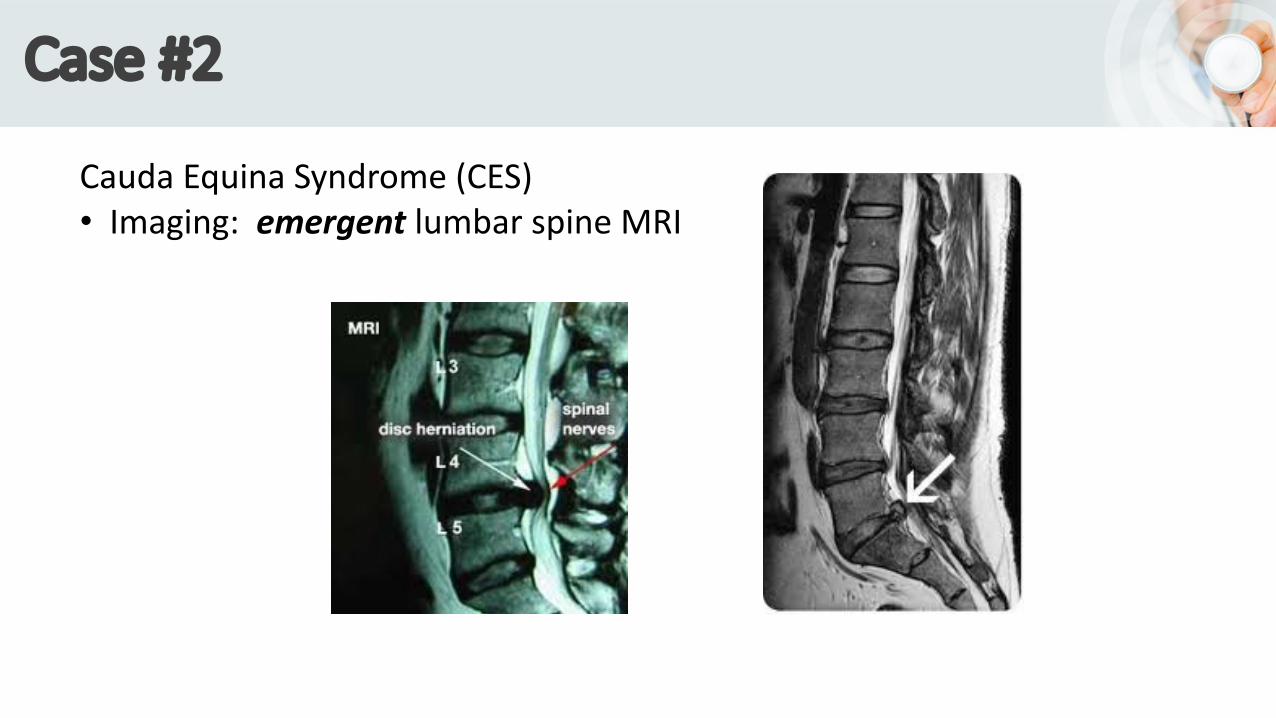
• Lumbar spine MRI: cauda equina syndrome (CES)
• Transferred for surgical decompression of disc herniation at L4 – L5, but developed foot drop on the right side and continued incontinence
Case #2
Now on disability and suffered "damage to his marital relationship”• Sued provider & hospital for $75,000
What went wrong?
Case #2
History (August 30 visit)• ED with worsening symptoms over past week• 10/10 throbbing pain• Numbness and tingling “from the waist down”• Trouble urinating
Case #2
Provider note specifically said:• History of herniated disc and chronic back pain, sees neurologist• Worsening over past week• Numbness from the waist down, with saddle paresthesia• Patient with numbness in legs before, but this is worse• Some difficulty urinating, but is able to urinate
Case #2
Review of Symptoms:• Genitourinary:• (+) Mild urinary retention
Physical Exam• Neurologic:• AAOx3• No focal neuro-deficits• Cranial nerves II-XII intact• Normal sensory. Normal motor• Normal speech. Normal coordination
Case #2
ED course• hydromorphone (Dilaudid) 1mg, IV x1• diazepam (Valium) 5mg, IV x1• ondansetron (Zofran) 4mg, IV x1
stenosis of the central canal with possibility of 2nd disc herniation
Case #2
Comforting Features• CT scan impression did not specifically state “cauda equina syndrome”• CT scan impression suggested elective MRI for further assessment
Case #2
Disconcerting Features• Lumbar spine MRI should have been the first study done• It should have been a stat study• If CT lumbar spine, at least do with contrast (CT myelogram)• Red flags:• worsening pain• urinary retention• saddle anesthesia• bilateral lower extremity symptoms
• All was listed multiple times on HPI, ROS, PE, and nursing notes
Case #2
Provider Deposition• Claimed that patient refused to see neurosurgeon, but nothing in
the chart about this• No AMA form• Furthermore, why would the provider consult neurosurgery
unless something acutely wrong?
Case #2
Other Notes• Lumbar spine MRI one of the only reasons for acute MRI in ED
• No rectal exam?
• No bladder scan?
• CES is true surgical emergency
Case #2
Other Notes• Radiologist report is poor (report said “chronic”, “elective MRI”)• may have led provider down wrong path…but shouldn't have• …is still not an excuse…it said central canal!
• Plaintiff claimed the patient should have “seen a physician”, and the lack of doing so broke the standard of care• not true, not required by state law
Case #2
Cauda Equina Syndrome (CES)• central disc protrusion• Typically occurs at L4-L5 or L5-S1
Case #3Diagnoses:1. Cervical paraspinal muscle strain2. Cervical radiculopathy
Discharge instructions:1. Rest & relax the muscles using comfortable pillows2. Heat, massage, or cold packs several times per day3. Acetaminophen or ibuprofen PRN4. Follow-up with Ortho spine specialists5. Return precautions6. Medications:• cyclobenzaprine (Flexeril)• hydrocodone/APAP (Lortab) 5/325mg, #20
Case #3
June 14, 0130 (really, later the night of the initial visit)• Patient's wife discovered him incontinent, unresponsive, and
with leg convulsions• Patient returned to ED via rescue squad• Had altered mental status• Combative & posturing - first decerebrate, then decorticate• BP: 242/147, Pulse: 102• Intubated. Stat head CT: diffuse subarachnoid hemorrhage• Transferred
Case #3
Patient suffered "permanent and profound brain damage”• Sued provider & hospital for $10 million• “Should have had a head CT the first time”
What went wrong?
Case #3
History (original ED visit):• Patient reported suffering from neck pain for prior 2-3 days, after a visit to
trampoline park• Also numbness in left arm & leg. Denied blurred vision, speech problems• Lightheaded with nausea, but no vomiting• Patient was “using iPad while in the room”• Denied LOC, back pain, fever, CP, SOB, & weakness• Admitted history of HTN but not on meds for it. No PCP• Reported "injury" at trampoline park. Using BC powder without relief
Case #3
Nursing notes:• Patient “speaking in full and logical sentences”• Patient with “steady gait”• BP elevated but “remainder of vital signs are within normal limits”• GCS 15
Case #3
Physical Exam• Full ROM of the neck with no signs of trauma• No swelling, contusions, abrasions, or tenderness to palpation• Normal neurologic exam, "without any motor or sensory deficits".
Comforting Features• No acute onset, no "thunderclap" - it was present x2-3 days• Related to an injury• No vomiting• No change in mental status• C-spine X-rays show possible stenosis (something to "hang your hat on”?)• No focal weakness, a relatively normal physical exam• No acute distress, using iPad
Case #3
Disconcerting Features• Numbness/tingling were in both the upper and lower extremities
• BC powder = aspirin (anti-platelet)
• C-spine X-rays• Show bilateral stenosis (not just left-sided)• Do not account for the leg numbness/tingling
Case #3
• Headaches• Did provider ask patient about history of headaches?• Did patient have history of migraine headaches? Was he previously
worked-up for migraines?
• Trampoline park• Was his head/neck simply sore from jumping on trampolines?• Or did he actually fall; have trauma to his head/neck?
• Why cervical radiculopathy diagnosis?• Pain radiated up to head/neck, not down to shoulder/arm
Case #3
Provider deposition• Admitted not knowing true history of trampoline park, did not know if
trauma occurred or not
• Said that she performed CN assessment, later admitted not testing visual acuity or visual fields (CN II, III, IV, & VI)
Case #3
Provider deposition• Admitted not doing cerebellar testing (did not actually see patient walk)
• Admitted subarachnoid hemorrhage was not on her differential diagnosis
• Stated that patient had lack of slurred speech, nuchal rigidity, and altered mental status…however all of these are very late findings
Case #3
Other notes• Description of the neurologic exam is very weak
• Collaborating MD saw the patient after the APP, just prior to discharge• BP was slightly higher than at triage• MD documented that patient was dizzy, but no change in the plan
Case #3
Other notes• Canadian head CT rules & New Orleans head CT rules do not apply
because this was not a trauma
• Even if head CT had been done at original visit, it may have missed subarachnoid hemorrhage• lumbar puncture is also needed• would have only been done if SAH suspected
Case #3
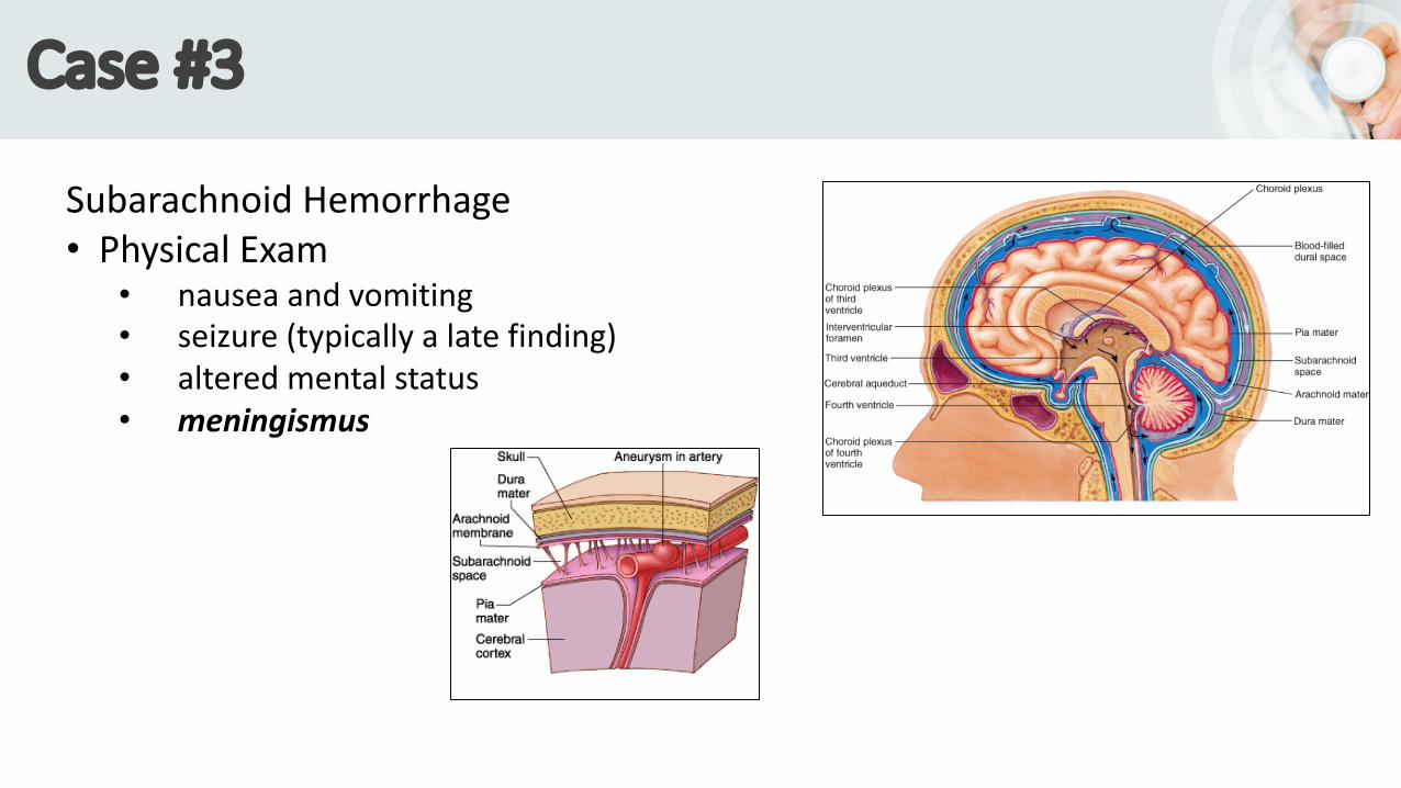
Subarachnoid Hemorrhage• Characteristics• often spontaneous, without trauma• with or without exertion• may present as sudden death• 10-15% patients die before reaching hospital
Discharge instructions:1. Follow-up with PCP within 10 days2. Medications:• albuterol inhaler, 2 inhalations q4-6 hours, PRN breathing• fluticasone (Flonase) nasal spray, 2 sprays each nostril, once/day• montelukast (Singulair), one PO daily
Case #4
April 18 (the next day)• Brought to ED by friend• While in ED, patient went into respiratory arrest and died• On autopsy found to have multiple bilateral pulmonary emboli
Case #4
Patient's family sued:• Urgent care• Provider• Collaborating physician• Emergency department
What went wrong?
Unfortunately, a lot went wrong!
Case #4
History (urgent care – original visit)• Chief complaint: Dyspnea on exertion• After one flight of stairs, patient had to “stop & catch her breath”• After walking dog for one block, felt SOB, had near syncope• Returned home to rest• Then mid-sternal chest tightness & difficulty taking deep breaths• Denied cough, fever/chills• Remote Hx of exercise-induced asthma, not requiring meds in some time• Former smoker (unsure how recent)
Case #4
Physical Exam• "vitals noted, appears well”• Able to speak in full, clear sentences. Talking with clear phonation• Eyes clear• Nasal turbinates with mild swelling, no drainage• Pharynx is not enlarged• TMs without erythema• Neck supple• Lungs with no wheezing or rhonchi. No increased work of breathing• No cyanosis or clubbing
Case #4
Labs/imaging• None
Notes: “peak flow reviewed and appears well performed”
Case #4
Further History• Patient came back to UC the next morning• Provider noted that patient appeared "anxious" and had “increased
work of breathing”• Pulse = 110. Respirations = 20• Lung exam: no wheezing
Further History, continued…• Later that afternoon, patient had syncopal episode while walking dog• Bystander called 911, patient called her PCP• Patient spoke with PCP over the phone, was convinced to go to a hospital
that was further away than where rescue squad would have taken her• Also convinced her to let friend drive her there• On route, patient had episode of "blankly staring x20 seconds”• Friend pulled over and patient vomited
Case #4
Further History, continued…• In emergency department:• Pulse: 107• Respirations: 24• SpO2: 94%
• ED physician noted that patient's mother had history of DVT• Lungs were clear with no wheezing
Case #4
ED workup:• EKG: sinus tachycardia
• CBC with diff: WNL• CMP: WNL• Troponin: 0.055 (elevated, normal is <0.040)• β natriuretic peptide: 2040 (elevated, normal is <125)• D-dimer: 10,462 (elevated, normal is <500)
• Chest CTA: bilateral pulmonary emboli with left basilar atelectasis
Case #4While in ED:• Respirations remained at 24, but SpO2 ↓ to 89%• Hospital admission was in process
• Patient got up to go to bathroom• While in bathroom she had a generalized tonic-clonic seizure• Was given Ativan 1mg, IV
• In postictal state had apneic breathing & bradycardia• Tried intubation, but difficulty• Tried video-assisted intubation, but when into respiratory arrest• Coded on/off x120 minutes but was ultimately declared deceased
Case #4
Comforting Features: none
Case #4
Disconcerting Features
Urgent Care - First visit• Simply did not have PE on differential• Risk factors:• BMI of 33.2 (obesity)• Former smoker (unsure of how recent)• Resting tachycardia (112)• Mid-sternal chest tightness• Family history (mother DVT)
• If diagnosis is asthma, why did patient have no cough and no wheezing?
Case #4
Disconcerting Features, continued
Urgent Care - Second visit• Still with resting tachycardia (110)• Borderline tachypnea (20)• Still no cough and no wheezing• “Increased work of breathing"• Appeared "anxious"
Case #4
Disconcerting Features, continued
Primary Care Physician• Giving advice over the phone? (cannot do a physical exam)
• Recommending a hospital that is further away?
• Recommending friend’s car instead of the ambulance?
Case #4
Disconcerting Features, continued
Emergency Department• CT scan findings communicated to ED physician at 8:30 PM• Heparin not ordered until 8:58 PM
• Patient found on bathroom floor at 10:20 PM• Pronounced dead at 12:44 AM• She was never given heparin while in the ED
Case #4
• Pulmonary embolus• clinical presentation is highly variable - must have high index of suspicion• absence of known risk factors does not rule out VTE
• poor correlation between PE size & symptom severity• i.e, large PEs can be asymptomatic
• Approximately 300,000 fatal PE’s occur per year (Murin 2002)
• in 25% of cases, sudden death is the first/only symptom• Close to 60% of patients die after undetected PE (Heit 2002)
Case #4
• Pulmonary embolus• Risk factors
INHERITED ACQUIREDfactor V Leiden recent “major” surgeryprothrombin gene mutation traumaprotein S deficiency presence of central venous catheterprotein C deficiency malignancyanti-thrombin deficiency pregnancydysfibrinogenemia limb immobilization