51
Breast Cancer Screening Carolyn Aoyama, CNM, MPH Mammography Measure Lead Indian Health Service
Breast Cancer Screening
Carolyn Aoyama, CNM, MPH
Mammography Measure Lead
Indian Health Service

Objectives Discuss differences in malignant breast disease in the
American Indian/Alaska Native population vs. the general population
Discuss Tillman / Myers study
Discuss how to improve mammography screening and GPRA mammography rates
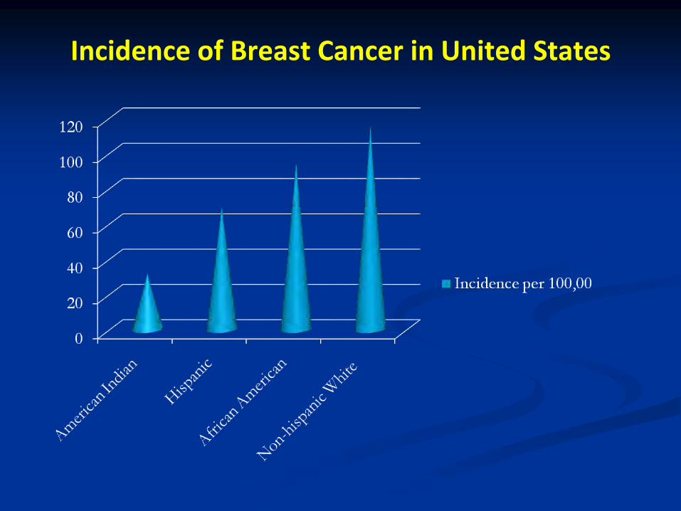
Incidence of Breast Cancer in United States

Compared with other ethnic/racial groups in the United States, AI/AN women have:
- the lowest incidence of breast cancer - the lowest breast cancer survival rate or any ethnic group in U.S.
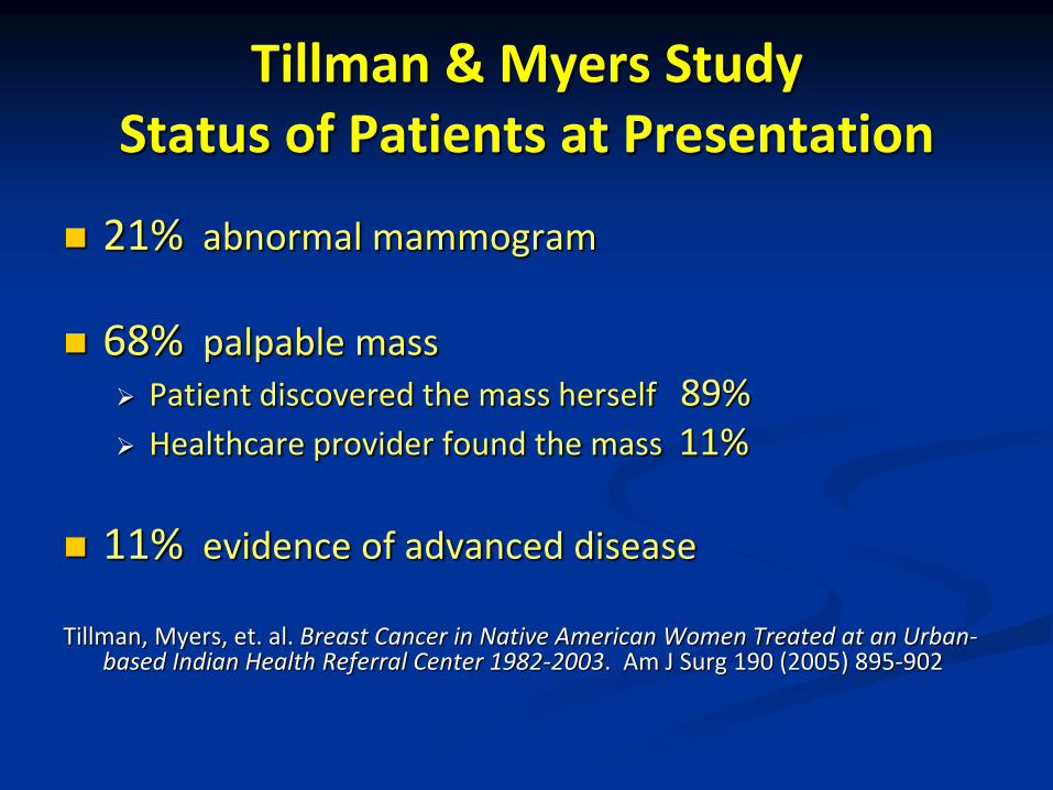
Tillman & Myers Study Status of Patients at Presentation
21% abnormal mammogram
68% palpable mass Patient discovered the mass herself 89% Healthcare provider found the mass 11%
11% evidence of advanced disease
Tillman, Myers, et. al. Breast Cancer in Native American Women Treated at an Urban-based Indian Health Referral Center 1982-2003. Am J Surg 190 (2005) 895-902
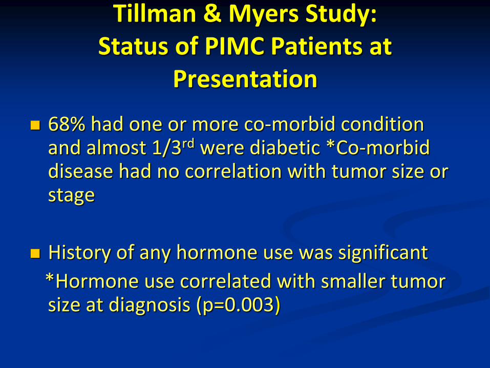
Tillman & Myers Study: Status of PIMC Patients at
Presentation
68% had one or more co-morbid condition and almost 1/3rd were diabetic *Co-morbid disease had no correlation with tumor size or stage
History of any hormone use was significant
*Hormone use correlated with smaller tumor size at diagnosis (p=0.003)

Status of PIMC Patients at Presentation
Over 80% were overweight or obese (increased risk of regional or metastatic disease - more serious disease - with higher BMI)
42.9% of overweight and 59% of obese patients (p=0.019) had more serious disease at presentation
80% of normal BMI patients had in situ or local disease (less serious) at presentation

Patient Outcomes
Average age at diagnosis was 54
Nationally, average age at diagnosis is 64
Average tumor size at diagnosis was 3.3 cm
Nationally, average tumor size is about 2 cm

Why are we diagnosing patients at a later stage?
We have lower screening rates
Our patients and providers may have the misconception that breast cancer is rare in the Native population
Why are we diagnosing patients at a later stage?
One study found that only 1/3rd of Native diabetic women aged 50-69 and living in Phoenix had ever had a mammogram, despite having a co-morbid condition for which they were seeing a healthcare provider
Not all of the IHS hospitals or larger clinics have fixed mammography units
Giroux J, Welty TK, Oliver FK, et al. Low National Breast and Cervical Cancer-Screening Rates in American Indian and Alaska Native Women with Diabetes. JABFP. 2000;13:239-245

5-year survival for PIMC patients 62%; compared to 86% nationally - why?
0
10
20
30
40
50
60
70
In situ/local
disease
Regional/distant
disease
PIMC
SEER
Tillman, Myers, et. al. Breast Cancer in Native American women treated at an urban-based Indian health referral center 1982-2003. Am J Surg 190 (2005) 895-902
AI/AN women in Tillman’s Study:
Presented at later stage of malignancy
Were more likely to undergo mastectomy
Had greater delays to seeking treatment
* This data suggests a need for increased breast cancer education for AI/AN women and their providers to facilitate earlier detection and adequate treatment

Effects of lower screening rates
From 1992 to 2002, death rates in the from breast cancer in the U.S. declined annually by:
2.4% for whites
1.8% for Hispanics
1.0% for African Americans and Asian Americans
0% for AI / AN
American Cancer Society, Breast Cancer Facts and Figures 2005- 2006 http://www.cancer.org/downloads/STT/CAFF2005BrF.pdf
Mammography Screening
Mammography is the best way to detect breast cancer in its earliest, most treatable stage—it takes an average of 1-3 years before a woman can feel a lump.
Mammography detects cancers too small to be felt during a clinical breast examination (CBE).
Mammography detects an average of 90% of breast cancers in women without symptoms!
Regular mammography screening reduces breast cancer mortality rates
Since the 1980’s, thanks to more widespread use of mammography and improved treatment, over-all breast cancer mortality rates in the United States have declined.
Between 1990 and 2002, the U.S Breast cancer death rate declined 2.3% each year.
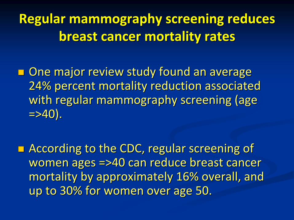
Regular mammography screening reduces breast cancer mortality rates
One major review study found an average 24% percent mortality reduction associated with regular mammography screening (age =>40).
According to the CDC, regular screening of women ages =>40 can reduce breast cancer mortality by approximately 16% overall, and up to 30% for women over age 50.

GPRA Mammography Measure
Denominator: All active female clinical patients aged 52 through 64, without a documented bilateral mastectomy or two separate unilateral mastectomies.
Numerator: Active female clinical patients with documented mammogram in the past two years.
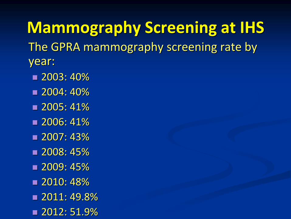
Mammography Screening at IHS The GPRA mammography screening rate by
year: 2003: 40%
2004: 40%
2005: 41%
2006: 41%
2007: 43%
2008: 45%
2009: 45%
2010: 48%
2011: 49.8%
2012: 51.9%
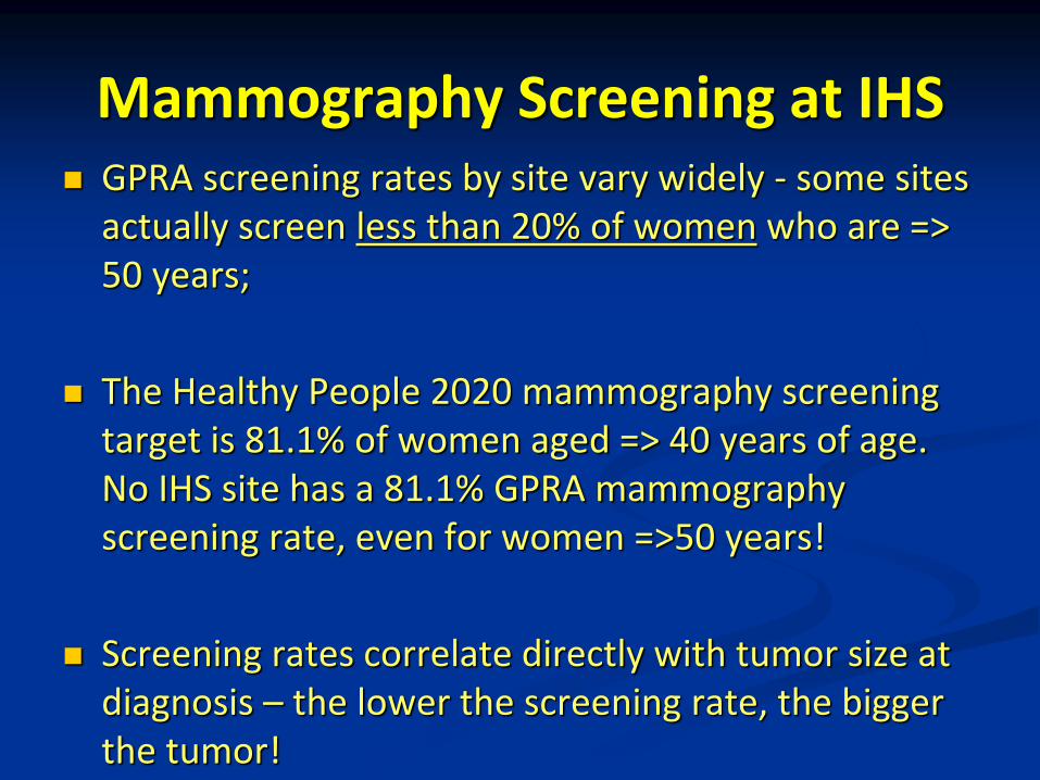
Mammography Screening at IHS GPRA screening rates by site vary widely - some sites
actually screen less than 20% of women who are => 50 years;
The Healthy People 2020 mammography screening target is 81.1% of women aged => 40 years of age. No IHS site has a 81.1% GPRA mammography screening rate, even for women =>50 years!
Screening rates correlate directly with tumor size at diagnosis – the lower the screening rate, the bigger the tumor!
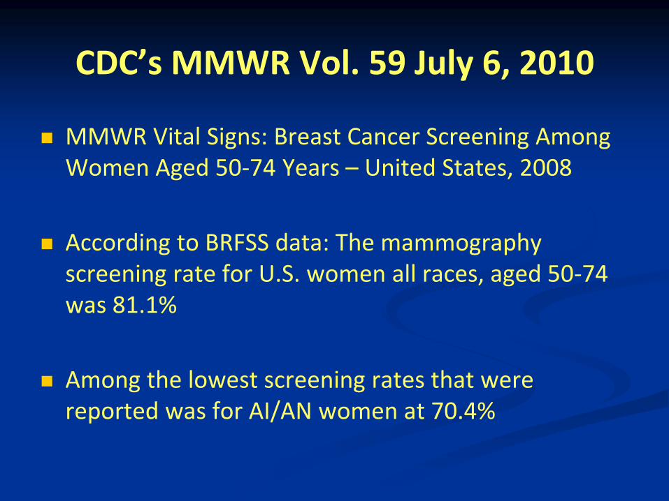
CDC’s MMWR Vol. 59 July 6, 2010
MMWR Vital Signs: Breast Cancer Screening Among Women Aged 50-74 Years – United States, 2008
According to BRFSS data: The mammography screening rate for U.S. women all races, aged 50-74 was 81.1%
Among the lowest screening rates that were reported was for AI/AN women at 70.4%
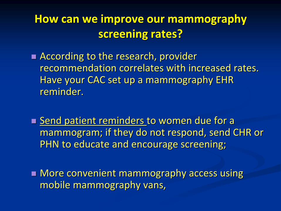
How can we improve our mammography screening rates?
According to the research, provider recommendation correlates with increased rates. Have your CAC set up a mammography EHR reminder.
Send patient reminders to women due for a mammogram; if they do not respond, send CHR or PHN to educate and encourage screening;
More convenient mammography access using mobile mammography vans,

How can we improve our mammography screening rates?
Ultimately, mammography screening needs to be accessible to busy women:
- Allow eligible women (50 yrs and over) to schedule their mammograms on demand, without a provider order;
- Engage CHRs to transport high risk women without wheels (50 yrs and over );
- Engage PHN & CHA to educate pts.

Provider recommendation is one of the strongest predictors of mammography use
One study found that “the most frequent reason cited by women for failure to have mammography is that their provider did not recommend one.”
Another study found that “94% of women whose provider had recommended mammograms had had one in the last 2 years, while only 36% of women whose provider had not made the recommendation had a mammogram.”

New U.S. Preventive Task Force Recommendations
Biennial (every other year) screening mammography for women aged 50 to 74 years;
Decision to initiate regular, biennial screening earlier is an individual one taking patient context into account, including patient values regarding benefits and harms;
Recommends against the BSE;
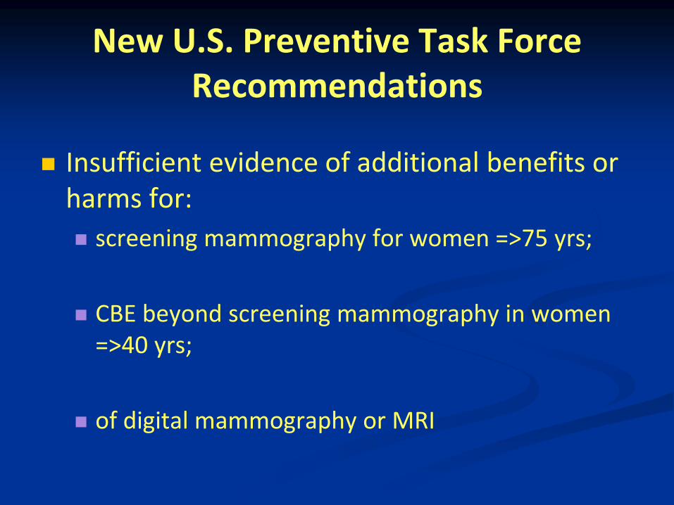
New U.S. Preventive Task Force Recommendations
Insufficient evidence of additional benefits or harms for:
screening mammography for women =>75 yrs;
CBE beyond screening mammography in women =>40 yrs;
of digital mammography or MRI

The GPRA Measure and the US Preventive Task Force Recommendations
The new recommendations do not mean women can’t be screened earlier
Women can request earlier screening
Clinicians can recommend earlier screening

So your patient’s screening
mammogram comes back
abnormal!
Now what?
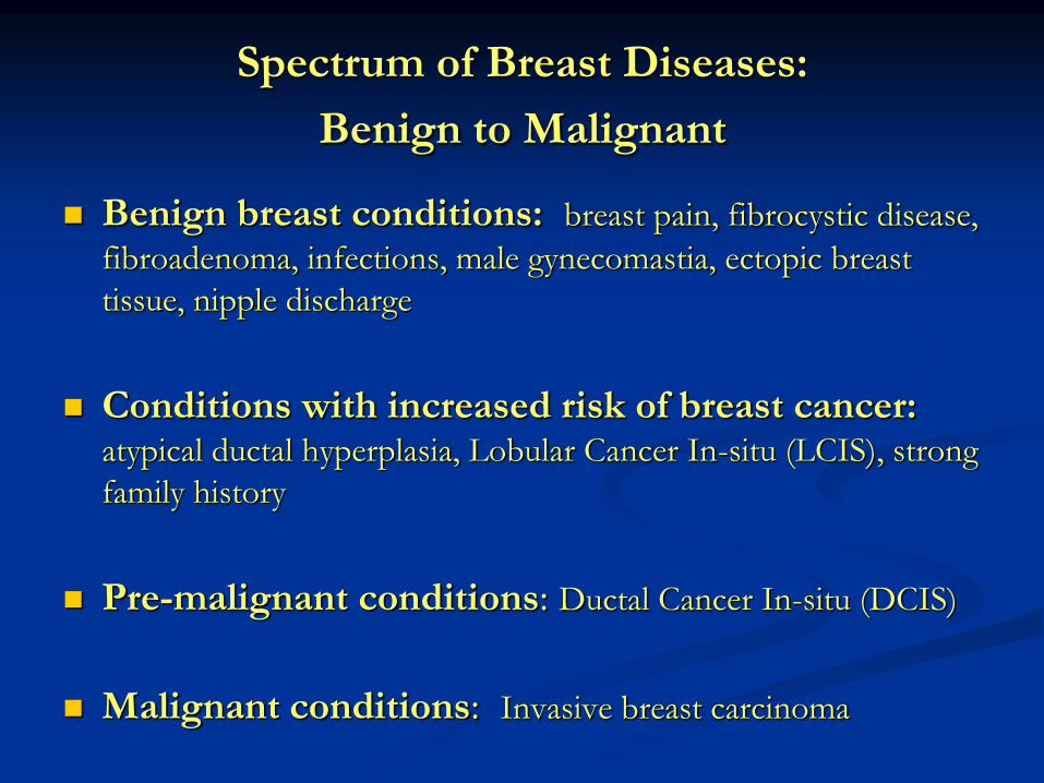
Spectrum of Breast Diseases:
Benign to Malignant
Benign breast conditions: breast pain, fibrocystic disease,
fibroadenoma, infections, male gynecomastia, ectopic breast
tissue, nipple discharge
Conditions with increased risk of breast cancer: atypical ductal hyperplasia, Lobular Cancer In-situ (LCIS), strong
family history
Pre-malignant conditions: Ductal Cancer In-situ (DCIS)
Malignant conditions: Invasive breast carcinoma

Benign Breast Conditions
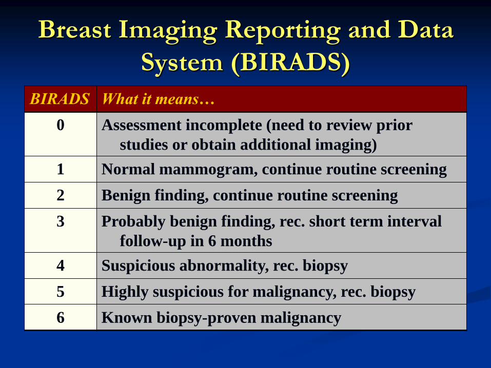
Breast Imaging Reporting and Data
System (BIRADS)
BIRADS What it means…
0 Assessment incomplete (need to review prior
studies or obtain additional imaging)
1 Normal mammogram, continue routine screening
2 Benign finding, continue routine screening
3 Probably benign finding, rec. short term
follow-up in 6 months
interval
4 Suspicious abnormality, rec. biopsy
5 Highly suspicious for malignancy, rec. biopsy
6 Known biopsy-proven malignancy
43 y/o female presents to ER c/o right
breast mass discovered on BSE PMHx: HTN, Type 2 DM, s/p cholecystectomy with
no breast cancero risk factors identified
Patient has never had mammogram or CBE (In a study from 2000, only 1/3rd of Native diabetic women living in Phoenix aged 50-69 had ever had a mammogram & less than 1/3rd had ever had a CBE, despite having a co-morbid condition for which they were seeing a healthcare provider!)
Patient scheduled for mammograms and referred to breast clinic
Giroux et al. Low National Breast and Cervical Cancer-Screening Rates in American Indian
and Alaska Native Women with Diabetes. JABFP. 2000;13:239-245

Ultrasound: lesion is
solid, wider than tall,
(BIRADS 3) Mammogram: smooth
round density, ultrasound
(BIRADS 0)

Physical exam in breast clinic reveals
a smooth, mobile 2 cm mass in the
right breast.
Imaging and exam are c/w fibroadenoma, but a
solid breast mass in a patient over 40 (or other
risk factors) needs tissue diagnosis to safely
observe
Core biopsy confirms diagnosis
of fibroadenoma, options of
excision versus observation
discussed with patient

Benign lesions can often be
completely removed with image
guided vacuum assisted biopsy; or
excisional biopsy with periareolar
incision
Patient opts for excision;
lesion is excised completely
under ultrasound guidance
with vacuum assisted device
Benign nipple discharge
In 2/3rd of non-lactating women fluid can be
expressed from the nipple ducts
Physiologic secretions can be white, yellow,
green, brown; may be from multiple ducts and
vary in color
Blood in nipple discharge during pregnancy or
lactation is benign, probably due to
hypervascularity of developing breast tissue
Galactorrhea
Galactorrhea is copious bilateral milky discharge
not associated with pregnancy or lactation
Careful drug history for drugs such as OCPs,
antihypertensives, or psychotropic agents that
can cause hyperprolactinemia
Elevated blood prolactin levels without drug
cause should prompt evaluation for pituitary
tumor
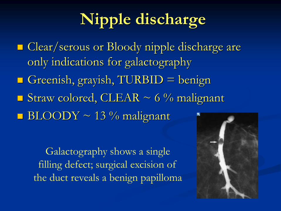
Nipple discharge
Clear/serous or Bloody nipple discharge are
only indications for galactography
Greenish, grayish, TURBID = benign
Straw colored, CLEAR ~ 6 % malignant
BLOODY ~ 13 % malignant
Galactography shows a single
filling defect; surgical excision of
the duct reveals a benign papilloma

Paget’s disease
Eczema-like patch of irritated skin starts at the nipple and
can spread onto areola
Very rarely bilateral
Will not improve with local treatment such as steriod
creams
Paget’s is almost always a sign of an underlying
malignancy, and treatment is that of the underlying
disorder
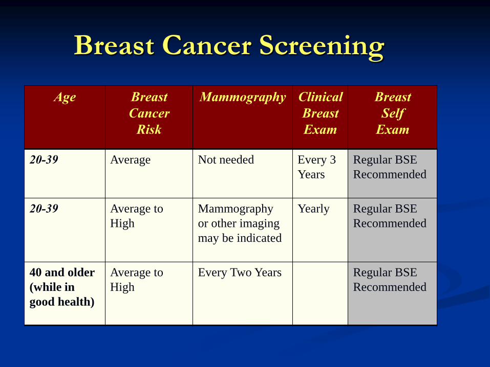
Breast Cancer Screening
Age Breast Cancer
Risk
Mammography Clinical Breast Exam
Breast Self
Exam
20-39 Average Not needed Every
Years
3 Regular BSE
Recommended
20-39 Average
High
to Mammography
or other imaging
may be indicated
Yearly Regular BSE
Recommended
40 and older
(while in
good health)
Average
High
to Every Two Years Regular BSE
Recommended
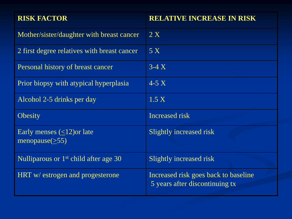
RISK FACTOR RELATIVE INCREASE IN RISK
Mother/sister/daughter with breast cancer 2 X
2 first degree relatives with breast cancer 5 X
Personal history of breast cancer 3-4 X
Prior biopsy with atypical hyperplasia 4-5 X
Alcohol 2-5 drinks per day 1.5 X
Obesity Increased risk
Early menses (<12)or late
menopause(>55)
Slightly increased risk
Nulliparous or 1st child after age 30 Slightly increased risk
HRT w/ estrogen and progesterone Increased risk goes back to baseline
5 years after discontinuing tx
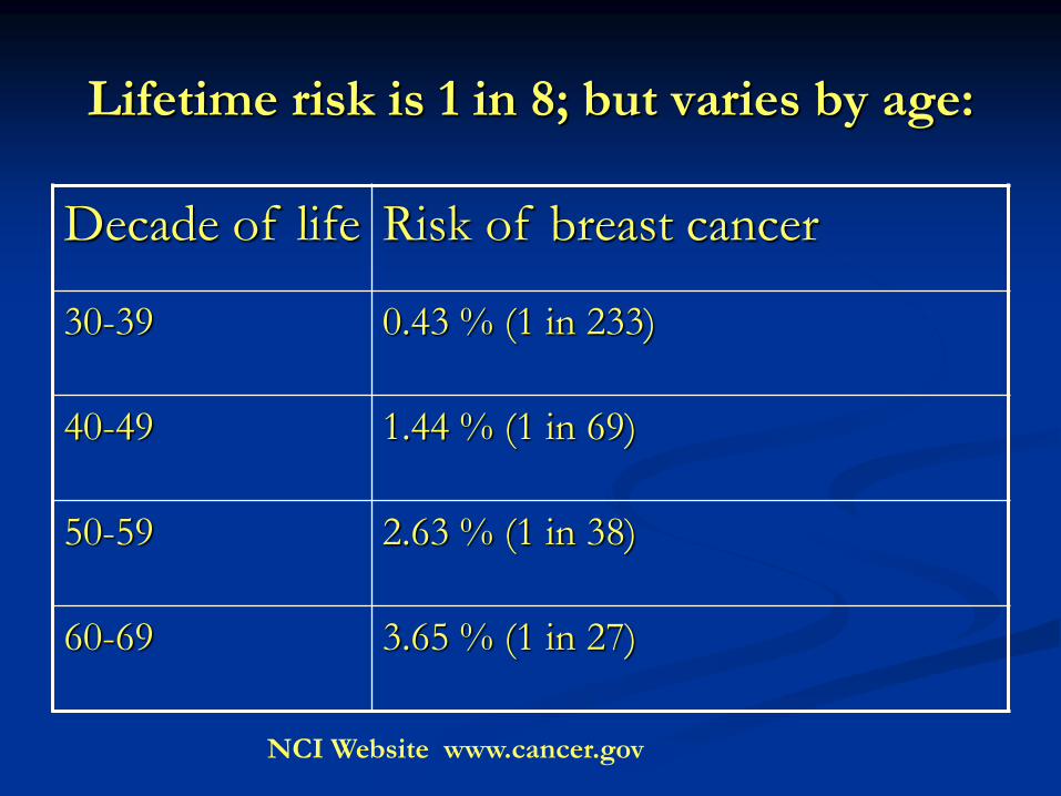
Lifetime risk is 1 in 8; but varies by age:
Decade of life Risk of breast cancer
30-39 0.43 % (1 in 233)
40-49 1.44 % (1 in 69)
50-59 2.63 % (1 in 38)
60-69 3.65 % (1 in 27)
NCI Website www.cancer.gov

Breast Cancer Risk Assessment
Tool: Gail model
Medical hx (age, number of prior breast biopsies,
presence of atypical hyperplasia)
Reproductive hx (age at 1st menses, age of 1st live birth)
Family hx (breast cancer in a mother, sister or daughter)
Calculates 5 year and lifetime risk compared to general
population

Breast Cancer Risk Assessment Tool:
Gail model
May underestimate risk for
some minority groups
Has not been validated in
AI/AN populations
Risk Calculator is available
on the NCI website: www.
Cancer.gov/bcrisktool
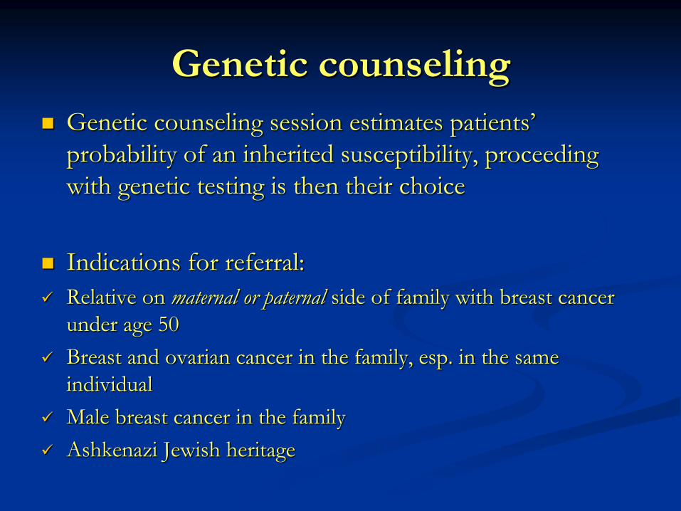
Genetic counseling
Genetic counseling session estimates patients’
probability of an inherited susceptibility, proceeding
with genetic testing is then their choice
Indications for referral:
Relative on maternal or paternal side of family with breast cancer
under age 50
Breast and ovarian cancer in the family, esp. in the same
individual
Male breast cancer in the family
Ashkenazi Jewish heritage
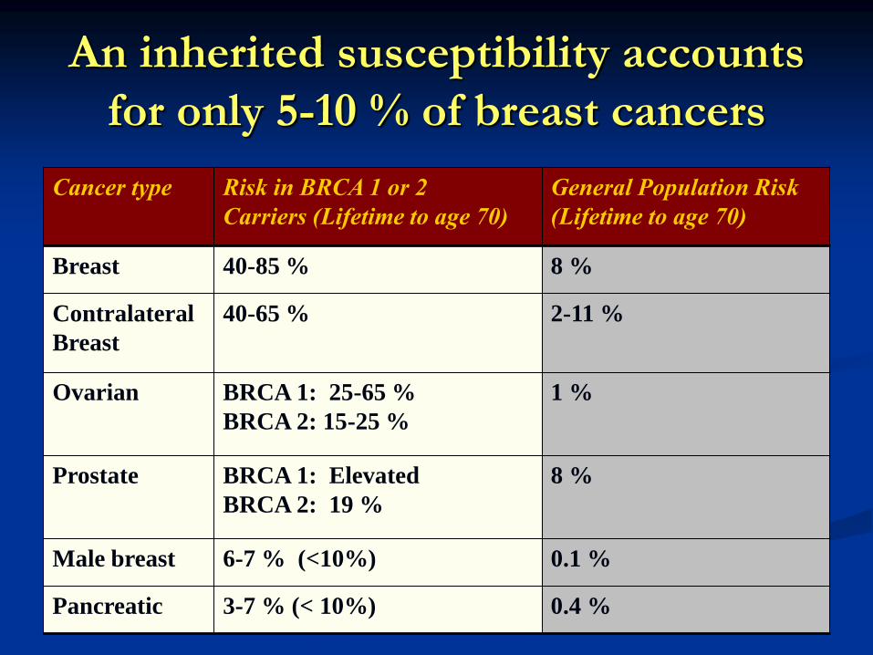
An inherited susceptibility accounts
for only 5-10 % of breast cancers
Cancer type Risk in BRCA 1 or 2
Carriers (Lifetime to age 70) General Population (Lifetime to age 70)
Risk
Breast 40-85 % 8 %
Contralateral
Breast
40-65 % 2-11 %
Ovarian BRCA
BRCA
1:
2:
25-65 %
15-25 %
1 %
Prostate BRCA
BRCA
1:
2:
Elevated
19 %
8 %
Male breast 6-7 % (<10%) 0.1 %
Pancreatic 3-7 % (< 10%) 0.4 %

Mammogram with clustered
microcalcifications
Patient sent for stereotactic biopsy which reveals
ductal carcinoma in situ, ER+
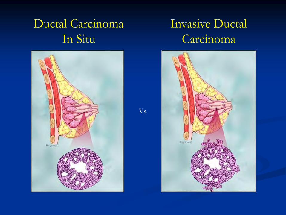
Ductal Carcinoma
In Situ
Vs.
Invasive Ductal
Carcinoma

Mammogram:
Spiculated mass left breast, 2 cm, BIRADS 4

Breast cancer staging
Primary Tumor Definitions Tis Carcinoma in situ
T0 No evidence of primary tumor
T1 Tumor 2 cm or less
T2 Tumor > 2 cm, < 5 cm
T3 Tumor more than 5 cm
T4 Any
wall
size tumor with direct extension into
or skin; inflammatory carcinoma
chest
Regional Nodes
N0 No regional lymph node metastasis
N1 Metastasis in 1-3 axillary lymph nodes
N2 Metastasis in 4-9 axillary lymph nodes
N3 Metastasis in 10 or more axillary lymph nodes,
Ipsilateral supraclavicular lymph nodes
Distant Mets
M0 No distant metastasis
M1 Distant metastasis

Breast cancer staging
Stage Definition 5 year Relative
Survival Rate
0 Tis N0 M0 100 %
I T1 N0 M0 100 %
IIA T0-1 N1 M0
T2 N0 M0
92 %
IIB T2
T3
N1
N0
M0
M0
81 %
IIIA T0-2 N2 M0
T3 N1-2 M0
67 %
IIIB T4 N0-2 M0 54 %
IV Any T any N M1 20 %
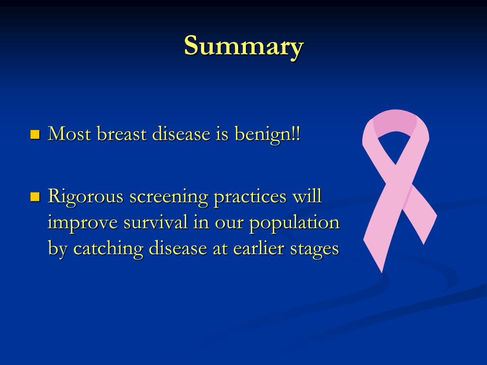
Summary
Most breast disease is benign!!
Rigorous screening practices will
improve survival in our population
by catching disease at earlier stages


