Guideline Breech – Management of Uncontrolled document when printed Published: (15/11/2018) Page 1 of 15 1. Purpose This document provides details of clinical management of women who have a diagnosis of breech presentation during pregnancy or intrapartum at the Women’s. This procedure outlines the decision and management process required for: breech presentation diagnosed antenatally planning for a vaginal breech birth managing a vaginal breech birth decision making in preterm breech birth neonatal management of the breech baby. The guiding principles of this procedure are informed consent and care by clinicians with suitable preparation and experience in vaginal breech birth. The RANZCOG guideline on Management of Breech at Term 12 states ‘Where a vaginal delivery of a breech presentation is planned, appropriate infrastructure must include: Continuous electronic fetal heart monitoring in labour Immediate availability of skilled anaesthetic staff, facilities for immediate caesarean section, and paediatric resuscitation Availability of a suitably experienced obstetrician for all of labour with arrangements in place to manage shift changes and fatigue arrangements. 2. Definitions The definition of breech presentation is when the buttocks, foot or feet are presenting instead of the head. There are 3 classifications of breech: Frank breech where the hips are flexed and legs extended Complete breech where the hips and knees are flexed and the feet are not below the level of the fetal buttocks Footling breech where one or both feet are presenting as the lowest part of the fetus.
Transcript
Guideline
Breech – Management of
Uncontrolled document when printed Published: (15/11/2018) Page 1 of 15
1. Purpose This document provides details of clinical management of women who have a diagnosis of breech presentation during pregnancy or intrapartum at the Women’s.
This procedure outlines the decision and management process required for:
breech presentation diagnosed antenatally
planning for a vaginal breech birth
managing a vaginal breech birth
decision making in preterm breech birth
neonatal management of the breech baby.
The guiding principles of this procedure are informed consent and care by clinicians with suitable preparation and experience in vaginal breech birth.
The RANZCOG guideline on Management of Breech at Term12 states ‘Where a vaginal delivery of a breech presentation is planned, appropriate infrastructure must include:
Continuous electronic fetal heart monitoring in labour
Immediate availability of skilled anaesthetic staff, facilities for immediate caesarean section, and paediatric resuscitation
Availability of a suitably experienced obstetrician for all of labour with arrangements in place to manage shift changes and fatigue arrangements.
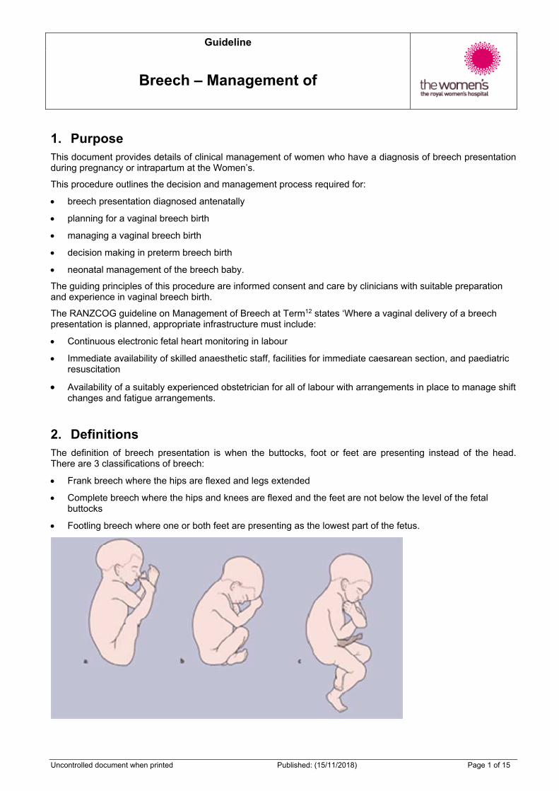
2. Definitions The definition of breech presentation is when the buttocks, foot or feet are presenting instead of the head. There are 3 classifications of breech:
Frank breech where the hips are flexed and legs extended
Complete breech where the hips and knees are flexed and the feet are not below the level of the fetal buttocks
Footling breech where one or both feet are presenting as the lowest part of the fetus.
Guideline
Breech – Management of
Uncontrolled document when printed Published: (15/11/2018) Page 2 of 15
3. Responsibilities Obstetric medical staff are responsible for the antenatal and intrapartum management decisions of women whose baby presents by the breech.
Midwifery staff are responsible for the midwifery care of the woman.
Around fifteen percent of pregnancies present as breech at 29-32 weeks. Breech presentation is a normal finding in preterm pregnancies, when the fetus is more mobile, and should not be considered abnormal until late pregnancy. Twenty-five percent of breech presentations will still undergo spontaneous version after 35 weeks gestation. Such changes occur with decreasing frequency as gestational age advances.
Spontaneous Rupture of Membranes carries a higher risk of cord prolapse. (link to cord prolapse guideline) Clinical care involves careful assessment including prompt confirmation of the presence of the fetal heart prior to transfer of the woman. Subsequent care is determined by clinical assessment.
In the event of a suspected cord prolapse, clinical staff will attend the woman insitu and call a Pink Alert. (link to procedure)
Abdominal palpation: if presenting part is irregular and not "ballotable", or if head ballotable at fundus of uterus
Pelvic examination: head not felt in pelvis (buttocks and or feet may be felt)
Very thick meconium present after rupture of membranes
Guideline
Breech – Management of
Uncontrolled document when printed Published: (15/11/2018) Page 3 of 15
Cord prolapse
Abnormal CTG
Fetal heart heard higher in abdomen.
Ultrasound scan will confirm diagnosis.
Note: In cases of extended breech, the breech may not be ballotable and the fetal heart may be in the same location as expected for a cephalic presentation.
4.3. Management
Antenatal care: Preterm (< 37 weeks):
Breech presentation is a normal finding in the preterm pregnancy. No further management in the uncomplicated pregnancy is required until 37 completed weeks of pregnancy are reached.
If elective preterm delivery is indicated the mode of birth will be dictated by clinical circumstances. For example, if the indication is for severe pre eclampsia then caesarean section would be the most appropriate mode, if however the indication was for fetal death in utero or lethal fetal anomaly, then induction of labour and vaginal birth may be appropriate.
Intrapartum care
The optimal mode of birth for preterm breech has not been fully evaluated in clinical trials and the relative risks for the preterm infant and mother remain unclear. Overall, decisions regarding mode of birth will need to be made on an individual basis. With the evidence available to us at this time, the Women's recommended practice is to perform emergency caesarean section for any woman presenting in preterm labour with breech presentation except where;
vaginal birth is imminent
The medical circumstances are such that survival (and least morbidity) of the fetus is assessed to be unchanged by mode of delivery (e.g. extremely premature infants-24-25 weeks, or lethal condition) and/or the maternal morbidity of caesarean section is judged to be too great for the relative potential fetal disadvantages.
Antenatal care: Term (> 37 completed weeks gestation):
When a breech presentation is suspected on abdominal palpation a real-time obstetric ultrasound should be performed to confirm diagnosis and exclude possible causative factors (e.g. polyhydraminos, low lying placenta, fetal anomaly).
4.4. External Cephalic Version (ECV)
The current evidence would suggest that ECV where there are no contraindications will reduce the number of breech presentations in labour and the number of caesarean sections for breech presentation with no increase in the perinatal, fetal or maternal morbidity1.
It is recommended practice at the Royal Women's Hospital to offer women with breech presentation at term ECV. If this is unsuccessful or the woman declines ECV, elective caesarean section should be booked for 39 weeks gestation.
Women who desire a vaginal breech birth should be referred to the Breech Clinic for assessment and decision-making with a clinician experienced in vaginal breech birth.
Guideline
Breech – Management of
Uncontrolled document when printed Published: (15/11/2018) Page 4 of 15
4.5. Elective caesarean section
The Term Breech Trial (TBT, 2000) found that compared to vaginal birth planned caesarean section was associated with:
lower rates of perinatal and neonatal death
lower rates of short term neonatal morbidity or perinatal death
fewer 5 minutes Apgar scores <7
lower risk of adverse perinatal outcomes
small increase in the short term maternal morbidity
decreased opportunity for spontaneous version.
There was no difference in outcome between the 2 groups at 2 year follow up. The advantage therefore in a policy of planned elective caesarean section for breech presentation at term is to decrease the short term perinatal and neonatal morbidity and mortality2.
However, the women selected for vaginal breech birth in the TBT has been questioned. The PREMODA study (2006) assessed French and Belgian practices in breech presentation including their consequences for mother and baby. The results indicated there was no significant difference between the 2 groups in perinatal mortality, neonatal mortality, and severe neonatal mortality.
A recent Dutch study found that the perinatal mortality was lower in the caesarean section group when compared with the vaginal breech group. However the relative safety of elective caesarean should be weighed against the consequences of a scarred uterus in future pregnancies11.In labour: Vaginal breech birth (unplanned/previously undiagnosed)
Confirm with ultrasound (RTS on Birth Centre)
Review history (risk factors such as placenta praevia, twins)
Full examination (including vaginal speculum and/or digital)
Continuous CTG monitoring until delivery.
If birth not imminent, caesarean section should be arranged unless a consultant skilled in vaginal breech birth is available. In addition, the woman must provide verbal consent to proceed with vaginal breech birth and the following criteria should be met.
Vaginal birth should only be undertaken where:
Birth is imminent
Obstetrician skilled in vaginal breech birth is available
Senior medical, neonatal and midwifery staff have been called to attend
There is no absolute contraindication to vaginal birth (e.g. placenta praevia)
Frank or complete breech with a flexed head.
4.6. Planned vaginal breech birth
The guiding principles of this procedure are informed consent and care by clinicians with suitable preparation and experience in vaginal breech birth.
Obstetric medical staff are responsible for:
counseling the woman about vaginal breech birth versus planned caesarean
making the decision with the woman for a planned vaginal breech birth and for obtaining consent
providing obstetric care during the intrapartum period
ensuring their own preparation and competency when undertaking a vaginal breech birth.
Guideline
Breech – Management of
Uncontrolled document when printed Published: (15/11/2018) Page 5 of 15
Clinicians supervising or attending vaginal breech births should have appropriate training, including simulation training and drills 1, 2, 3.
If the criteria regarding experienced clinicians cannot be met, it is reasonable to refer the woman who strongly desires a vaginal breech birth to another unit that can accommodate her request 2.
Preparation and consent
The woman should be informed of the benefits and risks for both current and future pregnancies, of planned caesarean birth versus planned vaginal breech birth at term 1,2 3,4.
The woman should be given sufficient information on the risks and benefits to make an informed decision. The woman’s right to change her mind and request a caesarean should be respected
Planned caesarean has a reduced risk of perinatal mortality and early morbidity compared with vaginal birth1
There is no evidence of long term health problems with babies with a breech presentation regardless of mode of birth1
Adverse outcomes are associated with labour augmentation, birth weight less than 2.8kg and more than 4 kg, delayed second stage and no experienced clinician at the birth 1,2,3,4
The rates of mortality and morbidity are significantly reduced when strict selection and management criteria are applied to singleton breech babies with a planned vaginal birth at term1, 2, 3
Planned caesarean carries a small increase in risk of serious immediate maternal complications compared with planned vaginal birth1
Planned caesarean does not carry extra risk to long-term health of the baby outside of pregnancy1.
Criteria for selection 1,2,3,4
Note: Diagnosis of breech for the first time during labour is not a contraindication for vaginal breech birth providing the criteria are met1
Appropriately prepared and experienced clinicians are available for the birth.
No contraindication to vaginal birth (e.g. placenta praevia, compromised fetus)
Frank or complete breech presentation (not footling or kneeling)
Baby expected to weigh more than 2500g and less than 4000g
Neck is not hyperextended in labour (by ultrasound)
No previous caesarean section
Emergency caesarean facilities are available
Considerations
Opinion on induction of labour varies 1, 3. It is preferable that labour be spontaneous. Individual cases must be discussed with a Consultant
Epidural analgesia is not routinely recommended; women have the same choice of analgesia as those having a cephalic birth
Continuous CTG monitoring is recommended
A scalp electrode can be applied to the breech. However, if fetal monitoring is required using this method, consideration must be given to the appropriateness of continuing.
Caesarean section should be considered if there is delay iin any stage of the labour1
Failure of the presenting part to descend may be a sign of relative cephalopelvic disproportion
Optimal maternal position for birth is in upright position e.g. hands and knees or kneeling
Guideline
Breech – Management of
Uncontrolled document when printed Published: (15/11/2018) Page 6 of 15
There is no clear evidence that selective episiotomy should differ from cephalic birth 1.Episiotomy should be considered to facilitate birth only when indicated1. The episiotomy should not be performed until the fetal anus is delivered2
Breech extraction should not be routinely used1.
4.7. Managing the birth
First Stage
All maternal and fetal observations, including diagnosis of labour and proactive planning apply as guided by the guideline Labour and Birth and Early Puerperium – Care during with the following exceptions:
Monitor progress of labour. Expect 1cm/hr from 4cm to full dilatation2
Second stage: good practice points
Confirm full dilatation and position of breech
Monitor fetal heart rate
Active pushing not encouraged until presenting part is distending the perineum
Spontaneous birth of the trunk and limbs by maternal effort should be awaited as breech extraction can cause extension of the arms and head1,2,3,4
Up to 60 minutes passive second stage, as defined by full dilatation without spontaneous urge to push, can be allowed for passive descent as long as there are no concerns for fetal wellbeing
Active second stage, as defined by full dilatation with spontaneous urge to push/directed pushing, should be within ½ hour for multigravida and 1 hour for primipara
If no descent or descent is poor then caesarean section is indicated
A Consultant should be present for the birth1,2,3,4
A neonatologist should be present at the birth
An anaesthetist should be available for the birth 1,2,3,4.
The pictures below demonstrate manoeuvres that are required only if necessary to aid the birth. Generally a breech birth is a ‘hands off’ birth.
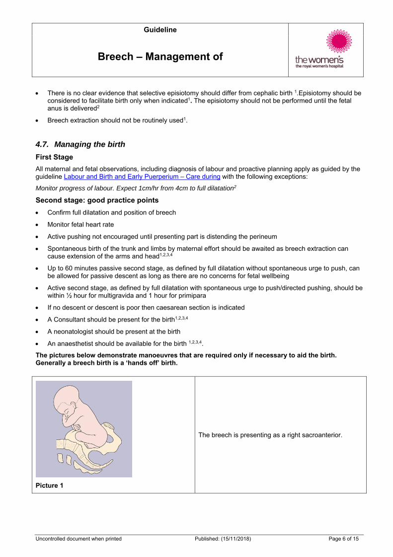
Picture 1
The breech is presenting as a right sacroanterior.
Guideline
Breech – Management of
Uncontrolled document when printed Published: (15/11/2018) Page 7 of 15
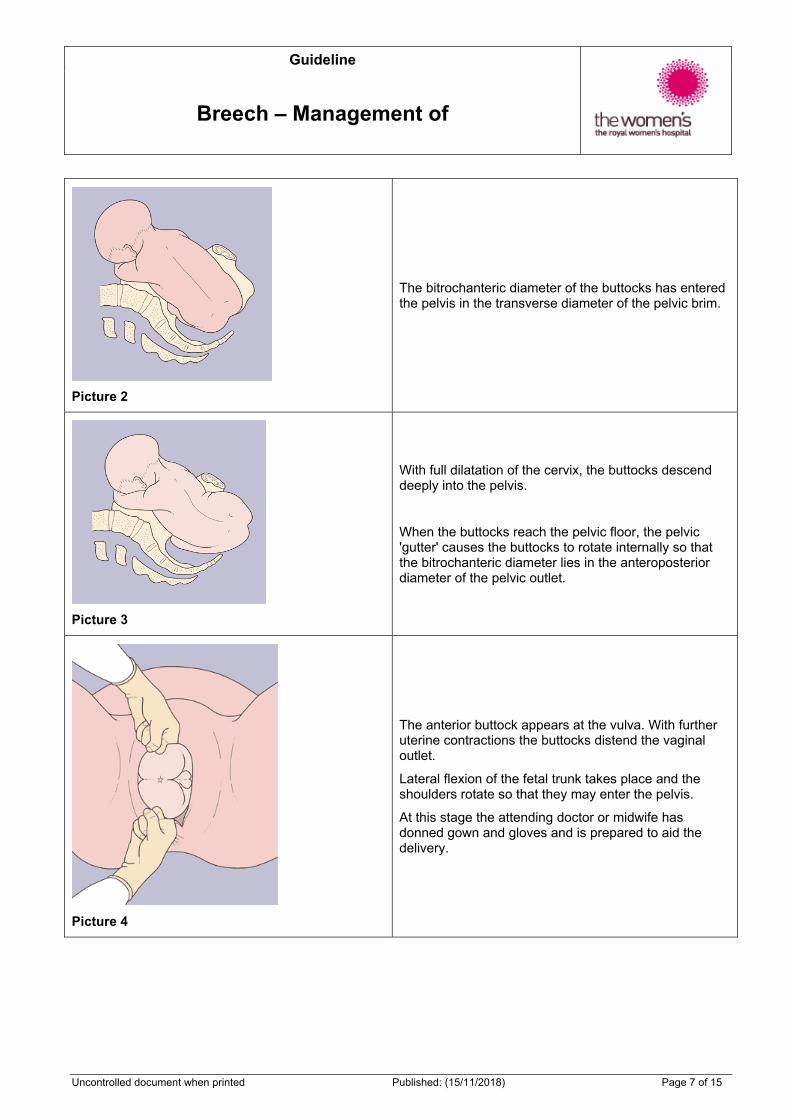
Picture 2
The bitrochanteric diameter of the buttocks has entered the pelvis in the transverse diameter of the pelvic brim.
Picture 3
With full dilatation of the cervix, the buttocks descend deeply into the pelvis.
When the buttocks reach the pelvic floor, the pelvic 'gutter' causes the buttocks to rotate internally so that the bitrochanteric diameter lies in the anteroposterior diameter of the pelvic outlet.
Picture 4
The anterior buttock appears at the vulva. With further uterine contractions the buttocks distend the vaginal outlet.
Lateral flexion of the fetal trunk takes place and the shoulders rotate so that they may enter the pelvis.
At this stage the attending doctor or midwife has donned gown and gloves and is prepared to aid the delivery.
Guideline
Breech – Management of
Uncontrolled document when printed Published: (15/11/2018) Page 8 of 15
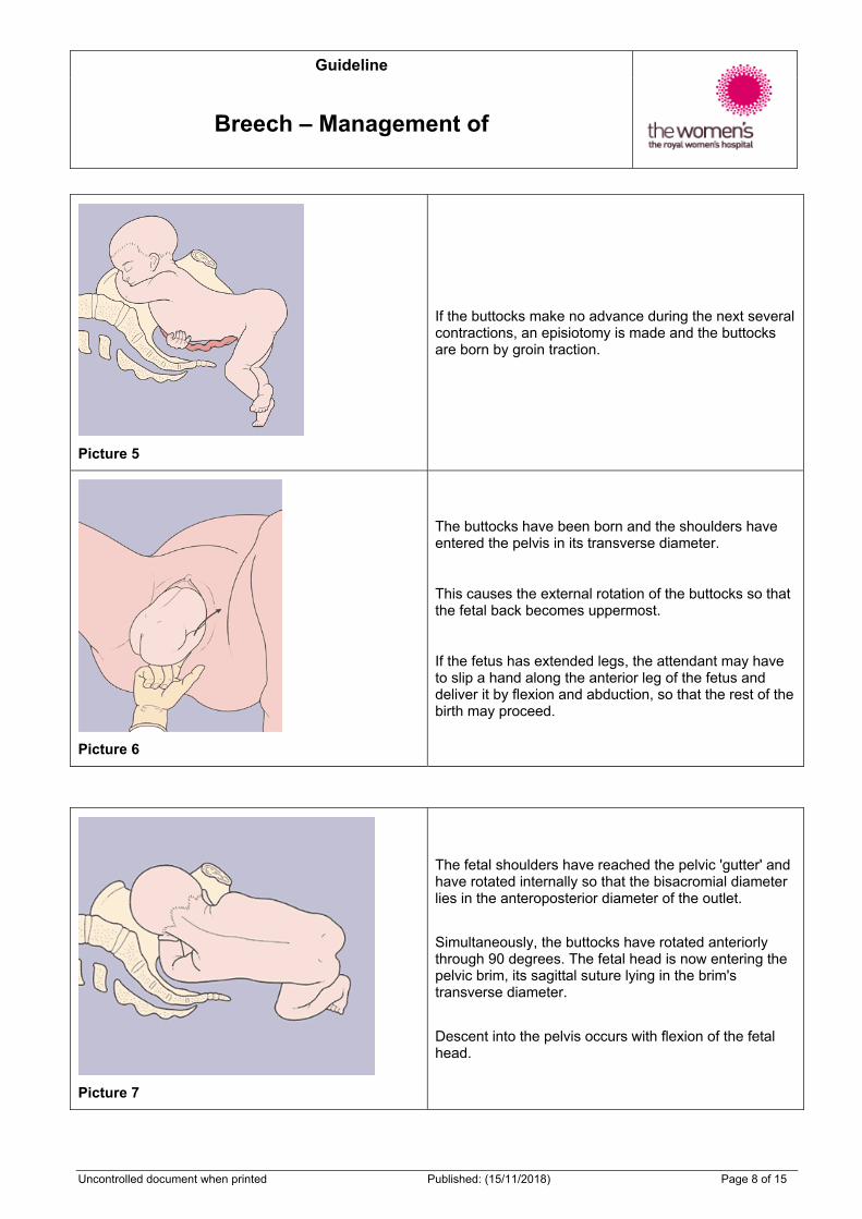
Picture 5
If the buttocks make no advance during the next several contractions, an episiotomy is made and the buttocks are born by groin traction.
Picture 6
The buttocks have been born and the shoulders have entered the pelvis in its transverse diameter.
This causes the external rotation of the buttocks so that the fetal back becomes uppermost.
If the fetus has extended legs, the attendant may have to slip a hand along the anterior leg of the fetus and deliver it by flexion and abduction, so that the rest of the birth may proceed.
Picture 7
The fetal shoulders have reached the pelvic 'gutter' and have rotated internally so that the bisacromial diameter lies in the anteroposterior diameter of the outlet.
Simultaneously, the buttocks have rotated anteriorly through 90 degrees. The fetal head is now entering the pelvic brim, its sagittal suture lying in the brim's transverse diameter.
Descent into the pelvis occurs with flexion of the fetal head.
Guideline
Breech – Management of
Uncontrolled document when printed Published: (15/11/2018) Page 9 of 15
Picture 8
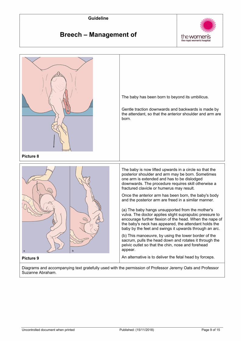
The baby has been born to beyond its umbilicus.
Gentle traction downwards and backwards is made by the attendant, so that the anterior shoulder and arm are born.
Picture 9
The baby is now lifted upwards in a circle so that the posterior shoulder and arm may be born. Sometimes one arm is extended and has to be dislodged downwards. The procedure requires skill otherwise a fractured clavicle or humerus may result.
Once the anterior arm has been born, the baby's body and the posterior arm are freed in a similar manner.
(a) The baby hangs unsupported from the mother's vulva. The doctor applies slight suprapubic pressure to encourage further flexion of the head. When the nape of the baby's neck has appeared, the attendant holds the baby by the feet and swings it upwards through an arc.
(b) This manoeuvre, by using the lower border of the sacrum, pulls the head down and rotates it through the pelvic outlet so that the chin, nose and forehead appear.
An alternative is to deliver the fetal head by forceps.
Diagrams and accompanying text gratefully used with the permission of Professor Jeremy Oats and Professor Suzanne Abraham.
Guideline
Breech – Management of
Uncontrolled document when printed Published: (15/11/2018) Page 10 of 15
Advanced Manoeuvres
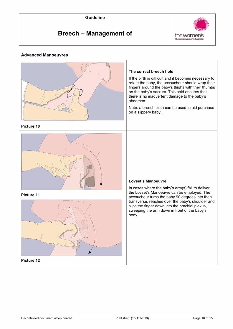
Picture 10
The correct breech hold
If the birth is difficult and it becomes necessary to rotate the baby, the accoucheur should wrap their fingers around the baby’s thighs with their thumbs on the baby’s sacrum. This hold ensures that there is no inadvertent damage to the baby’s abdomen.
Note: a breech cloth can be used to aid purchase on a slippery baby.
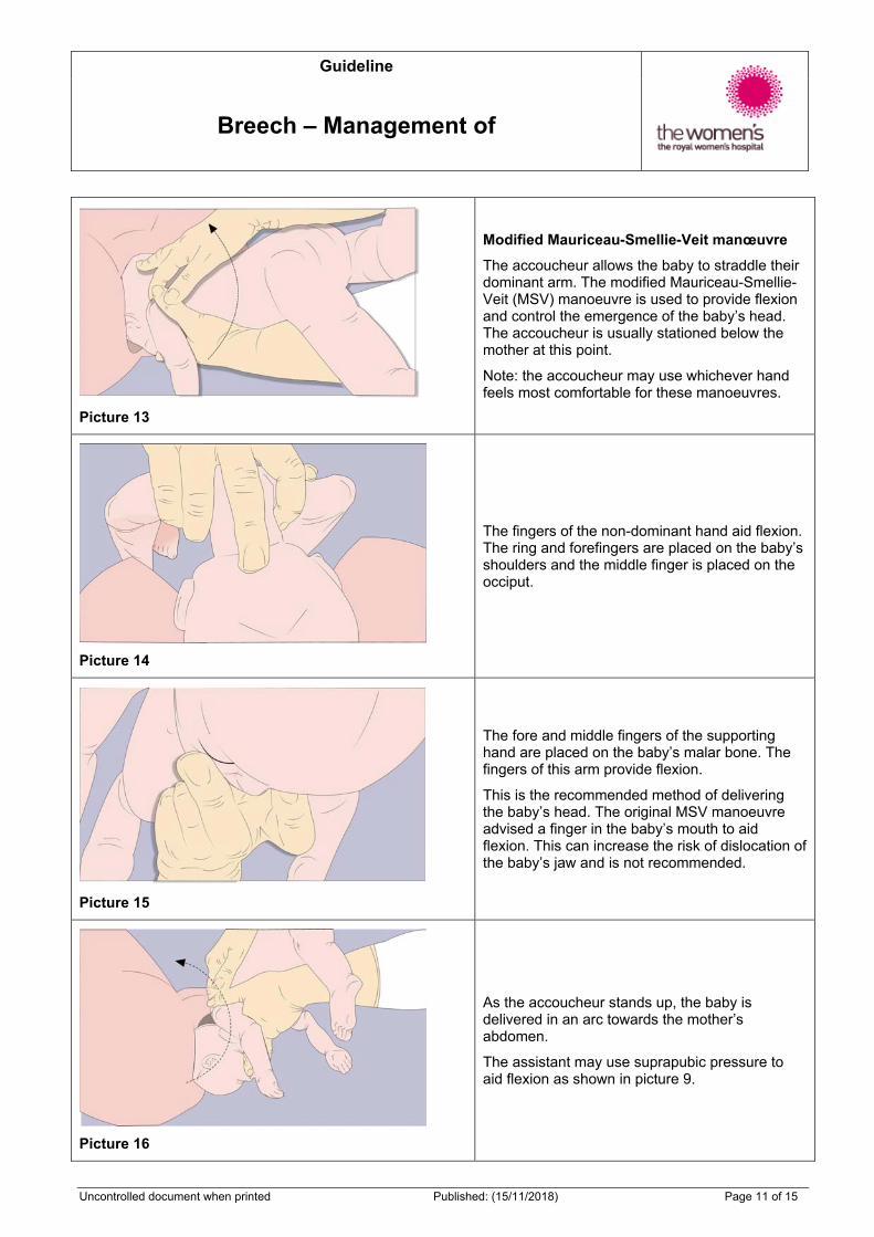
Picture 11
Picture 12
Lovset’s Manoeuvre
In cases where the baby’s arm(s) fail to deliver, the Lovset’s Manoeuvre can be employed. The accoucheur turns the baby 90 degrees into then transverse, reaches over the baby’s shoulder and slips the finger down into the brachial plexus, sweeping the arm down in front of the baby’s body.
Guideline
Breech – Management of
Uncontrolled document when printed Published: (15/11/2018) Page 11 of 15
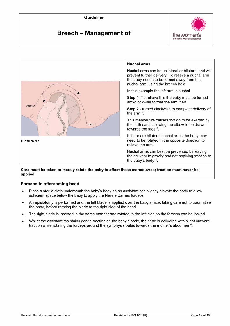
Picture 13
Modified Mauriceau-Smellie-Veit manœuvre
The accoucheur allows the baby to straddle their dominant arm. The modified Mauriceau-Smellie-Veit (MSV) manoeuvre is used to provide flexion and control the emergence of the baby’s head. The accoucheur is usually stationed below the mother at this point.
Note: the accoucheur may use whichever hand feels most comfortable for these manoeuvres.
Picture 14
The fingers of the non-dominant hand aid flexion. The ring and forefingers are placed on the baby’s shoulders and the middle finger is placed on the occiput.
Picture 15
The fore and middle fingers of the supporting hand are placed on the baby’s malar bone. The fingers of this arm provide flexion.
This is the recommended method of delivering the baby’s head. The original MSV manoeuvre advised a finger in the baby’s mouth to aid flexion. This can increase the risk of dislocation of the baby’s jaw and is not recommended.
Picture 16
As the accoucheur stands up, the baby is delivered in an arc towards the mother’s abdomen.
The assistant may use suprapubic pressure to aid flexion as shown in picture 9.
Guideline
Breech – Management of
Uncontrolled document when printed Published: (15/11/2018) Page 12 of 15
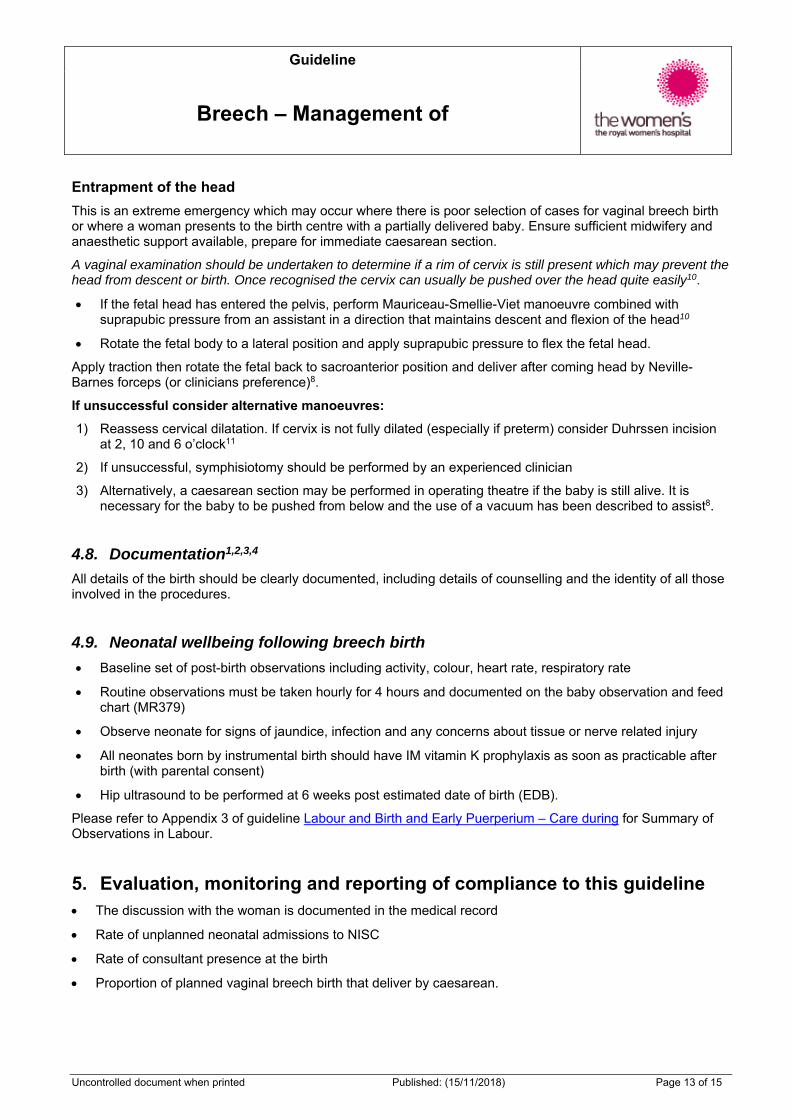
Picture 17
Nuchal arms
Nuchal arms can be unilateral or bilateral and will prevent further delivery. To relieve a nuchal arm the baby needs to be turned away from the nuchal arm, using the breech hold.
In this example the left arm is nuchal.
Step 1- To relieve this the baby must be turned anti-clockwise to free the arm then
Step 2 - turned clockwise to complete delivery of the arm11.
This manoeuvre causes friction to be exerted by the birth canal allowing the elbow to be drawn towards the face 9.
If there are bilateral nuchal arms the baby may need to be rotated in the opposite direction to relieve the arm.
Nuchal arms can best be prevented by leaving the delivery to gravity and not applying traction to the baby’s body11.
Care must be taken to merely rotate the baby to affect these manoeuvres; traction must never be applied.
Forceps to aftercoming head
Place a sterile cloth underneath the baby’s body so an assistant can slightly elevate the body to allow sufficient space below the baby to apply the Neville Barnes forceps
An episiotomy is performed and the left blade is applied over the baby’s face, taking care not to traumatise the baby, before rotating the blade to the right side of the head
The right blade is inserted in the same manner and rotated to the left side so the forceps can be locked
Whilst the assistant maintains gentle traction on the baby’s body, the head is delivered with slight outward traction while rotating the forceps around the symphysis pubis towards the mother’s abdomen10.
Guideline
Breech – Management of
Uncontrolled document when printed Published: (15/11/2018) Page 13 of 15
Entrapment of the head
This is an extreme emergency which may occur where there is poor selection of cases for vaginal breech birth or where a woman presents to the birth centre with a partially delivered baby. Ensure sufficient midwifery and anaesthetic support available, prepare for immediate caesarean section.
A vaginal examination should be undertaken to determine if a rim of cervix is still present which may prevent the head from descent or birth. Once recognised the cervix can usually be pushed over the head quite easily10.
If the fetal head has entered the pelvis, perform Mauriceau-Smellie-Viet manoeuvre combined with suprapubic pressure from an assistant in a direction that maintains descent and flexion of the head10
Rotate the fetal body to a lateral position and apply suprapubic pressure to flex the fetal head.
Apply traction then rotate the fetal back to sacroanterior position and deliver after coming head by Neville-Barnes forceps (or clinicians preference)8.
If unsuccessful consider alternative manoeuvres:
1) Reassess cervical dilatation. If cervix is not fully dilated (especially if preterm) consider Duhrssen incision at 2, 10 and 6 o’clock11
2) If unsuccessful, symphisiotomy should be performed by an experienced clinician
3) Alternatively, a caesarean section may be performed in operating theatre if the baby is still alive. It is necessary for the baby to be pushed from below and the use of a vacuum has been described to assist8.
4.8. Documentation1,2,3,4
All details of the birth should be clearly documented, including details of counselling and the identity of all those involved in the procedures.
4.9. Neonatal wellbeing following breech birth
Baseline set of post-birth observations including activity, colour, heart rate, respiratory rate
Routine observations must be taken hourly for 4 hours and documented on the baby observation and feed chart (MR379)
Observe neonate for signs of jaundice, infection and any concerns about tissue or nerve related injury
All neonates born by instrumental birth should have IM vitamin K prophylaxis as soon as practicable after birth (with parental consent)
Hip ultrasound to be performed at 6 weeks post estimated date of birth (EDB).
Please refer to Appendix 3 of guideline Labour and Birth and Early Puerperium – Care during for Summary of Observations in Labour.
5. Evaluation, monitoring and reporting of compliance to this guideline The discussion with the woman is documented in the medical record
Rate of unplanned neonatal admissions to NISC
Rate of consultant presence at the birth
Proportion of planned vaginal breech birth that deliver by caesarean.
Guideline
Breech – Management of
Uncontrolled document when printed Published: (15/11/2018) Page 14 of 15
6. References 1. Royal College of Obstetricians and Gynaecologists (RCOG) The Management of Breech Presentation
Guideline No. 20b December 2006 p1-13.
2. Women’s Hospitals Australasia (WHA) Breech presentation Clinical Practice Guideline June 2005 p1-15.
3. Kotasa et al, Journal Obstétrique et Gynécologie du Canada (JOGC) Vaginal Delivery of Breech Presentation Clinical Practice Guideline No.226 June 2009 p557-566.
4. King Edward Memorial Hospital Breech Presentation Clinical Guideline 2.10.1 Feb 2009 p1-3.
5. Taillefer, C and Dube, J (2010) Singleton Breech at Term: Two Continents, Two Approaches, JOGC, March, pp.238-243.
6. Jeremy Oats and Suzanne Abraham, Llewellyn-Jones Fundamentals of Obstetrics and Gynaecology, 9th Edition, Mosby Elsevier, United Kingdom 2010: 163-167.
7. Grady,K, Howell,C and Cox,C (2007) Managing Obstetric Emergencies and Trauma, 2nd Edition,RCOGPress,UnitedKingdom,p.273.
8. Cunningham,F.,Leveno,K.,Bloom,S.,Hauth,J.,Rouse,D and Spong, C, Williams Obstetrics,23rd Edition, The McGraw-Hill Companies, Inc, USA, 2010, Chapter 24, p.8.
9. Cronje, H., Cilliers, J. and Pretorius,M, Clinical Obstetrics a South African perspective, 3rd Edition, Van Schaik Publishers, Pretoria, 2011, pp. 371-372.
10. Advanced Life Support in Obstetrics (ALSO) Course Syllabus (2000) Fourth Edition, American Academy of Family Physicians.
11. Vlemmix F, Bergenhenegouwen L, Schaaf JM et al (2014) Term breech deliveries in the Netherlands: did the increased caesarean rate affect neonatal outcome? A population-based study. Acta Obstet Gynecol Scand 93: 888-896
12. Royal Australian and New Zealand College of Obstetricians and Gynaecologists (RANZCOG) The Management of Breech Presentation at Term C-Obs 11 (2013) available at; https://www.ranzcog.edu.au/doc/breech-management-term.html
7. Legislation/Regulations related to this guideline Not applicable.
8. Appendices Not applicable.
Guideline
Breech – Management of
Uncontrolled document when printed Published: (15/11/2018) Page 15 of 15
Please ensure that you adhere to the below disclaimer:
PGP Disclaimer Statement
The Royal Women's Hospital Clinical Guidelines present statements of 'Best Practice' based on thorough evaluation of evidence and are intended for health professionals only. For practitioners outside the Women’s this material is made available in good faith as a resource for use by health professionals to draw on in developing their own protocols, guided by published medical evidence. In doing so, practitioners should themselves be familiar with the literature and make their own interpretations of it.
Whilst appreciable care has been taken in the preparation of clinical guidelines which appear on this web page, the Royal Women's Hospital provides these as a service only and does not warrant the accuracy of these guidelines. Any representation implied or expressed concerning the efficacy, appropriateness or suitability of any treatment or product is expressly negated
In view of the possibility of human error and / or advances in medical knowledge, the Royal Women's Hospital cannot and does not warrant that the information contained in the guidelines is in every respect accurate or complete. Accordingly, the Royal Women's Hospital will not be held responsible or liable for any errors or omissions that may be found in any of the information at this site.
You are encouraged to consult other sources in order to confirm the information contained in any of the guidelines and, in the event that medical treatment is required, to take professional, expert advice from a legally qualified and appropriately experienced medical practitioner.
NOTE: Care should be taken when printing any clinical guideline from this site. Updates to these guidelines will take place as necessary. It is therefore advised that regular visits to this site will be needed to access the most current version of these guidelines.