Bridging Timescales and Length Scales: From Macroscopic Flux to the Molecular Mechanism of Antibiotic Diffusion through Porins Eric Hajjar, † Kozhinjampara R. Mahendran, ‡ Amit Kumar, † Andrey Bessonov, ‡ Mircea Petrescu, ‡ Helge Weingart, ‡ Paolo Ruggerone, † Mathias Winterhalter, ‡ and Matteo Ceccarelli † * † Department of Physics, Universita degli Studi di Cagliari and Sardinian Laboratory for Computational Materials Science, Monserrato, Italy; and ‡ School of Engineering and Science, Jacobs University, Bremen, Germany ABSTRACT Our aim in this study was to provide an atomic description of ampicillin translocation through OmpF, the major outer membrane channel in Escherichia coli and main entry point for b-lactam antibiotics. By applying metadynamics simulations, we also obtained the energy barriers along the diffusion pathway. We then studied the effect of mutations that affect the charge and size at the channel constriction zone, and found that in comparison to the wild-type, much lower energy barriers are required for translocation. The expected higher translocation rates were confirmed on the macroscopic scale by liposome-swelling assays. A microscopic view on the millisecond timescale was obtained by analysis of temperature-dependent ion current fluc- tuations in the presence of ampicillin and provide the enthalpic part of the energy barrier. By studying antibiotic translocation over various timescales and length scales, we were able to discern its molecular mechanism and rate-limiting interactions, and draw biologically relevant conclusions that may help in the design of drugs with enhanced permeation rates. INTRODUCTION Bacteria develop mechanisms of resistance that render the use of antibiotics ineffective (1). Moreover, an increase in multidrug-resistant pathogens is appearing at a time when only a few novel active antibacterial compounds are in clin- ical trials (2). To respond to this alarming situation, we need to reinforce and reinvent antibacterial research. Microscopi- cally based drug design, starting from molecular knowledge of resistant mechanisms, represents a potentially efficient way to bring new agents to the market (3). A key resistance mechanism in Gram-negative bacteria is the prevention of antibiotic uptake, mediated by outer-membrane porins. For example, the resistance of pathogenic bacteria to b-lactams has been attributed to alterations in the expression or the molecular structures of porins (4). The OmpF porin in Escherichia coli has an hourglass shape and the channel structure reveals a spatial constriction created by loop L3, which folds back into the channel. As shown in Fig. 1, this region is also characterized by a transversal electric field created by acidic residues on the L3 side (D113 and E117) facing a cluster of arginines (R42-R82-R132) (5). Several studies have raised questions concerning the role of these amino acids in diffusion processes through OmpF. For example, the single substitutions R132A and D113A were found to dramatically increase the uptake of b-lactams anti- biotics (6,7). Such findings provide investigators with an opportunity to tune the uptake of antibiotics based on only slight chemical modifications. This attractive strategy requires the development of better-tuned quantitative methods to elucidate the rate-limiting molecular interactions between drug and channel residues. In this work, we studied antibiotic diffusion by combining atomic-level descriptions provided by molecular-dynamics (MD) simulations (8), elec- trophysiology techniques at the single-molecule level (9), and liposome-swelling assays (10). Our findings reveal, for the first time to our knowledge, the complete pathways of ampicillin permeation through wild-type (WT) OmpF as well as D113N and R132A mutants, from their macroscopic flux down to their molecular mechanism. MATERIALS AND METHODS MD simulations For the MD simulations, we followed a previously described protocol (8), starting from the crystal structure (Protein Data Bank code: 2OMF) and residue protonation state as described by Im and Roux (11). We used the program ORAC and the Amber force field (12) for system setup and simu- lation (13). The porin mutants were obtained by substituting the single amino acid residue starting from the high-resolution structure of OmpF (2OMF) using the MD package ORAC. After the molecular replacement was completed, we further equilibrated the mutant system for ~2 ns of a stan- dard MD simulation. All simulated systems were validated for convergence and stabilization of energy, temperature, and root mean-square deviation (RMSD) with respect to the starting structure. Based on previous findings (7,8,14), we chose the following collective variables to simulate antibiotic translocation using metadynamics (15): 1), the distance Z, defined as the difference between the center of mass of the antibiotic and the center of mass of the system (porin þ detergent) along the z axis; and 2), the angle q, defined as the orientation of the long axis of the molecule with respect to the z axis. All simulated systems were validated for convergence and stabilization of energy, temperature, and RMSD with respect to the start- ing structure. Our choice of OmpF as a monomer is supported by previous studies that reported a mutual independence of the three monomers (i.e., no cooperativity) for ions, small-molecule transport, and antibiotics (16–18). Using this biased simulation strategy, we obtained translocation of ampi- cillin through WT OMPF, D113N, and R132A after 38, 27, and 15 ns, respectively. The metadynamics algorithm enables one to reconstruct the free energy in the subspace of the collective variables by integrating the Submitted August 20, 2009, and accepted for publication October 15, 2009. *Correspondence: [email protected]Editor: Benoit Roux. Ó 2010 by the Biophysical Society 0006-3495/10/02/0569/7 $2.00 doi: 10.1016/j.bpj.2009.10.045 Biophysical Journal Volume 98 February 2010 569–575 569
Transcript
Biophysical Journal Volume 98 February 2010 569–575 569
Bridging Timescales and Length Scales: From Macroscopic Fluxto the Molecular Mechanism of Antibiotic Diffusion through Porins
Eric Hajjar,† Kozhinjampara R. Mahendran,‡ Amit Kumar,† Andrey Bessonov,‡ Mircea Petrescu,‡ Helge Weingart,‡
Paolo Ruggerone,† Mathias Winterhalter,‡ and Matteo Ceccarelli†*†Department of Physics, Universita degli Studi di Cagliari and Sardinian Laboratory for Computational Materials Science, Monserrato, Italy;and ‡School of Engineering and Science, Jacobs University, Bremen, Germany
ABSTRACT Our aim in this study was to provide an atomic description of ampicillin translocation through OmpF, the majorouter membrane channel in Escherichia coli and main entry point for b-lactam antibiotics. By applying metadynamics simulations,we also obtained the energy barriers along the diffusion pathway. We then studied the effect of mutations that affect the chargeand size at the channel constriction zone, and found that in comparison to the wild-type, much lower energy barriers are requiredfor translocation. The expected higher translocation rates were confirmed on the macroscopic scale by liposome-swellingassays. A microscopic view on the millisecond timescale was obtained by analysis of temperature-dependent ion current fluc-tuations in the presence of ampicillin and provide the enthalpic part of the energy barrier. By studying antibiotic translocationover various timescales and length scales, we were able to discern its molecular mechanism and rate-limiting interactions,and draw biologically relevant conclusions that may help in the design of drugs with enhanced permeation rates.
INTRODUCTION
Bacteria develop mechanisms of resistance that render the
use of antibiotics ineffective (1). Moreover, an increase in
multidrug-resistant pathogens is appearing at a time when
only a few novel active antibacterial compounds are in clin-
ical trials (2). To respond to this alarming situation, we need
to reinforce and reinvent antibacterial research. Microscopi-
cally based drug design, starting from molecular knowledge
of resistant mechanisms, represents a potentially efficient
way to bring new agents to the market (3). A key resistance
mechanism in Gram-negative bacteria is the prevention of
antibiotic uptake, mediated by outer-membrane porins. For
example, the resistance of pathogenic bacteria to b-lactams
has been attributed to alterations in the expression or the
molecular structures of porins (4). The OmpF porin in
Escherichia coli has an hourglass shape and the channel
structure reveals a spatial constriction created by loop L3,
which folds back into the channel. As shown in Fig. 1, this
region is also characterized by a transversal electric field
created by acidic residues on the L3 side (D113 and E117)
facing a cluster of arginines (R42-R82-R132) (5). Several
studies have raised questions concerning the role of these
amino acids in diffusion processes through OmpF. For
example, the single substitutions R132A and D113A were
found to dramatically increase the uptake of b-lactams anti-
biotics (6,7). Such findings provide investigators with an
opportunity to tune the uptake of antibiotics based on
only slight chemical modifications. This attractive strategy
requires the development of better-tuned quantitative
methods to elucidate the rate-limiting molecular interactions
between drug and channel residues. In this work, we studied
Submitted August 20, 2009, and accepted for publication October 15, 2009.
ture-dependent blocking events of ampicillin with WT
OmpF and D113N. The continuous line represents the
exponential fit. (B) Effect of temperature on the antibiotic
residence time (t) for WT OmpF and D113N mutant.
572 Hajjar et al.
We then used additional methods to bridge the timescales
and length scales of transport, to clarify the case of R132A in
which only partial blocking events were observed at all
measured temperatures.
To elucidate the energetic details of transport, we per-
formed metadynamics simulations of ampicillin transloca-
tion through WT OmpF, D113N, and R132A mutants.
From the reconstructed 1D free-energy profiles (Fig. 4), we
observe that the effective barrier for ampicillin to translocate
is higher in the case of WT OmpF (14 kT) and lower for
R132A (9 kT) and D113N (5 kT). To compare these findings
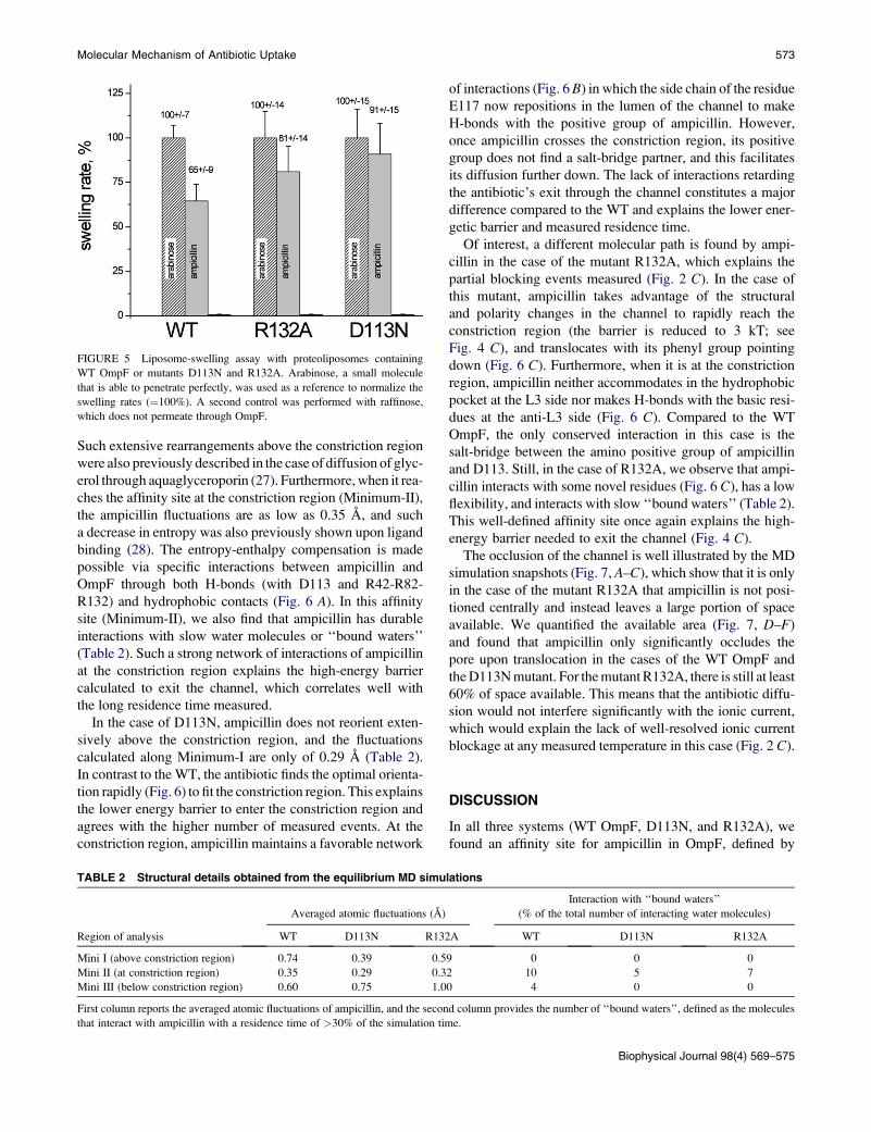
with our predictions, we quantified the in vitro macroscopic
flux of ampicillin using a liposome-swelling assay, a method
that has been successfully applied to such problems in
previous studies (10,25). The advantage of this technique
is that the penetration rates of ampicillin in proteoliposomes
generally mimic those of the intact cells, and swelling rates
are directly proportional to the permeability of the antibiotic.
As shown in Fig. 5, we find a higher diffusion rate of ampi-
cillin for both mutants, as it increases by 25% for R132A and
40% for D113N compared to the WT OmpF. The trend of the
FIGURE 4 One-dimensional free-energy profiles for the translocation of ampic
constriction region are highlighted in gray and the energy barriers are reported i
Biophysical Journal 98(4) 569–575
flux is in good agreement with that of the energy barriers
obtained from the molecular simulations. Taken together,
our results demonstrate that a single point mutation at the
constriction region can remarkably affect the kinetics of
ampicillin and thus its uptake.
To elucidate the molecular mechanism and rationalize our
findings, we then deciphered the physicochemical and struc-
tural properties of the diffusion process. Each relevant
minimum identified by the metadynamics (as labeled in
Fig. 4) was used as a starting point for the additional equilib-
rium MD simulations for which we analyzed the solvation,
flexibility, SASA, and interaction patterns between ampi-
cillin and the OmpF residues.
In the case of the WT, when ampicillin is above the con-
striction region (in the structures sampled along the equilib-
rium simulations at Minimum-I), it interacts only transiently
with channel residues, as the antibiotic undergoes numerous
reorientations and attempts to enter the constriction zone.
This is confirmed by the averaged atomic fluctuations of
ampicillin of 0.78 A (along Minimum-I), which is close to
the antibiotic fluctuation calculated in bulk water (Table 2).
illin through WT OmpF (A), D113N (B), and R132A (C). The minima at the
n kT. The ‘‘exit’’ label refers to the periplasmic side.
FIGURE 5 Liposome-swelling assay with proteoliposomes containing
WT OmpF or mutants D113N and R132A. Arabinose, a small molecule
that is able to penetrate perfectly, was used as a reference to normalize the
swelling rates (¼100%). A second control was performed with raffinose,
which does not permeate through OmpF.
Molecular Mechanism of Antibiotic Uptake 573
Such extensive rearrangements above the constriction region
were also previously described in the case of diffusion of glyc-
erol through aquaglyceroporin (27). Furthermore, when it rea-
ches the affinity site at the constriction region (Minimum-II),
the ampicillin fluctuations are as low as 0.35 A, and such
a decrease in entropy was also previously shown upon ligand
binding (28). The entropy-enthalpy compensation is made
possible via specific interactions between ampicillin and
OmpF through both H-bonds (with D113 and R42-R82-
R132) and hydrophobic contacts (Fig. 6 A). In this affinity
site (Minimum-II), we also find that ampicillin has durable
interactions with slow water molecules or ‘‘bound waters’’
(Table 2). Such a strong network of interactions of ampicillin
at the constriction region explains the high-energy barrier
calculated to exit the channel, which correlates well with
the long residence time measured.
In the case of D113N, ampicillin does not reorient exten-
sively above the constriction region, and the fluctuations
calculated along Minimum-I are only of 0.29 A (Table 2).
In contrast to the WT, the antibiotic finds the optimal orienta-
tion rapidly (Fig. 6) to fit the constriction region. This explains
the lower energy barrier to enter the constriction region and
agrees with the higher number of measured events. At the
constriction region, ampicillin maintains a favorable network
TABLE 2 Structural details obtained from the equilibrium MD simu
Averaged atomic fluctuations (A)
Region of analysis WT D113N R132
Mini I (above constriction region) 0.74 0.39 0.5
Mini II (at constriction region) 0.35 0.29 0.3
Mini III (below constriction region) 0.60 0.75 1.0
First column reports the averaged atomic fluctuations of ampicillin, and the secon
that interact with ampicillin with a residence time of >30% of the simulation tim
of interactions (Fig. 6 B) in which the side chain of the residue
E117 now repositions in the lumen of the channel to make
H-bonds with the positive group of ampicillin. However,
once ampicillin crosses the constriction region, its positive
group does not find a salt-bridge partner, and this facilitates
its diffusion further down. The lack of interactions retarding
the antibiotic’s exit through the channel constitutes a major
difference compared to the WT and explains the lower ener-
getic barrier and measured residence time.
Of interest, a different molecular path is found by ampi-
cillin in the case of the mutant R132A, which explains the
partial blocking events measured (Fig. 2 C). In the case of
this mutant, ampicillin takes advantage of the structural
and polarity changes in the channel to rapidly reach the
constriction region (the barrier is reduced to 3 kT; see
Fig. 4 C), and translocates with its phenyl group pointing
down (Fig. 6 C). Furthermore, when it is at the constriction
region, ampicillin neither accommodates in the hydrophobic
pocket at the L3 side nor makes H-bonds with the basic resi-
dues at the anti-L3 side (Fig. 6 C). Compared to the WT
OmpF, the only conserved interaction in this case is the
salt-bridge between the amino positive group of ampicillin
and D113. Still, in the case of R132A, we observe that ampi-
cillin interacts with some novel residues (Fig. 6 C), has a low
flexibility, and interacts with slow ‘‘bound waters’’ (Table 2).
This well-defined affinity site once again explains the high-
energy barrier needed to exit the channel (Fig. 4 C).
The occlusion of the channel is well illustrated by the MD
simulation snapshots (Fig. 7, A–C), which show that it is only
in the case of the mutant R132A that ampicillin is not posi-
tioned centrally and instead leaves a large portion of space
available. We quantified the available area (Fig. 7, D–F)
and found that ampicillin only significantly occludes the
pore upon translocation in the cases of the WT OmpF and
the D113N mutant. For the mutant R132A, there is still at least
60% of space available. This means that the antibiotic diffu-
sion would not interfere significantly with the ionic current,
which would explain the lack of well-resolved ionic current
blockage at any measured temperature in this case (Fig. 2 C).
DISCUSSION
In all three systems (WT OmpF, D113N, and R132A), we
found an affinity site for ampicillin in OmpF, defined by
lations
Interaction with ‘‘bound waters’’
(% of the total number of interacting water molecules)
A WT D113N R132A
9 0 0 0
2 10 5 7
0 4 0 0
d column provides the number of ‘‘bound waters’’, defined as the molecules
e.
Biophysical Journal 98(4) 569–575
FIGURE 6 Molecular details (side views) of ampicillin at the binding site of the constriction region of (A) WT OmpF, (B) D113N, and (C) R132A. The views and
orientations in this figure are the same as in Fig. 1 (the top is toward the vestibule, the bottom is toward the periplasmic space). The antibiotic is displayed in stick
representation and colored by atom type (blue for nitrogen, red for oxygen, cyan for carbon) where hydrogens are not shown. The backbone of OmpF is displayed in
gray cartoons to highlight its secondary structures. The constriction region is highlighted by loop L3 (colored in orange). Residues of OmpF that are seen as
strongly interacting with the antibiotic are labeled using the one-letter amino acid code; those making H-bonds are colored by residue type (positively charged
in blue, negatively charged in red, polar in green), and those making hydrophobic contacts are displayed with their molecular surface, highlighting their shape.
574 Hajjar et al.
specific interactions with key residues of the constriction
region and with strongly ordered water molecules. For the
concentration used here (far from saturation and within the
limit of the physiological dose), the only parameter that
FIGURE 7 Molecular details (top view) from equilibrium simulations started a
cillin is displayed in stick representation and colored according to atom type. The
residues making H-bonds are colored by residue type, and those making hydrop
SASA is reported for WT OmpF (D), D113N (E), and R132A (F) in the presen
Biophysical Journal 98(4) 569–575
makes a difference in the flux is kon. Of interest, we can con-
clude from our results that kon can be tuned by mutations at
the constriction region that affect, very locally, the molecular
interactions. The fact that ampicillin uptake can be tuned by
t Minima-II for WT OmpF (A), D113N (B), and R132A (C) mutants. Ampi-
backbone of OmpF is shown by gray cartoons (L3 is colored orange). The
hobic contacts are displayed by gray molecular surface. Below, the average
ce (black) and absence (red) of ampicillin.
Molecular Mechanism of Antibiotic Uptake 575
specific interactions inside the channel is in agreement with
previous theoretical and experimental studies (11,23,29–31).
Cornell et al. (12) assumed that kon is only dependent on the
diffusion coefficient of the molecule and the radius of attrac-
tion. This implies that, surprisingly, even very local interac-
tions at the constriction region must be taken into account in
the definition of this radius.
We conclude that the bottleneck for antibiotic transloca-
tion stems from the difficulty of overcoming the constriction
region. In the case of WT OmpF, ampicillin has to deal with
a particularly reduced size and a strong electrostatic field.
Using computer simulations, we were able to predict when
the presence of an ampicillin molecule would block the ion
current, and thus rationalize the ion current fluctuations
induced by antibiotics upon translocation.
By combining different approaches, we were able to
follow the ampicillin translocation process over various
timescales and length scales. This allowed us to reveal the
complete molecular mechanism of diffusion and relate it to
biologically relevant conclusions. The identification of
crucial antibiotic-channel interactions will benefit the design
of novel molecules with enhanced permeation rates. Finally,
we believe that our multiscale approach can be conveniently
employed to study porin-antibiotic interactions in other
enterobacterial pathogens (1,4), such as those involved in
persistent tuberculosis.
We thank Tivadar Mach, Malcom Page, and Jurg Dreier for their support and
productive discussions.
This study was supported by the European Union, FP6 grant MRTN-CT-
2005-019335 (Translocation), and by the computer center and consortiums
Cybersar, CASPUR, and CINECA through CPU hours.
REFERENCES
1. Arias, C. A., and B. E. Murray. 2009. Antibiotic-resistant bugs in the21st century—a clinical super-challenge. N. Engl. J. Med. 360:439–443.
2. Spellberg, B., J. H. Powers, ., J. E. Edwards, Jr. 2004. Trends in anti-microbial drug development: implications for the future. Clin. Infect.Dis. 38:1279–1286.
3. Barker, J. J. 2006. Antibacterial drug discovery and structure-baseddesign. Drug Discov. Today. 11:391–404.
4. Pages, J. M., C. E. James, and M. Winterhalter. 2008. The porin and thepermeating antibiotic: a selective diffusion barrier in Gram-negativebacteria. Nat. Rev. Microbiol. 6:893–903.
5. Cowan, S. W., T. Schirmer, ., J. P. Rosenbusch. 1992. Crystal struc-tures explain functional properties of two E. coli porins. Nature.358:727–733.
6. Simonet, V., M. Mallea, and J. M. Pages. 2000. Substitutions in theeyelet region disrupt cefepime diffusion through the Escherichia coliOmpF channel. Antimicrob. Agents Chemother. 44:311–315.
7. Vidal, S., J. Bredin, ., J. Barbe. 2005. b-Lactam screening by specificresidues of the OmpF eyelet. J. Med. Chem. 48:1395–1400.
8. Ceccarelli, M., C. Danelon, ., M. Parrinello. 2004. Microscopic mech-anism of antibiotics translocation through a porin. Biophys. J. 87:58–64.
9. Kozhinjampara, M., C. Chimerel, ., M. Winterhalter. 2009. Antibiotictranslocation through membrane channels: temperature-dependention current fluctuation for catching the fast events. Eur. Biophys. J.,in press.
10. Yoshimura, F., and H. Nikaido. 1985. Diffusion of b-lactam antibioticsthrough the porin channels of Escherichia coli K-12. Antimicrob.Agents Chemother. 27:84–92.
11. Im, W., and B. Roux. 2002. Ions and counterions in a biologicalchannel: a molecular dynamics simulation of OmpF porin from Escher-ichia coli in an explicit membrane with 1 M KCl aqueous salt solution.J. Mol. Biol. 319:1177–1197.
12. Cornell, W. D., P. Cieplak, ., P. Kollmann. 1995. J. Am. Chem. Soc.117:5179–5197.
13. Procacci, P., T. A. Darden, ., M. Marchi. 1997. ORAC: a moleculardynamics program to simulate complex molecular systems with realisticelectrostatic interactions. J. Comput. Chem. 18:1848–1862.
14. Danelon, C., E. M. Nestorovich, ., S. M. Bezrukov. 2006. Interactionof zwitterionic penicillins with the OmpF channel facilitates their trans-location. Biophys. J. 90:1617–1627.
15. Laio, A., and M. Parrinello. 2002. Escaping free-energy minima. Proc.Natl. Acad. Sci. USA. 99:12562–12566.
16. Robertson, K. M., and D. P. Tieleman. 2002. Orientation and interac-tions of dipolar molecules during transport through OmpF porin.FEBS Lett. 528:53–57.
17. Rostovtseva, T. K., E. M. Nestorovich, and S. M. Bezrukov. 2002.Partitioning of differently sized poly(ethylene glycol)s into OmpFporin. Biophys. J. 82:160–169.
18. Nestorovich, E. M., C. Danelon, ., S. M. Bezrukov. 2002. Designed topenetrate: time-resolved interaction of single antibiotic molecules withbacterial pores. Proc. Natl. Acad. Sci. USA. 99:9789–9794.
19. Laio, A., A. Rodriguez-Fortea, ., M. Parrinello. 2005. Assessing theaccuracy of metadynamics. J. Phys. Chem. B. 109:6714–6721.
20. Sterpone, F., M. Ceccarelli, and M. Marchi. 2001. Dynamics of hydra-tion in hen egg white lysozyme. J. Mol. Biol. 311:409–419.
21. Mach, T., P. Neves, ., P. Gameiro. 2008. Facilitated permeation ofantibiotics across membrane channels—interaction of the quinolonemoxifloxacin with the OmpF channel. J. Am. Chem. Soc. 130:13301–13309.
22. Humphrey, W., A. Dalke, and K. Schulten. 1996. VMD: visual molec-ular dynamics. J. Mol. Graph. 14:33–38, 27–28.
23. Berezhkovskii, A. M., and S. M. Bezrukov. 2005. Optimizing transportof metabolites through large channels: molecular sieves with andwithout binding. Biophys. J. 88:L17–L19.
24. Weingart, H., M. Petrescu, and M. Winterhalter. 2008. Biophysicalcharacterization of in- and efflux in Gram-negative bacteria. Curr.Drug Targets. 9:789–796.
25. Nikaido, H., and E. Y. Rosenberg. 1983. Porin channels in Escherichiacoli: studies with liposomes reconstituted from purified proteins. J. Bac-teriol. 153:241–252.
26. Nekolla, S., C. Andersen, and R. Benz. 1994. Noise analysis of ioncurrent through the open and the sugar-induced closed state of theLamB channel of Escherichia coli outer membrane: evaluation of thesugar binding kinetics to the channel interior. Biophys. J. 66:1388–1397.
27. Henin, J., E. Tajkhorshid, ., C. Chipot. 2008. Diffusion of glycerolthrough Escherichia coli aquaglyceroporin GlpF. Biophys. J. 94:832–839.
28. Jusuf, S., P. J. Loll, and P. H. Axelsen. 2003. Configurational entropyand cooperativity between ligand binding and dimerization in glycopep-tide antibiotics. J. Am. Chem. Soc. 125:3988–3994.
29. Berezhkovskii, A. M., M. A. Pustovoit, and S. M. Bezrukov. 2003.Channel-facilitated membrane transport: average lifetimes in thechannel. J. Chem. Phys. 119:3943–3951.
30. Berezhkovskii, A. M., G. Hummer, and S. M. Bezrukov. 2006. Identityof distributions of direct uphill and downhill translocationtimes for particles traversing membrane channels. Phys. Rev. Lett.97:020601.
31. Tieleman, D. P., and H. J. Berendsen. 1998. A molecular dynamicsstudy of the pores formed by Escherichia coli OmpF porin in a fullyhydrated palmitoyloleoylphosphatidylcholine bilayer. Biophys. J.74:2786–2801.