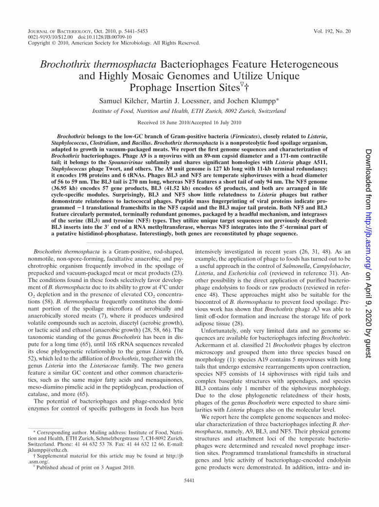
Prophage Insertion Sites�†Samuel Kilcher, Martin J. Loessner, and Jochen Klumpp*
Institute of Food, Nutrition and Health, ETH Zurich, 8092 Zurich, Switzerland
Received 18 June 2010/Accepted 16 July 2010
Brochothrix belongs to the low-GC branch of Gram-positive bacteria (Firmicutes), closely related to Listeria,Staphylococcus, Clostridium, and Bacillus. Brochothrix thermosphacta is a nonproteolytic food spoilage organism,adapted to growth in vacuum-packaged meats. We report the first genome sequences and characterization ofBrochothrix bacteriophages. Phage A9 is a myovirus with an 89-nm capsid diameter and a 171-nm contractiletail; it belongs to the Spounavirinae subfamily and shares significant homologies with Listeria phage A511,Staphylococcus phage Twort, and others. The A9 unit genome is 127 kb long with 11-kb terminal redundancy;it encodes 198 proteins and 6 tRNAs. Phages BL3 and NF5 are temperate siphoviruses with a head diameterof 56 to 59 nm. The BL3 tail is 270 nm long, whereas NF5 features a short tail of only 94 nm. The NF5 genome(36.95 kb) encodes 57 gene products, BL3 (41.52 kb) encodes 65 products, and both are arranged in lifecycle-specific modules. Surprisingly, BL3 and NF5 show little relatedness to Listeria phages but ratherdemonstrate relatedness to lactococcal phages. Peptide mass fingerprinting of viral proteins indicate pro-grammed �1 translational frameshifts in the NF5 capsid and the BL3 major tail protein. Both NF5 and BL3feature circularly permuted, terminally redundant genomes, packaged by a headful mechanism, and integrasesof the serine (BL3) and tyrosine (NF5) types. They utilize unique target sequences not previously described:BL3 inserts into the 3� end of a RNA methyltransferase, whereas NF5 integrates into the 5�-terminal part ofa putative histidinol-phosphatase. Interestingly, both genes are reconstituted by phage sequence.
Brochothrix thermosphacta is a Gram-positive, rod-shaped,nonmotile, non-spore-forming, facultative anaerobic, and psy-chrotrophic organism frequently involved in the spoilage ofprepacked and vacuum-packaged meat or meat products (23).The conditions found in these foods selectively favor develop-ment of B. thermosphacta due to its ability to grow at 4°C underO2 depletion and in the presence of elevated CO2 concentra-tions (58). B. thermosphacta frequently constitutes the domi-nant portion of the spoilage microflora of aerobically andanaerobically stored meats (7), where it produces undesiredvolatile compounds such as acetoin, diacetyl (aerobic growth),or lactic acid and ethanol (anaerobic growth) (28, 58, 66). Thetaxonomic standing of the genus Brochothrix has been in dis-pute for a long time (65), until 16S rRNA sequences revealedits close phylogenetic relationship to the genus Listeria (16,52), which led to the affiliation of Brochothrix, together with thegenus Listeria into the Listeriaceae family. The two generafeature a similar GC content and other common characteris-tics, such as the same major fatty acids and menaquinones,meso-diamino pimelic acid in the peptidoglycan, production ofcatalase, and more (65).
The potential of bacteriophages and phage-encoded lyticenzymes for control of specific pathogens in foods has been
intensively investigated in recent years (26, 31, 48). As anexample, the application of phage to foods has turned out to bea useful approach in the control of Salmonella, Campylobacter,Listeria, and Escherichia coli (reviewed in reference 31). An-other possibility is the direct application of purified bacterio-phage endolysins to foods or raw products (reviewed in refer-ence 48). These approaches might also be suitable for thebiocontrol of B. thermosphacta to prevent food spoilage. Pre-vious work has shown that Brochothrix phage A3 was able tolimit off-odor formation and increase the storage life of porkadipose tissue (28).
Unfortunately, only very limited data and no genome se-quences are available for bacteriophages infecting Brochothrix.Ackermann et al. classified 21 Brochothrix phages by electronmicroscopy and grouped them into three species based onmorphology (1): species A19 contains 5 myoviruses with longtails that undergo extensive rearrangements upon contraction,species NF5 consists of 14 siphoviruses with rigid tails andcomplex baseplate structures with appendages, and speciesBL3 contains only 1 member of the siphovirus morphology.Due to the close phylogenetic relatedness of their hosts,phages of the genus Brochothrix were expected to share simi-larities with Listeria phages also on the molecular level.
We report here the complete genome sequences and molec-ular characterization of three bacteriophages infecting B. ther-mosphacta, namely, A9, BL3, and NF5. Their physical genomestructures and attachment loci of the temperate bacterio-phages were determined and revealed novel prophage inser-tion sites. Programmed translational frameshifts in structuralgenes and lytic activity of bacteriophage-encoded endolysingene products were demonstrated. In addition, intra- and in-
* Corresponding author. Mailing address: Institute of Food, Nutri-tion and Health, ETH Zurich, Schmelzbergstrasse 7, CH-8092 Zurich,Switzerland. Phone: 41 44 632 53 78. Fax: 41 44 632 12 66. E-mail:[email protected].
† Supplemental material for this article may be found at http://jb.asm.org/.
terspecies comparative genomics led to the placement of phageA9 into the recently proposed Spounavirinae subfamily, andindicated relatedness of phages BL3 and NF5 to the P335quasispecies of Lactococcus phages.
MATERIALS AND METHODS
Bacterial strains and bacteriophages. The bacterial strains used in the presentstudy were B. thermosphacta HER1187 and HER1188 (Felix d’Herelle Refer-ence Center for Bacterial Viruses, Quebec, Canada [www.phage.ulaval.ca]) andE. coli XL1-Blue MRF� (Stratagene, Santa Clara, CA) and those listed in TableS1 in the supplemental material. Bacteria were grown at 24°C (Brochothrix), at30°C (Listeria), or at 37°C (Escherichia, Bacillus, Enterococcus, and Staphylococ-cus) under constant agitation in half-strength brain heart infusion (BHI; Difco/BD, Basel, Switzerland) (for Listeria and Brochothrix) or Luria-Bertani (LB)broth (Oxoid, Hampshire, United Kingdom) (for E. coli). Bacteriophages A9,NF5, and BL3 were purchased from the Felix d’Herelle Reference Center forBacterial Viruses as lysates and stored at 4°C.
Phage growth and multiplication parameters. Portions (10 ml) of a log-phaseculture (optical density at 600 nm of 0.5, �107 CFU/ml) of B. thermosphactaHER1187 were harvested by centrifugation and resuspended in 2.5 ml of freshBHI medium. Then, 5 � 104 PFU of phage A9 were added and allowed to adsorbfor 5 min at 24°C. Free phage was removed by centrifugation, and the cell pelletwas resuspended in 10 ml of fresh medium. Incubation was continued at 24°C,and samples were plated by soft-agar overlay (2) for free phage counts at varioustime points between 15 and 170 min postinfection. The log10 (PFU/ml) related totime (minutes) was plotted, and data points were fitted by using a sigmoidalfive-parameter approximation (SigmaPlot version 11; Systat Software, Inc., Chi-cago, IL). The burst size was determined from triplicate experiments by phagenumbers after the burst (t � 160 min) in relation to the initial titer at lag phase(t � 45 min).
Phage propagation and DNA isolation and manipulation. Phages were puri-fied and propagated by the standard soft-agar overlay method (2). SM buffer(100 mM NaCl, 8 mM MgSO4 and 50 mM Tris-HCl [pH 7.4]) was used to extractphages from plates. NF5 was propagated in B. thermosphacta HER1188 cells byusing LB bottom and LC (LB agar supplemented with 10 mM CaCl) top agar.Phages BL3 and A9 were propagated in B. thermosphacta HER1187 cells usingBHI-LC and LB-LC double layers, respectively. Phages were concentrated andpurified by polyethylene glycol precipitation (PEG 8000, 7% in the presence of1 M NaCl), stepped CsCl gradient ultracentrifugation (60), and dialysis againsta 1,000-fold excess of SM buffer at 4°C. DNA was extracted with organic solventsas described previously (37).
Phage DNA (0.5-�g samples) was digested with restriction enzymes accordingto the manufacturer’s instructions (Fermentas, St. Leon-Roth, Germany, or NewEngland Biolabs, Ipswich, United Kingdom). The products were analyzed elec-trophoretically. Bal31 nuclease treatment was performed by digestion of 40 �g ofphage DNA with 20 U of Bal31 (New England Biolabs) at 30°C. Samples wereremoved 0, 5, 10, 20, 40, 60, 90, and 120 min after enzyme addition and purifiedby organic extraction (60). An equal volume of each sample was digested with theindicated restriction enzyme, and products were electrophoresed in a GNA200horizontal electrophoresis apparatus (GE Healthcare, Switzerland) at 2 V/cm in1� Tris-acetate-EDTA buffer. Pulsed-field gel electrophoresis (PFGE) was per-formed in 0.5� Tris-borate-EDTA buffer with an initial switch time of 1 s and afinal switch time of 6 s at 5 V/cm, at an angle of 120° and a 14°C buffertemperature (CHEF DR III apparatus; Bio-Rad, Rainach, Switzerland) for 20 h.Lambda Mix 19 (Fermentas) and MidRange I PFG and MidRange II PFGmarkers (New England Biolabs) were used as DNA size standards.
Electron microscopy. Purified phage particles recovered from density gradi-ents were negatively stained with 2% sodium phosphotungstic acid (PTA), 2%uranyl acetate (UA), or 2% ammonium molybdate (AM) (67). Samples wereobserved in a Tecnai G2 Spirit instrument (at 120 kV) equipped with an Eaglecharge-coupled-device camera (FEI Company, Hillsboro, OR).
Genome sequencing. DNA shotgun-libraries were prepared after fragmenta-tion by sonication of purified genomic DNA (Sonopuls; Bandelin Electronics,Berlin, Germany). Fragments of the desired lengths were recovered from aga-rose gels, DNA ends were repaired (EndIt kit; Epicentre Biotechnologies, Mad-ison, WI), and ligated into EcoRV-digested pBluescript II SK(�) (Stratagene).Ligation products were transformed into E. coli XL1-Blue MRF�. Blue-whitescreening on ampicillin (100 �g/ml)-, IPTG (isopropyl-�-D-thiogalactopyrano-side)-, and X-Gal (5-bromo-4-chloro-3-indolyl-�-D-galactopyranoside)-contain-ing plates was used to identify plasmid-bearing clones. The presence of insertswas verified by PCR, and inserts were sequenced by using M13forward and
M13reverse primers (see Table S2 in the supplemental material). Obtainednucleotide sequences were edited and aligned by using CLC Genomics Work-bench (version 3.5; CLC Bio, Aarhus, Denmark) and SeqMan Pro (LasergeneSeqMan Pro version 8.1; DNASTAR, Madison, WI) software. Gaps betweencontigs were closed by a primer-walking strategy using purified genomic DNA asa template. Primers were derived from the contig sequences as they becameavailable. Sequences with insufficient coverage or ambiguities were verified byprimer walking.
Determination of the physical genome structure and packaging mode. Thepresence of putative pac sites was evaluated by restriction profiling, which wasalso used to verify the assembled genome sequences. To exclude the presence ofcohesive DNA ends, 0.5 �g of purified genomic DNA was digested with restric-tion enzymes, heated to 62°C for 10 min, and immediately separated by electro-phoresis. A nonheated sample was used as a control. Digestion of genomic phageDNA with Bal31 exonuclease, prior to restriction digestion, was performed asdescribed previously (see above and reference 37). Large terminase amino acidsequences of 86 phages with known DNA structure were compared as describedpreviously (13); sequences were aligned by using the built-in alignment tool ofCLC Genomics Workbench, and phylogenetic trees were generated with theneighbor-joining algorithm (1,000 bootstrap replicates).
Mass spectrometry (MS) and peptide mass fingerprinting. Phage proteinswere separated by horizontal sodium dodecyl sulfate-polyacrylamide gel electro-phoresis (SDS-PAGE) on gradient gels (ExcelGel SDS, 8 to 18% gradient; GEHealthcare, Glattbrugg, Switzerland) as previously described (37, 74). Majorprotein bands were excised and, after tryptic in-gel digestion, analyzed byliquid chromatography-tandem MS and electrospray ionization-tandem MSto determine masses of the fragments (37, 74). The data were analyzed byusing the Scaffold Proteome Software (version 2.0; Proteome Software, Inc.,Portland, OR).
Identification of attB and attP. In order to obtain stable lysogens, the respec-tive Brochothrix host cells were infected with phage NF5 or BL3 for 1 h at amultiplicity of infection (MOI) of 1,000 in liquid culture at 24°C. Serial dilutionswere plated, and surviving colonies were restreaked onto fresh agar plates.Selected lysogens were assayed for immunity against infection with the samephage (homoimmunity screening) and for the presence of phage-specific DNAsequences by PCR. B. thermosphacta HER1187::BL3 and HER1188::NF5 chro-mosomal DNA was purified (60). BL3 and NF5 integration sites (one transitionposition) were identified as previously described (54), using two divergent prim-ers BL3_iPCR_fwd/BL3_iPCR_rev and NF5_iPCR_fwd/NF5_iPCR_rev, derivedfrom the putative BL3 and NF5 phage integrase genes, respectively. Primers arelisted in Table 2 in the supplemental material. Lysogen DNA was digested withPvuII (BL3) and HhaI (NF5) and self-ligated using T4 DNA ligase (New EnglandBiolabs), and inverse PCR products were sequenced. Nonphage sequenceswere identified by alignments with the phage genomes. Second transition positionsfrom phage to host were identified accordingly by using the primer pairsBL3_iPCR_2_rev/BL3_iPCR_2_fwd and NF5_iPCR_2_rev/NF5_iPCR_2_fwd, re-spectively. To confirm prophage locations, the transition regions between phage andhost were PCR amplified and sequenced by using the primer pairs BL3_iPCR_to_int/BL3_iPCR_rev and BL3_iPCR_2_fwd/BL3_iPCR_to_ORF27_c2 for the BL3prophage and NF5_iPCR_rev/NF5_iPCR_to_int and NF5_iPCR_2_fwd/NF5_iPCR_to_ORF24_v2 for the NF5 prophage. For final identification of attP,PCR using the primers BL3_iPCR_to_int/BL3_iPCR_to_ORF27_c2 andNF5_iPCR_to_int/NF5_iPCR_to_ORF24_v2 was performed, using genomic DNAof phage-free hosts as templates.
Bioinformatics and sequence submission. Nucleotide and amino acid se-quence manipulations were performed by using CLC Genomics Workbench(version 3.5; CLC Bio). Local sequence alignments were conducted by using theBLASTn, BLASTx, and BLASTp programs available at the National Center forBiotechnology Information website (4) or the CLC Genomics Workbenchbuilt-in blast engine. The alignment and pairwise comparison tools of CLCGenomics Workbench were used for multiple, global protein sequence align-ments. The protein signatures of selected gene products were identified byInterProScan (http://www.ebi.ac.uk/Tools/InterProScan) (72) or by comparisonto the Pfam database (Pfam 24.0) (21). tRNAs were predicted with tRNAScanSE (51), and GC content was calculated by using OligoCalculator (http://mbcf.dfci.harvard.edu/docs/oligocalc.html). Rho-independent bacterial transcriptionterminators were predicted by using Softberry FindTerm (http://www.softberry.ru/berry.phtml?topic�findterm&group�programs&subgroup�gfindb) using anenergy threshold value of �11 (default setting). Hairpin stem-loop structureswere predicted by using DNAsis Max 2.6 (Miraibio, San Francisco, CA), andRNA secondary structure was calculated by using CLC Genome Workbench.Nucleotide sequences of NF5 (HM144385), BL3 (HM144386), and A9(HM242243), and partial sequences obtained from B. thermosphacta HER1187
(HM569776) and HER1188 (HM569777) have been deposited at GenBankunder the accession numbers listed above.
RESULTS
Bacteriophages BL3, NF5, and A9 feature distinct morphol-ogies. Transmission electron micrographs of phages A9, BL3,and NF5 were obtained from purified lysates (Fig. 1). A9features an isometric head (89 nm) and a contractile, nonflex-ible tail (171 nm) with a complex baseplate structure (1). Uponcontraction, the tail tube is exposed and a double baseplatestructure becomes visible, as described earlier (1). Such exten-sive rearrangements of the distal tail structure have been de-scribed for a group of phages, such as A511, P100, K (37),LP65 (14), Bastille (35), �812 (57), and others (38). PurifiedA9 particles all exhibited contracted tails; the phage seems
sensitive to the force generated by ultracentrifugation. Elec-tron micrographs of noncontracted virus particles were ob-tained from a filtered lysate (Fig. 1, left side of panel B).Phages BL3 and NF5 are members of the Siphoviridae family,featuring icosahedral heads and noncontractile tails. BL3 has ahead diameter of 58.9 2.4 nm and a long, flexible tail (length,269.9 4.8 nm; diameter, 10.4 0.2 nm) with a single tail fiber(58.4 2.0 nm) (Fig. 1F). The overall architecture is verybasic, without recognizable fine structures. NF5 exhibits asmaller head diameter (55.7 0.8 nm) with a short and rigidtail (length, 94.0 2.0 nm; diameter, 8.7 0.2 nm). In contrastto BL3, NF5 features a complex baseplate structure with ap-pendages (Fig. 1C and D, Table 1).
Phage ecology and infection. BL3 and NF5 were inducedfrom lysogenic strains in France (1), whereas A9 was isolated
FIG. 1. Electron micrographs of A9, NF5, and BL3. (A) A9 virions with contracted tails show double baseplate structures. Head symmetry ishexagonal; the darker heads are empty. Samples are negatively stained with phosphotungstic acid (PTA). (B) Close-up views of phage A9 withuncontracted (left panel, uranyl acetate [UA] stain) and contracted (right panel, PTA stain) tails; the thin whiskers are visible in the contractedstate. Ammonium molybdate (AM) (C)- and PTA (D)-stained NF5 virions reveal a siphovirus morphology, with noncontractile tails and complexbaseplates with appendages. (E) Phage BL3 (AM stained) is a siphovirus with a long flexible tail. (F) A single tail fiber with three characteristicnodes is visible in PTA-stained BL3 samples (indicated with arrows).
TABLE 1. General characteristics of B. thermosphacta bacteriophages
Name Virus family Viralspecies Lifestyle Capsid size
from spoiled beef in Canada (27). A limited host range analysiswas performed using the propagation strains HER1187 andHER1188, six B. thermosphacta strains isolated from pork loins(24), 13 strains isolated from beef loins (24), and 7 strainsisolated from fish (see Table S1 in the supplemental material).As expected for temperate phages, BL3 and NF5 display aquite narrow host range. In contrast, phage A9 was able to lyse12 of the 28 tested strains. Its broader host range is typical forvirulent phages and has been reported earlier (27). None of thephages were able to infect any of the other tested Gram-positive species (see Table S1 in the supplemental material).A9 features a latency period of 85.3 1.5 min, with a burst sizeof 81.6 13.1.
Complete nucleotide sequences, genomic organization, andbioinformatics. A graphical representation of the phage ge-nomes is shown in Fig. 2, detailed annotations are provided asTables S3 (BL3), S4 (NF5), and S5 (A9) in the supplementalmaterial. Table 1 presents an overview.
BL3 genome. Shotgun sequencing and primer walking pro-duced a genome length of 41,518 bp, which is in agreementwith restriction enzyme digests. One tRNA gene and 65 openreading frames (ORFs) were identified, which cover 95.1% ofthe genome. Ribosomal binding sites (RBS) were identified formost ORFs, located 6 to 10 nucleotides (nt) upstream of thestart codon and resembling those known for Listeria (AGGAGGTG [25, 61]). Significant BLAST hits were found for 46gene products, and a putative function could be assigned to 22(see Table S3 in the supplemental material). As expected for
members of the Siphoviridae, their genome is organized intodefined functional clusters, reflecting the phage life cycle.Starting from the small terminase subunit gene (gp1), theseclusters comprise: (i) DNA packaging and structural proteincluster flanked by terminase and tail fiber (gp19) genes and (ii)cell lysis function consisting of a holin (gp22) with two pre-dicted transmembrane domains and an endolysin (gp23) withN-terminal N-acetylmuramoyl-L-alanine amidase domain(InterPro ID IPR002502) and a C-terminal bacterial SH3 do-main (IPR003646). The endolysin gene was cloned and puri-fied (50), and the mureolytic activity of the product against B.thermosphacta HER1187 was demonstrated (data not shown).(iii) The lysogeny control module, encoded on the negativestrand, includes integrase (gp29), repressor (gp32), and anti-repressor (gp34), and (iv) the DNA replication, recombina-tion, and modification cluster is located between gp41 (with aHTH motif, SM00530) and gp61.
NF5 genome. The genome of phage NF5 is 36,953 bp in size,in agreement with restriction profiles, and 57 ORFs werefound (see Table S4 in the supplemental material). It featuresa coding capacity of 98.4% and is also organized into func-tional modules. (i) DNA packaging and structural genes en-compass all genes between the small terminase subunit (gp1)and gp18. Three large ORFs (orf16, orf17, and orf18) reflectthe more complex baseplate and tail appendage architecturecompared to phage BL3 (Fig. 1C and D). (ii) The lysis cassette(gp22-23) contains a holin protein (gp22) with a phage_holin_1family domain (Pfam ID PF04531 [30]) and an endolysin
FIG. 2. Genome map alignments of Brochothrix phage A9, Listeria phage A511, and Enterococcus phage EF24C (A) and Brochothrix phagesBL3 and NF5, Lactococcus phage Tuc2009, and prophage SK11_4 (B). Arrows indicate identified ORFs and are drawn to scale. Gene productswith sequence identities �20% are linked by shaded areas. Color codes for functional clusters apply to Brochothrix bacteriophages only. Putativefunctions of selected gene products are indicated (see Tables S3 to S5 in the supplemental material). Abbreviations: terS and terL, small and largeterminase subunits; scf, scaffold; cps, major capsid; tsh, tail sheath; tmp, tape measure; int, integrase; rep, repressor; ssb, single strand binding.
(gp23) with an N-terminal N-acetylmuramoyl-L-alanine ami-dase domain, which is almost identical to the BL3 amidase,whereas the C-terminal cell wall binding domain is distinct.Again, the lytic activity of recombinant NF5 endolysin againsthost cells could be experimentally confirmed (data not shown).(iii) The lysogeny control region (gp26-30) is flanked by theintegrase (gp26) and the antirepressor gene (gp30), and (iv)the replication, recombination, and modification cluster is de-fined by gp34, containing an N-terminal RecF/RecN/SMC_Ndomain (PF02463) and by gp53, a putative transcription regu-lator.
A9 genome. Sequencing of phage A9 revealed an informa-tion genome of �127 kb, which is significantly shorter than thatestimated by PFGE (Fig. 3A). A total of 198 ORF and 6 tRNAgenes were annotated, resulting in a coding capacity of 91.2%.Significant homologies were found for 88 gene products andputative functions were assigned to 34 (see Table S5 in thesupplemental material). The overall A9 genome is structuredin three blocks with opposite transcription directions: orf3-28,orf29-76, and orf77-196. The second block is interrupted bythree short regions with inverted transcription direction (orf49-52, orf56-60, and orf66-71) (Fig. 2A). Two major functionalclusters can be defined. (i) The DNA packaging and structural
gene cluster is flanked by gp83 (portal protein) and gp108, aputative tail protein with homology to phage Lb338-1 gp134(3). Surprisingly, the putative large terminase genes seems tobe located away from the structural gene cluster and encodedon the opposite strand. Three gene products (gp63, gp65, andgp72) display homology to large terminase proteins of SPO1-related phages (Spounavirinae). (ii) The replication and DNAmetabolism cluster is flanked by a putative helicase (gp112)and a thymidylate synthetase (gp165). gp171 displays homol-ogy to toxin components of toxin-antitoxin systems and con-tains an N-terminal PemK-like protein domain (PF02452) alsofound in other toxin components such as MazF (73). Theendolysin gene product (gp52) with an N-terminal N-acetyl-muramoyl-L-alanine amidase domain was identified, and thelytic activity of the recombinant protein was experimentallydemonstrated (data not shown).
The sequence between nucleotide positions 34900 and 36100is highly ambiguous. It is characterized by the presence of largeinverted repeats and long TAAA repetitive sequences locatedaround nt 35420. Interestingly, when this fragment is PCRamplified and sequenced, it is �1 kb shorter than the corre-sponding restriction fragment and lacks most of the repeats.When sequencing directly on genomic DNA, said repeats are
FIG. 3. Structure analysis of the DNA molecules of phage A9, NF5, and BL3. (A) PFGE of full-length phage DNA. Size markers: �mix, lambdamix marker 19 (Fermentas); M1 and M2, PFG midrange markers I and II (NEB). BL3 and NF5 genomic DNAs run at slightly larger sizes thantheir unit genome lengths of 41.3 and 37 kb. A9 DNA exhibits approximately the same physical size as Listeria phage A511 DNA (137.62 kb) (37).(B) Gel electrophoresis of PacI-digested A9 DNA after Bal31 nuclease treatment for the indicated time intervals. Terminal fragments of thenonpermuted genomes disappear over time (indicated by arrows). (C) Restriction enzyme profiles of PstI-digested NF5 DNA and SwaI-digestedBL3 DNA; the putative pac fragments are indicated by black arrowheads. (D and E) EcoRI (NF5)- and Van91I (BL3)-digested genomic DNA afterBal31 nuclease treatment for the indicated time intervals. Arrows with square heads indicate restriction fragments which were not predicted insilico from a circular molecule (pac fragment, 4.2 kb; smeary band, �10 kb). The “submolar” pac fragment disappears after 10 min of Bal31treatment. Arrows with pointed heads indicate restriction fragments “downstream” from the putative pac site-containing fragment. Theirconsecutive appearance within the respective genome is indicated by numbering and correlates with the order of disappearance, indicatingincomplete permutation of the DNA molecules based on the limited number of genomes in each replication concatemer.
VOL. 192, 2010 B. THERMOSPHACTA BACTERIOPHAGES 5445
present, but the sequencing quality is poor, making it impos-sible to sequence the complete region from the flanking non-repetitive sections. Therefore, the size of the A9 physical ge-nome could only be estimated with �1 kb uncertainty.
BL3 and NF5 feature partially permuted genomes, while theA9 genome is nonpermuted and terminally redundant. Eluci-dation of the DNA packaging mechanism was approachedexperimentally and in silico. Heating of restriction fragmentsdid not alter the separation pattern, suggesting the absence ofcohesive (cos) genome ends (data not shown). For phages BL3and NF5, PFGE also indicated the presence of short terminalrepeats (Fig. 3A). Restriction fragment patterns of NF5 werein agreement with in silico predictions from a circular mole-cule, except for some “submolar” bands observed, which indi-cate so-called pac site-containing fragments (Fig. 3C). Theseare likely generated from the first series initiation cleavage ofthe concatemeric phage DNA molecule during headful pack-aging, and the presence of such fragments is diagnostic for pacphages which feature partially permuted genomes (12). Similarevidence found in restriction patterns of phage BL3 DNA (Fig.3C) also strongly suggests the usage of a headful packagingmechanism, although the predicted pac fragment was mostlyoverlaid by other restriction fragments or too small to be vis-ible in gel electrophoresis (12). The pattern observed in aVan91I digest of Bal31 treated BL3 DNA (Fig. 3E) is charac-teristic for a partial permutation, which is to be expected, whenthe size of the terminal redundancy and the length of theconcatemer is limited (12). An EcoRI digest of Bal31-treatedNF5 DNA indicates similar findings for this phage (Fig. 3D).
Putative pac fragments of NF5 and BL3 were extracted fromrestriction digests and sequencing confirmed the assumed lo-cation, at the small terminase subunit-encoding gene, as de-scribed for other phages (12; data not shown). The finding thatboth phages are related to Sfi11-type pac site phages of Strep-tococcus thermophilus (see below) and feature the characteris-tic presence of a scaffolding protein gene between portal andmajor head protein genes, as well as the absence of proteolyticprocessing of the major capsid protein (see below), yields fur-ther support for the proposed genome structure (9) (see Fig.S2 in the supplemental material).
The A9 DNA molecule has a structure and size similar tothat of Listeria phage A511 (137.62 kb, Fig. 3B), strongly sug-gesting redundant termini. Exonuclease treatment, followed bydigestion of genomic A9 DNA with XcmI, Van91I, or PacI,revealed that two fragments are consecutively shortened (Fig.3), indicating a DNA molecule structure with invariable, fixedends. The estimated 11-kb terminal redundancy is in perfectagreement with restriction patterns (37).
The genome structure and packaging strategy of bacterio-phages largely depends on the function of the terminase ho-loenzyme. Large terminase amino acid sequences reliably clus-ter into groups with similar packaging strategies when aligned(13) (Fig. 4). Both BL3 and NF5 clustered with P22-like head-ful packaging phages, whereas all three putative gene productsannotated as large terminase fragments in phage A9 clusteredwith terminases of SPO1-related phages, also confirming theexperimentally determined genome structures for all threephages.
FIG. 4. Phylogenetic relationships of the large terminase proteins of the three sequenced Brochothrix phages, including other phages withknown packaging mechanisms (13). Groups with similar packaging strategies are indicated. The tree is based upon neighbor joining with 1,000bootstrap replicates, calculated from an alignment with the following parameters: gap open cost, 10; gap extension cost, 1; and end gap cost, free(CLC Genomics Workbench).
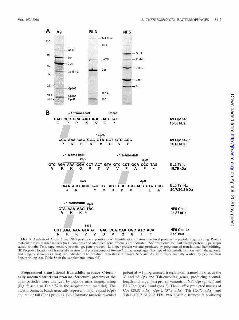
Programmed translational frameshifts produce C-termi-nally modified structural proteins. Structural proteins of thevirus particles were analyzed by peptide mass fingerprinting(Fig. 5; see also Table S7 in the supplemental material). Themost prominent bands generally represent major capsid (Cps)and major tail (Tsh) proteins. Bioinformatic analysis revealed
potential �1 programmed translational frameshift sites at the3� end of Cps and Tsh-encoding genes, producing normal-length and larger (-L) protein variants of NF5 Cps (gp 6.1) andBL3 Tsh (gp14.1 and gp14.2). The in silico predicted masses ofCps (28.87 kDa), Cps-L (37.9 kDa), Tsh (15.75 kDa), andTsh-L (20.7 or 20.8 kDa, two possible frameshift positions)
FIG. 5. Analysis of A9, BL3, and NF5 protein composition. (A) Identification of virus structural proteins by peptide fingerprinting. Proteinmolecular mass marker masses (in kilodaltons) and identified gene products are indicated. Abbreviations: Tsh, tail sheath protein; Cps, majorcapsid protein; Tmp, tape measure protein; gp, gene product; -L, longer protein variants produced by programmed translational frameshifting.(B) Proposed locations of frameshifts in structural protein genes of Brochothrix bacteriophages. The type of frameshift, location within the genome,and slippery sequences (lines) are indicated. The putative frameshifts in phages NF5 and A9 were experimentally verified by peptide massfingerprinting (see Table S6 in the supplemental material).
VOL. 192, 2010 B. THERMOSPHACTA BACTERIOPHAGES 5447
correspond to the observed masses (Fig. 5). The Cps �1frameshift in phage NF5 occurs at the 3� end of the transcriptwhen the ribosome reaches the AAA lysine codon of the con-sensus heptanucleotide slippery sequence (A.AAA.AAG 3AAA.AAA.G; Fig. 5B) (56) and was experimentally confirmedby identification of tryptic fragments specifically generatedfrom the longer protein variant Cps-L (see Table S6 in thesupplemental material). Two potential �1 frameshift positionsidentified at the 3� end of the tsh gene of phage BL3 do notfollow the standard consensus heptanucleotide slippery se-quence, and only a small amount of frameshift product wasobserved in the virion (Fig. 5). In phage A9, gp154 and gp155could be identified as structural components, although locatedoutside the structural gene cluster. A similar situation has beenreported for other phages (20, 37). A perfect heptanucleotideslippery sequence (Fig. 5B) is present at the 3� end of gp154,indicating a translational frameshift that results in a gp154/155fusion protein (gp154-L). gp154 (15.8 kDa)- and gp154-L(34.10 kDa)-specific peptides were present in the correspond-ing protein bands, confirming the occurrence of this type re-coding event.
Phages BL3 and NF5 utilize novel prophage insertion sites.BL3 and NF5 integration sites were identified by an inversePCR approach (54), since a Brochothrix genome sequence isnot available. Analysis of the prophage flanking host sequencesrevealed that BL3 integrates into the 3�-end of a conservedRNA methyltransferase, partially reconstituting the remainingC-terminal amino acid residues (HVEVVTAFTLVD in thehost versus HVEVVCALTLIE in the prophage). Similar meth-yltransferases are found in related Gram-positive organismssuch as Bacillus mycoides (NZ_CM000744) or Listeria mono-cytogenes EGD-e (25), with similar C-terminal ends (HVECVAWLELV and HIEAVTVLHLN). The BL3 integrase recog-nizes a core region of only two identical nucleotides, flanked byinverted repeats both in the phage and in the host genome(Fig. 6A). NF5 integrates at the 5� end of a putative histidinol-phosphatase gene with homology (over the available 24 aminoacids) to corresponding proteins from Lactobacillus and Liste-ria (Fig. 6B). Integration reconstitutes the gene including itsribosomal binding site. The site-specific recombinase of NF5recognizes a region of identity extending over 45 nt betweenattP of NF5 and attB of B. thermosphacta HER1188. The in-tergenic sequence downstream of the NF5 integrase was not
predicted to be able to form any potential secondary structure(stem-loop) that could function as a transcription terminator.This was a surprising observation, since other phages (BL3,A006, A500, A118, B025, ul36, and Tuc2009) generally featuretranscription terminators located in between the 3� end of theintegrase gene and attP, with a free energy G of �18.6 to�40.3 kcal/mol (details not shown).
BL3 and NF5 share homologies and are related to the P335quasispecies of Lactococcus phages. Genomes of BL3 and NF5were compared to each other and to a set of six Listeria phages(18, 49, 74), four Lactococcus phages (8, 40, 41), and fourLactococcus prophages (70) (see Fig. S1 and S2 in the supple-mental material). Although NF5 and BL3 feature homologiesprimarily in the early gene region, no similarities to any of theListeria phages could be identified, even though there is re-markable synteny in the genome organization. Instead, bothBrochothrix phages reveal homology to distinct subgroups ofthe heterogeneous P335 quasispecies of Lactococcus phages(see Fig. S2 in the supplemental material). This also is re-stricted to the structural gene cluster and includes gene prod-ucts of prophage SK11_4 and t712 for BL3, as well as phagesTuc2009, TP901-1, P335, ul36, and prophages SK11_2 andSK11_3 for NF5. With respect to their overall genome orga-nization, all of these lactococcal phages and prophages werepreviously shown to be Sfi11-like Siphoviridae or demonstratedto be related to these phages (9, 40, 70). Therefore, we proposethat BL3 and NF5 are also members of this group, whichagrees well with the proposed headful packaging mechanism.Global protein sequence alignment confirmed the homology toP335 phage Tuc2009 (for NF5) and prophage SK11_4 (forBL3) (Fig. 2B). BL3 and NF5 share amino acid homologiesmainly in early gene products and in TerL, endolysin (con-served amidase domain) and repressor. The homologies arenot randomly distributed but rather focused in connected clus-ters, indicating a genome mosaicism, which is typical for manybacteriophage genomes, and likely originating from extensivehorizontal gene transfer during evolution and adaptation (32).
A9 is a member of the large, virulent SPO1-related Myoviri-dae (Spounavirinae). Assignment of phage A9 to the SPO1-related Myoviridae of the proposed new subfamily Spounaviri-nae (38, 44) is based on the following evidence: A9 (i) infectsa low GC content, Gram-positive host (Firmicutes), (ii) isstrictly virulent, (iii) displays a relatively broad host-range
FIG. 6. Detail view of BL3 (A) and NF5 (B) integration site in the HER1187 and HER1188 genome, respectively. Regions of identity betweenphage and host sequence (core sequences) are boxed.
within the bacterial genus, and (iv) features a double-strandedDNA genome with invariable terminal repeats. Members ofthis subfamily share a common morphology, comparable ge-nome sizes, marked synteny in their genome organization, andpronounced protein sequence relatedness (37, 38, 44). In A9,these homologies are largely confined to structural and fewearly gene products (see Table S5 in the supplemental mate-rial). An alignment of phage A9 with the Spounavirinae A511(37) and EF24C (69) indicates striking colinearity in genomeorganization (Fig. 2A).
DISCUSSION
Genome sequencing and molecular characterization of rep-resentatives of each of the three known Brochothrix bacterio-phage species provided basic information on morphology, ge-nome organization, mode of DNA packaging, and integrationinto the host chromosome and revealed interesting details suchas the occurrence of programmed translational frameshifts forsynthesis of C-terminally modified structural proteins. The in-formation genomes of the two small temperate siphovirusesBL3 and NF5 span 41,518 and 36,953 bp, respectively. Theviral DNA is incorporated into the empty capsids via a headfulpackaging mechanism, producing a collection of circularly per-muted, terminally redundant DNA molecules. The overall or-ganization into defined functional modules is similar in bothphages and resembles the well-known genome arrangementsfound in most temperate phages infecting the Firmicutes, mir-roring the phage’s life cycles (9, 18). The clearly differentmorphology of BL3 and NF5 (Fig. 1) is also reflected in a lackof amino acid sequence homology in the structural proteins(Fig. 2B). Sequence homologies dominate among the earlygenes, suggesting analogous host takeover and genome repli-cation mechanisms. Such gene products are encoded by groupsof adjacent genes, rather than being randomly dispersed overthe genome (Fig. 2B). This pattern is indicative for a horizontalexchange of modules during evolution of these phages, whichtypically results in highly mosaic virus genomes (11, 33, 34).Both phages seem to lack genes coding for DNA polymerasesand helicases and therefore depend on host enzymes for theirreplication. DNA and amino acid sequence alignments and theoverall organization of structural genes indicate that both BL3and NF5 are members of the Sfi11-like, pac site containingSiphoviridae (9, 59), which are structurally related to differentsubgroups of the heterogeneous P335 quasispecies of Lacto-coccus phages (40, 70). Brochothrix phages are strictly genusspecific, and the cross-genus sequence homologies thereforesuggest that any such genetic exchange probably occurred inthe distant past or that the relationship is based on divergencefrom a common ancestor. Interestingly, only very limitedamino acid sequence homologies to Listeria phages were ob-served, even though the host bacteria are closely related (16,52) (see Tables S3 and S4 in the supplemental material). As-suming that horizontal gene transfer and exchange mecha-nisms are more important than vertical passage during theevolution of these phage genomes, it is tempting to speculatethat phages sharing a common ecological niche are more likelyto show relatedness than phages infecting phylogenetically re-lated hosts. This might hold true for the relatedness of Bro-chothrix phages to phages infecting Lactococcus. However,
other findings suggest that phage relatedness reflects the re-latedness of their bacterial hosts (suggesting coevolution), atleast for members of the Sfi21-like genus (9). Moreover, be-cause these homologies are mostly restricted to structural pro-teins, relatedness may also reflect various degrees of a diverg-ing (but still similar) morphology rather than being the resultof constant selection based on functional requirements.
Integration of viral DNA into the host chromosome by site-specific recombination is essential for propagation of theprophage. Recombination is mediated by phage-encoded inte-grases (29, 64). The occurrence of highly conserved aminoacids indicate that the BL3 integrase is a member of the serinerecombinase family, which recognizes the only 2-bp core se-quence shared in attB and attP but may also require additionalrecognition signals such as inverted repeats flanking the coresequence (64; Margaret Smith, unpublished data). In contrast,the NF5 integrase belongs to the tyrosine recombinases, whichgenerally recognize longer core sequence, in this case 45 bp. Ofparticular interest is that both phages integrate into unique locinot previously known to be utilized for phage insertion. PhageNF5 inserts into the 5� end of a putative histidinol-phosphatasegene, which first appeared to disrupt the gene. Upon closerinspection, however, it became evident that the 45-bp attach-ment site overlaps with the start codon, and phage integrationdoes reconstitute the functional coding sequence with only onenucleotide exchange (silent) at the opposite end of the pro-phage. Moreover, since the NF5 integrase gene is apparentlynot followed by any transcription terminator, it appears that inthe lysogenized host, expression of the histidinol-phosphataseis then driven by transcriptional coupling of the host gene tothe phage integrase expression system.
Integration of the BL3 genome occurs close to the 3� end ofa putative RNA-methyltransferase gene but reconstitutes theinterrupted reading frame to yield a product highly similar tothe original polypeptide, probably resulting in a functionalgene product. Such “reconstitutive” integration has been de-scribed for other temperate phages (10, 18, 53), including Lis-teria phage PSA, which inserts into the 3� end of a tRNAcoding sequence (74), and for B054, which integrates into the3� end of a transcription elongation factor gene (18). It isreasonable to assume that reconstitutive integration into spe-cific and conserved sequences has evolved to secure insertionsite sequences and, at the same time, maintains host fitnessupon prophage insertion, which is beneficial for both partici-pants.
Programmed translational frameshifting is not uncommonamong bacteriophages, and alternative decoding is used togenerate variant proteins utilized in phage morphogenesis.The mechanism has been reported for various phages, includ-ing A2, T3, and T7 (reviewed in reference 6), and a number ofListeria phages (18, 74). Recoding is quite frequent in sipho-viruses and might have evolved to keep genomes small byincreasing the coding capacity, which is surprisingly high inboth phages. An elegant explanation for the functional role incapsid morphogenesis has been proposed for Listeria phagePSA. The longer frameshift variants are believed to producepentameric ring structures of the capsomers, while the shortervariants produce the hexameric structures (74). We observedhere a �1 translational frameshift site in the NF5 cps gene,featuring a classic X.XXY.YYZ heptanucleotide mRNA slip-
VOL. 192, 2010 B. THERMOSPHACTA BACTERIOPHAGES 5449
pery sequence at which the ribosome can slip back on themRNA (Fig. 5B) (71). However, the precise role of the longerpolypeptide in head morphogenesis remains to be elucidated.
Programmed translational frameshifts in phage tail proteingenes have also been frequently reported, and a role of themodified products in correct assembly of the tail was suggested(15, 18, 46, 71). We found a �1 frameshift site in tsh of BL3(orf14; Fig. 5B), which does not follow the classic X.XXY.YYZslippery sequence. However, not all phages with a verified �1translational frameshift feature this consensus sequence. Forinstance, phage C31 features a slippery sequence of the typeX.XXX.YYX (G.GGG.AAG) (63, 71), which is similar to theBL3 motif (A.AAA.GGA; Fig. 5B). Thus, it is likely that aframeshift in BL3 tsh occurs at the slippery sequence locatedbetween nt 9573 to 9579. The presence of a nonconsensus sitemight explain why only a small amount of frameshift product isdetected in the mature virion (Fig. 5A). Again, the exact func-tion of the frameshift products in the BL3 tail remains unclear,
and yet an essential role for correct tail assembly may beassumed, similar to the situation in phages Lambda (46) andSPP1 (5).
Bacteriophage A9 is different from BL3 and NF5; it is avirulent (obligately lytic) virus and features an informationgenome of 127 kb with a large terminal redundancy of �11 kb.Genome organization, protein homologies, morphology, andlifestyle of the phage clearly place it within the recently pro-posed Spounavirinae subfamily of the Myoviridae. The termi-nally redundant, nonpermuted genome of A9 further strength-ens the hypothesis that this genome structure is a characteristichallmark of the Spounavirinae (37, 68). As previously shownfor other phages of this subfamily (14, 37, 68, 69), the genomeis organized into two clusters with discrete function, the struc-ture-associated genes (gp83-gp108), and DNA synthesis- orreplication-associated genes (gp112-gp165). When A9 is com-pared to other Spounavirinae, the gene synteny among the twoclusters is clearly visible and yet not exhaustive; the loci of
FIG. 7. Identification of intervening sequences (IVS) in the A9 genome. (A) Splicing variants producing the most significant database hits wereannotated. The putative intein was identified by Blastp. RNR, ribonucleotide reductase. (B) Alignment of the spliced proteins with homologoussequences which do not contain IVS (a, RNR� subunit of Granulicatella elegans ATCC 700633; b, RNR� subunit of Enterococcus casseliflavusEC30; A511 TerL, large terminase of Listeria phage A511). Amino acid sequence identities and gaps in the alignment are indicated. Small trianglesmark the putative processing sites.
genes coding for tRNAs, endolysin, terminase, and ribonucle-otide reductase appear to be particularly variable. The ribonu-cleotide reductase genes of A9 and A511 are located at similarsites within their genomes, which, however, are different fromother family members such as phages phiEF24C (69) (Fig. 2A)or SPO1 (68).
Another striking observation was the presence of large ter-minase subunits encoded in opposite transcription directionupstream of the portal protein in phage A9. Recent analyses inour lab have revealed such an unusual genome structure to beconserved among several SPO1-related Bacillus phages (un-published data). Considering the strict genus specificity of A9,any cross-genus genetic exchange events probably occurred inthe distant past, or represent the result of infrequent illegiti-mate recombination. Interestingly, genes exhibiting such vari-able positions within phage genomes are frequently inter-rupted by intervening sequences, often observed within genesinvolved in DNA metabolism (19). Several Spounavirinae havebeen shown to carry self-splicing group I introns or inteins inDNA-polymerase genes (phages G1, K, and SPO1), ribonucle-otide-reductase genes (phage Twort), DNA helicase genes(phage Twort, intein), endolysin genes (phages G1 and K), andlarge terminase subunits (phage Twort) (39, 42, 43, 55). Withrespect to A9, sequence alignments indicated intervening se-quences in both the large terminase and the ribonucleotidereductase (RNR) genes (Fig. 2A and 7A). Both are located atpositions not previously described for SPO1-related phages. Infact, the putative A9 large terminase gene possibly containstwo introns, which would explain the presence of three sepa-rate ORFs with partial terL homologies. A similar situation wasreported for phage Twort (39), where the large terminase geneis also interrupted by two introns. The putative type I RNR ofA9 is encoded on ORFs 125, 127, 128, 130, 132, and 134, withamino acid sequence homologies to the RNR alpha (nrdE-like) and beta (nrdF-like) subunits, respectively (Fig. 7; seeTable S5 in the supplemental material). In addition, the phageencodes an nrdI-like (orf124) and an nrdH-like protein (orf135)(Table S5 in the supplemental material), all of which are typ-ically found in type Ib RNR enzymes (62). The alpha subunitappears to be interrupted by three introns and one intein,located within orf128, and encoding a homing endonuclease.One putative intron could be identified in the RNR beta-subunit gene. Introns and inteins in RNR genes have previ-ously been observed in phage (e.g., Twort and several Bacillusprophages [42, 45]) and in bacterial genomes (47). Whetherthe occurrence of introns and inteins has a regulatory role orwhether these genes are particularly susceptible to the inser-tion of intervening sequences is not known (17).
A �1 translational frameshift is programmed into the codingsequence and mRNA for gp154, a protein identified as a com-ponent of the A9 virion. This alternative recoding event isespecially interesting, since the coding sequence is not locatedwithin the structural gene cluster, and its expression thereforeis probably not driven by a late promoter. In addition, gp154-Lcontains a bacterial Ig-like domain (PF02368 [36]), raising thequestion of whether it might be involved in nonspecific bindingof host surface structures involved in phage adsorption. Therole of virion-associated Ig-like domains in other tailed phageswas recently reconsidered, and an accessory role during infec-
tion was proposed (22). Apparently, several of these domainsare produced by translational frameshifting (22).
In conclusion, the characterization and genome sequencingof three different phages infecting the genus Brochothrix re-vealed a diversity of morphotypes, genetic make-up, and rela-tionship to phages of other Firmicutes. Our data support theclassification of Brochothrix phages into at least three families,as proposed by Ackermann et al. (1). Although phage A9 is amember of the large but poorly characterized group of SPO1-related large myoviruses (Spounavirinae), NF5 and BL3 aresmall temperate siphoviruses, primarily related to lactococcalphages. All phages feature a high degree of flexibility andmosaicism among their genomes. The availability of more ge-nome sequences will facilitate the development of a reliableclassification scheme and enable substantiated evolutionarycomparisons between bacteriophages infecting Firmicutes. Thepresented data on phage A9, NF5, and BL3 highlight thephylogenetic relationships between bacteriophages across ge-nus boundaries from a new angle.
ACKNOWLEDGMENTS
We are grateful to George Nychas (University of Athens, Athens,Greece) and to Bryan Dilts (Agriculture and Agri-Food Canada, La-combe, Canada) for the gift of B. thermosphacta strains. We thankHans-Wolfgang Ackermann (University of Laval, Quebec City, Que-bec, Canada) for fruitful discussions; Margaret Smith (University ofAberdeen, Aberdeen, United Kingdom) for helpful discussions onphage integrases; and Rudi Lurz (Max Planck Institute for MolecularGenetics, Berlin, Germany) for electron microscopic images of A9,BL3, and NF5. We are grateful to Vanessa Bernbach for assistancewith some experiments.
REFERENCES
1. Ackermann, H.-W., G. G. Greer, and J. Rocourt. 1988. Morphology ofBrochothrix thermosphacta phages. Microbios 56:19–26.
2. Adams, M. H. 1959. Methods of study of bacterial viruses, p. 443–457. InBacteriophages. Interscience Publishers, Inc., New York, NY.
3. Alemayehu, D., R. P. Ross, O. O’Sullivan, A. Coffey, C. Stanton, G. F.Fitzgerald, and O. McAuliffe. 2009. Genome of a virulent bacteriophageLb338-1 that lyses the probiotic Lactobacillus paracasei cheese strain. Gene448:29–39.
4. Altschul, S. F., T. L. Madden, A. A. Schaffer, J. Zhang, Z. Zhang, W. Miller,and D. J. Lipman. 1997. Gapped BLAST and PSI-BLAST: a new generationof protein database search programs. Nucleic Acids Res. 25:3389–3402.
5. Auzat, I., A. Droge, F. Weise, R. Lurz, and P. Tavares. 2008. Origin andfunction of the two major tail proteins of bacteriophage SPP1. Mol. Micro-biol. 70:557–569.
6. Baranov, P. V., O. Fayet, R. W. Hendrix, and J. F. Atkins. 2006. Recoding inbacteriophages and bacterial IS elements. Trends Genet. 22:174–181.
7. Borch, E., M. L. Kant-Muermans, and Y. Blixt. 1996. Bacterial spoilage ofmeat and cured meat products. Int. J. Food Microbiol. 33:103–120.
8. Brondsted, L., S. Ostergaard, M. Pedersen, K. Hammer, and F. K. Vo-gensen. 2001. Analysis of the complete DNA sequence of the temperatebacteriophage TP901-1: evolution, structure, and genome organization oflactococcal bacteriophages. Virology 283:93–109.
9. Brussow, H., and F. Desiere. 2001. Comparative phage genomics and theevolution of Siphoviridae: insights from dairy phages. Mol. Microbiol. 39:213–222.
10. Bruttin, A., S. Foley, and H. Brussow. 1997. The site-specific integrationsystem of the temperate Streptococcus thermophilus bacteriophage phiSfi21.Virology 237:148–158.
11. Casjens, S. R. 2005. Comparative genomics and evolution of the tailed-bacteriophages. Curr. Opin. Microbiol. 8:451–458.
12. Casjens, S. R., and E. B. Gilcrease. 2009. Determining DNA packagingstrategy by analysis of the termini of the chromosomes in tailed-bacterio-phage virions, p. 91–111. In M. R. J. Clokie and A. M. Kropinski (ed.),Bacteriophages: methods and protocols, vol. 2. Molecular and applied as-pects. Humana Press, Inc., New York, NY.
13. Casjens, S. R., E. B. Gilcrease, D. A. Winn-Stapley, P. Schicklmaier, H.Schmieger, M. L. Pedulla, M. E. Ford, J. M. Houtz, G. F. Hatfull, and R. W.Hendrix. 2005. The generalized transducing Salmonella bacteriophage ES18:complete genome sequence and DNA packaging strategy. J. Bacteriol. 187:1091–1104.
VOL. 192, 2010 B. THERMOSPHACTA BACTERIOPHAGES 5451
14. Chibani-Chennoufi, S., M. L. Dillmann, L. Marvin-Guy, S. Rami-Shojaei,and H. Brussow. 2004. Lactobacillus plantarum bacteriophage LP65: a newmember of the SPO1-like genus of the family Myoviridae. J. Bacteriol. 186:7069–7083.
15. Christie, G. E., L. M. Temple, B. A. Bartlett, and T. S. Goodwin. 2002.Programmed translational frameshift in the bacteriophage P2 FETUD tailgene operon. J. Bacteriol. 184:6522–6531.
16. Collins, M. D., S. Wallbanks, D. J. Lane, J. Shah, R. Nietupski, J. Smida, M.Dorsch, and E. Stackebrandt. 1991. Phylogenetic analysis of the genus Lis-teria based on reverse transcriptase sequencing of 16S rRNA. Int. J. Syst.Bacteriol. 41:240–246.
17. Derbyshire, V., and M. Belfort. 1998. Lightning strikes twice: intron-inteincoincidence. Proc. Natl. Acad. Sci. U. S. A. 95:1356–1357.
18. Dorscht, J., J. Klumpp, R. Bielmann, M. Schmelcher, Y. Born, M. Zimmer,R. Calendar, and M. J. Loessner. 2009. Comparative genome analysis ofListeria bacteriophages reveals extensive mosaicism, programmed transla-tional frameshifting, and a novel prophage insertion site. J. Bacteriol. 191:7206–7215.
19. Edgell, D. R., M. Belfort, and D. A. Shub. 2000. Barriers to intron promis-cuity in bacteria. J. Bacteriol. 182:5281–5289.
20. Eyer, L., R. Pantucek, Z. Zdrahal, H. Konecna, P. Kasparek, V. Ruzickova,L. Hernychova, J. Preisler, and J. Doskar. 2007. Structural protein analysisof the polyvalent staphylococcal bacteriophage 812. Proteomics 7:64–72.
21. Finn, R. D., J. Mistry, J. Tate, P. Coggill, A. Heger, J. E. Pollington, O. L.Gavin, P. Gunasekaran, G. Ceric, K. Forslund, L. Holm, E. L. Sonnhammer,S. R. Eddy, and A. Bateman. 2010. The Pfam protein families database.Nucleic Acids Res. 38:D211–D222.
22. Fraser, J. S., Z. Yu, K. L. Maxwell, and A. R. Davidson. 2006. Ig-like domainson bacteriophages: a tale of promiscuity and deceit. J. Mol. Biol. 359:496–507.
23. Gardner, G. A. 1981. Brochothrix thermosphacta (Microbacterium thermo-sphactum) in the spoilage of meats: a review, p. 139–173. In T. A. Roberts,G. A. Hobbs, J. H. B. Christian, and N. Skovgaard (ed.), Psychrotrophicmicroorganisms in spoilage and pathogenicity. Academic Press, London,England.
24. Gardner, G. A. 1966. A selective medium for the enumeration of Microbac-terium thermosphactum in meat and meat products. J. Appl. Bacteriol. 29:455–460.
25. Glaser, P., L. Frangeul, C. Buchrieser, C. Rusniok, A. Amend, F. Baquero,P. Berche, H. Bloecker, P. Brandt, T. Chakraborty, A. Charbit, F. Chetouani,E. Couve, A. de Daruvar, P. Dehoux, E. Domann, G. Dominguez-Bernal, E.Duchaud, L. Durant, O. Dussurget, K. D. Entian, H. Fsihi, F. Garcia-delPortillo, P. Garrido, L. Gautier, W. Goebel, N. Gomez-Lopez, T. Hain, J.Hauf, D. Jackson, L. M. Jones, U. Kaerst, J. Kreft, M. Kuhn, F. Kunst, G.Kurapkat, E. Madueno, A. Maitournam, J. M. Vicente, E. Ng, H. Nedjari, G.Nordsiek, S. Novella, B. de Pablos, J. C. Perez-Diaz, R. Purcell, B. Remmel,M. Rose, T. Schlueter, N. Simoes, A. Tierrez, J. A. Vazquez-Boland, H. Voss,J. Wehland, and P. Cossart. 2001. Comparative genomics of Listeria species.Science 294:849–852.
26. Greer, G. G. 2005. Bacteriophage control of food-borne bacteria. J. FoodProt. 68:1102–1111.
27. Greer, G. G. 1983. Psychrotrophic Brochothrix thermosphacta bacteriophagesisolated from beef. Appl. Environ. Microbiol. 46:245–251.
28. Greer, G. G., and B. D. Dilts. 2002. Control of Brochothrix thermosphactaspoilage of pork adipose tissue using bacteriophages. J. Food Prot. 65:861–863.
29. Groth, A. C., and M. P. Calos. 2004. Phage integrases: biology and applica-tions. J. Mol. Biol. 335:667–678.
30. Grundling, A., M. D. Manson, and R. Young. 2001. Holins kill withoutwarning. Proc. Natl. Acad. Sci. U. S. A. 98:9348–9352.
31. Hagens, S., and M. J. Loessner. 2007. Application of bacteriophages fordetection and control of food-borne pathogens. Appl. Microbiol. Biotechnol.76:513–519.
32. Hatfull, G. F. 2008. Bacteriophage genomics. Curr. Opin. Microbiol. 11:447–453.
33. Hendrix, R. W. 2003. Bacteriophage genomics. Curr. Opin. Microbiol. 6:506–511.
34. Hendrix, R. W., G. F. Hatfull, and M. C. Smith. 2003. Bacteriophages withtails: chasing their origins and evolution. Res. Microbiol. 154:253–257.
35. Jarvis, A. W., L. J. Collins, and H.-W. Ackermann. 1993. A study of fivebacteriophages of the Myoviridae family which replicate on different gram-positive bacteria. Arch. Virol. 133:75–84.
36. Kelly, G., S. Prasannan, S. Daniell, K. Fleming, G. Frankel, G. Dougan, I.Connerton, and S. Matthews. 1999. Structure of the cell-adhesion fragmentof intimin from enteropathogenic Escherichia coli. Nat. Struct. Biol. 6:313–318.
37. Klumpp, J., J. Dorscht, R. Lurz, R. Bielmann, M. Wieland, M. Zimmer, R.Calendar, and M. J. Loessner. 2008. The terminally redundant, nonper-muted genome of Listeria bacteriophage A511: a model for the SPO1-likemyoviruses of gram-positive bacteria. J. Bacteriol. 190:5753–5765.
38. Klumpp, J., R. Lavigne, M. J. Loessner, and H.-W. Ackermann. 2010. TheSPO1-related bacteriophages. Arch. Virol. 155:1547–1561.
39. Kwan, T., J. Liu, M. DuBow, P. Gros, and J. Pelletier. 2005. The completegenomes and proteomes of 27 Staphylococcus aureus bacteriophages. Proc.Natl. Acad. Sci. U. S. A. 102:5174–5179.
40. Labrie, S. J., and S. Moineau. 2002. Complete genomic sequence of bacte-riophage ul36: demonstration of phage heterogeneity within the P335 quasi-species of lactococcal phages. Virology 296:308–320.
41. Labrie, S. J., J. Josephsen, H. Neve, F. K. Vogensen, and S. Moineau. 2008.Morphology, genome sequence, and structural proteome of type phage P335from Lactococcus lactis. Appl. Environ. Microbiol. 74:4636–4644.
42. Landthaler, M., U. Begley, N. C. Lau, and D. A. Shub. 2002. Two self-splicinggroup I introns in the ribonucleotide reductase large subunit gene of Staph-ylococcus aureus phage Twort. Nucleic Acids Res. 30:1935–1943.
43. Landthaler, M., and D. A. Shub. 1999. Unexpected abundance of self-splicing introns in the genome of bacteriophage Twort: introns in multiplegenes, a single gene with three introns, and exon skipping by group Iribozymes. Proc. Natl. Acad. Sci. U. S. A. 96:7005–7010.
44. Lavigne, R., P. Darius, E. J. Summer, D. Seto, P. Mahadevan, A. S. Nilsson,H. W. Ackermann, and A. M. Kropinski. 2009. Classification of Myoviridaebacteriophages using protein sequence similarity. BMC Microbiol. 9:224.
45. Lazarevic, V. 2001. Ribonucleotide reductase genes of Bacillus prophages: arefuge to introns and intein coding sequences. Nucleic Acids Res. 29:3212–3218.
46. Levin, M. E., R. W. Hendrix, and S. R. Casjens. 1993. A programmedtranslational frameshift is required for the synthesis of a bacteriophagelambda tail assembly protein. J. Mol. Biol. 234:124–139.
47. Liu, X. Q., J. Yang, and Q. Meng. 2003. Four inteins and three group IIintrons encoded in a bacterial ribonucleotide reductase gene. J. Biol. Chem.278:46826–46831.
48. Loessner, M. J. 2005. Bacteriophage endolysins-current state of research andapplications. Curr. Opin. Microbiol. 8:480–487.
49. Loessner, M. J., R. B. Inman, P. Lauer, and R. Calendar. 2000. Completenucleotide sequence, molecular analysis and genome structure of bacterio-phage A118 of Listeria monocytogenes: implications for phage evolution.Mol. Microbiol. 35:324–340.
50. Loessner, M. J., A. Schneider, and S. Scherer. 1996. Modified Listeria bac-teriophage lysin genes (ply) allow efficient overexpression and one-step pu-rification of biochemically active fusion proteins. Appl. Environ. Microbiol.62:3057–3060.
51. Lowe, T. M., and S. R. Eddy. 1997. tRNAscan-SE: a program for improveddetection of transfer RNA genes in genomic sequence. Nucleic Acids Res.25:955–964.
52. Ludwig, W., K.-H. Schleifer, and E. Stackebrandt. 1984. 16S rRNA analysisof Listeria monocytogenes and Brochothrix thermosphacta. FEMS Microbiol.Lett. 25:6.
53. McShan, W. M., Y. F. Tang, and J. J. Ferretti. 1997. Bacteriophage T12 ofStreptococcus pyogenes integrates into the gene encoding a serine tRNA.Mol. Microbiol. 23:719–728.
54. Ochman, H., A. S. Gerber, and D. L. Hartl. 1988. Genetic applications of aninverse polymerase chain reaction. Genetics 120:621–623.
55. O’Flaherty, S., A. Coffey, R. Edwards, W. Meaney, G. F. Fitzgerald, and R. P.Ross. 2004. Genome of staphylococcal phage K: a new lineage of Myoviridaeinfecting gram-positive bacteria with a low GC content. J. Bacteriol. 186:2862–2871.
56. Olia, A. S., and G. Cingolani. 2008. A shifty stop for a hairy tail. Mol.Microbiol. 70:549–553.
57. Pantucek, R., A. Rosypalova, J. Doskar, J. Kailerova, V. Ruzickova, P.Borecka, S. Snopkova, R. Horvath, F. Gotz, and S. Rosypal. 1998. Thepolyvalent staphylococcal phage phi 812: its host-range mutants and relatedphages. Virology 246:241–252.
58. Pin, C., G. D. Garcia de Fernando, and J. A. Ordonez. 2002. Effect ofmodified atmosphere composition on the metabolism of glucose by Brocho-thrix thermosphacta. Appl. Environ. Microbiol. 68:4441–4447.
59. Proux, C., D. van Sinderen, J. Suarez, P. Garcia, V. Ladero, G. F. Fitzgerald,F. Desiere, and H. Brussow. 2002. The dilemma of phage taxonomy illu-strated by comparative genomics of Sfi21-like Siphoviridae in lactic acidbacteria. J. Bacteriol. 184:6026–6036.
60. Sambrook, J., and D. W. Russell. 2001. Molecular cloning, vol. 1 to 3. ColdSpring Harbor Laboratory Press, Cold Spring Harbor, NY.
61. Schurr, T., E. Nadir, and H. Margalit. 1993. Identification and character-ization of Escherichia coli ribosomal binding sites by free energy computa-tion. Nucleic Acids Res. 21:4019–4023.
62. Sjoberg, B.-M. 1997. Ribonucleotide reductases: a group of enzymes withdifferent metallosites and a similar reaction mechanism, p. 139–173. In P. J.Sadler and A. J. Thomson (ed.), Structure and bonding: metal sites inproteins and models. Springer, Berlin, Germany.
63. Smith, M. C., R. N. Burns, S. E. Wilson, and M. A. Gregory. 1999. Thecomplete genome sequence of the Streptomyces temperate phage straight�C31: evolutionary relationships to other viruses. Nucleic Acids Res. 27:2145–2155.
64. Smith, M. C., and H. M. Thorpe. 2002. Diversity in the serine recombinases.Mol. Microbiol. 44:299–307.
65. Stackebrandt, E., and D. Jones. 2006. The genus Brochothrix, p. 477–491. In
S. Falkow, E. Rosenberg, K.-H. Schleifer, E. Stackebrandt, and M. Dworkin(ed.), The prokaryotes. Springer, New York, NY.
66. Stanley, G., K. J. Shaw, and A. F. Egan. 1981. Volatile compounds associatedwith spoilage of vacuum-packaged sliced luncheon meat by Brochothrix ther-mosphacta. Appl. Environ. Microbiol. 41:816–818.
67. Steven, A. C., B. L. Trus, J. V. Maizel, M. Unser, D. A. Parry, J. S. Wall, J. F.Hainfeld, and F. W. Studier. 1988. Molecular substructure of a viral recep-tor-recognition protein. The gp17 tail-fiber of bacteriophage T7. J. Mol. Biol.200:351–365.
68. Stewart, C. R., S. R. Casjens, S. G. Cresawn, J. M. Houtz, A. L. Smith, M. E.Ford, C. L. Peebles, G. F. Hatfull, R. W. Hendrix, W. M. Huang, and M. L.Pedulla. 2009. The genome of Bacillus subtilis bacteriophage SPO1. J. Mol.Biol. 388:48–70.
69. Uchiyama, J., M. Rashel, I. Takemura, H. Wakiguchi, and S. Matsuzaki.2008. In silico and in vivo evaluation of bacteriophage �EF24C, a candidatefor treatment of Enterococcus faecalis infections. Appl. Environ. Microbiol.74:4149–4163.
70. Ventura, M., A. Zomer, C. Canchaya, M. O’Connell-Motherway, O. Kuipers,F. Turroni, A. Ribbera, E. Foroni, G. Buist, U. Wegmann, C. Shearman,M. J. Gasson, G. F. Fitzgerald, J. Kok, and D. van Sinderen. 2007. Com-parative analyses of prophage-like elements present in two Lactococcus lactisstrains. Appl. Environ. Microbiol. 73:7771–7780.
71. Xu, J., R. W. Hendrix, and R. L. Duda. 2004. Conserved translational frame-shift in dsDNA bacteriophage tail assembly genes. Mol. Cell 16:11–21.
72. Zdobnov, E. M., and R. Apweiler. 2001. InterProScan-an integration plat-form for the signature-recognition methods in InterPro. Bioinformatics 17:847–848.
73. Zhang, J., Y. Zhang, L. Zhu, M. Suzuki, and M. Inouye. 2004. Interferenceof mRNA function by sequence-specific endoribonuclease PemK. J. Biol.Chem. 279:20678–20684.
74. Zimmer, M., E. Sattelberger, R. B. Inman, R. Calendar, and M. J. Loessner.2003. Genome and proteome of Listeria monocytogenes phage PSA: an un-usual case for programmed 1 translational frameshifting in structural pro-tein synthesis. Mol. Microbiol. 50:303–317.
VOL. 192, 2010 B. THERMOSPHACTA BACTERIOPHAGES 5453