Bronchoscopic Treatment of Emphysema: State of the Art
Stefano Gasparini a Lina Zuccatosta a Martina Bonifazi a Chris T. Bolliger b
a Pulmonary Diseases Unit, Department of Immunoallergic and Respiratory Diseases, AziendaOspedaliero-Universitaria ‘Ospedali Riuniti’, Ancona , Italy; b Lung Unit, Department of Internal Medicine,Tygerberg Hospital, Cape Town , South Africa
while, the bronchoscopic treatment of emphysema cannot yet be considered a standard of care and patients should be treated in the context of clinical trials or controlled registries, with well-defined programs of evaluation and follow-up.
The first attempts of surgical lung volume reduction (LVRS) for the treatment of emphysema were performed in the 1950s [1, 2] , but it was only in 2003, with the pub-lication of the results of the National Emphysema Treat-ment Trial (NETT) [3] that this procedure demonstrated its ability to improve the clinical and functional status of selected patients affected by emphysema. In the NETT study, a total of 1,218 patients with severe emphysema were randomized to receive LVRS (608 patients) or best medical treatment (610 patients). Subjects with forced ex-piratory volume in 1 s (FEV 1 ) or with diffusion capacity less than 20% of predicted, and subjects with homoge-neous emphysema were excluded because of a high surgi-cal risk and a low probability of benefitting from the sur-gery. The results of this trial showed an improvement of exercise capacity of more than 10 W in 28, 22 and 15% of
In recent years, different bronchoscopic techniques have been proposed for the treatment of emphysema, with the aim of obtaining the same clinical and functional advantag-es of lung volume reduction surgical techniques while re-ducing risks and costs. Such techniques can be classified into: methods employing devices that block the airways (e.g. spigots and unidirectional valves), methods that have a di-rect effect on the lung parenchyma (polymeric lung volume reduction, coils and thermal vapor ablation) and procedures that facilitate the expiration of trapped air from the emphy-sematous lung (airway bypass). This review aimed to evalu-ate the indications, outcomes and safety of the different techniques, based on the evidence from the available litera-ture. Results obtained by these methods are encouraging, but they are still based mainly on studies with small groups of patients. However, several trials are ongoing and in the near future we will acquire more knowledge which should lead to a better optimization of these procedures. Mean-
Received: June 11, 2012 Accepted: June 13, 2012 Published online: July 23, 2012
Bronchoscopic Treatment of Emphysema Respiration 2012;84:250–263 251
patients, respectively, at 6, 12 and 24 months after surgery in comparison to an improvement in 4, 5 and 3% of pa-tients in the control arm. The mortality rate in the 90 days after surgery was 7.9%, significantly higher than the 1.3% for the group treated with medical therapy. However, if the analysis of the results is limited to the subgroup of patients with predominant upper-lobe emphysema and low basal exercise capacity, mortality was lower in the surgical group (2.9%) than in the control group (3.3%). This study identified the characteristics of emphysema patients who might benefit from LVRS (subjects with pre-dominant upper-lobe emphysema and low exercise ca-pacity) and receive not only a functional and clinical im-provement, but also an increase in survival.
However, some concerns remain regarding LVRS, in-cluding its long-term efficacy (24 months after the proce-dure there is a trend of functional parameters to return towards baseline values) [3] , safety (prolonged air-leak is described in 30–48% of cases after LVRS) [4] and costs related not only to the surgical procedure, but also to the long hospital stay (in the NETT study, 28.1% of patients were still in hospital 1 month after the intervention) [3] . A recent long-term analysis of the NETT study confirmed a 5-year survival benefit for the surgically treated patients in the subgroup with heterogeneous upper-lobe emphy-sema (70 vs. 60% in the medical group) (p = 0.02), where-as this was not the case for the patients with homoge-neous emphysema [5] . The considerable morbidity and mortality associated with LVRS prompted reflection on other less invasive ways to achieve LVR, such as endo-scopic techniques.
A growing enthusiasm has permeated the world of in-terventional pulmonology since the beginning of the last decade when the first studies were published on achiev-ing LVR in emphysematous patients via bronchoscopic procedures [6, 7] . The development of bronchoscopic techniques is an attempt to obtain the same results as sur-gery for the treatment of emphysema, using procedures that are less invasive, potentially reversible and feasible in an outpatient setting at reduced risks and costs. Further-more, bronchoscopic techniques could also be more suit-able for patients who might not be good candidates for surgery, such as patients with predominant lower-lobe emphysema.
In recent years, several new technologies have been in-troduced, thereby pressuring interventional pulmonolo-gists to treat patients with new devices that have not yet fully been evaluated in terms of their efficacy and safety. Indications, outcomes, contraindications, patient-selec-tion criteria and the costs of these new treatment modal-
ities have not been well defined. Several studies are ongo-ing and new results continue to appear in the literature. The topic is fascinating and it seems interesting and time-ly to attempt to answer, on the basis of the available lit-erature results, the following questions. (1) What is the current state of the bronchoscopic management of em-physema? (2) What are the technical characteristics, the advantages and limits of each procedure? (3) What are the implications for the daily clinical practice?
Table 1 shows the different bronchoscopic techniques that have been proposed, classified on the basis of the un-derlying mechanism into 3 main groups: blocking devic-es that act at proximal bronchi level with the aim to pro-duce bronchial occlusion and atelectasis, devices that work at the pulmonary parenchymal level and methods that create extra-anatomical airways to facilitate lung de-flation.
Bronchial Blocking Devices
Plugs The only bronchial plugs available on the market to-
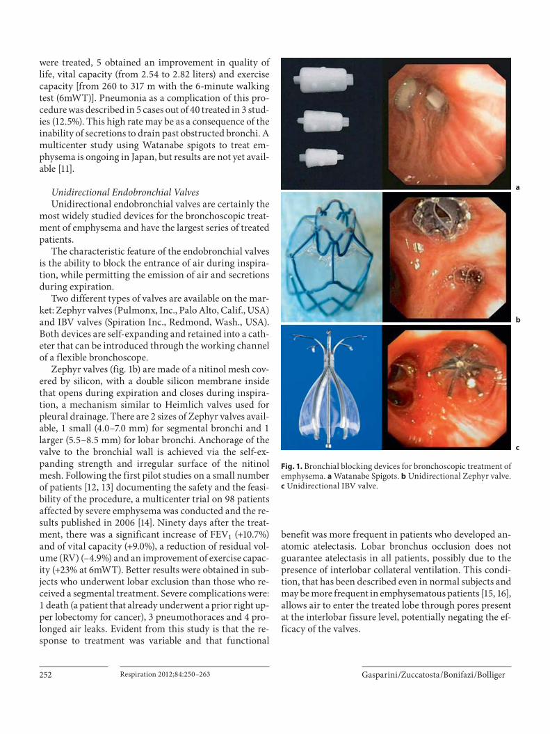
day are the so-called ‘Watanabe Spigots’, named after the Japanese pulmonologist who proposed them [8] . They are made of silicon and have a truncated conical shape and lateral studs that facilitate anchorage to the bronchial wall. Watanabe Spigots (Novatech, La Ciotat, France) are available in 3 different sizes (5, 6 and 7 mm) ( fig. 1 a). In effect, such devices were initially introduced into the clinical practice for the treatment of pulmonary fistula and persistent pneumothorax with continuous air leak-age. They have also been used to achieve LVR in cases of emphysema, but the results, limited to a small number of patients, have only been published in abstract form [9–11] . In a study by Miyazawa [10] , out of 7 patients who
Table 1. T echniques for the bronchoscopic treatment of emphy-sema
were treated, 5 obtained an improvement in quality of life, vital capacity (from 2.54 to 2.82 liters) and exercise capacity [from 260 to 317 m with the 6-minute walking test (6mWT)]. Pneumonia as a complication of this pro-cedure was described in 5 cases out of 40 treated in 3 stud-ies (12.5%). This high rate may be as a consequence of the inability of secretions to drain past obstructed bronchi. A multicenter study using Watanabe spigots to treat em-physema is ongoing in Japan, but results are not yet avail-able [11] .
Unidirectional Endobronchial Valves Unidirectional endobronchial valves are certainly the
most widely studied devices for the bronchoscopic treat-ment of emphysema and have the largest series of treated patients.
The characteristic feature of the endobronchial valves is the ability to block the entrance of air during inspira-tion, while permitting the emission of air and secretions during expiration.
Two different types of valves are available on the mar-ket: Zephyr valves (Pulmonx, Inc., Palo Alto, Calif., USA) and IBV valves (Spiration Inc., Redmond, Wash., USA). Both devices are self-expanding and retained into a cath-eter that can be introduced through the working channel of a flexible bronchoscope.
Zephyr valves ( fig. 1 b) are made of a nitinol mesh cov-ered by silicon, with a double silicon membrane inside that opens during expiration and closes during inspira-tion, a mechanism similar to Heimlich valves used for pleural drainage. There are 2 sizes of Zephyr valves avail-able, 1 small (4.0–7.0 mm) for segmental bronchi and 1 larger (5.5–8.5 mm) for lobar bronchi. Anchorage of the valve to the bronchial wall is achieved via the self-ex-panding strength and irregular surface of the nitinol mesh. Following the first pilot studies on a small number of patients [12, 13] documenting the safety and the feasi-bility of the procedure, a multicenter trial on 98 patients affected by severe emphysema was conducted and the re-sults published in 2006 [14] . Ninety days after the treat-ment, there was a significant increase of FEV 1 (+10.7%) and of vital capacity (+9.0%), a reduction of residual vol-ume (RV) (–4.9%) and an improvement of exercise capac-ity (+23% at 6mWT). Better results were obtained in sub-jects who underwent lobar exclusion than those who re-ceived a segmental treatment. Severe complications were: 1 death (a patient that already underwent a prior right up-per lobectomy for cancer), 3 pneumothoraces and 4 pro-longed air leaks. Evident from this study is that the re-sponse to treatment was variable and that functional
benefit was more frequent in patients who developed an-atomic atelectasis. Lobar bronchus occlusion does not guarantee atelectasis in all patients, possibly due to the presence of interlobar collateral ventilation. This condi-tion, that has been described even in normal subjects and may be more frequent in emphysematous patients [15, 16] , allows air to enter the treated lobe through pores present at the interlobar fissure level, potentially negating the ef-ficacy of the valves.
Fig. 1. Bronchial blocking devices for bronchoscopic treatment of emphysema. a Watanabe Spigots. b Unidirectional Zephyr valve. c Unidirectional IBV valve.
a
b
c
Bronchoscopic Treatment of Emphysema Respiration 2012;84:250–263 253
To evaluate the effectiveness and the safety of Zephyr valves for the treatment of emphysema, an international, multicenter, prospective, randomized study (Endobron-chial Valve for Emphysema Palliation Trial; VENT) was performed [17] . The trial was conducted at 31 centers in the USA and at 23 sites in Europe with a total number of 492 randomized patients (321 in USA and 171 in Europe). Af-ter a period of 6–8 weeks in a rehabilitation program, pa-tients were randomly assigned (2: 1) to the valve-treatment arm or the control arm (best standard medical care). The results of the US and European cohorts were evaluated and published separately in two different papers [18, 19] .
In the US study [18] , after 6 months, the endobron-chial valve treatment had induced a modest improvement in lung function (FEV 1 = +4.3%) and exercise tolerance (6mWT = +2.5%) and a small change in patients’ quality of life, as evaluated by the St. George Respiratory Ques-tionnaire (SGRQ) (–2.8 points). Even if these changes were statistically significant in comparison to the control group, they are hardly of clinical relevance. However, if the analysis were limited to patients with a higher CT scan evidence of emphysema heterogeneity, there was a greater improvement in both the FEV 1 (+10.7%) and the 6mWT (+12.4%). The other characteristic that predicted a better response to treatment was the fissure integrity as evaluated by CT scan, ostensibly because the patients with evidence of a complete fissure have a lower probabil-ity for collateral ventilation. The subgroup of patients with fissure integrity showed an improvement of 16.2% for FEV 1 and 7.7% for 6mWT.
In the European study [19] , the global results were similar, showing a modest benefit in the valve-treatment arm after 6 months in comparison to the control group for FEV 1 (+7 vs. 0.5%), cycle ergometry workload (+2 vs. –3 W) and SGRQ (–5 vs. 0.3 points). Also in the Euro-pean cohort, fissure integrity, present in about one third of the patients, was a good indicator for a better response (FEV 1 = +16%; cycle workload = +4 W; 6mWT = +11%).
Another factor evaluated in the European study is the CT evidence of complete lobar occlusion provided by the valve placement. Incomplete occlusion provides a con-duit for air to enter upon inspiration and this was identi-fied by the presence of air between the valve and the bron-chial wall, suggesting a leak. CT evidence of lobar occlu-sion was found in less than half of the valve-treated patients and in this subgroup the clinical outcomes were better than subjects with incomplete occlusion [FEV 1 = +26 vs. +6%; cycle ergometry workload = +8 vs. 0%; 6mWT = +22 vs. –2%; SGRQ = –10 vs. –2; RV/total lung capacity (TLC) = –14 vs. –1%].
Both reports of the VENT study demonstrated the safety of the valve treatment, showing a small incidence of complications. In the US study [18] , the most common complication was pneumonia distal to the valves (4.2% at 12 months, resolved in 6 patients without valve removal and in 6 patients after valve removal). Other complica-tions were hemoptysis (5.6% in the first 6 months after treatment and 6.1% 6–12 months after treatment) and pneumothorax (4.2%). Exacerbations of COPD were more frequent in the valve-treated group than in the con-trol arm in the first 6 months (7.9 vs. 1.1%), but occurred at a similar rate during the period 6 –12 months after. In the European study [19] , the incidence of pneumothorax was 4.5%, pneumonia distal to the valve 3.6% and hemop-tysis 5.4%. Occurrence of COPD exacerbations did not differ significantly between the treated patients and the control group.
These studies demonstrate the strong influence of an-atomic characteristics on valve treatment outcomes, such as fissure integrity and emphysema heterogeneity and the importance of technical factors such as the achievement of a complete lobar occlusion, underscoring the impor-tance of both optimal procedural technique and careful patient selection.
The second model of valve available on the market is the IBV valve, an umbrella-shaped device made by a ni-tinol mesh covered by a polyurethane membrane ( fig. 1 c). The valve is secured to the bronchial wall by 5 hook-like anchors and can be removed by grasping and pulling on its proximal central rod with forceps. It is available in 3 different sizes (5, 6 and 7 mm).
A pilot multicenter study with IBV valves was per-formed in the US on 91 patients affected by heteroge-neous predominant upper-lobe emphysema [20] . It is in-teresting to note that in this trial, following the observa-tion of a higher incidence of pneumothorax occurring with complete lobar occlusion (especially of the left upper lobe), the therapeutic strategy was modified during the study. For this reason, the trial was carried on with the bilateral treatment of both upper lobes, keeping open the lingula and avoiding complete lobar occlusion. The aim of this treatment strategy, which is not dependent on lo-bar atelectasis, is to shift the ventilation to the untreated healthier lobes, improving ventilation/perfusion match-ing and to reduce dynamic hyperinflation. The results of this trial showed no modification of functional parame-ters (FEV 1 , total lung volume and exercise tests were un-changed) but a significant improvement of quality of life (SGRQ = –8.2 points at 6 months). The same modality of treatment was evaluated in a prospective, randomized,
Gasparini /Zuccatosta /Bonifazi /Bolliger
Respiration 2012;84:250–263254
multicenter, sham-controlled European study [21] car-ried out on 73 patients with predominant upper-lobe em-physema. Thirty-seven patients were randomized to re-ceive valve treatment (implantation of IBV valves in the bilateral upper lobes, without complete lobar occlusion) and 36 patients underwent bronchoscopy with a sham procedure. No functional improvement was observed in the treatment group of this study either; however, a vol-ume reduction evaluated by CT scan and a significant improvement of the quality of life in comparison to the control group were reported. In particular, 8 out of 33 (24%) in the treatment group exceeded the minimum threshold changes for CT lung volumes and SGRQ total score, compared with no subjects (out of 35) in the control group [21] .
The strategy to keep open a segmental bronchus has been questioned by a study published by Eberhardt et al. [22] . Twenty-two patients were randomized to receive complete unilateral occlusion or bilateral upper-lobe treatment with incomplete occlusion. The functional im-provement was significantly greater in the group that un-derwent a complete lobar treatment (FEV 1 = +21.4 vs. –0.03%), with just 1 case of pneumothorax in this group. This study demonstrated that the unilateral procedure aimed at obtaining complete lobar occlusion is more ef-fective than the bilateral incomplete treatment and that, given the superior outcome, the increased risk of pneu-mothorax may be acceptable.
More recently, a third valve model has been proposed (Endobronchial Miyazawa Valve, Novatech, La Ciotat, France). The Miyazawa valve is a silicon device covered with small studs to prevent migration and a duckbill mechanism that permits exhalation of air from the distal lung segment but does not allow reinflation. The first study with the Miyazawa valve [23] on 12 patients affect-ed by advanced emphysema revealed a reduction of vol-ume in the treated lobe (–17.7% at 1 month and –12% at 6 months) and an improvement of quality of life and exer-cise capacity (6mWT = +47% at 1 month and +57% at 6 months). Another positive experience with this valve was published as a case report [24] , but studies on large num-bers of patients are still lacking.
Our experience with the use of unidirectional valves for the treatment of emphysema, based on the patients treated at the Azienda Ospedaliero-Universitaria ‘Os-pedali Riuniti’, Ancona (Italy), is summarized in figure 2 . From 2005 to August 2011 we treated 34 patients af-fected by heterogeneous emphysema. The treatment was performed unilaterally with the aim of obtaining com-plete lobar occlusion. Thirty-two patients were treated on
the upper lobe and 2 patients on the lower lobe. Four pa-tients (11.7%) benefited greatly, with a functional (FEV 1 increase of more than 40%) and clinically relevant im-provement. All these patients showed an atelectasis of the treated lobe. In 10 patients (29.4%), we observed some small benefit (FEV 1 = +7%), but atelectasis was not pres-ent. In 20 patients (58.8%), there were neither functional nor clinical improvements and the valves were removed in 16 patients after 6 months. It must be observed that some patients in our series were treated before the aware-ness that fissure integrity is an important predictive fac-tor for the outcome, so that some were not evaluated for this condition. This factor could potentially explain the high incidence of nonresponders.
The collective experience chronicled above allows us to draw some reasonable conclusions on the use of unidi-rectional valves, as summarized below.
(1) There is no comparative study demonstrating the advantages of one model of valve over another.
(2) Valves can also be used for predominant lower-lobe emphysema ( fig. 3 ).
(3) The best clinical and functional results seem to be correlated with the development of atelectasis, and there-fore a true volume reduction – without atelectasis, the improvement is generally modest or absent.
(4) Atelectasis occurs in a minority of patients. The main reason why valves do not work in some patients is the presence of collateral ventilation. Another important factor could be the technical failure of the valves to ac-complish a complete occlusion of the target bronchus.
Unidirectional valves(29 pts: Zephyr and 5 pts: IBV)
Fig. 2. Experience from the use of unidirectional valves for the treatment of emphysema, based on the patients treated at the Azienda Ospedaliero-Universitaria, Ospedale Riuniti, Ancona, Italy. pts = Patients.
Bronchoscopic Treatment of Emphysema Respiration 2012;84:250–263 255
(5) The evaluation of collateral ventilation is a crucial step in selecting patients that might benefit from valve treatment. The interlobar fissure integrity evaluated by CT scan seems to be a good predictor of the absence of collateral ventilation. The assessment of collateral ven-tilation can also be performed with the use of an endo-bronchial catheter system (Chartis System, Pulmonx Inc., Redwood, Calif., USA) that can be inserted through a flexible bronchoscope [25] . The Chartis catheter has at its tip a balloon that after inflation, blocks the air en-trance into the target bronchus. The catheter is connect-ed to an external console that detects the flow and pres-sure of air coming from the balloon-occluded bronchus. When airflow from the target lobe trends over time to-wards zero, collateral ventilation is assumed to be lim-
ited. On the contrary, when airflow from the targetlobe persists, significant cross-communication between lobes may be present. In a study performed on 20 pa-tients, the resistance measurements assessed by Chartis correlated with post-implantation atelectasis in 90% of cases [26] .
(6) The incidence of complications related to the pro-cedure is quite low and valve implantation can be consid-ered safe. Pneumonia (3.6–4.2%), pneumothorax (4.2–4.5%), hemoptysis (5.4–6.1%) and exacerbation of COPD (7.9%) are the most frequent complications. However, one potential advantage of valves is their easy removability, even a long time after implantation.
Fig. 3. A case of a 62-year-old woman with predominant left lower-lobe emphysema. Left: CT scan before the procedure. Two Zephyr valves were positioned (1 in the left segment No. 6 and the other at the level of the ori-fice of the left lower lobe). Right: CT scan 30 days after the procedure shows a complete atelectasis of the left lower lobe and an evident volume reduction of the left hemithorax. Functional evaluation before and after the procedure shows a significant improvement of all the parameters.
Gasparini /Zuccatosta /Bonifazi /Bolliger
Respiration 2012;84:250–263256
Devices That Work at the Pulmonary Parenchymal
Level
Sealant First-generation products used in bronchoscopic LVR
were biological substances (so-called ‘biological lung vol-ume reduction’) aimed at obtaining atelectasis and sub-sequent fibrosis of the lung parenchyma. After the first pilot studies in animal models and emphysematous pa-tients [27–29] had demonstrated the safety of the tech-nique and the efficacy in inducing LVR, a large multi-center phase-2 dose-ranging trial was conducted in 50 patients affected by heterogeneous predominant upper-lobe emphysema [30] . The treatment consisted of bron-choscopic instillation of a fibrinogen biopharmaceutical suspension and thrombin solution that polymerized in situ to form a hydrogel able to initiate a localized inflam-matory reaction that collapsed the lung region over 4–6 weeks. Twenty-eight patients were treated at 8 subseg-mental sites (4 in each lung) with low-dose (LD) hydrogel (10 ml) and 22 with high-dose (HD) hydrogel (20 ml) per subsegment. Fourteen patients in the LD group and 10 in the HD group received 2 treatment sessions separated by 6–12 weeks, while 14 in the LD and 12 in the HD group
underwent a single session. The results of this trial showed a reduction in the ratio RV/TLC at 12 weeks in both the LD (–6.4%) and HD (–5.5%) groups. At 3 months, FEV 1 had improved by 9.9% in the LD group and by 17.7% in the HD-treated patients. There was also a significantimprovement in forced vital capacity (FVC) (+9.8% for LD and 11.9 for HD patients) and in exercise capacity (6mWT = +38.6 and +6.4%, respectively, in the LD and HD groups). Six months after treatment, all the function-al, measured parameters remained significantly higher than baseline for the HD group, while in the LD group the values of RV, RV/TLC and FVC did not maintain a significant difference with respect to baseline.
First-generation biological substances have now been replaced by synthetic polymeric foam (Aeris Therapeu-tics, Woburn, Mass., USA), administered to the subseg-mental bronchi. The foam flows into the peripheral air-ways and acts as a glue that seals the target regions and produces consequent airway collapse and atelectasis ( fig. 4 , 5 ). In a multicenter study conducted on 25 patients with advanced heterogeneous emphysema in Germany [31] , synthetic polymer sealant was instilled initially at 2–4 subsegments. After 12 weeks, patients were eligible for repeat treatment at a total of 6 sites. After 24 weeks,
a b
c d
Fig. 4. The AeriSeal System. a , b Synthetic polymeric substance and cross-linker that must be mixed before instillation through different syringes to initiate polymeriza-tion. c The foam sealant is delivered in a subsegmental bronchus through a catheter with its tip positioned 2 cm beyond the bronchoscope. d The foam sealant fills the subsegmental bronchus after delivering.
Bronchoscopic Treatment of Emphysema Respiration 2012;84:250–263 257
there was an improvement in FEV 1 (+10.0 8 19.8%), FVC (+15.8 8 22.2%) and 6mWT (+24.6 8 58.9 m), whileRV/TLC decreased (–4.7 8 9.5%), but only the improve-ment in FVC was statistically significant. Results were better in the 14 GOLD-stage-III patients (FEV 1 = +15.9 8 22.6%; FVC = +24.1 8 22.7%; RV/TLC = –7.4 8 10.3%; 6mWT = +28.7 8 59.6 m) in comparison to the 11 GOLD-stage-IV subjects for whom the benefit was less relevant (FEV 1 = +2.3 8 12.3%; FVC = +2.6 8 21.1%; RV/TLC = –0.5 8 6.4%; 6mWT = +28.3 8 58.4 m). There were no serious procedural or immediate postprocedur-al complications and no treatment-related deaths. The treatment was associated with a ‘flu-like’ reaction with elevated inflammatory markers, dyspnea, fever and leu-kocytosis. On chest radiograph, 16 patients had infiltrates and 12 had chest pain. These symptoms were generally self-limited and resolved within 24–96 h. COPD exacer-bations occurred in 6 GOLD-stage-III patients and in 4 GOLD-stage-IV patients in the period after treatment. These study results are promising, but the small number of patients makes it necessary to conduct additional tri-
als, to be able to draw final conclusions about the safety and effectiveness of polymeric sealant.
Based on the above evidence regarding polymeric lung sealant, we present the following considerations.
(1) This treatment acts at the alveolar rather than the airway level and for this reason should be not influenced by collateral ventilation.
(2) The procedure appears easy to perform, but, in contrast to endobronchial valves, it is not reversible. Op-timal patient and target site selection is therefore crucial.
(3) The procedure is not indicated if there are large bullae ( 1 5 cm) and for predominant lower-lobe emphy-sema.
(4) The literature concerns a small number of patients and further studies on larger populations are required.A multicenter, international, controlled, phase-IV, ran-domized study (ASPIRE trial) is currently ongoing to provide more data on this kind of treatment [32] .
(5) The applicability of this technique to patients with homogeneous emphysema is also under evaluation in pi-lot studies, but the results are not yet available.
FEV1 (liters/s):VC (liters):RV (liters):6mWT (m):
0.741.916.18128
0.79 (+6%)2.47 (+29%)5.37 (–13%)154 (+20%)
Fig. 5. CT scan of a 67-year-old patient treated with the AeriSeal System on the left upper lobe before (left) and 1 month after the procedure (right). Functional param-eters before and after the treatment are re-ported. Three months later, the patient was treated on the right side with further func-tional improvement.
Gasparini /Zuccatosta /Bonifazi /Bolliger
Respiration 2012;84:250–263258
Coils Coils (PneumRx Inc., Mountain View, Calif., USA)
are nitinol devices designed to behave as spring elements capable of retracting lung parenchyma and consequently reducing volume, restoring lung tissue tension and re-storing radial suspension of the peripheral airways ( fig. 6 ). The preformed nitinol wire coils are bronchoscopically inserted, in an elongated straightened position into sub-segmental airways, out into the lung periphery, recover-ing their predetermined coil shape upon deployment. The insertion process requires first the advance of a guidewire into the selected airway up to 15 mm from the pleura surface. A catheter is then inserted over the guide-wire and the straightened coil is pushed through the catheter under fluoroscopic guidance. After removal of the catheter, the coil recovers its original shape and bun-dles up the surrounding lung parenchyma. The coils are made in a range of lengths (70–200 mm) and on average, 10 coils per treated lobe are deployed ( fig. 7 ).
A pilot study involving 8 animals and 2 human iso-lated lungs each implanted with 6 coils, demonstrated an average volume reduction of 466 ml [33] . Preliminary studies on patients were mainly designed to assess the safety of the procedure. In the first 11 patients treated with coils, no cases of death, pneumonia or pneumotho-rax were recorded [34] . Adverse events were: an increase in dyspnea (6 cases), cough (5 cases), exacerbation of COPD (3 cases) and thoracic pain (1 case). Efficacy data showed meaningful improvements only in patients af-fected by heterogeneous emphysema while there were no significant improvements in patients with homogeneous emphysema [35] .
Recently, the results of coil implant in 16 patients with severe heterogeneous emphysema were published [36] . Twelve patients were treated bilaterally in two se-quential procedures and 4 patients received coils in one lung. Two hundred and sixty coils were implanted (a median of 10 per procedure). Six months after the pro-cedure, there was a significant improvement in FEV 1 (+14.9%), FVC (+13.4%) and exercise capacity (+84.4 m at 6mWT), while RV was reduced (–11.4%). A significant improvement in quality of life evaluated with the SGRQ was also reported (–14.9 points). No life-threatening complications related to coil implant occurred. The ad-verse events were pneumothorax 1 h after the procedure (1 case), mild hemoptysis in 75% of the procedures (spontaneously resolved in all cases), and transient chest pain in 4 cases.
The published data on coils supports the following tentative conclusions.
(1) The coils seem to confer benefit to patients with heterogeneous emphysema, independent of collateral ventilation.
(2) It is uncertain that this device can be removed long after its deployment.
(3) The coils require an incompletely defined minimal amount of tissue for optimal performance, and for this reason their use is not indicated if the lung parenchyma is too destroyed or if there are large bullae.
(4) The number of treated patients is still small and larger studies are warranted to better define the efficacy and safety profile of this device. Recently, PneumRx re-ceived FDA approval to commence a pivotal clinical trial on more than 300 patients in the USA.
Uptake Medical, Seattle, Wash., USA) is a technique that uses high-temperature water vapor delivered into the tar-get lung segments through a catheter at a precise amount of energy (calories/gram of lung tissue). The heated vapor induces thermal damage and an inflammatory reaction that is followed by permanent fibrosis. In addition to cel-lular responses to heat damage, the blood flow reduction inducing ischemia may play a large role in determining LVR with this technique [37] .
The efficacy of bronchoscopic thermal vapor ablation was demonstrated in animal studies with normal lungs or with papain-induced emphysema. The volume of tar-get areas was reduced by up to 80% and the volume reduc-tion entity was proportional to the dose of administered vapor [38] .
Fig. 6. Coil for the bronchoscopic treatment of emphysema in its predetermined shape which it recovers after deployment into the airways.
Bronchoscopic Treatment of Emphysema Respiration 2012;84:250–263 259
The first feasibility study in humans was performed in 11 patients with severe heterogeneous emphysema who underwent unilateral upper-lobe bronchoscopic applica-tion of vapor thermal energy at a low dose (5 cal/g of lung tissue) [39] . Efficacy results at 6 months were modest and showed no changes in FEV 1 or RV, but there was an im-provement in diffusion capacity (+16%), in dyspnea score and in quality of life evaluated with the SGRQ (from 64.4 to 49.1 points). Serious adverse complications included 5 cases of probable bacterial pneumonia and 2 cases of COPD exacerbation.
In a following larger study on 44 patients with hetero-geneous upper-lobe emphysema, a higher dose of vapor was administered (10 cal/g of lung tissue) [40] . A total of 72 and 58 segments were treated in the right upper lobe (n = 24) and left upper lobe (n = 20), respectively. At 6 months, there was a significant improvement in FEV 1 (+140.8 8 26.3 ml) and FVC (+ 271.0 8 71.9 ml) and a reduction in RV (–406.0 8 112.9). There was also a sig-nificant improvement in quality of life, dyspnea index and exercise capacity (6mWT = +46.5 8 15.0 meters). After 6 months, the HRCT measurement of lobar volume was reduced by 48%. Lobar fissure integrity had no or minimal influence on LVR and improvements in clinical outcome [41] . The major total adverse events observed were: COPD exacerbations (10 cases), pneumonia [6] , re-spiratory tract infections [5] and hemoptysis [3] . All the adverse events resolved with medical therapy, except for 1 patient who died secondary to a COPD exacerbation that occurred 67 days after treatment.
For vapor thermal ablation, many of the same consid-erations discussed above for sealant apply and are pre-sented here.
(1) The treatment is not influenced by collateral venti-lation.
(2) The procedure is not reversible. (3) The technique was utilized only in patients with
heterogeneous predominant upper-lobe emphysema and no data are available for predominant lower-lobe emphy-sema or for patients with homogeneous emphysema.
FEV1 (liters/s):RV (liters):6mWT (m):
0.467.62090
0.79 (+41%)5.340 (–29%)120 (+25%)
Fig. 7. Chest X-ray of a 67-year-old patient treated with 8 coils on the left upper lobe. Functional data before the treatment and 1 month later are reported. This patient was previously treated with a valve im-plant without any benefit, and so the valves were removed.
Fig. 8. Flow chart for the bronchoscopic treatment of emphysema followed at the Pulmonary Diseases Unit of the Azienda Os-pedaliero-Universitaria ‘Ospedali Riuniti’, Ancona. Vapor is not yet available in Italy.
Gasparini /Zuccatosta /Bonifazi /Bolliger
Respiration 2012;84:250–263260
(4) Results are available only in a small number of pa-tients and further studies in larger populations are re-quired.
Airway Bypass
The technique of airway bypass (Exhale Emphysema Treatment System, Broncus Technologies Inc., Mountain View, Calif., USA) is based on the creation of extra-ana-tomic passages between the hyperinflated lung paren-chyma and larger airways, with the aim of facilitating ex-piration and to decrease air trapping.
The system is based on different components: (1) a Doppler flexible probe with an ultrasonic transducer at its tip, to identify blood vessel-free areas at the level of segmental bronchi, (2) a 25-gauge needle that is used to perforate the bronchial wall, associated with a 2.5-mm dilation balloon, (3) a delivery catheter that is used to place in the hole a paclitaxel-coated stent (length 2 mm, inner diameter 3.3 mm and outer diameter 5.3 mm), de-signed to reduce granulation and to hold the passage open.
Preliminary pilot studies, on excised lungs and on pa-tients who were already scheduled to undergo lobectomy or lung transplantation, demonstrated the safety of the technique and showed an improvement in functional pa-rameters after the creation of bypasses [42–44] .
In a multicenter study of 35 patients [45] , 33 with ho-mogeneous emphysema characterised by severe hyperin-flation (RV 1 220% predicted), a total of 264 stents were implanted (median 8 stents per patient and range 2–12) [43] . One month after the procedure, the results showed a significant reduction in RV (–12.4%) and an improve-ment in FEV 1 (+7.3%), vital capacity (+17.2%) and 6mWT (+37.2%). At 6 months, there was a trend for the function-al parameters to return towards baseline values and only changes in RV and in dyspnea index remained statisti-cally different from the baseline.
The best short-term and long-term results were ob-served in patients with a high degree of hyperinflation (RV/TLC 1 0.67). In this study, a death from massive he-moptysis was observed. This adverse event led the au-thors to recommend that a standby balloon blocker be placed into the main bronchus during the procedure, and to repeat the Doppler scanning after the creation of the hole and before the placement of the stent.
Recently, the results of a randomized, double-blind, sham-controlled study (EASE trial) on 315 patients with emphysema and severe hyperinflation (RV/TLC 1 0.67)
were published [46] . Two hundred and eight patients were treated with airway bypass and 107 control patients un-derwent sham procedures. An immediate improvement was observed after the procedure, but at 6 months no dif-ferences were seen between the treatment arm and the control group. The authors concluded that airway bypass is unable to provide a long-term sustainable benefit in patients with severe homogeneous emphysema.
As a consequence of this study, Broncus Technologies Inc. is now exploring new ways to extend the procedure benefit, but at the moment the procedure has been aban-doned and there are currently no clinical trials underway with airway bypass [47] .
Conclusions
Within the last decade, the systems for bronchoscopic emphysema treatment have roused great interest among pulmonologists, becoming one of the most exciting tech-nological innovations in the field of bronchoscopy. Even though the results are promising (the functional im-provements are greater than those obtained in any phar-macological trial using bronchodilator and anti-inflam-matory drugs), the indications and the real long-term ef-ficacy and safety outcomes have not yet been well defined.
Table 2 summarizes the effects of different procedures on functional parameters.
A direct comparison between different techniques is not feasible given the heterogeneity of the study popula-tions and the small sample size of most of the trials.
Since the effects of these bronchoscopic techniques may be more than LVR, as other potential mechanisms may be involved (redistribution of air flow, restoring of lung tissue tension and influence on ventilation/perfu-sion relationship), it seems that ‘bronchoscopic treat-ment of emphysema’ instead of ‘bronchoscopic lung vol-ume reduction’ would be a more appropriate descriptive term.
The emerging scenario is characterized by the defini-tion of different emphysema phenotypes, since not all the procedures are indicated in all the cases and each tech-nique appears to provide greater benefit to specific sub-groups of patients. The assessments of collateral ventila-tion, emphysema heterogeneity and distribution, the de-gree of hyperinflation and lung tissue consistency are all elements that must be carefully considered to identify the best technique for each individual patient.
Patient selection, therefore, is key to a successful treat-ment and close cooperation between bronchoscopists,
Bronchoscopic Treatment of Emphysema Respiration 2012;84:250–263 261
pulmonary pathophysiologists and radiologists is an es-sential step in achieving this aim.
According to these considerations, the bronchoscopic treatment of emphysema should be performed in selected centers with expertise in various treatment modalities. Such centers should also have the expertise to carefully select subjects based on clinical, functional and imaging
characteristics and have the ability to follow up the pa-tients, providing alternative therapies in the case of bron-choscopic treatment failure.
The high cost of all these systems is another crucial point that underscores the need for careful patient selec-tion to best identify those who will and will not benefit from these procedures.
Table 2. Results obtained with the different techniques on functional parameters, exercise capacity and quality of life in the most rep-resentative studies published in the literature
Technique First author/study Number ofpatients
FEV1 RV VC 6mWT QoL(SGRQ)
Notes
Zephyr valves Wan [14] 98 +10.7% –4.9% +9% +23% n.a. results at 3 months
Zephyr valves VENT study – US cohort [18]
220 +4.3% –1.29% n.a. +2.5% –2.8 results at 6 months (as a% change from baseline)
Zephyr valves VENT study – US cohort [18] (patients with high heterogeneity)
91 +10.7% NA n.a. +12.4% n.a. results at 6 months (as abetween-group difference in change from baseline)
Zephyr valves VENT study – US cohort [18] (patients with fissure integrity)
68 +16.2% NA n.a. +7.7% n.a. results at 6 months (as abetween-group difference in change from baseline)
Zephyr valves VENT study – European cohort [19]
111 +7.0% NA n.a. +4.4% –5.0 results at 6 months (as a% change from baseline)
Zephyr valves VENT study – European cohort [19] (patients with fissure integrity)
44 +16.0% NA n.a. +11.0% –6.0 results at 6 months (as a% change from baseline)
Zephyr valves VENT study – European cohort [19] (patients with fissure integrity andcomplete lobar occlusion)
20 +26.0% –14% n.a. +22.0% –10.0 results at 6 months (as a% change from baseline)
IBV valves Sternman [20] 91 no changes
no changes
no changes
no changes
–8.2 results at 6 months (as a% change from baseline)(bilateral treatment withincomplete lobar occlusion)
Sealant Herth [31] 21 +10.0 –4.7% +15.8% +8.3% –7.5 results at 6 months (as a% change from baseline)
Sealant Herth [31] GOLD IIIpatients only
14 +15.9% –7.4% +24.1% +8.8% –9.9 results at 6 months (as a% change from baseline)
Coils Slebos [36] 16 +14.9% –11.4% +13.4% +24.9% –14.9 results at 6 months (as a% change from baseline)(treatment: 12 patients bilateral and 4 patients unilateral)
Vapor Snell GI [40] 44 +17.0% –6.0% +11.0% +2.9% –14.0 results at 6 months (as a% change from baseline)
n.a. = Not available; QoL = quality of life; VC = vital capacity.
Gasparini /Zuccatosta /Bonifazi /Bolliger
Respiration 2012;84:250–263262
Figure 8 shows the flow chart for the bronchoscopic treatment of emphysema at our institution that we follow and propose for further evaluation. Symptomatic pa-tients with heterogeneous emphysema and severe hyper-inflation (RV greater than 150% predicted) are evaluated for treatment. If there is evidence of absent collateral ven-tilation (fissure integrity on CT scan and/or by Chartis evaluation), unidirectional valves are considered the pre-ferred option (because this treatment is fully reversible and has been demonstrated, even if only in a minority of patients, to provide a huge improvement in functional parameters and quality of life). If there is improvement, the valves are kept in place and the patient is followed up. If there is no improvement after 3 months, they are re-moved and other modalities of treatment are considered. If there is evidence of collateral ventilation (no fissure in-
tegrity on CT scan and/or by Chartis evaluation), other treatment modalities are considered. If lung tissue is deemed adequate, we consider coils, sealant or vapor; if deemed inadequate, sealant or vapor are the first options.
In conclusion, it must be underlined that the evidence on the efficacy and safety of bronchoscopic emphysema treatment is still based mainly on studies with small groups of patients. However, several trials are ongoing and in the near future we will acquire more knowledge which should lead to better optimization of these proce-dures. Meanwhile, the bronchoscopic treatment of em-physema cannot yet be considered a standard of care and patients should be treated in the context of clinical trials or controlled registries, with well-defined programs of evaluation and follow-up.
References
1 Brantigan OC, Mueller EA, Kress MB: A sur-gical approach to pulmonary emphysema. Am Surg 1957; 23: 789–804.
2 Brantigan OC, Kress MB, Mueller EA: The surgical approach to pulmonary emphyse-ma. Dis Chest 1961; 39: 485–501.
3 National Emphysema Treatment Trial Re-search Group: A randomized trial compar-ing lung-volume-reduction surgery with medical therapy for severe emphysema. N Engl J Med 2003; 348: 2059–2073.
4 McKenna RJ Jr, Fischel RJ, Brenner M, et al: Use of the Heimlich valve to shorten hospital stay after lung reduction surgery for emphy-sema. Ann Thorac Surg 1996; 61: 1115–1117.
5 Sanchez PG, Charles Kucharczuk J, Su S, et al: National Emphysema Treatment Trial redux: accentuating the positive. J Thorac Cardiovasc Surg 2010; 140: 564–572.
6 Ingenito EP, Reilly JJ, Mentzer SJ, et al: Bron-choscopic volume reduction: a safe and ef-fective alternative to surgical therapy for em-physema. Am J Respir Crit Care Med 2001; 164: 295–301.
7 Toma TP: The flexible bronchoscopic ap-proach to lung volume reduction. Pneumo-logia 2001; 2: 97–100.
8 Watanabe Y, Matsuo K, Tamaoki A, et al: Bronchial occlusion with endobronchial Watanabe spigots. J Bronchol 2003; 10: 264–267.
9 Toma TP, Matsuo K, Tamaoki A, et al: Endo-scopic bronchial occlusion with spigots in patients with emphysema (abstract). Am J Respir Crit Care Med 2002; 165:B9.
10 Miyazawa H, Noto H, Tniguchi H, et al: Bronchoscopic lung volume reduction using silicone spigots in patients with severe em-physema. XIII World Congress for Bron-chology, Barcelona, June 2004, abstract p 17.
11 Watanabe Y, Fujii H, Morichika D, et al: Bronchial occlusion with endoscopic Wata-nabe spigots on pulmonary diseases and bronchoscopic lung volume reduction. 16th World Congress for Bronchology, Budapest, June 2010, abstract p 66.
12 Snell GI, Holsworth L, Borill ZL, et al: The potential for bonchoscopic lung volume re-duction using bronchial prostheses: a pilot study. Chest 2003; 124: 1073–1080.
13 Toma TP, Hopkinson NS, Hiller J, et al: Bronchoscopic volume reduction with valve implants in patients with severe emphysema. Lancet 2003; 361: 931–933.
14 Wan IYP, Toma TP, Geddes DM, et al: Bron-choscopic lung volume reduction for end-stage emphysema. Report on the first 98 pa-tients. Chest 2006; 129: 518–526.
15 Mitnzer W: Collateral ventilation; in Crystal RG (ed): The Lung: Scientific Foundations. New York, Raven Press, 1991, pp 1053–1063.
16 Morrell NW, Wignall BK, Biggs T, et al: Col-lateral ventilation and gas exchange in em-physema. Am J Respir Crit Care Med 1994; 150: 635–641.
17 Strange C, Herth FJ, Kovitz KL, et al: Design of the Endobronchial Valve for Emphysema Palliation Trial (VENT): a nonsurgical method of lung volume reduction. BMC Pulm Med 2007; 7: 10.
18 Sciurba FC, Ernst A, Herth FJF, et al: A ran-domized study of endobronchial valves for advanced emphysema. N Engl J Med 2010; 363: 1233–1244.
19 Herth FJF, Noppen M, Valipour A, et al: Ef-ficacy predictors of lung volume reduction with Zephyr valves in a European cohort. Eur Respir J 2012; 39: 1334–1342.
20 Sternman DH, Mehta AC, Wood DE, et al: A multicenter pilot study of a bronchial valve for the treatment of severe emphysema. Res-piration 2010; 79: 222–233.
21 Ninane V, Geltner C, Bezzi M, et al: Multi-centre European study for the treatment of advanced emphysema with bronchial valves. Eur Respir J 2012; 39: 1319–1325.
22 Eberhardt R, Gompelmann D, Schuhmann M, et al: Complete unilateral versus partial bilateral endoscopic lung volume reduction in patients with bilateral lung emphysema. Chest 2012, E-pub ahead of print.
23 Miyazawa H, Yamamoto Y, Shinno H, et al: Bronchoscopic lung volume reduction with new silicone valve in patients with advanced emphysema and giant bulla. 16th World Congress for Bronchology, Budapest, June 2010,abstract p 68.
24 Galluccio G, Lucantoni G: Bronchoscopic lung volume reduction for pulmonary em-physema: preliminary experience with a new Novatech endobronchial silicone one-way valve. Interact CardioVasc Thorac Surg2010; 11: 213–215.
25 Mantri S, Macaraeg C, Shetty S, et al: Mea-surement of collateral f low in the lung with a dedicated endobronchial catheter system. J Bronchol Intervent Pulmonol 2009; 16: 141–144.
26 Gompelmann D, Eberhardt R, Michaud G, et al: Predicting atelectasis by assessment of collateral ventilation prior to endobronchial lung volume reduction: a feasibility study. Respiration 2010; 80: 419–425.
Bronchoscopic Treatment of Emphysema Respiration 2012;84:250–263 263
27 Ingenito EP, Berger RL, Henderson AC, et al: Bronchoscopic lung volume reduction using tissue engineering principles. Am J Respir Crit Care Med 2003; 167: 771–778.
28 Pinto-Plata V, Reilly J, Rafaely Y, et al: Bio-logic lung volume reduction for advanced emphysema. Chest 2006, Meeting of the American College of Chest Physicians, Salt Lake City, October 2006.
29 Reilly J, Washko G, Pinto-Plata V, et al: Bio-logical lung volume reduction. A new bron-choscopic therapy for advanced emphysema, Chest 2007; 131: 1108–1113.
30 Crinier GJ, Pinto-Plata V, Strange C, et al: Biologic lung volume reduction in advanced upper lobe emphysema: phase 2 results. Am J Respir Crit Care Med 2009; 179: 791–798
31 Herth FJF, Gompelmann D, Stanzel F, et al: Treatment of advanced emphysema with emphysematous lung sealant (AeriSeal). Respiration 2011; 82: 36–45.
33 Ost D, Ernst R, Maxfield F, et al: Evaluation of a bronchoscopically delivered non-valve implant that mechanically compresses dis-eased lung for the treatment of emphysema. European Respiratory Society Meeting, Ber-lin, October 2008, abstract 1588.
34 Herth F, Eberhardt R, Ernst A: Pilot study of an improved lung volume reduction coil for the treatment of emphysema. Am J Respir Crit Care Med 2009; 179:A6160.
35 Herth FJF, Eberhard R, Gompelmann D, et al: Bronchoscopic lung volume reduction with a dedicated coil: a clinical pilot study. Ther Adv Respir Dis 2010; 4: 225–231.
36 Slebos DJ, Klooster K, Ernst A, et al: Bron-choscopic lung volume reduction coil treat-ment of patients with severe heterogeneous emphysema. Chest 2011, E-pub ahead of print.
37 Emery MJ, Eveland RL, Eveland K, et al: Am J Respir Crit Care Med 2010; 182: 1282–1291.
39 Snell G, Hopkins P, Westall G, et al: A feasi-bility and safety study of bronchoscopic thermal vapor ablation: a novel emphysema therapy. Ann Thorac Surg 2009; 88: 1993–1998.
40 Snell G, Herth FJF, Hopkins P, et al: Bron-choscopic thermal vapor ablation therapy in the management of heterogeneous emphy-sema. ERJ 2012; 39: 1326–1333.
41 Gompelmann D, Heussel CP, Eberhardt R, et al: Efficacy of bronchoscopic thermal vapor ablation and lobar fissure completeness in patients with heterogeneous emphysema. Respiration 2012; 83: 400–406.
42 Rendina EA, De Giacomo T, Venuta F, et al: Feasibility and safety of the airway bypass procedure for patients with emphysema. J Thorac Cardiovasc Surg 2003; 125: 1294–1299.
43 Lausberg HF, Chino K, Patterson GA, et al: Bronchial fenestration improves expiratory flow in emphysematous human lung. Ann Thorac Surg 2003; 75: 393–397.
44 Choong CK, Macklem PT, Pierce JA, et al: Airway bypass improves the mechanical properties of explanted emphysematous lungs. Am J Respir Crit Care Med 2008; 178: 902–905.
45 Cardoso PF, Snell GI, Hopkins P, et al: Clini-cal application of airway bypass with pacli-taxel-eluting stents: early results. J Thorac Cardiovasc Surg 2007; 134: 974–981.
46 Shah PL, Slebos DJ, Cordoso PFG, et al: Bronchoscopic lung-volume reduction with Exhale airways stents for emphysema (EASE trial): randomised, sham controlled, multi-centre trial. Lancet 2011; 378: 997–1005.