Sangari et al. BMC Microbiology 2010, 10:107http://www.biomedcentral.com/1471-2180/10/107
Open AccessR E S E A R C H A R T I C L E
Research articleBrucella abortus ure2 region contains an acid-activated urea transporter and a nickel transport systemFélix J Sangari*, Ana M Cayón, Asunción Seoane and Juan M García-Lobo
AbstractBackground: Urease is a virulence factor that plays a role in the resistance of Brucella to low pH conditions, both in vivo and in vitro. Brucella contains two separate urease gene clusters, ure1 and ure2. Although only ure1 codes for an active urease, ure2 is also transcribed, but its contribution to Brucella biology is unknown.
Results: Re-examination of the ure2 locus showed that the operon includes five genes downstream of ureABCEFGDT that are orthologs to a nikKMLQO cluster encoding an ECF-type transport system for nickel. ureT and nikO mutants were constructed and analyzed for urease activity and acid resistance. A non-polar ureT mutant was unaffected in urease activity at neutral pH but showed a significantly decreased activity at acidic pH. It also showed a decreased survival rate to pH 2 at low concentration of urea when compared to the wild type. The nikO mutant had decreased urease activity and acid resistance at all urea concentrations tested, and this phenotype could be reverted by the addition of nickel to the growth medium.
Conclusions: Based on these results, we concluded that the operon ure2 codes for an acid-activated urea transporter and a nickel transporter necessary for the maximal activity of the urease whose structural subunits are encoded exclusively by the genes in the ure1 operon.
BackgroundUrease catalyzes the chemical hydrolysis of the urea mol-ecule into CO2 and ammonia. These equilibrate in watercausing a rise of the pH of the medium. Accordingly, bac-terial ureases serve two main purposes: to neutralizeacidic conditions, and to provide a source of assimilablenitrogen.
Pathogenic bacteria exploit urease activity in differentways along the infectious process. In Brucella spp, as wellas in Helicobacter pylori, Klebsiella and Yersinia, ureaseallows bacteria to survive the acidic conditions encoun-tered in the stomach during the gastrointestinal infection[1-5]. The role of bacterial ureases in infectious diseasehas been recently reviewed [6].
Ureases are complex enzymes generally composed ofthree structural subunits (UreABC). To assemble a func-
tional urease, the cooperation of several accessory pro-teins is required (UreEFGD) and, as a consequence, largegene clusters are needed to encode for functional ureases.Brucella contains two urease operons, both located inchromosome I. The Brucella ure1 operon contains thegenes ureDABCEFG, and the Brucella ure2 locus showsthe structure ureABCEFGDT [1]. The last gene of ure2,ureT, encodes a putative urea transporter homologous toYut from Yersinia pseudotuberculosis [7]. Most Brucellaspecies show a strong urease activity, derived from ure1but not from ure2, and this activity is responsible for theability of Brucella to survive stomachal transit and toestablish a systemic infection [1,2]. B. ovis is not able toinfect the host by the gastrointestinal route, a fact thathas been linked to its lack of urease activity [8]. Further-more, purification and characterization of urease from B.suis showed the presence of urease subunits from ure1but not from ure2 [9]. Strikingly, ure2 genes are tran-scribed in vivo [1,2], suggesting that they play a role inBrucella.
* Correspondence: [email protected] Departamento de Biología Molecular, Universidad de Cantabria, and Instituto de Biomedicina y Biotecnología de Cantabria (IBBTEC), UC-CSIC-IDICAN, Santander, SpainFull list of author information is available at the end of the article
Sangari et al. BMC Microbiology 2010, 10:107http://www.biomedcentral.com/1471-2180/10/107
Page 2 of 12
Urease is one of the few enzymes known to containnickel atoms in their active centers [10]. Because of this,the bacteria needs nickel uptake systems and a mecha-nism to incorporate the metal into the active center of theenzymes. Transition metal atoms are toxic and they can-not be free in the bacterial cytoplasm. Nickel should bedelivered from the transport systems to chaperones thatstore the metal until needed for assembly. Chaperonesand folding-assisting proteins are encoded by the ureaseaccessory genes ureDEFG that form part of both Brucellaurease operons. High affinity nickel transport systems ofbacteria fall into several categories: the ATP-binding cas-sette (ABC) systems represented by NikABCDE of E. coli[11], the newly described Energy-Coupling Factor (ECF)transporters like NikMNQO [12] and secondary trans-porters from different families that include NiCoT [13],UreH [14], and HupE/UreJ [14,15]. The ECF transporterNickMNQO consist of substrate-specific module (S com-ponents, NikMN), which are integral membrane pro-teins, and an energy-coupling module that contains anATPase typical of the ATP binding cassette (ABC) super-family (A component, NikO) and a characteristic trans-membrane protein (T component, NikQ). It may containadditional components like NikL, which is an integralmembrane protein, or NikK, a periplasmic protein[12,16]. In Brucella suis, a nickel ABC transporter codedby the nikABCDE gene cluster has been identified. Thisgene cluster has been shown to contribute towards theurease activity of the bacteria when Ni ions are chelatedwith EDTA in the growth medium, but not in controlmedia without EDTA. This implies, as noted by theauthors, that there is at least another functional nickeltransport system in this bacteria [17].
Urease activity is also dependent on the supply of urea.There are at least three urea uptake systems in bacteria.The ABC-type urea transporter is energy-dependent andrequires ATP to transport urea across the cytoplasmicmembrane. The other two urea transporters, Yut andUreI, are energy-independent and appear to be channel-like structures that allow urea to enter the cytoplasmthrough a pore powered by a favorable concentration gra-dient that is maintained by rapid hydrolysis of the incom-ing urea by intrabacterial ureases. The recentdetermination of the crystal structure of the Desulfovibriovulgaris urea transporter [18] confirms the existence ofan unoccluded channel for urea, with a 'molecular coin-slot' mechanism that allows urea to pass through thetransporter in preference to other small molecules. Thisselective filter consists of two hydrophobic slots in series,just wide enough to permit the coin-shaped urea mole-cule to enter. Each slot is formed by two phenylalanineamino-acid residues, an "oxygen ladder" lying along oneside of the slot, and several hydrophobic phenylalanineand leucine residues lining the pore opposite to each of
the oxygen ladders. The oxygen ladder provides electro-static interactions for the urea molecules, helping toextract them from the aqueous environment outside thechannel. The urea channels are composed of differentnumbers of membrane-spanning helices (six for Helico-bacter UreI, ten for Yersinia Yut), that in the case of Yutand UreT form two repeated domains linked by a largeperiplasmic loop. However, the most important differ-ence between UreI and Yut is their response to acidic pH.While Yut shows similar activity at a range of different pH[7], UreI shows a 6- to 10-fold activation at pH 5.0 com-pared to pH 7.5 [19]. The presence of protonable residues(histidines or carboxylates) in the periplasmic loops ofUreI seems to be responsible for this activation, and themechanism of proton-gating presumably is a conforma-tional change in the membrane domains of UreI inducedby a change in the state of protonation of those residues[20]. Both nickel and urea transport systems are requiredin order to reach maximum levels of urease activity.
The evidence presented here shows that the ureaseoperon ure2 includes genes for the transport of urea andnickel, and that these genes are expressed and active, con-tributing to urease activity and to resistance to the acidicconditions present in the oral route of infection.
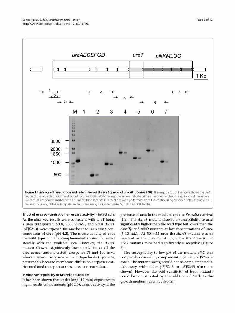
ResultsEvidence of transcription and redefinition of the ure2 operon of Brucella abortus 2308We have previously reported that the Brucella ureaseoperon ure2 did not contribute to the urease activity ofthe bacteria [1]. The ure2 operon of Brucella abortus2308 was considered to be composed of eight genes ure-ABCEFGDT (BAB1_1376-1383). A re-evaluation of thechromosomal region suggested that some genes immedi-ately downstream of ureT could be part of the sameoperon, because: 1) the distance between ureT and thecontiguous gene nikM was only 26 bp, 2) there was agood ribosome binding site upstream the putative startcodon of nikM, and 3) there was no obvious transcrip-tional terminator between the two genes. PCR amplifica-tion of reverse transcribed Brucella RNA using the pairsof primers indicated in Table 1 was conducted to assessthe continuity of the transcript until we reached the firstgene annotated on the opposite strand (BAB1_1389).Genomic DNA and total RNA were used as positive andnegative controls, and the results are shown in Figure 1.Five additional genes (BAB1_1384-1388) were found tobe cotranscribed with the first eight genes, and theirfunctional gene annotation was performed using theSEED comparative genomics resource [21]. The proposedrole of these genes (nikKMLQO) was to code for a nickeltransport system belonging to the novel ECF class ofmodular transporters [12]. According to this classifica-tion, NikM would be the substrate-specific component,
Sangari et al. BMC Microbiology 2010, 10:107http://www.biomedcentral.com/1471-2180/10/107
Sangari et al. BMC Microbiology 2010, 10:107http://www.biomedcentral.com/1471-2180/10/107
Page 4 of 12
while NikQ and NikO would be the transmembrane andATPase components, respectively, of the energizing mod-ule. NikK and NikL would be additional components.Their role in nickel transport is also supported by thegenomic context, as urease is a nickel-containing enzyme,although we could not find other supporting evidence(e.g. NickR-binding sites in the region). The completeure2 operon is thus composed of thirteen genes puta-tively involved in three different functions, namely ureaseproduction, urea transport, and nickel transport.
Construction of chromosomal mutants in the ure2 operonIn order to analyze the impact of the ure2 genes on ureaseactivity, we constructed three mutants as described in theMethods section: i) a polar mutant created by replacingpart of ureT with a kanamycin resistance gene that has atranscriptional termination signal (ΔureTp), ii) a non-polar mutant lacking the aph transcriptional terminator,which only affects ureT function (ΔureT), and iii) a ΔnikOmutant, affecting the ATP binding protein of the putativenickel transport system encoded by nikO, the last gene ofthe operon, and predicted to have the biggest impact onthe correct function of the transporter while still main-taining basal activity [16].
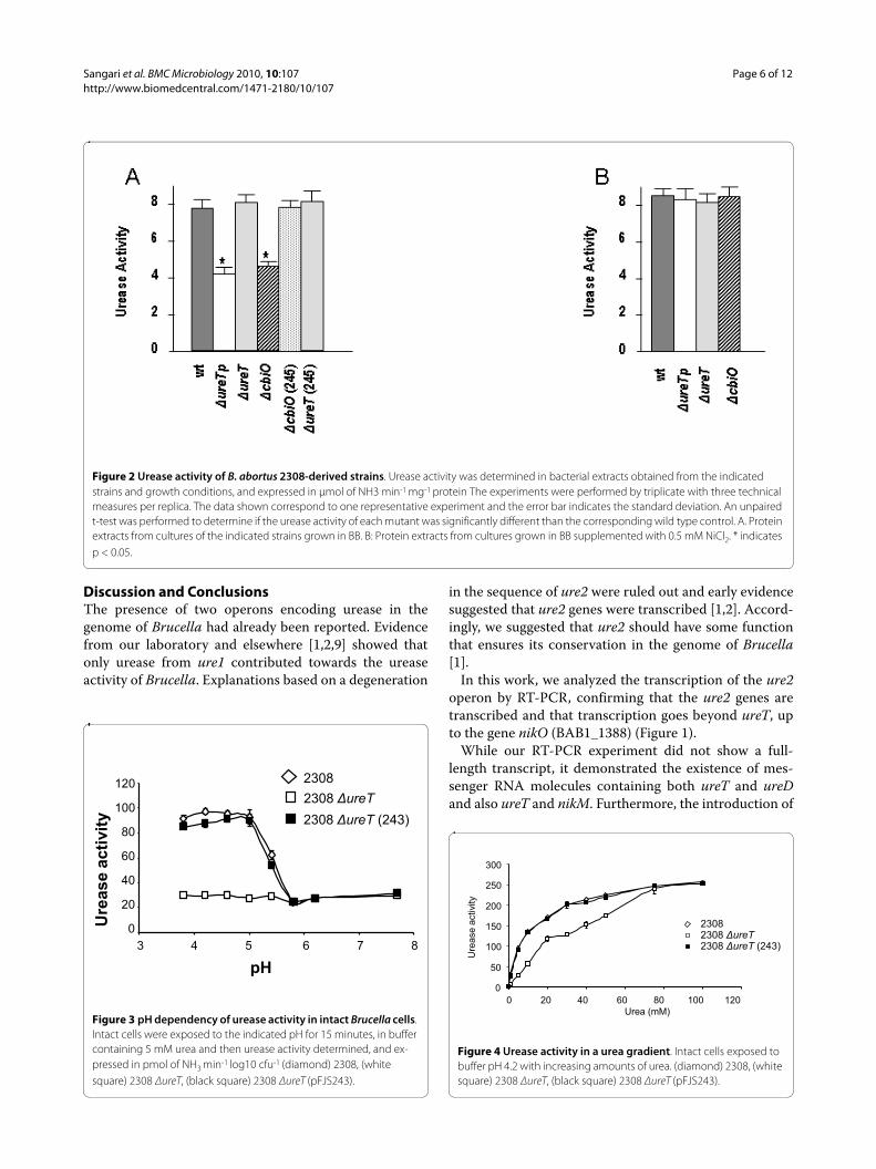
Urease activity of the different ure2 mutantsUrease activity was measured in crude protein extractsfrom the mutants and the wild type strain. The results inFigure 2A show that extracts of both the ΔureTp andΔnikO mutants had their urease activity reduced to about50% of the activity observed in the wild type strain 2308,while the urease activity was rather unaffected in theΔureT mutant. To confirm that the observed effects weredirect consequence of the introduced mutations, mutantswere complemented with plasmids carrying either ureTor nikO genes. Mutants ΔureTp and ΔnikO were comple-mented by the nikO containing plasmid pFJS245 (Figure2A), but no effect on urease activity was observed whenpFJS243 (containing ureT) was used to complement theΔureTp, ΔureT, or ΔnikO mutants (data not shown).
Effect of nickel addition on urease activity of different Brucella strainsNickel and cobalt are transition metals that can share thesame bacterial import systems [16]. The genes nikKM-LQO, currently annotated in the Brucella genomes ascomponents of a cobalt transport system, are founddownstream of the ure2 genes, and form part of the sameoperon, so we tested whether they were involved in thetransport of nickel, which is essential for urease activity.The addition of an excess of nickel in the form of NiCl2would supply the metal needed for urease assembly inspite of the inactivation of the nik transport system. Wetested the urease activity of the different strains grown inthe presence or absence of 0.5 mM NiCl2 in the culturemedium. The results in Figure 2B indicate that the ureaseactivity of all the mutants reverted to normal values whenthe culture medium was supplemented with nickel, thusconfirming the suspected role of the products of the nikgenes of the ure2 operon in nickel transport. Theseresults are also a further evidence for the extension of theoperon until the nikO gene; that is a polar ureT mutationhas a lower urease activity than the corresponding non-polar mutant, and identical activity to that of the nikOmutant, suggesting that the observed phenotype is theresult of a polar effect on the genes downstream of ureT.
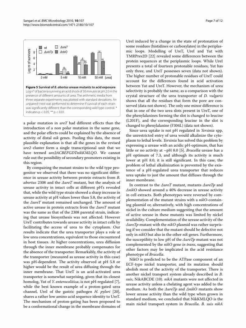
Effect of pH on urease activity in intact cellsBrucella urease assayed in vitro shows a pH-dependentactivity that is maximal at pH 7.3 [1]. When urease activ-ity was assayed in intact B. abortus 2308 cells, the activitywas higher at low pH values and dropped to near onethird as the pH of the medium reached a value of 6 (Fig-ure 3). ΔureT intact cells showed very similar activity towild type cells at pH values above 6, but they lost theacid-dependent induction of urease activity at lower pHvalues. The increased urease activity of the ΔureT mutantdue to enhanced urea uptake at low pH could be restoredby complementation with the ureT containing plasmidpFJS243.
Sangari et al. BMC Microbiology 2010, 10:107http://www.biomedcentral.com/1471-2180/10/107
Page 5 of 12
Effect of urea concentration on urease activity in intact cellsAs the observed results were consistent with UreT beinga urea transporter, 2308, 2308 ΔureT, and 2308 ΔureT(pFJS243) were exposed for one hour to increasing con-centrations of urea (pH 4.2). The urease activity of boththe wild type and the complemented strains increasedsteadily with the available urea. However, the ΔureTmutant showed significantly lower activities at all theurea concentrations tested, except for 75 and 100 mM,where urease activity reached wild type levels (Figure 4),presumably because membrane diffussion surpasses car-rier mediated transport at these urea concentrations.
In vitro susceptibility of Brucella to acid pHIt has been shown that under long (15 min) exposures tohighly acidic environments (pH 2.0), urease activity in the
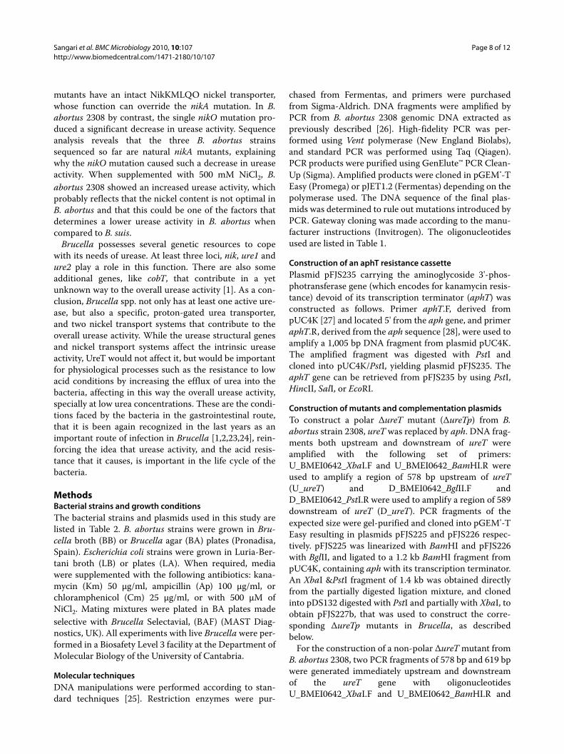
presence of urea in the medium enables Brucella survival[1,2]. The ΔureT mutant showed a susceptibility to acidsignificantly higher than the wild type but lower than theΔureTp and nikO mutants at low concentrations of urea(5-10 mM). At 50 mM urea the ΔureT mutant was asresistant as the parental strain, while the ΔureTp andnikO mutants remained significantly susceptible (Figure5).
The susceptibility to low pH of the mutant nikO wascompletely reversed by complementing it with pFJS245 intrans. The mutant ΔureTp could not be complemented inthis assay with either pFJS243 or pFJS245 (data notshown). However the acid sensitivity of both mutantscould be compensated by the addition of NiCl2 to thegrowth medium (data not shown).
Figure 1 Evidence of transcription and redefinition of the ure2 operon of Brucella abortus 2308. The map on top of the figure shows the ure2 region of the large chromosome of Brucella abortus 2308. Below the map the arrows indicate primers designed to check transcription of the region. For each pair of primers marked with a number, three separate PCR reactions were performed: a positive control using genomic DNA as template; a test reaction using cDNA as template, and a control using RNA as template. M, 1 Kb Plus DNA ladder.
12
3
45
6
7
ureABCEFGD ureT nikKMLQO
1 2 3 4 5 6 7
1 Kb
M
1000
500
16502000
3000
Sangari et al. BMC Microbiology 2010, 10:107http://www.biomedcentral.com/1471-2180/10/107
Page 6 of 12
Discussion and ConclusionsThe presence of two operons encoding urease in thegenome of Brucella had already been reported. Evidencefrom our laboratory and elsewhere [1,2,9] showed thatonly urease from ure1 contributed towards the ureaseactivity of Brucella. Explanations based on a degeneration
in the sequence of ure2 were ruled out and early evidencesuggested that ure2 genes were transcribed [1,2]. Accord-ingly, we suggested that ure2 should have some functionthat ensures its conservation in the genome of Brucella[1].
In this work, we analyzed the transcription of the ure2operon by RT-PCR, confirming that the ure2 genes aretranscribed and that transcription goes beyond ureT, upto the gene nikO (BAB1_1388) (Figure 1).
While our RT-PCR experiment did not show a full-length transcript, it demonstrated the existence of mes-senger RNA molecules containing both ureT and ureDand also ureT and nikM. Furthermore, the introduction of
Figure 2 Urease activity of B. abortus 2308-derived strains. Urease activity was determined in bacterial extracts obtained from the indicated strains and growth conditions, and expressed in μmol of NH3 min-1 mg-1 protein The experiments were performed by triplicate with three technical measures per replica. The data shown correspond to one representative experiment and the error bar indicates the standard deviation. An unpaired t-test was performed to determine if the urease activity of each mutant was significantly different than the corresponding wild type control. A. Protein extracts from cultures of the indicated strains grown in BB. B: Protein extracts from cultures grown in BB supplemented with 0.5 mM NiCl2. * indicates p < 0.05.
Figure 3 pH dependency of urease activity in intact Brucella cells. Intact cells were exposed to the indicated pH for 15 minutes, in buffer containing 5 mM urea and then urease activity determined, and ex-pressed in pmol of NH3 min-1 log10 cfu-1 (diamond) 2308, (white square) 2308 ΔureT, (black square) 2308 ΔureT (pFJS243).
2308 ΔureT2308 ΔureT (243)
2308
3 4 5 6 7 8
120
100
80
60
40
20
0
pH
Ure
ase
activ
ity
Figure 4 Urease activity in a urea gradient. Intact cells exposed to buffer pH 4.2 with increasing amounts of urea. (diamond) 2308, (white square) 2308 ΔureT, (black square) 2308 ΔureT (pFJS243).
2308 ΔureT2308 ΔureT (243)
2308
0 20 40 60 80 100 120 Urea (mM)
0
50
100
150
200
250
300
Ure
ase
activ
ity
Sangari et al. BMC Microbiology 2010, 10:107http://www.biomedcentral.com/1471-2180/10/107
Page 7 of 12
a polar mutation in ureT had different effects than theintroduction of a non polar mutation in the same gene,and the polar effects could be explained by the absence ofactivity of distal nik genes. Pooling this data, the mostplausible explanation is that all the genes in the revisedure2 cluster form a single transcriptional unit that wehave termed ure2ACBEFGDTnikKMLQO. We cannotrule out the possibility of secondary promoters existing inthis region.
By compairing the mutant strains to the wild type pro-genitor we observed that there was no significant differ-ence in urease activity between protein extracts from B.abortus 2308 and the ΔureT mutant, but the analysis ofurease activity in intact cells at different pH's revealedthat, while the wild type strain showed a sharp increase inurease activity at pH values lower than 5.8, the activity ofthe ΔureT mutant remained unchanged. The amount ofactive urease in protein extracts from the ΔureT mutantwas the same as that of the 2308 parental strain, indicat-ing that urease biosynthesis was not affected. HoweverUreT contributes towards urease activity in intact cells byfacilitating the access of urea to the cytoplasm. Ourresults indicate that the urea transporter plays a role atlow urea concentrations, equivalent to those encounteredin host tissues. At higher concentrations, urea diffusionthrough the inner membrane probably compensates forthe absence of the transporter. Remarkably, the activity ofthe transporter (measured as urease activity in this case)was pH-dependent. The activity observed at pH 5.8 orhigher would be the result of urea diffusing through theinner membrane. That UreT is an acid-activated ureatransporter is somewhat surprising, given that its closesthomolog, Yut of Y. enterocolitica, is not pH-regulated [7],while the best known example of a proton-gated ureachannel, UreI of the gastric pathogen H. pylori [20],shares a rather low amino acid sequence identity to UreT.The mechanism of proton-gating has been proposed tobe a conformational change in the membrane domains of
UreI induced by a change in the state of protonation ofsome residues (histidines or carboxylates) in the periplas-mic loops. Modelling of UreT, UreI and Yut withTMRPres2D [22] revealed some differences between theprotein sequences at the periplasmic loops. While UreIpresents a total of fourteen protonable residues, Yut hasonly three, and UreT possesses seven (data not shown).The higher number of protonable residues of UreT couldaccount for the differences found in acid activationbetween Yut and UreT. However, the mechanism of ureaselectivity is probably the same, as a comparison with thecrystal structure of the urea transporter of D. vulgarisshows that all the residues that form the pore are con-served (data not shown). The only one minor difference isthat in one of the two urea slots present in UreT, one ofthe phenylalanines forming the slot is changed to leucine(L201F), and the corresponding leucine in the slot ischanged to phenylalanine (F304L) (data not shown).
Since urea uptake is not pH regulated in Yersinia spp,the unrestricted entry of urea would alkalinize the cyto-plasm to lethal levels. Yersinia has solved this problem byexpressing a urease with an acidic pH-optimum, that haslittle or no activity at ~pH 8.0 [5]. Brucella urease has apH optimum of 7.3, and although its activity is muchlower at pH 8.0, it is still significant. In this case, theproblem of lethal alkalinization is prevented by the exis-tence of a pH-regulated urea transporter that reducesurea uptake to just the amount that diffuses through theinner membrane.
In contrast to the ΔureT mutant, mutants ΔureTp andΔnikO showed around a 40% decrease in urease activityin cell extracts. Both phenotypes were reversed by com-plementation of the mutant strains with a nikO-contain-ing plasmid or, alternatively, with high concentrations ofnickel in the culture medium suggesting that the amountof active urease in these mutants was limited by nickelavailability. Complementation of the urease activity of theΔureTp mutant with the nikO plasmid was rather surpris-ing if we consider that the mutant should be defective notonly in nikO but also in the other nik genes. Furthermore,the susceptibity to low pH of the ΔureTp mutant was notcomplemented by the nikO gene in trans, suggesting thatother factors may be implicated in the acid resistancephenotype of Brucella.
NikO is predicted to be the ATPase component of anECF-type nickel transporter, and its mutation shouldabolish most of the activity of the transporter. There isanother nickel transport system already described in B.suis, NikABCDE (10). nikA mutants were not affected inurease activity unless a chelating agent was added to themedium. As both the ΔureTp and ΔnikO mutants showlower urease activity than the wild type when grown instandard medium, we concluded that NikKMLQO is themain nickel transport system in Brucella. B. suis nikA
Figure 5 Survival of B. abortus urease mutants to acid exposure. Log n° of bacteria surviving an acid shock of 30 minutes at pH 2.0 in the presence of different amounts of urea. The arithmetic media from three separate experiments was plotted with standard deviations. An unpaired t-test was performed to determine if survival of each strain was significantly different than the corresponding wild type control. * indicates p < 0.05, ** p < 0.01.
Log n
r of s
urviv
ors
2
4
6
8
5 mM 10 mM 50 mM [urea]0 mM
wtΔureTp ΔcbiO
ΔureT
*****
*****
** **
Sangari et al. BMC Microbiology 2010, 10:107http://www.biomedcentral.com/1471-2180/10/107
Page 8 of 12
mutants have an intact NikKMLQO nickel transporter,whose function can override the nikA mutation. In B.abortus 2308 by contrast, the single nikO mutation pro-duced a significant decrease in urease activity. Sequenceanalysis reveals that the three B. abortus strainssequenced so far are natural nikA mutants, explainingwhy the nikO mutation caused such a decrease in ureaseactivity. When supplemented with 500 mM NiCl2, B.abortus 2308 showed an increased urease activity, whichprobably reflects that the nickel content is not optimal inB. abortus and that this could be one of the factors thatdetermines a lower urease activity in B. abortus whencompared to B. suis.
Brucella possesses several genetic resources to copewith its needs of urease. At least three loci, nik, ure1 andure2 play a role in this function. There are also someadditional genes, like cobT, that contribute in a yetunknown way to the overall urease activity [1]. As a con-clusion, Brucella spp. not only has at least one active ure-ase, but also a specific, proton-gated urea transporter,and two nickel transport systems that contribute to theoverall urease activity. While the urease structural genesand nickel transport systems affect the intrinsic ureaseactivity, UreT would not affect it, but would be importantfor physiological processes such as the resistance to lowacid conditions by increasing the efflux of urea into thebacteria, affecting in this way the overall urease activity,specially at low urea concentrations. These are the condi-tions faced by the bacteria in the gastrointestinal route,that it is been again recognized in the last years as animportant route of infection in Brucella [1,2,23,24], rein-forcing the idea that urease activity, and the acid resis-tance that it causes, is important in the life cycle of thebacteria.
MethodsBacterial strains and growth conditionsThe bacterial strains and plasmids used in this study arelisted in Table 2. B. abortus strains were grown in Bru-cella broth (BB) or Brucella agar (BA) plates (Pronadisa,Spain). Escherichia coli strains were grown in Luria-Ber-tani broth (LB) or plates (LA). When required, mediawere supplemented with the following antibiotics: kana-mycin (Km) 50 μg/ml, ampicillin (Ap) 100 μg/ml, orchloramphenicol (Cm) 25 μg/ml, or with 500 μM ofNiCl2. Mating mixtures were plated in BA plates madeselective with Brucella Selectavial, (BAF) (MAST Diag-nostics, UK). All experiments with live Brucella were per-formed in a Biosafety Level 3 facility at the Department ofMolecular Biology of the University of Cantabria.
Molecular techniquesDNA manipulations were performed according to stan-dard techniques [25]. Restriction enzymes were pur-
chased from Fermentas, and primers were purchasedfrom Sigma-Aldrich. DNA fragments were amplified byPCR from B. abortus 2308 genomic DNA extracted aspreviously described [26]. High-fidelity PCR was per-formed using Vent polymerase (New England Biolabs),and standard PCR was performed using Taq (Qiagen).PCR products were purified using GenElute™ PCR Clean-Up (Sigma). Amplified products were cloned in pGEM®-TEasy (Promega) or pJET1.2 (Fermentas) depending on thepolymerase used. The DNA sequence of the final plas-mids was determined to rule out mutations introduced byPCR. Gateway cloning was made according to the manu-facturer instructions (Invitrogen). The oligonucleotidesused are listed in Table 1.
Construction of an aphT resistance cassettePlasmid pFJS235 carrying the aminoglycoside 3'-phos-photransferase gene (which encodes for kanamycin resis-tance) devoid of its transcription terminator (aphT) wasconstructed as follows. Primer aphT.F, derived frompUC4K [27] and located 5' from the aph gene, and primeraphT.R, derived from the aph sequence [28], were used toamplify a 1,005 bp DNA fragment from plasmid pUC4K.The amplified fragment was digested with PstI andcloned into pUC4K/PstI, yielding plasmid pFJS235. TheaphT gene can be retrieved from pFJS235 by using PstI,HincII, SalI, or EcoRI.
Construction of mutants and complementation plasmidsTo construct a polar ΔureT mutant (ΔureTp) from B.abortus strain 2308, ureT was replaced by aph. DNA frag-ments both upstream and downstream of ureT wereamplified with the following set of primers:U_BMEI0642_XbaI.F and U_BMEI0642_BamHI.R wereused to amplify a region of 578 bp upstream of ureT(U_ureT) and D_BMEI0642_BglII.F andD_BMEI0642_PstI.R were used to amplify a region of 589downstream of ureT (D_ureT). PCR fragments of theexpected size were gel-purified and cloned into pGEM®-TEasy resulting in plasmids pFJS225 and pFJS226 respec-tively. pFJS225 was linearized with BamHI and pFJS226with BglII, and ligated to a 1.2 kb BamHI fragment frompUC4K, containing aph with its transcription terminator.An XbaI &PstI fragment of 1.4 kb was obtained directlyfrom the partially digested ligation mixture, and clonedinto pDS132 digested with PstI and partially with XbaI, toobtain pFJS227b, that was used to construct the corre-sponding ΔureTp mutants in Brucella, as describedbelow.
For the construction of a non-polar ΔureT mutant fromB. abortus 2308, two PCR fragments of 578 bp and 619 bpwere generated immediately upstream and downstreamof the ureT gene with oligonucleotidesU_BMEI0642_XbaI.F and U_BMEI0642_BamHI.R and
Sangari et al. BMC Microbiology 2010, 10:107http://www.biomedcentral.com/1471-2180/10/107
Page 9 of 12
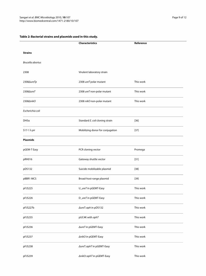
Table 2: Bacterial strains and plasmids used in this study.
Characteristics Reference
Strains
Brucella abortus
2308 Virulent laboratory strain
2308ΔureTp 2308 ureT polar mutant This work
2308ΔureT 2308 ureT non-polar mutant This work
2308ΔnikO 2308 nikO non-polar mutant This work
Escherichia coli
DH5α Standard E. coli cloning strain [36]
S17-1 λ pir Mobilizing donor for conjugation [37]
Plasmids
pGEM-T Easy PCR cloning vector Promega
pRH016 Gateway shuttle vector [31]
pDS132 Suicide mobilizable plasmid [38]
pBBR1 MCS Broad-host-range plasmid [39]
pFJS225 U_ureT in pGEMT-Easy This work
pFJS226 D_ureT in pGEMT-Easy This work
pFJS227b ΔureT::aph in pDS132 This work
pFJS235 pUC4K with aphT This work
pFJS236 ΔureT in pGEMT-Easy This work
pFJS237 ΔnikO in pGEMT-Easy This work
pFJS238 ΔureT::aphT in pGEMT-Easy This work
pFJS239 ΔnikO::aphT in pGEMT-Easy This work
Sangari et al. BMC Microbiology 2010, 10:107http://www.biomedcentral.com/1471-2180/10/107
Page 10 of 12
oligonucleotides D_BMEI0642.F andD_BMEI0642_PstI.R respectively. The reaction condi-tions for both PCRs were 30 cycles at 55°C, and 45 sec-onds at 72°C, using Vent polymerase. Both fragments(containing complementary regions) were ligated byoverlapping PCR using oligonucleotidesU_BMEI0642_XbaI.F and D_BMEI0642_PstI.R and Taqpolymerase from Qiagen, for 25 cycles at 55°C and exten-sion time of 1 minute at 72°C. The resulting fragmentcontaining the ureT deletion allele was gel-purified andcloned into pGEM®-T Easy to obtain pFJS236. A BamHIfragment from pFJS235 containing aphT was introducedinto the BamHI site of pFJS236, resulting in plasmidpFJS238. An XbaI & PstI fragment from this plasmid con-taining the replaced ureT gene was cloned into pDS132digested with PstI and partially with XbaI, resulting inplasmid pFJS241b, that was used to create the corre-sponding Brucella mutant as described below.
For the construction of a ΔnikO non-polar mutant, twoPCR fragments of 501 bp and 499 bp were generatedimmediately upstream and downstream of the nikO genewith oligonucleotides BAB1_1388_XbaI.F andRT_BAB1_1388.R, and oligonucleotidesBAB1_1388_BglII.F and BAB1_1388_PstI.R respectively,using Vent polymerase. Both fragments (containing com-plementary regions) were ligated by overlapping PCRusing oligonucleotides BAB1_1388_XbaI.F andBAB1_1388_PstI.R and Taq polymerase, and the resultingfragment containing the deleted nikO allele was clonedinto pGEM®-T Easy (pFJS237). A BamHI fragment frompFJS235 containing aphT was introduced into the BglIIsite of pFJS237, resulting in plasmid pFJS239. An XbaI&PstI fragment from this plasmid containing the replacednikO gene was cloned into pDS132 digested with PstI andpartially with XbaI, resulting in plasmid pFJS242b, thatwas used to create the corresponding Brucella mutant asdescribed below.
To construct the different mutants, replacement plas-mids were transformed into E. coli S17-1 λ pir, and mobi-lized to the corresponding Brucella recipient strain, by
mixing equal volumes (100 μl) of liquid cultures of bothdonor and recipient cells on a 0.22-μm-pore-size filter.The filter was left for 4 h on a BA plate without antibiot-ics, soaked in PBS, and then different dilutions wereplated onto BAF plates containing Cm and Km. Coloniesgrowing in this medium represented single-crossoverevents. Five colonies of each construct were pooled andgrown in BB, and 108 CFU were plated on BA containing5% sucrose to select for the double crossover. Sucrose-resistant colonies were replicated in BA Cm plates, andCmS colonies were selected and analyzed by PCR andsouthern blot to ensure that the right mutant had beenconstructed.
To complement the different mutants complementa-tion plasmids were constructed as follows: ureT wascloned by using the Gateway recombination cloning tech-nology (Invitrogen) [29]. The entry vector was obtainedfrom the Brucella ORFeome generated previously [30].The destination vector, pRH016 [31], carries a chloram-phenicol resistance marker, and the toxic cassette isflanked by attR1 and attR2 recombinational sites. Therecombinational cloning procedure was performed asrecommended by the manufacturer, to produce pFJS243.nikO was amplified by PCR with oligonucleotidesnikO_SalI.F and nikO_PstI.R, cloned into pGEM®-T Easyto obtain pFJS244, and then subcloned into pBBR1 MCS/SalI &PstI to give pFJS245. Both pFJS243 and pFJS245were transformed into E. coli S17-1 λ pir to be mobilizedto Brucella. Complemented strains were selected in BAFCm.
In vitro susceptibility of Brucella to acid pHB. abortus strains were grown in BB until the end of theexponential phase, washed in sterile water and resus-pended at a concentration of 108 CFU/ml in citrate bufferpH 2.0 for 30 min in the presence or absence of differentconcentrations of urea. Bacteria were washed three timesin phosphate-buffered saline (PBS), and survivorscounted after dilution and plating.
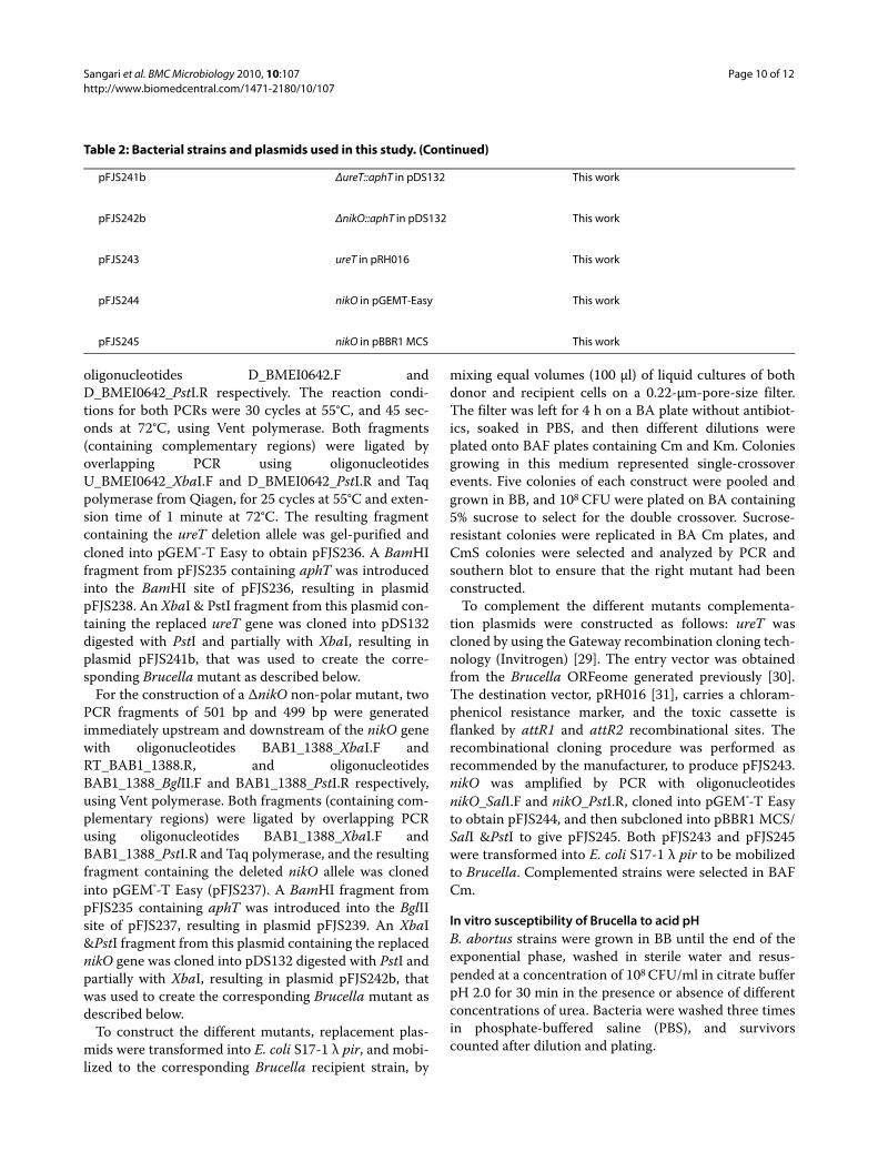
pFJS241b ΔureT::aphT in pDS132 This work
pFJS242b ΔnikO::aphT in pDS132 This work
pFJS243 ureT in pRH016 This work
pFJS244 nikO in pGEMT-Easy This work
pFJS245 nikO in pBBR1 MCS This work
Table 2: Bacterial strains and plasmids used in this study. (Continued)
Sangari et al. BMC Microbiology 2010, 10:107http://www.biomedcentral.com/1471-2180/10/107
Page 11 of 12
Measurement of urease activityUrease activity was determined by measuring the amountof ammonia released from urea. Exponential cultures ofbacteria grown in BB, supplemented or not with 500 μMof NiCl2 as indicated, were recovered by centrifugation,washed, and resuspended in PBS to a concentration of108 CFU/ml. The preparations were then lysed usingthree 10-s cycles with a FastPrep system (Bio 101, Vista,CA) at the maximum setting, cooled on ice, and centri-fuged for 5 min at 25,000 × g at 4°C to remove the celldebris. Crude extracts were stored at -80°C until theywere used. For standard urease reactions, 5 to 10 μl ofextract were added to a tube containing 200 μl of 50 mMurea in PBS and incubated for 5 min at 37°C. Ureaseactivitiy was also measured in intact cells, in this case thepelleted bacteria were resuspended in 200 μl of eitherPBS (pH 7.7) or citrate buffer at different pH (3.8, 4.2, 4.6,5.0, 5.4, 5.8, and 6.2), supplemented or not with urea atdifferent concentrations (0, 1, 5, 10, 20, 30, 40, 50, 75, and100 mM), and incubated at 37°C for 1 hour. The amountof ammonia released from urea hydrolysis was deter-mined colorimetrically by the modified Berthelot reac-tion [32], and the total protein concentration wasmeasured by a Bradford assay [33]. Urease specific activ-ity was expressed in μmol of NH3 min-1mg-1 protein (forcrude extracts) and pmol of NH3 min-1 log10 cfu-1 (forintact cells).
RNA isolation and reverse transcriptase PCR (RT-PCR)3 ml of a bacterial culture in mid-log phase (OD600 = 0.6-0.7) were stabilized with RNAprotect Bacteria Reagent(Qiagen). After harvesting the cells, they were resus-pended in 300 μl of TE containing lysozyme 1 mg/ml, andincubated for 15 min at room temperature. They weretreated with 15 μl of 10% Zwittergent 3-16 (Calbiochem)and 6 μl of 20 mg/ml of proteinase K for 1 h at 37°C, andthen total RNA was extracted with the RNeasy Mini Sys-tem (Qiagen) in combination with the RNase-Free DNaseSet (Qiagen). cDNA was generated by using SuperscriptIII RT (Invitrogen) according to the manufacturer's pro-tocol. 1 μl of the resulting cDNA was used for each PCR.As a negative control, reactions were also run on RNAtemplates without RT treatment, and as a positive con-trol, each reaction was also made with purified genomicDNA as template. The cycling parameters were 30 cyclesof 94°C for 30 s, 55°C for 30 s, and 72°C for 1.5 min. Theresulting amplicons were analyzed in 0.8% agarose gels.Primers were designed with Primer3 software [34].
Genomic data and analysisThe complete genome sequence and annotation of the B.abortus 2308 strain was obtained fron GenBank (Acces-sion numbers AM040264 and AM040265 for chromo-somes I and II respectively).
Blast comparisons against the microbial genome data-base were performed via web at the NCBI Blast server[35].
Statistical analysisA statistical analysis was performed using Prism3, version3.0(GraphPad Software, San Diego, CA). Statistical signif-icance wascalculated using either a nonparametricMann-Whitney test or an unpaired t test. A P value of <0.05 was considered statistically significant.
Authors' contributionsFJS designed and supervised the work and wrote the paper. AC performed allthe microbiological work and the different urease activity assays. AS did thetranscriptional analysis of the urease operon. JMGL performed the genomicanalysis and bioinformatic work and also wrote the paper.
AcknowledgementsThis work was supported by grants BIO2007-63656 from the Spanish Ministerio de Educación y Ciencia, and API 07/01 from Fundación Marqués de Valdecilla to FJS.We thank Matxalen Llosa and Olga Draper for critical reading and copyediting of the manuscript, Regis Hallez and Xavier de Bolle for providing plasmid pRH016, and Dominique Schneider for providing plasmid pDS132.
Author DetailsDepartamento de Biología Molecular, Universidad de Cantabria, and Instituto de Biomedicina y Biotecnología de Cantabria (IBBTEC), UC-CSIC-IDICAN, Santander, Spain
Characterization of the urease operon of Brucella abortus and assessment of its role in virulence of the bacterium. Infect Immun 2007, 75(2):774-780.
2. Bandara AB, Contreras A, Contreras-Rodriguez A, Martins AM, Dobrean V, Poff-Reichow S, Rajasekaran P, Sriranganathan N, Schurig GG, Boyle SM: Brucella suis urease encoded by ure1 but not ure2 is necessary for intestinal infection of BALB/c mice. BMC Microbiol 2007, 7:57.
3. Marshall BJ, Barrett LJ, Prakash C, McCallum RW, Guerrant RL: Urea protects Helicobacter (Campylobacter) pylori from the bactericidal effect of acid. Gastroenterology 1990, 99(3):697-702.
4. Maroncle N, Rich C, Forestier C: The role of Klebsiella pneumoniae urease in intestinal colonization and resistance to gastrointestinal stress. Res Microbiol 2006, 157(2):184-193.
5. Young GM, Amid D, Miller VL: A bifunctional urease enhances survival of pathogenic Yersinia enterocolitica and Morganella morganii at low pH. J Bacteriol 1996, 178(22):6487-6495.
6. Burne RA, Chen Y-YM: Bacterial ureases in infectious diseases. Microbes and Infection 2000, 2(5):533-542.
7. Sebbane F, Bury-Mone S, Cailliau K, Browaeys-Poly E, De Reuse H, Simonet M: The Yersinia pseudotuberculosis Yut protein, a new type of urea transporter homologous to eukaryotic channels and functionally interchangeable in vitro with the Helicobacter pylori UreI protein. Mol Microbiol 2002, 45(4):1165-1174.
8. Tsolis RM, Seshadri R, Santos RL, Sangari FJ, Lobo JM, de Jong MF, Ren Q, Myers G, Brinkac LM, Nelson WC, et al.: Genome degradation in Brucella ovis corresponds with narrowing of its host range and tissue tropism. PLoS One 2009, 4(5):e5519.
9. Contreras-Rodriguez A, Quiroz-Limon J, Martins A, Peralta H, Avila-Calderon E, Sriranganathan N, Boyle S, Lopez-Merino A: Enzymatic, immunological and phylogenetic characterization of Brucella suis urease. BMC Microbiology 2008, 8(1):121.
Sangari et al. BMC Microbiology 2010, 10:107http://www.biomedcentral.com/1471-2180/10/107
Page 12 of 12
11. Navarro C, Wu LF, Mandrand-Berthelot MA: The nik operon of Escherichia coli encodes a periplasmic binding-protein-dependent transport system for nickel. Mol Microbiol 1993, 9(6):1181-1191.
12. Rodionov DA, Hebbeln P, Eudes A, ter Beek J, Rodionova IA, Erkens GB, Slotboom DJ, Gelfand MS, Osterman AL, Hanson AD, et al.: A novel class of modular transporters for vitamins in prokaryotes. J Bacteriol 2009, 191(1):42-51.
13. Hebbeln P, Eitinger T: Heterologous production and characterization of bacterial nickel/cobalt permeases. FEMS Microbiol Lett 2004, 230(1):129-135.
14. Eitinger T, Suhr J, Moore L, Smith JA: Secondary transporters for nickel and cobalt ions: theme and variations. Biometals 2005, 18(4):399-405.
15. Hidalgo E, Palacios JM, Murillo J, Ruiz-Argueso T: Nucleotide sequence and characterization of four additional genes of the hydrogenase structural operon from Rhizobium leguminosarum bv. viciae. J Bacteriol 1992, 174(12):4130-4139.
16. Rodionov DA, Hebbeln P, Gelfand MS, Eitinger T: Comparative and functional genomic analysis of prokaryotic nickel and cobalt uptake transporters: evidence for a novel group of ATP-binding cassette transporters. J Bacteriol 2006, 188(1):317-327.
17. Jubier-Maurin V, Rodrigue A, Ouahrani-Bettache S, Layssac M, Mandrand-Berthelot M-A, Kohler S, Liautard J-P: Identification of the nik Gene Cluster of Brucella suis: Regulation and Contribution to Urease Activity. J Bacteriol 2001, 183(2):426-434.
18. Levin EJ, Quick M, Zhou M: Crystal structure of a bacterial homologue of the kidney urea transporter. Nature 2009, 462(7274):757-761.
19. Weeks DL, Eskandari S, Scott DR, Sachs G: A H+-gated urea channel: the link between Helicobacter pylori urease and gastric colonization. Science 2000, 287(5452):482-485.
20. Weeks DL, Gushansky G, Scott DR, Sachs G: Mechanism of proton gating of a urea channel. J Biol Chem 2004, 279(11):9944-9950.
21. Overbeek R, Begley T, Butler RM, Choudhuri JV, Chuang HY, Cohoon M, de Crecy-Lagard V, Diaz N, Disz T, Edwards R, et al.: The subsystems approach to genome annotation and its use in the project to annotate 1000 genomes. Nucleic Acids Res 2005, 33(17):5691-5702.
22. Spyropoulos IC, Liakopoulos TD, Bagos PG, Hamodrakas SJ: TMRPres2D: high quality visual representation of transmembrane protein models. Bioinformatics 2004, 20(17):3258-3260.
23. Delpino MV, Marchesini MI, Estein SM, Comerci DJ, Cassataro J, Fossati CA, Baldi PC: A bile salt hydrolase of Brucella abortus contributes to the establishment of a successful infection through the oral route in mice. Infect Immun 2007, 75(1):299-305.
24. Paixao TA, Roux CM, den Hartigh AB, Sankaran-Walters S, Dandekar S, Santos RL, Tsolis RM: Establishment of systemic Brucella melitensis infection through the digestive tract requires urease, the type IV secretion system, and lipopolysaccharide O-antigen. Infect Immun 2009, 77(10):4197-4208.
25. Sambrook J, Fritsch EF, Maniatis T: Molecular cloning: a laboratory manual. 2nd edition. Cold Spring Harbor, NY.: Cold Spring Harbor Laboratory Press; 1989.
26. Sangari FJ, Aguero J: Identification of Brucella abortus B19 vaccine strain by the detection of DNA polymorphism at the ery locus. Vaccine 1994, 12(5):435-438.
27. Vieira J, Messing J: The pUC plasmids, an M13 mp7-derived system for insertion mutagenesis and sequencing with synthetic universal primers. Gene 1982, 19(3):259-268.
28. Oka A, Sugisaki H, Takanami M: Nucleotide sequence of the kanamycin resistance transposon Tn903. J Mol Biol 1981, 147(2):217-226.
29. Walhout AJ, Temple GF, Brasch MA, Hartley JL, Lorson MA, Heuvel S van den, Vidal M: GATEWAY recombinational cloning: application to the cloning of large numbers of open reading frames or ORFeomes. Methods Enzymol 2000, 328:575-592.
30. Dricot A, Rual JF, Lamesch P, Bertin N, Dupuy D, Hao T, Lambert C, Hallez R, Delroisse JM, Vandenhaute J, et al.: Generation of the Brucella melitensis ORFeome Version 1.1. Genome Res 2004, 14(10B):2201-2206.
31. Hallez R, Letesson JJ, Vandenhaute J, De Bolle X: Gateway-based destination vectors for functional analyses of bacterial ORFeomes: application to the Min system in Brucella abortus. Appl Environ Microbiol 2007, 73(4):1375-1379.
32. Senior BW, Bradford NC, Simpson DS: The ureases of Proteus strains in relation to virulence for the urinary tract. J Med Microbiol 1980, 13(4):507-512.
33. Bradford MM: A rapid and sensitive method for the quantitation of microgram quantities of protein utilizing the principle of protein-dye binding. Anal Biochem 1976, 72:248-254.
34. Rozen S, Skaletsky H: Primer3 on the WWW for general users and for biologist programmers. Methods Mol Biol 2000, 132:365-386.
35. BLAST with microbial genomes [http://www.ncbi.nlm.nih.gov/sutils/genom_table.cgi]
36. Hanahan D: Studies on transformation of Escherichia coli with plasmids. J Mol Biol 1983, 166(4):557-580.
37. Herrero M, de Lorenzo V, Timmis KN: Transposon vectors containing non-antibiotic resistance selection markers for cloning and stable chromosomal insertion of foreign genes in gram-negative bacteria. J Bacteriol 1990, 172(11):6557-6567.
38. Philippe N, Alcaraz JP, Coursange E, Geiselmann J, Schneider D: Improvement of pCVD442, a suicide plasmid for gene allele exchange in bacteria. Plasmid 2004, 51(3):246-255.
doi: 10.1186/1471-2180-10-107Cite this article as: Sangari et al., Brucella abortus ure2 region contains an acid-activated urea transporter and a nickel transport system BMC Microbiol-ogy 2010, 10:107