32
Course: B.Sc. (Agri.) Subject:- Principles of Plant Pathology Unit-5.1 Topic: Plant Parasitic Nematodes
| Date post: | 15-Jul-2015 |
| Category: |
Education |
| Upload: | rai-university |
| View: | 323 times |
| Download: | 2 times |

Course: B.Sc. (Agri.)
Subject:- Principles of Plant Pathology
Unit-5.1
Topic: Plant Parasitic Nematodes
Nematology is an important branch of biological science, which deals
with a complex, diverse group of round worms known as Nematodes that
occur worldwide in essentially allenvironments. Nematodes are also known
as eelworms in Europe, nemas in the United States and round worms by
zoologists. Many species are important parasites of plants and animals,
whereas others are beneficial to agriculture and the environment.
Nematodes that are parasites of man and animals are called helminthes and
the study is known as Helminthology. The plant parasitic forms are called
nematodes and the study is known as Plant Nematology. The name
nematode was derived from Greek words nema (thread) and oides
(resembling).Annual crop losses due to these obligate parasites have been
estimated to be about $ 78 billion wordwide and $ 8 billion for U.S.
growers. The estimated annual crop loss in Tamil Nadu is around Rs. 200
crores.
The soils in a hectare of all agro ecosystem typically contain billions
of plant parasitic as well as beneficial nematodes. The damage to plants
caused by nematodes is often overlooked because the associated symptoms,
including slow growth, stunting and yellowing, can also be attributed to
nutritional and water related disorders

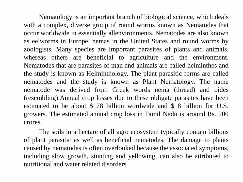
General Characteristics of Nematodes
1. The nematodes possess elongate, unsegmented, cylindrical or worm like body tapering towards
both ends, unciliated and circular in cross section.
2. Body is bilaterally symmetrical.
3. They are aquatic, terrestrial and parasitic or free living.
4. The body is covered by tough and resistant cuticle secreted by epidermal (hypodermal) cells.
5. Terminal oral aperture (mouth) surrounded with lips and papillae.
6. Digestive system consist of feeding apparatus, oesophagus, intestine and rectum.
7. Body consist of two tubes.
8. The nervous system consist of circum-oesophageal nerve ring and longitudinal nerves.
9. Primitive excretory system, is devoid of protonephridial cilia or matanephridial funnel.
10. The circulatory and respiratory systems completely absent.
11. The females have separate genital pore and males have a common opening known cloaca and
well developed copulatory apparatus consisting of spicules and gubernaculum.
12. Females are oviparous or ovoviviparous or viviparous. The cleavage is terminate and growth
is accompanied by molting.
13. Life cycle is direct and there are four juvenile stages.
Fig-1

Fig-2
Even though nematodes occupy nearly every habitat on earth, they are
remarkably similar in morphology and life stages. Despite their structural
complexity, certain basic principles are common to all nematodes. Nematodes are
triploblastic, bilaterally symmetrical, unsegmented, Pseudocoelomate, vermiform
and colourless animals. The plant parasitic nematodes are slender elongate,
spindle shaped or fusiform, tapering towards both ends and circular in cross
section. The length of the nematode may vary from 0.2 mm (Paratylenchus) to
about 11.0mm (Paralongidorus maximus). Their body width vary from 0.01 to
0.05 mm. In few genera, the females on maturity assume pear shape
(Meloidogyne), globular shape (Globodera), reniform (Rotylenchulus reniformis)
or saccate (Tylenchulus semipenetrans). The swelling increases the reproductive
potential of the organism. Radially symmetric traits (triradiate, tetraradiate and
hexaradiate) exist in the anterior region. The regions of intestine, excretory and
reproductive systems show tendencies towards symmetry. The nematodes have
one or two tubular gonads which open separately in the female and into the
rectum in the male which also have the copulatory spicules.The free living
saprophytic nematodes are generally larger in size. The animal and human
parasitic helminthes may have length of few centimeters to even a meteer or
more. The helminth parasitising whale fish is about 27 feet long. The study on
these animal and human parasites are known as Helminthology

•The following are some examples of Helminths
•1. Filarial worm - Wucheria bacrofti
•2. Guinea worm - Dracunculus medinesis
•3. Round worm - Ascaris lumricoides
•4. Tape worm - Taenia solium
The nematode body is not divided into definite parts, but certain sub – divisions are
given for convenience. The anterior end starts with the head, which consists of mouth and pharynx
bearing the cephalic papillae or setae. The portion between the head and the oesophagus is known
as the neck. Beginning at the anus and extending to the posterior terminus is the tail.
Longitudinally the body is divided into four regions as dorsal, right lateral, left and ventral. All the
natural openings like vulva, excretory pore and anus are located in the ventral region. The
nematode body is made up of several distinct body systems. They are the body wall, nervous
system, secretory – excretory system, and digestive system and reproductive system. Nematodes
do not posses a specialized circulatory or respiratory system. The exchange of gases is thought to
occur through the cuticle and circulation proceeds through the movement of fluids within the
pseudocolelom and by simple diffusion across membranes.

• The nematode body is divided into three regions. They are the outer body
tube or body wall, inner body tube and body cavity or pseudocoelome.
The outer body tube
• The outer body tube or body wall includes the cuticle, hypodermis, and
somatic muscles. The body wall protects the nematode from the harsh
external environment, serves as the exoskeleton and provides the
mechanism for movement of the organism through the soil and plant tissue.
The body wall also contains much of the nervous and secretory – excretory
systems, and it plays a role in the exchange of gases
Exoskeleton or cuticle: • It is outermost covering of body wall which is non-cellular, semipermeable and
tough layer secreted by the epidermal cells. It invades all natural opening ofbody including the mouth, rectum, cloaca, vagina, excretory pore, amhids andphasmids.
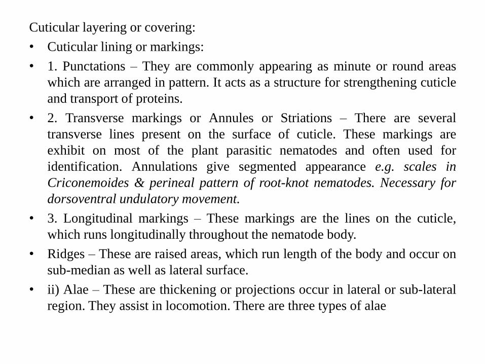
• The cuticle of many nematode species has markings on the surface. They arevaried and complex and often used by taxonomist in identification of nematodespecies. The cuticular lining/markings are categorized in different types are asfollows
• Cuticular lining or markings:
• Punctations – They are commonly appearing as minute or round areas which are arranged in pattern. It acts as a structure for strengthening cuticle and transport of proteins.
• 2. Transverse markings or Annules or Striations – There are several transverse lines present on the surface of cuticle. These markings are exhibit on most of the plant parasitic nematodes and often used for identification. Annulations give segmented appearance e.g. scales in Criconemoides & perineal pattern of root-knot nematodes. Necessary for dorsoventral undulatory movement.
• 3. Longitudinal markings – These markings are the lines on the cuticle, which runs longitudinally throughout the nematode body

• i) Ridges – These are raised areas, which run length of the body and occur
on sub-median as well as lateral surface.
• ii) Alae – These are thickening or projections occur in lateral or sub-lateral
region. They assist in locomotion. There are three types of alae
• Caudal alae – These are found in the posterior region and restricted to
males as copulatory bursa.
• Cervical alae – These are confined to anterior part of the nematode body.
Cervical alae are found in some species of marine nematodes.
• Longitudinal alae – These are limits to the lateral fields. They are
transverse by striations or furrows varying in number from one to twelve
which provide locomotion and may permit slight change in the width of
nematode.

Fig-3
Cuticular layering or covering:
• Cuticular lining or markings:
• 1. Punctations – They are commonly appearing as minute or round areas
which are arranged in pattern. It acts as a structure for strengthening cuticle
and transport of proteins.
• 2. Transverse markings or Annules or Striations – There are several
transverse lines present on the surface of cuticle. These markings are
exhibit on most of the plant parasitic nematodes and often used for
identification. Annulations give segmented appearance e.g. scales in
Criconemoides & perineal pattern of root-knot nematodes. Necessary for
dorsoventral undulatory movement.
• 3. Longitudinal markings – These markings are the lines on the cuticle,
which runs longitudinally throughout the nematode body.
• Ridges – These are raised areas, which run length of the body and occur on
sub-median as well as lateral surface.
• ii) Alae – These are thickening or projections occur in lateral or sub-lateral
region. They assist in locomotion. There are three types of alae

Cuticular layering or covering:
The nematode cuticle is basically three layer structure and composed of(a) Cortical layer, (b) Median layer and (c) Basal layer.
(a) Cortical layer – It is often divided into external cortical layer andinternal cortical layer. The surface of external cortical layer is exposedto the environment. This layer is very thin measuring about 25 to 40mμ. The external layer has been considered to be kertatine (protein)chemically. In cyst nematode the cuticle of the female on maturitybecomes tough and leathery to form cyst which protect eggs under dryconditions.
b) Median layer – The average thickness of the median layer is 0.1 μ inthe larva of Meloidogyne and Heterodera. Chemically the median layerconsist of protein, which resembles collagen (Non osmophilic collagenprotein).
(c) Basal layer – It consist of regularly arranged vertical rods orstriations. It is composed of protein with very close linkage between themolecules, resulting in resistant layer which protect the nematode fromouter environment. The thickness of basal layer varies from 125 to 500mμ (Osmophilic protein close to keratine)
Functions of cuticle:
1) Protects the nematode from harsh environment.
2) Serves as exoskeleton
3) Provide mechanism of movement of the nematode through the soil and plant
tissue.
(B)Hypodermis –
The hypodermis is cellular or partially cellular layer. It secretes the cuticle. It
lies between cuticle and somatic muscle layer. It is important metabolic active
part of the nematode. Forms 4 cords (dorsal, ventral and two lateral). Contains
hypodermal glands
(C) Muscle layer -
It is arranged in a single layer. The muscle cells are spindal shaped and
attached to the hypodermis throughout their length. It is well connected to the
nervous system. The stimulation of the muscles by dorsal and ventral nerves
cause contractions in the dorso-ventral plane and result in the characteristic
scinusodial movement of nematode.

• On the basis of arrangement of basic cells identified following three typesare identified: :
• a. Holomyarian: Having two muscle cells in each zone.
• b. Meromyarian: Two or five muscle cells in each interchordal zone.
• c. Polymyarian: More than five muscle cells in each zone
Specialized muscles:
Digestive System
Fig-4

• Stomodaeum: It includes the mouth and lips, the stoma and the
oesophagus.
• Mouth and lips: The mouth and lips are also associated with the feeding
activity of the nematode. Generally, there are 6 lips (two sub dorsal, two
sub ventral and two lateral) which surround the mouth. In some cases they
may be reduced by partial fusion to 3 or by complete fusion to form a
united ring around the mouth.
• Stoma or Buccal cavity: The stoma, which is also called as mouth cavity
or buccal cavity forms the feeding apparatus and lies between the mouth
and the oesophagus. The simple stoma is found in many bacterial feeding
nematodes, takes the form of a cylindrical or triangular tube, terminating in
a valve like glottoid apparatus, which may bear the minute teeth. The
cuticular lining of stoma may form teeth. Plant parasitic nematodes are
armed with a protrusible stylet which is usually hallow and functions like a
hypodermic needle. Stylet with basal knob are called as Stomatostylet
e.g.Tylenchida and the stylet without basal knob are called as odontostylet
or ononiostyle e.g. Dorylaimida.
• Oesophagus or pharynx: The oesophagus is a muscular pumpingorgan attached to the posterior portion of the stylet and lined withcuticle. It is the largest part of stomodaeum and found betweenstoma and intestine. Internally, pharynx lined with cuticle andexternally by membrane (basal lamella). It contains radial muscles,oesophageal glands and valves, which prevents the regurgitation offood. In some nematodes median and posterior part of pharynxswollen to form muscular bulb. The cylindrical oesophagus hasthree well defined regions are as follows.
• i) Corpus- The corpus may further divided to form pro and metacorpus, which is swollen contain muscle cells, supporting cells,nerve cells, gland cells (one dorsal and two sub ventral).
• ii) Isthumus
• iii) Basal bulb
• Intestine or midgut : The midgut is endodermal in origin. It issimple, hallow, straight tube consisting of a single layer of epithelialcells. The intestine is generally divided in to three region whichmerge in to each other without any perceptible boundaries. They areanterior or ventricular region, the mid intestinal region and posteriorpre-rectal region.

• 3. Proctodeum : The proctodeam or hind gut consist of Rectum and anus in
female and cloaca in male.
• Rectum is cuticular linings and invaginated in to rectal gland on par in
nematodes. Female nematodes consist of simple tube leading to anus,
whereas reproductive system opened in to it and form cloaca in male contain
spicules and other copulatory structure.
• Anus consist of slit structure on ventral side. The control of anus opening is
by unicellular, H shaped depressor muscle, which acts by raising dorsal wall
of the rectum and pulling posterior lip of anus to open it.
• Glands :
• 1) Pharyngeal or Esophageal – There are three uninucleated glands are
present. One is dorsal & other two ventro-lateral or sub ventral position. The
glands connected with lumen of oesophagus by means of terminal ampulla or
swelling.
• Function: Hatching, host penetration and digestion
• 2) Rectal - Rectal glands are varies from species to species or male and
female of same species. Copious production of gelatinous
mucopolusaccharide matrix in the eggs of deposited as mass. Which range to
protect the eggs.

Function of Digestive system
• Digestive juices which is secreted from dorsal oesophageal glands are
injected into the host plant cell by means of the stylet. During feeding, a
distinct zone develop around the feeding site in the host cell. There are two
feeding phases- 1) Injection phase or salivation phase and 2) Ingestion
phase.
• 1) Injection phase or salivation phase: During this phase, the flow of
salivary juices into the host cell occurs due to contraction of lateral muscle
of the median bulb.
• 2) Ingestion phase: During this phase, rhythmical contraction of the
posterior part of oesophagus associated with the median bulb occurs and in
some forms, the oesophageo-intestinal valve or cardia is responsible for
ingestion of material from the host.

Reproductive system of nematode
Female Reproductive system:-
Monodelphic- The nematodes may have a single ovary the female is called as
monodelphic.
Didelphic- The nematodes may have two ovaries then the female is called as
didelphic.
Prodelphic- When a single gonad is present, it may be either directed towards
anterior to vulva then female is called as prodelphic.
Opisthodelphic- The gonad either directed towards posterior to vulva then
female is opisthodelphic.
Amphidelphic- The two ovaries are opposite to one another, such as one is
anteriorly directed and other posteriorly directed
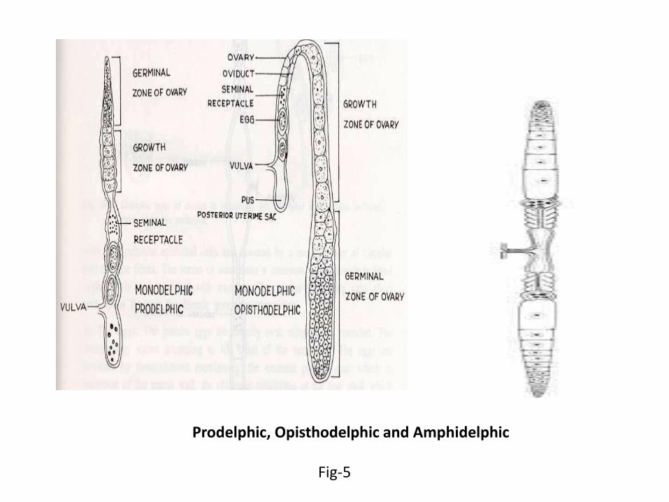
Prodelphic, Opisthodelphic and Amphidelphic
Fig-5

• Monarchic- The nematode may have one testis are called monarchic.
• • Diarchic- The nematode may have two testis are called diarchic.
• The male reproductive system generally consists of three primordial parts:
the testis, seminal vesicle, and vas deference.
• (i) The Testis-
• In the testis the germinal and growth zone can be easily distinguished. In
germinal zone Spermatogonial division takes place, while in growth zone,
spermatocytes increases in size. The spermatocytes are arranged in single
or double rows.
• (ii) Vas deference-
• It consist of an anterior glandular region and posterior muscular region and
containing the ejaculatory duct at the posterior end.

Excretory system of Nematodes
The excretory system is not well developed in nematodes. The excretory poreis located in midventral line close to the nerve ring. The excretory system innematodes are two types.
a. Glandular type
b. Tubular type

Nervous system of Nematodes:-
In nematodes, a central nervous system and a peripheral nervous
system can be described.
Central nervous system-
It is also known as brain consist of nerve ring associated with ganglia
and nerves. The nerve ring or circum-oesophageal commissure is belt which
may be broad and flat. It is present around the oesophagus in majority of
nematodes. In Tylenchida it encircles the isthumus while in Dorylaimida it is
present around the narrow anterior part of oesophagus. The nerve ring is
placed obliquetly with dorsal side most anterior. Towards the anterior end of
nerve ring six ganglia are present (2 sub-dorsal, 2 sub-ventral and 2- lateral)
known as papillary ganglia which are very small in size. Towards the posterior
side of nerve ring nerves arise in the dorsal, lateral and ventral side of the
body. Transverse commissure connecting the nerves are also present in
different regions of the body.
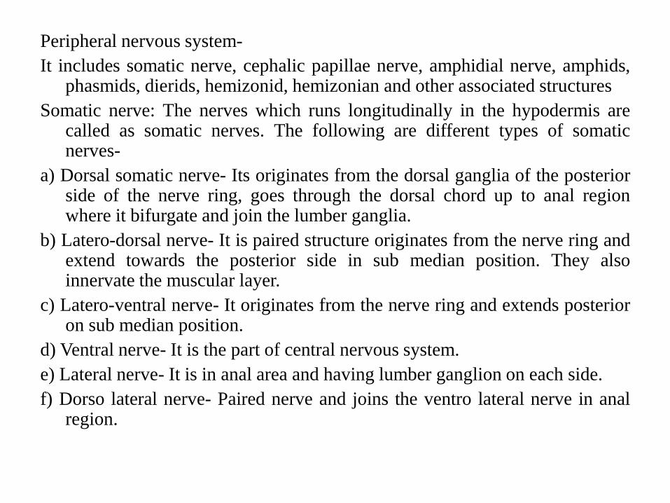
Peripheral nervous system-
It includes somatic nerve, cephalic papillae nerve, amphidial nerve, amphids,phasmids, dierids, hemizonid, hemizonian and other associated structures
Somatic nerve: The nerves which runs longitudinally in the hypodermis arecalled as somatic nerves. The following are different types of somaticnerves-
a) Dorsal somatic nerve- Its originates from the dorsal ganglia of the posteriorside of the nerve ring, goes through the dorsal chord up to anal regionwhere it bifurgate and join the lumber ganglia.
b) Latero-dorsal nerve- It is paired structure originates from the nerve ring andextend towards the posterior side in sub median position. They alsoinnervate the muscular layer.
c) Latero-ventral nerve- It originates from the nerve ring and extends posterioron sub median position.
d) Ventral nerve- It is the part of central nervous system.
e) Lateral nerve- It is in anal area and having lumber ganglion on each side.
f) Dorso lateral nerve- Paired nerve and joins the ventro lateral nerve in analregion.

• 2) Cephalic papillae nerve: These nerves goes through the body cavity.
These are nerve fibers arising from cephalic papillae ganglion from the
cephalic nerve near the lips.
• 3) Amphidial nerve: In above the papillary ganglia are directly connected
with nerve ring, while in this case the connection is indirect i.e. through
sub-ventral trunk by lateral ventro commissure. Anteriorly each amphideal
nerve enters to amphideal glands and its processes (nerves) break up in an
elongate sac, which represent the neuron are called terminals and pouch.
Sensory elements which represent the neuron are called terminals and the
group of such terminal is called as sensilla. The has an amphid aperture
situated either on the lips (labial) or post labial and opening to the exterior.
Internally the aperture is connected to a pouch (fovea) which leads to
sensilla pouch or fusus through an amphid duct or canalis amphidianlis.
The sensilla pouch is connected to the amphidial nerve through the nerve
process
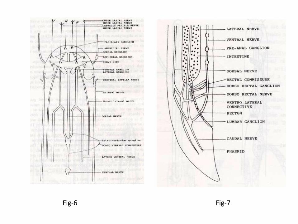
Fig-6 Fig-7
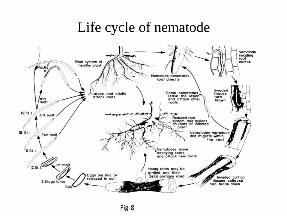
Life cycle of nematode
Fig-8

All nematodes pass through an embryonic stage, four juvenil stages
(J1–J4) and an adult stage. Juvenile Meloidogynes parasites hatch from eggs as
vermiform, second-stage juveniles (J2), the first moult having occurred within
the egg. Newly hatched juveniles have a short free-living stage in the soil, in
the rhizosphere of the host plants. They may reinvade the host plants of their
parent or migrate through the soil to find a new host root. J2 larvae do not feed
during the free-living stage, but use lipids stored in the gut.
Briefly, second stage juveniles invade in the root elongation region
and migrate in the root until they became sedentary. Signals from the J2
promote parenchyma cells near the head of the J2 to become multinucleate to
form feeding cells, generally known as giant cells, from which the J2 and later
the adults feed. Concomitant with giant cell formation, the surrounding root
tissue gives rise to a gall in which the developing juvenile is embedded.
Juveniles first feed from the giant cells about 24 hours after becoming
sedentary.
After further feeding, the J2s undergo morphological changes and
become saccate. Without further feeding, they moult three times and
eventually become adults. In females, which are close to spherical, feeding
resumes and the reproductive system develops. The life span of an adult female
may extend to three months, and many hundreds of eggs can be produced.
Females can continue egg laying after harvest of aerial parts of the plant and
the survival stage between crops is generally within the egg.
The length of the life cycle is temperature-dependent. The
relationship between rate of development and temperature is linear over much
of the root-knot nematode life cycle, though it is possible the component stages
of the life cycle, e.g. egg development, host root invasion or growth, have
slightly different optima. Species within the Meloidogyne genus also have
different temperature optima. In M. javanica, development occurs between 13
and 34 °C, with optimal development at about 29 °C.

Reference
Books
Plant Pathology by G. N. Agrios
Web resources
http://agridr.in/tnaueagri

Image References
Fig-1 http://coursewares.mju.ac.th:81/elearning47/PP300/0016sugarteam1014/5605nematode/002%20anatomy/m450818d161544_p013.jpg
Fig-2 http://www.ucmp.berkeley.edu/phyla/ecdysozoa/nematodexs.gif
Fig-3 http://www.pnas.org/content/104/44/17376/F1.large.jpg
Fig-4 http://www.uic.edu/classes/bios/bios100/labs/roundworm.jpg
Fig-5 http://coursewares.mju.ac.th:81/elearning47/PP300/0016sugarteam1014/5605nematode/002%20anatomy/m450818d161544_p031.jpg
Fig-6 http://coursewares.mju.ac.th:81/e-learning47/PP300/0016sugarteam1014/5605nematode/002%20head/m450818d161544_p026.jpg
Fig-7 http://coursewares.mju.ac.th:81/e-learning47/PP300/0016sugarteam1014/5605nematode/002%20bottom/m450818d161544_p027.jpg
Fig-8 http://www.sardi.sa.gov.au/__data/assets/image/0003/91830/lifecycle_prat_large.jpg


