2’, 3’, 4’-trihydroxychalcone is an Estrogen Receptor Alpha Coagonist By Candice Blair Herber A dissertation submitted in partial satisfaction of the requirements for the degree of Doctor of Philosophy in Endocrinology in the Graduate Division of the University of California, Berkeley Committee in charge: Professor Dale C. Leitman, Co-Chair Professor Gary L. Firestone, Co-Chair Professor Jen-Chywan Wang Professor Joseph L. Napoli Spring 2014
Transcript
2’, 3’, 4’-trihydroxychalcone is an Estrogen Receptor Alpha Coagonist
By
Candice Blair Herber
A dissertation submitted in partial satisfaction of the
requirements for the degree of
Doctor of Philosophy
in
Endocrinology
in the
Graduate Division
of the
University of California, Berkeley
Committee in charge:
Professor Dale C. Leitman, Co-Chair Professor Gary L. Firestone, Co-Chair
Professor Jen-Chywan Wang Professor Joseph L. Napoli
Spring 2014
1
Abstract
2’, 3’, 4’-trihydroxychalcone is an Estrogen Receptor Alpha Coagonist
By
Candice Blair Herber
Doctor of Philosophy in Endocrinology
University of California, Berkeley
Professor Dale C. Leitman, Co-Chair Professor Gary L. Firestone, Co-Chair
Estrogens in hormone replacement therapy (HRT) decrease menopausal symptoms, but increase the risks of reproductive cancers. The beneficial effects of estrogen on peripheral tissues and the adverse proliferative effects on the uterus and mammary gland are mediated by ERα. Currently HRT is approved only for short-term use. Short-term HRT works for decreasing symptoms associated with menopause, however, long-term usage is needed to prevent subclinical diseases. Because estradiol-bound ERα is an agonist in all tissues there is a need for development of more tissue-selective estrogens that can be used for both short and long-term HRT. Chalcones display antiproliferative activity through ERβ and may have benefits on menopause-induced hot flashes, however activity through ERα and their effects on both estradiol gene regulation and physiology are less known. The present study aimed at identifying a chalcone compound which could change the activity of ERα in the presence of estradiol as a coagonist, thereby modulating the response of ERα on gene regulation and increasing its tissue specificity. After screening a panel of five chalcone compounds for estrogenic activity in cells cotransfected with ERα and an ERE upstream of tk-luciferase, 2’, 3’, 4’-trihydroxychalcone was identified as a unique ERα coagonist. 2’, 3’, 4’-THC displayed no estrogenic activity on its own, but synergized the activation of the ERE in the presence of estradiol. Competitive binding assays with [3H]-estradiol demonstrated that 2’, 3’, 4’-THC binds to both ERα and ERβ. Estradiol and SERM-induced genes, KRT-19 and NKG2E, were not regulated by 2’, 3’, 4’-THC alone. Both KRT-19 and NKG2E were synergized with the combination of 2’, 3’, 4’-THC and estradiol. Tamoxifen and raloxifene induced expression of NKG2E, but did not synergize the expression in the presence of estradiol. The data demonstrates that 2’, 3’, 4’-THC behaves as a novel coagonist and not a SERM on gene regulation. A unique gene expression profile was induced in U2OSα cells treated with a combination of estradiol and
2
2’, 3’, 4’-THC for 24 hours with doses that would allow binding of both ligands to ERα at the same time. Functional analysis utilizing the binding affinities of estradiol, 2’, 3’, 4’-THC and another ERα binding chalcone, 2, 2’, 4’-THC, demonstrated that a heteroligand complex consisting of estradiol and 2’, 3’, 4’-THC in ERα is possible. Despite the synergistic activation of estradiol regulated genes in U2OSα cells, the combination of 2’, 3’, 4’-THC and estradiol did not induce proliferation of MCF-7 cells. In the same cells 2’, 3’, 4’-THC blocked estradiol-induced G1 to S phase cell cycle transition without blocking proliferative genes regulated by estradiol. In female ovariectomized mice on a soy-free chow diet treated for four weeks (n=5, per group), 2’, 3’, 4’-THC did not cause uterine proliferation and blocked estradiol-induced proliferation and gene expression. Although 2’, 3’, 4’-THC blocked estradiol gene expression in uterine tissue, it regulated and modulated estradiol-induced genes in adipose tissue. Because 2’, 3’, 4’-THC displays unique coagonist activity through ERα without causing proliferation, it may be useful for future HRT and expanding our knowledge of ERα regulation and ligand interaction.
i
In dedication to my Grammy, Wilma James Blair, and my mom Tami Susan Blair, who taught me to believe in myself and the importance of passion and perseverance.
I would like to start out by thanking my family for giving me the love and support I always needed to follow my dreams and my pursuit of higher education. I first and foremost acknowledge my mom and her creative spirit for always filling my life with love and teaching me the art of happiness and creativity. Mom, you were always there for me with a supportive and loving ear, and for that I am truly grateful. Dad, thank you for instilling in me the importance of education and hard work. Without your example I would not be the individual I am today. To my brother Shane, thank you for being the best brother a sister could ask for. You have always been my rock. Although you are my younger brother, you have taught me so much about life and the importance of patience and love and I could not imagine my life without you. To my grandparents, Joe and Pauline Herber, thank you for your constant support and acceptance of my career goals. To my grandparents, Bob and Wilma Blair, words cannot describe how your love and support carried me through the Ph.D. process. Although you are not here with me now, your spirit lives on. I carry with me all of the life lessons you bestowed in me along the way. Love you always.
To my second family: Laila, Sandra, Howard, Woozle, Ashley and Doug, thank you for all of the Thanksgivings and Christmases full of love and laughs. Thank you for all of the support. Laila, thank you for being there for me always no matter how crazy graduate school got. You always understood and were supportive, and for that I am always grateful. I’m glad to have a person like you in my life. Margit, thank you for always supporting me and my decisions. It’s nice to have someone in my life that always has my back and understands my craziness. Carolyn, John, Lydia and Aurelio, thank you so much for taking me into your family, accepting me, and always being both supportive and positive figures in my life. Through all the tough days of graduate school you were always there with hugs, smiles and support. Cesar, I am grateful to you for being there always as my other half. You were there for me and gave me strength even on those rough and stressful evenings when I felt nothing was working. I could not have made it out of graduate school without you by my side. Thanks for all of the love, support and much needed late night hugs.
To all the lab members of the Leitman laboratory, thank you for being a great support system. Chandi, thank you for teaching me the importance of the little things in science and for your friendship. Sreeni, thank you for teaching me so much about bench science and taking a risk on a crazy, very chatty undergrad to process your samples! Thanks to Aleksandra for always helping me with any questions I might have, and for giving me someone to look up to. Thank you Judy, Elise and Andrea for teaching me how to work with animals. Rachel, I am grateful to you for being a good friend and someone I could go to for emotional support. Without your tinctures, herbal tonics, and emotional support I would have never finished this dissertation! Chaoshen, I am grateful to you for your support and technical guidance. Isaac and Emma, thank you for all of your support over the years and your words of wisdom. They will not be forgotten. To my amazing undergraduate students: Sally, Priya, Julie, Misha and Shawn for all of the help and especially the laughs along the way.
Last I would like to acknowledge my advisor, Dale Leitman, for his support over the years. Thank you for all of your help and creative ideas for developing new and innovative ways of looking for novel estrogens to help women. I appreciated all of your guidance and support through the Ph.D. process.
1
CHAPTER ONE
Introduction and Literature Review
2
Role of Estrogens in Reproductive Physiology
Estrogens were the first human steroid hormones isolated [1] that have both a morphological and reproductive function. Estradiol is the main estrogen produced by the ovaries. In reproductive tissues, estrogens are required for tissue maturation at puberty. The female reproductive tract includes the ovaries, fallopian tubes, uterus and vagina, which require estrogen-mediated actions for both maturation and function. Puberty requires circulating estrogen levels and is a tightly regulated process controlled by a feedback loop between the hypothalamus, pituitary and ovaries. The onset of puberty is initiated when a certain body mass index is reached, which leads to the production and release of a hormone, kisspeptin. Kisspeptin binds and activates specific neurons in the hypothalamus to release the peptide hormone, GnRH [2]. The neurons also exhibit intrinsic activity that causes GnRH to be released in a pulsatile manner, which is essential to stimulate the anterior pituitary to release two hormones, LH and FSH.
LH and FSH are required not only for estradiol secretion, but follicular proliferation and subsequent ovulation. FSH, a trophic hormone, causes proliferation of ovarian cells and induces aromatase expression and activation required for estradiol production. LH stimulates the production of testosterone in the theca cells in ovarian follicles. Testosterone then enters adjacent granulosa cells, where it is aromatized to estradiol by the enzyme aromatase. The release of estradiol from the ovaries provides feedback to the hypothalamus and pituitary to control LH and FSH secretion, as well as induce proliferation of endometrial, myometrial and luminal glandular cells that line the uterine wall [3]. Endometrial cell proliferation causes an increase in cell layers that line the ducts of the uterine wall and prepares it for implantation of a fertilized oocyte and the growth and maintenance of a fetus during pregnancy. The release of estradiol during puberty is also critical for the development of the mammary gland by promoting the proliferation of epithelial cells that line the lobular and ducts that synthesize and transport milk during pregnancy.
In the absence of ERα, the formation of the reproductive tract in males and females is unaltered. The formation of rudimentary reproductive tract structures of the female mouse in utero appears normal and is not dependent on the presence of estradiol-mediated action through ERα. At puberty, the maturation of the uterus is dependent on the presence of estradiol secreted from the ovaries. In the ERα knockout (ERKO) mouse uterine tissue consisting of myometrial, endometrial and luminal glandular epithelial cells, becomes hypoplastic at puberty and only rudimentary structures are present [4]. The cells of the vagina are atrophied due to no ERα-mediated cellular proliferation, and the follicles of the ovaries are enlarged and anovulatory [4]. Because the reproductive tract is rudimentary and there is no ovulation the ERKO female mice are infertile.
Without the presence of ERα, not only is female reproduction impaired, but male reproduction as well. The male reproductive tract in the ERKO mouse in utero is normal, however, by 20 weeks of age the testis weight is low. Male ERKO mice are also infertile and are characterized by atrophied seminiferous tubules, a low sperm count (13% of the wild type), and a decrease in sexual behavior [4].
The ERβKO (BERKO) mice have shown different reproductive phenotypes dependent on the laboratory where the mice were produced. One strain of BERKO displayed subfertility in female
3
mice characterized by a low litter number and increased follicular atresia compared to wild-type litter mates [5]. Another laboratory found the BERKO female and male mice were sterile [6].
Estrogen and Mammary Maturation and Development
Estradiol is also essential for normal mammary gland development. The mammary gland has five stages of development: embryonic/fetal, preburtal, pubertal, sexually mature adult and the pregnancy/lactational development stage. Estradiol is important in regulating the maturation of the mammary gland during the pubertal and sexually mature adult phases [4]. During the preburtal and pubertal phases of gland development MMPs break down proteins that make up the extracellular matrix of the mammary gland to clear a path for ductal elongation [7]. At puberty, estradiol in combination with specific growth factors induces the proliferation and migration of epithelial cells of the mammary gland ductal tree towards the mammary fat pad. These ductal trees fill up the mammary fat pad. During pregnancy two hormones, progesterone and prolactin, initiate the formation of alveolar lobules from the apical stems of the ductal trees to produce and secrete milk [8, 9]. Without estradiol maturation and remodeling of the mammary gland tissue is significantly impaired.
The ERKO mouse is characterized by underdeveloped mammary gland tissue. Remodeling of the mammary gland and epithelial cell branching is dependent on the expression of ERα in both the stroma and the epithelial cells of the gland. In the absence of ERα there is no branching of the ductal tree towards the mammary fat pad [4]. A similar phenotype is not observed in the BERKO mice, which show normal ductal proliferation at adulthood, demonstrating that ERα has a dominant role in mammary gland ductal formation and proliferation [5]. The essential role of ERα in regulating cell proliferation has also been demonstrated in human breast cancer cells. Exposure to estradiol results in cell proliferation in breast cancer cells that express only ERα. In contrast, the expression of ERα produces an anti-proliferative effect in cells in the presence of estradiol.
Since estrogens produce many important physiological functions in reproductive tissues the loss of estrogen secretion that occurs during menopause leads to both hormonal and reproductive tissue structural changes. The decrease in estradiol secretion leads to the loss of a tightly regulated hormonal feedback loop between the brain and the ovaries, severe fluctuations of LH and FSH occur in women at menopause which ultimately lead to the end of ovulation and menstrual cycles. High circulating levels of LH and FSH contribute to an overall increase in androgen levels and low estrogen levels lead to uncomfortable menopausal symptoms such as hot flashes, vaginal dryness, mood swings and sleep disorders. To combat menopausal symptoms, women have been prescribed estrogens for over sixty years.
Estrogen and Brain Physiology
In addition to its reproductive effects, estrogens also produce important physiological effects in many non-reproductive tissues. The brain is one of the most studied non-reproductive tissues
4
responsive to estrogens. Some of the regulatory actions of estrogens include sexual dimorphism of the female and male brain during prenatal development, the increase of synapse formation in the arcuate nucleus during postnatal development, regulating important feedback loops between the hypothalamus and pituitary, protecting the brain from cognitive decline, and participating in the regulation of body temperature control [10, 11, 12, 13, 14, 15].
Even though the mechanism of action is unknown, estrogens participate in the tight control of body temperature regulation. With low and fluctuating estrogen levels during menopause, women often have hot flashes due to thermoregulatory dysfunction [15]. The role of estrogen in maintaining the thermal neutral zone is unknown, however, women taking estrogens for hormone replacement therapy have reduced hot flashes, demonstrating that estrogen is an important factor in maintaining proper body thermoregulation [16]. Both ERα and β are expressed in the thermoregulatory regions of the hypothalamus brain, but it is unclear which receptor is the major mediator of estrogen action on hot flash regulation. In vitro studies with mouse neurons suggest that ERβ has a major role. However, other studies have shown that the ERα agonist PPT reduces hot flashes in a rodent model [17, 18]. Clinically, women taking estrogen alone, or ERβ agonist MF-101, show a decrease in quantity and severity of hot flashes [19]. These studies indicate that both ERα and ERβ might be involved in thermoregulation and hot flash prevention. Estrogen is important for maintaining equilibrium not only in the temperature control center of the brain, but also functions to maintain the equilibrium of other non-reproductive tissues such as the bone and adipose tissue through anti-inflammatory mechanisms.
Anti-inflammatory Role of Estrogen in the Bone and Adipose Tissue
Estrogen and the Bone
Estrogens are important in maintaining bone mineral density by inhibiting many inflammatory factors that promote bone resorption. For healthy dense bones, bone remodeling is imperative. The process of bone remodeling requires the activity of two different cell types, the bone resorbing osteoclasts and the bone forming osteoblasts. Osteoclast cells secrete acid which breaks down the extracellular matrix of bone cells, allowing osteoblast cells to lay down new bone. The relative activity of osteoclasts to osteoblasts cells is imperative for maintaining sufficient bone mineral density [20]. Estradiol is important in maintaining the balance of activity between these two cell types by regulating genes and apoptosis of the osteoclast cells [21, 22] and promoting the bone building activity of the osteoblast cell type. In ovariectomized rodent models, low circulating estrogen levels stimulate local inflammation in the bone contributing to an increase in secretion of cytokines including IL-6, IL-7, TNF and IFN-γ, all of which contribute to activation of osteoclast cell activity and bone resorption [23, 24, 25, 26, 27]. Estradiol functions to increase the release of TGF-β from osteoblast cells decreasing the activity of T-cells responsible for secretion of cytokines such as TNF [28, 29]. By decreasing cytokine secretion and T-cell activation in the bone, estradiol has a protective role by inhibiting bone turnover due to inflammation.
Although the mechanisms of ERα and ERβ in the bone are not fully understood, it is thought that ERα is responsible for the protective effects of estradiol, whereas ERβ may play an opposite role in bone formation by repressing ERα action. ERKO mice exhibit a smaller length and diameter of
5
femoral bones, as well as an increase in bone resorption in female mice [30, 31], whereas BERKO have increased thickness of cortical bone. Furthermore, males with a mutant ERα develop severe osteoporosis [32]. Postmenopausal women with low levels of circulating estrogens have an increased risk of osteoporosis due to an increase in osteoclast activity and bone resorption. The decrease of bone mineral density in postmenopausal women is reversed in women taking estrogens for hormone replacement therapy. The reversal is thought to be mediated by the anti-inflammatory effects of ERα. Estrogen and Adipose Tissue
Estrogen’s anti-inflammatory effects also occur in adipose tissue by promoting lipolysis and fat oxidation and decreasing both fat storage and weight gain. Adipocytes produce a variety of cytokines and adipokines that have proinflammatory activity, which contributes to obesity and the metabolic syndrome. In ovariectomized rodent models, low circulating estrogen levels lead to an increase in gonadal and visceral adiposity compared to wild-type controls, demonstrating estrogens role in adipose regulation [33]. Although estrogen’s role in fat storage regulation is not fully understood, it is known that estrogen increases fat oxidation in muscle and decreases lipogenesis in adipose tissue, muscle and liver. The effects of estrogens on the muscle are mediated by the up-regulation of PPARδ, which increases the capacity of muscle to oxidize fat [34]. ERKO mice phenotypically have increased truncal adiposity with increased fat accumulation in both the gonadal and visceral fat depots. Along with overall increased adiposity, ERKO mice have impaired glucose tolerance and insulin resistance [35]. BERKO mice do not present with an obese phenotype, suggesting that ERα mediates estrogen effects in white adipose tissue. In women, estrogen mediates differential TG storage between subcutaneous and visceral adipose depots by increasing α2A-adrenergic receptor expression in subcutaneous fat tissue, but not visceral [36]. Women at menopause have an increase in both abdominal and visceral fat accumulation which is associated with an increased risk of developing diabetes and metabolic syndrome and is reversed with estrogen replacement.
Mechanism of Estrogen Action
Estrogen plays a prominent role in the maintenance and function of both reproductive and non-reproductive tissues making its replacement at menopause imperative to maintain tissue function, decrease the risk of disease, and to increase the quality of life for women. Estrogen in hormone replacement therapy works to decrease symptoms and diseases associated with menopause. However, estrogens in menopausal hormone therapy can produce serious side-effects, which makes it important for development of safer more selective estrogens for HRT. To develop more selective estrogens, the mechanism of estrogen action must first be understood. The best characterized mode of action of estrogens is its actions on gene transcription, but it is clear that some biological effects are mediated through non-genomic pathways.
6
Nuclear Estrogen Receptors ERα and ERβ
Estrogen action is mediated by estrogen receptors, ERα and ERβ. Both proteins are encoded by separate genes found on different chromosomes [37, 38]. ERα is encoded by the ESR1 gene found on human chromosome 6, whereas ERβ is encoded by ESR2 on chromosome 14. ERα was discovered and characterized in 1973 by Elwood Jensen after preliminary research done with an estrogen binding protein isolated from the rat uterus in 1966 by J Gorski [39, 40]. The existence of a second estrogen binding protein (ERβ) was not discovered until 1996 by Gustafsson [41]. Both estrogen receptors are members of the nuclear receptor superfamily. The nuclear receptor superfamily includes proteins which are ligand-activated nuclear receptor transcription factors, which regulate gene transcription by binding and activating hormone regulatory elements.
The full length ERα and ERβ receptors both contain 6 highly conserved functional domains (A-F). Domain A/B, found within the N-terminus, contains the ligand-independent activation function-1, important in non-classical gene regulation. This A/B domain is 20% homogenous and is the least conserved domain between the receptors. The DNA binding domain is 95% conserved and is important for protein-DNA interaction. Both ERα and ERβ contain a dimerization domain, a ligand binding domain which is 60% conserved, and a ligand-dependent activation function-2 (AF-2). The AF-2 sequence is important for recruiting and binding coactivator proteins necessary for gene regulation [42].
ERα and β Activation of Genes
ERα and ERβ are nuclear receptors which function classically as transcriptional regulators, by binding to sequence specific regulatory elements in target genes through their zinc finger binding proteins. Both ERα and β contain type II zinc fingers which are characterized by a zinc ion coordinated with 4 cysteine molecules [43, 44]. Each of the two zinc fingers makes a high specificity contact with a half site of the estrogen receptor element contained within the promoter or enhancer sequences of regulated genes, upstream or downstream of the transcriptional start site.
The estrogen receptor can bind to multiple hormone regulatory elements within the genome. The estrogen response element is the most studied binding site for ERs, which consists of an inverted palindromic sequence with a three nucleotide spacer, GGTCAnnnTGACC [45]. Many genes within the genome contain an ERE, or a variant ERE containing only one half site or two half sites with differential nucleotide spacing. It was thought that ERE elements were primarily located within the promoter regions of regulated genes, but after the birth of ChIP-Sequencing technology it was found that the majority of EREs are located within distal enhancer or cis-regulatory regions [46, 47]. When ER binds to an enhancer region it is thought that the chromatin then loops around to make contact with transcriptional machinery at the promoter of estrogen regulated genes to induce or enhance transcription of those genes.
ER also can interact with other regulatory elements by tethering to specific proteins. By tethering to proteins which directly bind to DNA elements, ER is able to regulate transcription of genes that do not contain an ERE without having to directly bind to DNA itself, therefore increasing the complexity of estrogen receptor regulated transcription. ER has been shown to directly interact with proteins Fos/Jun (AP-1) and Sp1. ERα interacts with the C-terminal DNA binding domain of Sp1 allowing it to become tethered to the promoters of Sp1 regulated genes including cyclin D1,
7
which is a gene known to be involved in estradiol- induced MCF-7 breast cancer proliferation [48]. It has been shown that the A/B domain of ERα is responsible for interacting with Sp1 in response to estradiol, and that the activation of Sp1 elements by ER is dependent on: cell type, cellular protein expression, type of ligand bound to the ER, and the characteristics of the regulated promoters. Due to a highly distinct sequence in the A/B domain, ERβ does not have the same effect on estrogen regulated genes which contain a Sp1 motif.
ER also tethers to AP-1 elements by interacting with two proteins, Fos and Jun [49].Both ERα and ERβ interact with the Fos/Jun complex, also known as AP-1, allowing them to become tethered in promoters of regulated genes. AP-1 elements are classically found in genes which regulate proliferation and cell growth, and activation of these elements by ERs is cell type and ligand specific. In both breast cancer (MCF-7) and cervical cancer (HeLa) cell lines, ERβ activates AP-1 elements when bound by classical selective estrogen receptor modulators (SERMS) raloxifene and tamoxifen, but not when bound by estradiol. However, ERα is able to activate an AP-1 reporter gene in the presence of both SERMS and estradiol in HeLa cells, demonstrating a difference in functional activity of ligand bound ERα and ERβ, and how different ER bound ligands can induce different cellular responses [49].
Coactivator Recruitment
Estrogen receptor transcriptional regulation is achieved not only by receptor binding to hormone regulatory elements, but also by the differential recruitment of transcriptional regulatory machinery which is both ligand and cell- type specific. When estradiol binds to the ligand binding domain of the estrogen receptor it causes a conformational change [50]. The conformational change exposes the DNA binding domain of the receptor while increasing its affinity for genomic binding, and allowing ER to homo or heterodimerize [51]. During the conformational change there is a rotation in helix 12 (H12) which allows binding with coactivator or corepressors depending on whether the gene is activated or repressed. Upon estradiol binding, H12 swings over the ligand binding pocket trapping estradiol and exposing a specific LXXLL motif that is recognized by other transcription factor proteins needed for ER regulated gene transcription. For transactivation, a LXXLL motif is present in a specific family of proteins, known as the p160 family of coactivators that interacts with the LBD.
The p160 family of coactivators includes steroid receptor coactivators, SRC-1, 2 and 3. SRC-1 and SRC-3 are amplified in breast cancer tumors and are associated with cancer initiation and metastasis [52, 53]. SRCs function to enhance the transcriptional activity of both ERα and ERβ. Coactivator proteins recognize a docking surface in the AF-2 of ERs. [54]. After receptor interaction, coactivators recruit histone acetyl transferases (HAT), p300 and CBP, along with other chromatin remodeling complexes. The HATs cause acetylation of lysine residues in histones, which reduces the charged interaction between histones and the phosphate backbone of DNA. This causes histone wound DNA to unwind, exposing promoter sequences and allowing basal transcription factor recruitment to the promoter [55]. Binding of basal transcription factors to promoters of regulated genes activates RNA polymerase II by phosphorylation, and induces transcription. Although estrogen receptors require steroid receptor coactivators for transactivation, they also bind with corepressors NCOR/SMRT to repress gene transcription. The ratio of protein expression of coactivators to corepressors in different tissue or cell types dictates the preferential
8
binding of the coregulators to the estrogen receptor, and therefore the response on gene expression. Corepressors contain a LXX I/H I XXX I/L helix motif that allow it to interact with the LBD of ER. Once the corepressor is bound to ER it recruits histone deacetylases (HDACs), which remove acetyl groups from histones leading to enhanced winding of the DNA, preventing the formation of the basal transcriptional machinery and recruitment of RNA polymerase resulting in the repression of the target gene.
ERα and β Repression of Inflammatory Genes
One unappreciated action of estrogens is their anti-inflammatory effects. A number of diseases, including osteoporosis, cardiovascular disease, Alzheimer’s disease, obesity and atrophic vaginitis which occur during menopause have an important inflammatory component. A greater understanding of the molecular mechanisms whereby estrogens inhibit inflammation could lead to safer estrogens for inflammatory diseases. In contrast to gene activation much less is known about the mechanisms whereby estrogens repress genes. The repression of inflammatory genes by estrogens has been studied the most. Estrogens repress inflammatory genes, such as TNFα by recruiting ERα to AP-1 like or NFΚB elements. Once the ER is tethered to these elements through transcription factors ER recruits the coactivator, which can function as a corepressor at these sites. Whereas ERα is more potent at activating target and reporter genes, ERβ is more potent than ERα at repressing proinflammatory genes. Non-Genomic ER Functions
ERs also have the ability to produce non-genomic effects by causing rapid cell signaling and crosstalk with growth factor signaling cascades through membrane bound estrogen receptors. Estrogen receptors can activate the PI3K/AKT pathway leading to the phosphorylation of extracellular signal regulated kinase 1 and 2, which can then phosphorylate ERα [56, 57] to up-regulate the expression of genes involved in cell cycle progression and survival. These genes include cyclinD1 [48] and c-Myc [58, 59]. There is recent evidence to support the idea that the estrogen receptor is both a sequestered and an integral membrane protein. Studies show that truncated forms of ERα, such as ERα-36, are shuttled to the cellular membrane after palmitoylation to associate with proteins such as calveolin-1 [60, 61, 62]. Membrane bound ERs are able to activate the MAPK signaling cascade for cell growth, and in the case of ERα-36 are speculated to be important in tumor progression and tamoxifen resistance in breast cancer.
The phosphorylation of ERα is also important for enhancing the transcriptional activity. MAPK phosphorylation of serine 118 of ERα increases its binding to EREs and recruitment of coactivators. ERα and ERβ both bind estradiol with the same affinity, however, each receptor regulates its own set of genes. ERα has been shown to be involved in the up-regulation of genes for proliferation and cell survival. ERβ down regulates proliferative genes and can heterodimerize with ERα, blocking the regulation of its target genes. These observations show that ERα and ERβ have distinct functions and gene regulatory mechanisms [63, 51].
9
Pharmacological Indications for Estrogens
Because of their physiological importance there are many pharmacological indications for estrogens. There are two types of estrogens used clinically, agonists and antagonists. Estrogen receptor agonists work by binding to the estrogen receptor in the ligand binding pocket and inducing an active conformation of the receptor, whereas antagonist bind and block estrogen receptor function. Estrogen receptor agonists, such as conjugated estrogens (CE) used in hormone replacement therapy, bind and activate the estrogen receptor and are clinically used to treat menopausal symptoms while decreasing a woman’s risk of developing osteoporosis. Although the agonist activity of conjugated estrogens is useful in the brain and bone, it causes an increased risk of endometrial cancer in postmenopausal women.
SERMS, tamoxifen and raloxifene, are used clinically as both estrogen receptor antagonists and partial agonists. Tamoxifen and raloxifene are approved by the FDA for prevention of breast cancer in high risk women. Tamoxifen has also been the major treatment for ERα positive breast tumors. These SERMS are antagonists in the breast tissue, but they have partial agonist activity in the bone and uterus. In breast tissue, both tamoxifen and raloxifene bind to the estrogen receptor inducing an inactive conformation which is unable to recruit needed coactivators for estrogen-mediated transcription that lead to cell proliferation. In bone tissue, SERMS have agonist activity and raloxifene has been approved for the prevention of fractures in postmenopausal women. Although clinically useful, SERMS come with side effects. The partial agonist activity of tamoxifen in the uterus increases a woman’s risk for developing endometrial cancer, and both raloxifene and tamoxifen increase the incidents of hot flashes and blood clots in postmenopausal women.
Conjugated estrogens used for short-term hormone replacement therapy work well for decreasing menopausal symptoms and long-term can decrease a woman’s risk of developing subclinical diseases such as osteoporosis and type 2 diabetes [64, 65, 66]. Many benefits of estrogens used in both short and long-term hormone replacement therapy are mediated through ERα. ERα is an agonist in all tissues when bound by conjugated estrogens, and it is because of this agonistic activity that there are side effects associated with the use of traditional menopausal hormone therapy.
Traditional HRT and the One-Ligand, One-Receptor Mechanism
Traditional estrogen replacement therapy uses the one-ligand, one-receptor mechanism. Conjugated estrogens bind to the ligand binding domain of the estrogen receptor and occupy the binding site on each monomer of ERα [50]. In order for ERα to be transcriptionally active it homodimerizes with another subunit of itself and binds to response elements in estrogen regulated genes [43]. Estrogen regulates a variety of genes, some of which are responsible for increasing proliferation of the mammary gland and uterine tissue. Because ERα is an agonist in all tissues in response to estrogen, observational studies have shown that traditional hormone therapy causes an increased risk of uterine cancer development in women taking estrogen alone. Because of the increased risk, menopausal women who have an intact uterus are urged to use combinational hormone replacement therapy consisting of both conjugated estrogens and progesterone [67].
10
Current HRT and the Two-Ligand, Two-Receptor Mechanism
The effects of estrogen and progesterone are seen through a two- ligand two- receptor mechanism. Progesterone binds to the progesterone receptor and can inhibit the proliferative actions of estrogens in the uterine tissue [67]. Women taking combined hormone replacement therapy decrease their risk of developing uterine cancer compared to women taking estrogen alone, while maintaining short and long term benefits of hormone replacement. Although the combined regimen has benefits, progesterone has effects on its own through the progesterone receptor. The combination of estrogens and progesterone lead to an increase in breast cancer incidence and cardiovascular disease [68].
The women’s health initiative trial (WHI), a 15 year long double blind placebo study done to assess the risks and benefits of hormone replacement therapy, concluded that HRT causes a 33% decrease in hip fractures, but an increase in breast cancer risk of 26% [68]. Both the increased risk of endometrial and breast cancer is associated with ERα-mediated regulation of proliferative genes in both tissues. The conclusion of the WHI was that the risks of hormone therapy exceed the benefits. After these findings hormone therapy is recommended only for short-term use to treat menopausal and vaginal symptoms. Because short-term administration of estrogens is not useful for preventing osteoporosis, weight gain, or diabetes, which requires long-term, continuous therapy, there is a need to discover safer more tissue-selective estrogens. An ideal estrogen for treating menopausal symptoms would be one which is inactive alone, however, able to synergize the effects of low circulating endogenous estrogen levels to treat symptoms such as hot flashes and vaginal dryness, while preserving the positive effects on the bone without causing an increased risk of breast or endometrial cancer.
Tissue-Selective Estrogen Complexes for HRT
With the need to develop safer HRT regimens researchers are turning to the combinations of both novel and classical SERMS and exploiting their ER-mediated tissue selective properties to make more tissue-selective estrogens in the form of selective estrogen complexes known as TSECs. Observational studies using classical SERMS tamoxifen or raloxifene, have shown that they decrease the risk of fractures in postmenopausal women, however, they increase hot flashes, venous thromboembolic events and in the case of tamoxifen, increase the risk of endometrial cancer [69, 70]. Because of the side effects associated with SERMS, clinically they are not ideal drugs to use for treating menopausal symptoms. Clinical trials with a novel SERM, Bazedoxifene (BZE), have shown that it is possible to add estrogen and a SERM together to make a TSEC. Upcoming clinical research suggests BZE/CE maintains certain benefits of estrogens on the bone by decreasing bone turnover and even decreasing hot flushes without increasing reproductive cancer risk [71, 72]. Although BZE/CE has shown promise both in vitro and in clinical studies, this TSEC still requires giving women exogenous estrogens and therefore possibly increasing reproductive cancer risk by raising circulating estrogen levels. Further studies on the mechanism of BZE and reproductive cancer risk must be done, and this combination also increases the risk of serious blood clots and strokes. Research must still be undertaken to find compounds which behave as inducible estrogen receptor modulators. Estrogen receptor modulators alone will synergize the effects of low circulating estrogens without the need to expose women to exogenous estrogens. In cases where endogenous estrogens are insufficient, the addition of a second compound could be
11
instituted to lower the dose of exogenous estrogens, potentially reducing the side-effects of estrogens. In this way, a compound could bind to ERα simultaneously with an estrogen in a novel two-ligand one-receptor mechanism, modulating ERα to maintain benefits of estrogens without increasing the side effects seen with the two-ligand two-receptor mechanism. Potential sources for such unique estrogen-like compounds are plants that have been used in Chinese medicine.
ERα Coagonists for Future HRT and a Two-Ligand, One-Receptor Mechanism
For centuries women have used traditional Chinese herbal medicine to treat menopausal symptoms. Plants and herbs contain combinations of phytoestrogens including isoflavanoid compounds and derivatives such as chalcones, which are known to bind and activate ERs [73]. Many isoflavanoids and chalcone compounds are ERβ selective, which may account for their antiproliferative activity [74]. ERβ selective compounds may be useful for developing drugs to treat hot flashes, but it is unlikely that they will maintain the positive effects on the bone and obesity since studies indicate these beneficial effects are mediated by ERα. By screening compounds isolated from plants it may be possible to find ligands which bind and activate ERα as coagonists, which could cause ERα to regulate a different set of genes that could mediate beneficial effects.
Based on clinical studies, the existing agonists and antagonists can produce serious side-effects that limit their therapeutic use. Currently there are no options for women to take long-term continuous estrogens necessary to reduce osteoporosis, weight gain, and diabetes. There are three potential strategies for long-term therapy. First, as noted above estrogens have been combined with a SERM, such as bazedoxifene. However, this regimen is approved for only short-term use for the treatment of hot flashes and is a second-line treatment option for osteoporosis, and it also causes adverse side effects such as venous thromboembolism and strokes. Second, ERβ-selective agonist have developed, which are very promising because of the antiproliferative action of ERβ. Unfortunately, it is unlikely that these will be effective for osteoporosis, weight gain, and diabetes because ERα is the major receptor in bone and adipose tissue. A third possibility is to discover compounds that act as ERα coagonists. These compounds could work by binding to ERα simultaneously with estradiol. In this case, ERα will be bound with two different ligands, which could alter the coregulatory proteins that the ERα interacts with to regulate gene transcription. By altering the pattern of genes expressed the ERα coagonist could change the physiology of the cell to produce different clinical effects. To discover estrogen receptor coagonists, unique estrogen screening methods will have to be utilized. Classical screening methods for estrogens include screening compounds for their ability to bind to the estrogen receptor and induce ERE activity in reporter assays. Compounds that bind to and activate an ERE through ERα are classified as estrogenic, which are potentially harmful by causing proliferation in reproductive tissues. By using classical screening techniques many novel, and potentially useful estrogens, including ERα coagonists may go undiscovered because estrogens that activate ERα are often not pursued for clinical effects. The key to finding an estrogen receptor coagonist is to identify a compound that changes the transcriptional effects of the ERα in response to estradiol. In this dissertation I demonstrate that 2’, 3’, 4’-trihydroxychalcone is a novel ERα coagonist which might be useful for preventing and treating diseases associated with menopause.
12
REFERENCES
1. Schwenk, E., and F. Hildebrandt, Naturwissenschaften, 1933. 21: pg. 177. 2. Williams, W.P., III, S. Jarjisian, J.D. Mikkelsen, L.J. Kriegsfeld, Circadian Control of
Kisspeptin and a Gated GnRH Response Mediate the Preovulatory Luteinizing Hormone Surge. Endocrinology, 2011. 152(2): pg. 595-606.
3. Kang, H., W. Anderson, E. Desombre, Modulation of Uterine Morphology and Growth by Estradiol-17β and an Estrogen Antagonist. Journal of Cell Biology, 1975. 64: pg. 682-691.
4. Lubahn, D.B., J.S. Moyer, T.S. Golding, J.F. Couse, K.S. Korach, O. Smithies, Alteration of Reproductive Function but not Prenatal Sexual Development after Insertional Disruption of the Mouse Estrogen Receptor Gene. Proceedings of the National Academy of Science U.S.A., 1993. 90: pg. 11162-11166.
5. Krege, J., J.B. Hodgin, J.F. Couse, E. Enmark, M. Warner, J.F. Mahler, M. Sar, K.S. Korach, J. Gustafsson, O. Smithies, Generation and Reproductive Phenotypes of Mice Lacking Estrogen Receptor β. Proceedings of the National Academy of Science U.S.A., 1998. 95(26): pg. 15677–15682.
6. Antal, M.C., A. Krust, P. Chambon, M. Mark, Sterility and Absence of Histopathalogical Defects in Non-Reproductive Organs of a ERbeta-null Mutant. Proceedings of the Nationals Academy of Sciences U.S.A., 2008. 105(7): pg. 2433-2438.
7. Simian, M., Y. Hirai, M. Navre, Z. Werb, A. Lochter, M. Bissell, The Interplay of Matrix Metalloproteinases, Morphogens and Growth Factors is Necessary for Branching of Mammary Epithelial Cells. Development, 2001. 128: pg. 3117-3131.
8. Graham, J.D., C.L. Clarke, Physiological Action of Progesterone in Target Tissues. Endocrinology Review, 1997. 18: pg. 502–519.
9. Brisken, C., K. Sarabjeet, T. Chavarria, N. Binart, R. Sutherland, R. Weinberg, P. Kelly, C. Ormandy, Prolactin Controls Mammary Gland Development via Direct and Indirect Mechanisms. Developmental Biology, 1999. 210 (1): pg. 96-106.
10. Naftolin, F., K.J. Ryan, I.J. Davies, Z. Petro, M. Kuhn, The Formation and Metabolism of Estrogens in Brain Tissues. Advances in Bioscience, 1975. 15: pg. 105-121.
11. Arai, Y., A. Matsumoto, Synapse Formation of the Hypothalamic Arcuate Nucleus during Post-Natal Development in the Female Rat and its Modification by Neonatal Estrogen Treatment. Psychoneuroendocrinology, 1978. 3: pg. 31-45.
12. Kalra, S.P., P.S. Kalra, Neural Regulation of Luteinizing Hormone Secretion in the Rat. Endocrinology Review, 1983. 4: pg. 311-351.
13. Luine, V.N., S.T. Richards, V.Y. Wu, K.D. Beck, Estradiol Enhances Learning and Memory in a Spatial Memory Task and Effects Levels of Monoaminergic Neurotransmitters. Hormones and Behavior, 1998. 34: pg. 149-162.
14. Simpkins, J.W., P.S. Green, K.E. Gridley, M. Singh, N.C. De Fiebre, G. Rajakumar, Role of Estrogen Replacement Therapy in Memory Enhancement and the Prevention of Neuronal Loss Associated with Alzheimer’s Disease. American Journal of Medicine, 1997. 103: pg. 19S-25S.
15. Deecher, D.C., K. Dorries, Understanding the Pathophysiology of Vasomotor Symptoms (Hot Flushes and Night Sweats) that Occur in Perimenopause, Menopause, and
13
Postmenopause Life Stages. Archives of Women’s Mental Health, 2007. 10(6): pg. 247-257.
16. Brockie, J., Managing Menopausal Symptoms: Hot Flashes and Night Sweats. Nursing Standard, 2013. 23(12): pg. 48-53.
17. Zang, L., B.E. Blackman, M.D. Schonemann, T. Zogovic-Kapsalis, X. Pan, M. Tagliaferri, H.A. Harris, I. Cohen, R.A. Pera, S.H. Mellon, R.I. Weiner, D.C. Leitman, Estrogen Receptor Beta-Selective Agonists Stimulate Calcium Oscillations in Human and Mouse Embryonic Stem Cell-Derived Neurons. Public Library of Science One, 2010. 5(7): pg. 11791.
18. Harris, H.A., J.A. Katzenellenbogen, B.S. Katzenellenbogen, Characterization of the Biological Roles of the Estrogen Receptors, ERalpha and ERbeta, in Estrogen Target Tissues In Vivo Through the use of an ERalpha-Selective Ligand. Endocrinology, 2002. 143(11): pg. 4172-4177.
19. Stovall, D.W., J.V. Pinkerton, MF-101, an Estrogen Receptor Beta Agonist for the Treatment of Vasomotor Symptoms in Peri-and Postmenopausal Women. Current Opinion in Investigational Drugs, 2009. 10(4): pg. 365-371.
20. Sims, N.A., T.J. Martin, Coupling the Activities of Bone Remodeling and Resorption: A Multitude of Signals within the Basic Multicellular Unit. BoneKEy Reports, 2014. 3: pg. 481.
21. Garcia, A.J., C. Tom, M. Guemes, G. Polanco, M.E. Mayorga, K. Wend, G.A. Miranda-Caroboni, S.A. Krum, ERα Signaling Regulates MMP3 Expression to Induce FasL Cleavage and Osteoclast Apoptosis. Journal of Bone and Mineral Research, 2013. 28(2): pg. 283-290.
22. Chen, F., Y. Ouyang, T. Ye, B. Ni, A. Chen, Estrogen Inhibits RANKL-Induced Osteoclastic Differentiation by Increasing the Expression of TRPV5 Channel. Journal of Cell Biochemistry, 2014. 115(4): pg. 651-658.
23. Baker, P.J., et al, CD4(+) T Cells and the Proinflammatory Cytokines Gamma Interferon and Interleukin-6 Contribute to Alveolar Bone Loss in Mice. Journal of Infection Immunology, 1999. 67: pg. 2804–2809.
24. Weitzmann, M.N., S. Cenci, L. Rifas, C. Brown, R. Pacifici, Interleukin-7 Stimulates Osteoclast Formation by Up-Regulating the T- Cell Production of Soluble Osteoclastogenic Cytokines. Blood, 2000. 96: pg. 1873–1878.
25. Weitzmann, M.N., C. Roggia, G. Toraldo, L. Weitzmann, R. Pacifici, Increased Production of IL-7 Uncouples Bone Formation from Bone Resorption During Estrogen Deficiency. Journal of Clinical Investigation, 2002. 110: pg. 1643–1650.
26. Cenci, S., et al, Estrogen Deficiency Induces Bone Loss by Enhancing T-Cell Production of TNF-α. Journal of Clinical Investigation, 2000. 106: pg. 1229–1237.
27. Cenci, S., et al, Estrogen Deficiency Induces Bone Loss by Increasing T Cell Proliferation and Lifespan through IFN-gamma-Induced Class II Transactivator. Proceedings of the National Academy of Science U. S. A., 2003. 100: pg. 10405–10410.
28. Gao, Y., et al, Estrogen Prevents Bone Loss through Transforming Growth Factor Beta Signaling in T cells. Proceedings of the National Academy of Science U. S. A., 2004. 101: pg. 16618–16623.
29. Hughes, D.E., et al, Estrogen Promotes Apoptosis of Murine Osteoclasts Mediated by TGF-beta. Nature Medicine, 1996. 2: pg. 1132–1136.
14
30. Korach, K.S., M. Taki, K.S. Kimbro, The Effects of Estrogen Receptor Gene Disruption on Bone. Women’s Health and Menopause, 1997. 11: pg. 69-73.
31. Pan, L.C., H.Z. Ke, H.A. Simmons, D.T. Crawford, K.L. ChidseyFrink, S.P. McCurdy, J.R. Schafer, K.S. Kimbro, M. Taki, K.S. Korach, D.D. Thompson, Estrogen Receptor α Knockout (ERKO) Mice Lose Trabecular and Cortical Bone Following Ovariectomy. Journal of Bone Mineral Research, 1997. 12: pg. 126.
32. Grumbach, M.M., R.J. Ruchus, Estrogen: Consequences and Implications of Human Mutations in Synthesis and Action. The Journal of Endocrinology and Metabolism, 1999. 84(12): pgs. 4677-4694.
33. Rogers, N.H., J.W. Perfield, K.J. Strissel, M.S. Obin, A.S. Greenberg, Reduced Energy Expenditure and Increased Inflammation are Early Events in the Development of Ovariectomy-induced Obesity. Endocrinology, 2009. 150: pg. 2161–2168.
34. D'Eon, T.M., S.C. Souza, M. Aronovitz, M.S. Obin, S.K. Fried, A.S. Greenberg, Estrogen Regulation of Adiposity and Fuel Partitioning. Evidence of Genomic and Non-genomic Regulation of Lipogenic and Oxidative Pathways. Journal of Biological Chemistry, 2005. 280: pg. 35983-35991.
35. Heine, P.A., J.A. Taylor, G.A. Iwamoto, D.B. Lubahn, P.S. Cooke, Increased Adipose Tissue in Male and Female Estrogen Receptor-α Knockout Mice. Proceedings from the National Academy of Science, 2000. 97(23): pg. 12729–12734.
36. Pedersen, S.B., K. Kristensen, P.A. Hermann, J.A. Katzenellenbogen, B. Richelsen, Estrogen Controls Lipolysis by Up-regulating Alpha2A-adrenergic Receptors Directly in Human Adipose Tissue Through the Estrogen Receptor Alpha. Implications for the Female Fat Distribution. Journal of Clinical Endocrinology and Metabolism, 2004. 89: pg. 1869-1878.
37. Enmark, E., M. Pelto-Huikko, K. Grandien, S. Lagercrantz, L.P. Menasce, G.R. White, C.J. Harrison, J.M. Boyle, Localization of the Estrogen Receptor Locus (ESR) to Chromosome 6q25.1 by FISH and a Simple Post-FISH Banding Technique. Genomics, 1993. 17: pg. 263–265.
38. Lagercrantz, J., G. Fried, M. Nordenskjold, J.A. Gustafsson, Human Estrogen Receptor Beta Gene Structure, Chromosomal Localization, Expression Pattern. Journal of Clinical Endocrinology and Metabolism, 1997. 82: pg. 4258-4265.
39. Jensen, E.V., On the Mechanism of Estrogen Action. Perspectives in Biology and Medicine, 1962. 6: pg. 47–54.
40. Toft, D. and J. Gorski, A Receptor Molecule for Estrogens: Isolation from the Rat Uterus and Preliminary Characterization. Proceeding of the National Academy of Science U. S. A., 1966. 55(6): pg. 1574–1581.
41. Kuiper, G.G., E. Enmark, M. Pelto-Huikko, S. Nilsson, J.A. Gustafsson, Cloning of a Novel Receptor Expressed in Rat Prostate and Ovary. Proceedings of the National Academy of Science U.S.A., 1996. 93: pg. 5925–5930.
42. Kumar, V., S. Green, G. Stack, M. Berry, J.R. Jin, P. Chambon, Functional Domains of the Human Estrogen Receptor. Cell, 1987. 6: pg. 941-51.
43. Khorasanizadeh, S., F. Rastinejad, Nuclear-Receptor Interactions on DNA-Response Elements. Trends in Biochemical Sciences, 2001. 26: pg. 384-390.
44. Claessens, F., D.T. Gewirth, DNA Recognition by Nuclear Receptors. Essays in Biochemistry: Nuclear Receptor Superfamily, 2004. 40: pg. 59-72.
15
45. Burch, J.B., M. Evans, T.M. Friedman, P.J. O’ Malley, Two Functional Estrogen Response Elements are Located Upstream of the Major Chicken Vitellogenin Gene. Molecular and Cell Biology, 1988. 8(3): pg. 1123-1131.
46. Carroll, J.S., C.A. Meyer, J. Song, W. Li, T.R. Geistlinger, J. Eeckhoute, A.S. Brodsky, E.K. Keeton, K.C. Fertuck, G.F. Hall, Q. Wang, S. Bekiranov, V. Sementchenko, E.A. Fox, P.A. Silver, T.R. Gingeras, X.S. Liu, M. Brown, Genome-Wide Analysis of Estrogen Receptor Binding Sites. Nature Genetics, 2006. 38(11): pg. 1289-97.
47. Bourdeau, V., J. Deschênes, R. Métivier, Y. Nagai, D. Nguyen, N. Bretschneider, F. Gannon, J. White, S. Mader, Genome-Wide Identification of High-Affinity Estrogen Response Elements in Human and Mouse. Molecular Endocrinology, 2004. 18(6): pg. 1411-1427.
48. Prall, O., B. Sarcevic, E.A. Musgrove, C. Watts and R. L. Sutherland, Estrogen-Induced Activation of Cdk4 and Cdk2 during G1-S Phase Progression is Accompanied by Increased Cyclin D1 Expression and Decreased Cyclin-dependent Kinase Inhibitor Association with Cyclin E-Cdk2. The Journal of Biological Chemistry, 1997. 272: pg. 10882-10894.
49. Safe, S., K. Kyoungkim, Nonclassical Genomic ER/Sp and ER/AP-1 Signaling Pathways. Journal of Molecular Endocrinology, 2008. 41(5): pg. 263-75.
50. Brzozowski, A.M., A. Pike, Z. Dauter, R. E. Hubbard, T. Bonn, O. Engstrom, L. Ohman, G.L. Greene, J.A. Gustafsson, M. Carlquist, Molecular Basis of Agonism and Antagonism in the Oestrogen Receptor. Nature, 1997. 389: pg. 753-758.
51. Cowley, S.M., S. Hoare, S. Mosselman, M.G. Parker, Estrogen Receptors Alpha and Beta Form Heterodimers on DNA. Journal of Biological. Chemistry, 1997. 272: pg. 19858–19862.
52. Anzick, Sarah et al, AIB1 a Steroid Receptor Coactivator Amplified in Breast and Ovarian Cancer. Science, 1997. 277: pg. 965.
53. Walsh, C.A., L. Qin, J.C. Tien, L.S. Young, J. Xu, The Function of Steroid Receptor Coactivator-1 in Normal Tissues and Caner. International Journal of Biological Sciences, 2012. 8(4): pg. 470-85.
54. Savkur, R.S., T.P. Burris, The Coactivator LXXLL Nuclear Receptor Recognition Motif. Journal of Peptide Research, 2003. 63: pg. 207-212.
55. Karmakar, S., E.A. Foster, C.L. Smith, Unique Roles of p160 Coactivators for Regulation of Breast Cancer Cell Proliferation and Estrogen Receptor alpha Transcriptional Activity. Endocrinology, 2009. 150: pg. 1588-1596.
56. Kelly, M.J., E.R. Levine, Rapid Action of Plasma Membrane Estrogen Receptors. Trends in Endocrinology and Metabolism, 2001. 12: pg. 152-156.
Associated with Estrogen- Induced Proliferation of Human Breast Cancer Cells. Cancer Research, 1987. 47: pg. 6517-6521.
59. Dubik, D. and R.P. Shiu, Transcriptional Regulation of c-Myc Oncogene Expression by Estrogen in Hormone Responsive Human Breast Cancer Cells. The Journal of Biological Chemistry, 1988. 263: pg. 12705-12708.
60. Chaudhri, R.A., N. Schwartz, K. Elbaradie, Z. Schwartz, B.D. Boyan, Role of ERα-36 in Membrane-Associated Signaling by Estrogen. Steroids, 2013. 81: pg. 71-80.
16
61. Acconcia, F., P. Ascenzi , A. Bocedi, E. Spisni, V. Tomasi, A. Trentalance, P. Visca, M. Marino, Palmitoylation-Dependent Estrogen Receptor Alpha Membrane Localization: Regulation by 17beta-Estradiol. Molecular Biology of the Cell, 2005. 16(1): pg. 231-237.
62. Pappas, T.C., B. Gametchu, C.S. Watson, Membrane Estrogen Receptors Identified by Multiple Antibody Labeling and Impeded-Ligand Binding. The Journal of the Federation of American Societies for Experimental Biology, 1995. 9: pg. 404–410.
63. Kuiper G.G., B. Carlsson, K. Grandien, E. Enmark, J. Haggblad, S. Nilsson, J.A. Gustafsson, Comparison of the Ligand Binding Specificity and Transcript Tissue Distribution of Estrogen Receptors Alpha and Beta. Endocrinology, 1997. 138: pg. 863–870.
64. North American Menopause Society, The 2012 Hormone Therapy Position Statement of the North American Menopause Society. Menopause, 2012. 19(3): pg. 257–271.
65. Zhu, L., W.C. Brown, Q. Cai, et al, Estrogen Treatment after Ovariectomy Protects Against Fatty Liver and May Improve Pathway-Selective Insulin Resistance. Diabetes, 2013. 62(2): pg. 424–434.
66. Maki, P.M, Critical Window Hypothesis of Hormone Therapy and Cognition: a Scientific Update on Clinical Studies. Menopause, 2013. 20(6): pg. 695–709.
67. Anderson, G.L., H.L. Judd, A.M. Kaunitz, D.H. Barad, S.A. Beresford, M. Pettinger, J. Jiu, S.G. McNeeley, A.M. Lopez, “Effects of Estrogen plus Progestin on Gynecologic Cancers and Associated Diagnostic Procedures: The Women's Health Initiative Randomized Trial." The Journal of the American Medical Association, 2003. 290: pg. 1739-1748.
68. Gann, P.H., M. Morrow, Combined Hormone Therapy and Breast Cancer a Single-Edged Sword. The Journal of the American Medical Association, 2003. 289(24): pg. 3304-3306.
69. Gambacciani, M., Selective Estrogen Modulators in Menopause. Minerva Ginecologica, 2013. 65(6): pg. 621-30.
70. Runowicz, C.D., J.P. Costantino, D.L. Wickerham, R.S. Cecchini, W.M. Cronin, L.G. Ford, V.G. Vogel, N. Wolmark, Gynecologic Conditions in the Participants in the NSABP Breast Cancer Prevention Study of Tamoxifen and Raloxifene (STAR). American Journal of Obstetrics and Gynecology, 2011. 205(6): pg. 535.
71. Mirkin, S., J.H. Pickar, Management of Osteoporosis and Menopausal Symptoms: Focus on Bazedoxifene/Conjugated Estrogen Combination. International Journal of Women’s Health, 2013. 5: pg. 465-475.
72. Moore, A., Advances in Menopausal Therapy: the Tissue-Selective Estrogen Complex. Journal of American Association of Nurse Practitioners, 2013. 25(3): pg. 126-33.
73. Wuttke, W., H. Jarry, S. Westphalen, V. Christoffel, D. Seidlova-Wuttke, Phytoestrogens for Hormone Replacement Therapy? Journal of Steroid Biochemistry and Molecular Biology, 2002. 83: pg. 133–147.
74. Leclercq, G., Y. Jacquot, Interactions of Isoflavones and Other Plant Derived Estrogens with Estrogen Receptors for Prevention and Treatment of Breast Cancer-Considerations Concerning Related Efficacy and Safety. Journal of Steroid Biochemistry and Molecular Biology, 2014. 139: pg. 237-44.
17
CHAPTER TWO
Materials and Methods
18
Compounds
Compounds 2’, 3’, 4’-trihydroxychalcone (T-501) and 2, 2’, 4’-trihydroxychalcone (T-502) were obtained from INDOFINE Chemical Company (Hillsborough, NJ). Compounds were stored at room temperature and protected from light. Workings solutions were made by dissolving compounds in ETOH. All other compounds were obtained from Sigma Aldrich (St. Louis, MO).
Preparation of Stable Cell Lines
Human osteosarcoma cell lines expressing a tetracycline-regulated ERα (U2OS-ERα) and ERβ (U2OS-ERβ) cDNA were prepared, characterized, and maintained as previously described [1]. Cells were maintained in DMEM/ F-12 supplemented with 5% stripped fetal bovine serum (Gemini Bio-Products), 100U/mL of penicillin and streptomycin, 50μg/mL fungizone and 2mM glutamine. All cells were continuously maintained in phenol red-free media supplemented with 50µg/mL of hygromycin B and 500µg/mL of zeocin.
Breast Cancer Cell Line Maintenance
MCF-7 breast cancer cell lines were obtained from ATCC and maintained in DMEM/F-12 supplemented with 10% fetal bovine serum (Gemini Bio-Products), 100U/mL of penicillin and streptomycin, 50μg/mL fungizone and 2mM glutamine. All cells were continuously maintained in phenol red-free media.
Transfection and Luciferase Assays
2μgs of a plasmid containing the ERE upstream of the minimal thymidine kinase luciferase promoter and 3μgs either CMV-ERα or CMV-ERβ were transfected into U2OS cells carried out by electroporation [2]. Cells were treated for a period of 24 hours then lysed and assayed for luciferase activity according to the manufacturer’s protocol (Promega Corp., Madison, WI) using the Lumat LB 9507 (EG&G Berthold Technologies; Wildbad, Germany). MCF-7 cells were only transfected with the ERE-tk luciferase plasmid.
Competitive Estrogen Receptor Binding Assays
U2OS-ERα and ERβ Cells
U2OS-ERα or U2OS-ERβ stable cells grown in 12-well dishes were treated for 24 hours with and without 1µg/ml doxycycline. After the treatment, cells were incubated [37°C, 1 h] with 5nM [3H]-estradiol [specific activity 87.6 Ci/mmol; PerkinElmer Life Science, Boston, MA] in the presence of increasing concentrations of 2’, 3’, 4’-THC or 2, 2’, 4’-THC. After washing with 0.1% bovine serum albumin in PBS, 100% ETOH was added and cells were frozen then thawed after 1 hour. Specific binding of [3H]-estradiol was calculated as the difference between total and nonspecific binding in CPM (counts per minute) using the Wallac 1409 DSA Liquid Scintillation Counter.
19
MCF-7 Cells
MCF-7 cells were grown in 12-well dishes in phenol red-free DMEM/ F-12 supplemented with 5% stripped FBS. Cells were incubated [37°C, 1 h] with 5nM [3H]-estradiol [specific activity 87.6 Ci/mmol; PerkinElmer Life Science, Boston, MA] in the presence of increasing concentrations of 2’, 3’, 4’-THC. After washing with 0.1% bovine serum albumin in PBS, 100% ETOH was added and cells were frozen then thawed after 1 hour. Specific binding of [3H]-estradiol was calculated as the difference between total and nonspecific binding in CPM (counts per minute) using the Wallac 1409 DSA Liquid Scintillation Counter.
RNA Extraction and Quantitative Real-Time PCR
Total RNA was extracted and then treated with DNAse using the Aurum Total RNA Mini Kit (Bio-Rad Laboratories, Hercules, CA). Reverse transcription reactions were performed using the iScript cDNA Synthesis Kit with 1 μg of total RNA according to Bio-Rad protocol. Quantitative PCR was performed with a Bio-Rad CFX96 Thermal Cycler System using SsoFast Eva Green Supermix (Bio-Rad). Mean ± SEM was calculated using Prism curve-fitting program (GraphPad Software, Inc., San Diego, CA).
Microarray and Data Analysis
Total cellular RNA was isolated utilizing the Aurum RNA isolation kit (Bio-Rad, Hercules, CA) per the manufacturer's directions. RNA isolates were first quantified by nanodrop, and then qualitatively evaluated by the Bio-Rad Experion system per the manufacturer's instruction. Biotin-labeled cRNA samples were prepared using 750 ng of total RNA. Biotin-labeled samples were evaluated by both 260/280 absorbance spectrophotometry and capillary electrophoresis. Labeled cRNA samples were hybridized overnight against Human genome HG U133A-2.0 GeneChip arrays, (Affymetrix, Santa Clara, CA). All treatments were done in triplicate and the same batch of microarrays were used for all treatments. Cluster software was used to perform the hierarchical clustering based on Pearson correlation coefficients to find clusters of genes with similar expression patterns. TreeView was then used to visualize the clusters and produce the figures.
Chromatin Immunoprecipitation (ChIP)
Cells were plated at 80% confluency and treated for 24 hours with 1μg/μl doxycycline to induce receptor expression followed by treatment with control ETOH, 10nM estradiol, 5μM 2’, 3’, 4’-THC or the combination of estradiol and 2’, 3’, 4’-THC for 1 and 2 hours. Cells were fixed with 11X formaldehyde solution for 10 minutes at 37˚ and the reaction was quenched for 2 minutes with 1.25M glycine solution. Cells were washed with ice cold PBS supplemented with protease inhibitor cocktail (Roche Distribution, Indianapolis, USA) and collected in collecting buffer (100mM Tris-HCL pH 9.4 and 10mM DTT) supplemented with protease inhibitor cocktail and centrifuged at 1200 rpm 20 minutes at 4˚. Pellets were frozen overnight at -80˚. Cell pellets were thawed on ice and 4mL of lysis buffer (50mM Tris pH 7.4, 150mM NaCl, 10mM EDTA, 0.5mM EGTA, 0.5% Triton X-100 and 1X protease inhibitor cocktail) was added. Cell lysate was spun down at 2000 rcf 5 minutes at 4˚ and then supernatant was discarded and pellet was resuspended
20
in 1mL RIPA buffer ( 10mM Tris pH 8.0, 150mM NaCl, 1mM EDTA, 0.5mM Na-DOC, 0.5% Triton X-100, 0.075% SDS, 1X protease inhibitor cocktail). Samples were sonicated for 2 minutes 40 seconds on and 40 seconds off. Samples were spun down at 10,000 rpm for 10 minutes at 4˚ and supernatant was collected. Samples were diluted 1:6 in dilution buffer (0.59% Triton X-100, 0.12% Na-DOC, 2.9mM Tris, and 176.5mM NaCl) and 5% of each sample was taken for input and stored at 4˚. Samples were split into 3 groups and 2µg of antibody was added. Samples were rotated overnight at 4˚. Immune complexes were collected using magnetic sepharose beads (GE Healthcare, Pittsburgh, PA) equilibrated in RIPA buffer. Complexes were collected over a period of 3 to 4 hours rotating at 4˚. After complexes were collected, the magnetic beads were washed 5 times and the DNA was eluted overnight with elution solution (1% SDS, 0.1M NaHCO3) at 65˚. Eluted DNA was cleaned and concentrated using the ChIP DNA Clean and Concentrator (Zymo Research, Irvine, CA). Anti-ERα antibody (HC-20) and normal mouse IgG (sc-2025) were purchased from Santa Cruz Biotechnology, Santa Cruz, CA. Anti-SRC-2 (ab-9261 (NCOA2)) was purchased from Abcam, Burlingame, CA.
Western Blot
U2OS-ERα cells were plated at a density of 800,000 cells per well of 6-well dishes and treated for 24 hours with 1μg/μL doxycycline. Cells were then treated for 1, 3, 6 and 24 hours with control ETOH, 10nM estradiol, 5μM 2’, 3’, 4’-THC or the combination of 2’, 3’, 4’-THC and estradiol. Cells were washed with 1X PBS and scraped on ice in 200μL lysis buffer containing 150mM NaCl, 0.5% NP-40, 0.5% Na-DOC, 0.1% SDS, 50mM Tris pH 8.0, 1mM EDTA and 1X protease inhibitor cocktail (Roche Distribution, Indianapolis, USA). For western blots involving phosphorylated antibodies, a phosphatase inhibitor cocktail was added at a 1X concentration (Roche Distribution, Indianapolis, USA). Cell lysate was centrifuged at 13.5 rpm for 15 minutes at 4˚. Supernatant was collected and total protein was measured using the BIO-RAD SmartSpec 3000 after staining protein with Coomassie Plus Protein Assay Reagent (Thermo Scientific, Rockford, IL). Protein lysate samples were mixed with 1X Nupage LDS Sample Buffer and Reducing agent and run using the Invitrogen system in MOPS SDS Running Buffer supplemented with Nupage antioxidant. PVDF membrane was activated with methanol and protein was transferred in Nupage transfer buffer (Life Technologies, Grand Island, NY) supplemented with 10% methanol. Membranes were soaked in primary antibody overnight at 4˚ followed by secondary for 1 hour at room temperature. Membranes were visualized using the ECL Prime Western Blotting Detection Reagent (Amersham, UK). Primary antibodies used include anti-ERα sc-543 (Santa Cruz Biotechnology, Santa Cruz, CA) ERα phosphorylated Serine 118 from Bethyl Laboratories BL1641, β-actin from Santa Cruz Biotechnology, sc-1615-R. Secondary antibodies include anti-rabbit IgG-HRP (Cell Signaling Technology, Beverly, MA) and goat anti-mouse IgG-HRP conjugated sc-2055 from Santa Cruz Biotechnology.
Point Mutation Constructs of ERα LBD
All constructs were obtained from the Leitman Laboratory (44 Morgan Hall, University of California, Berkeley) ESR1 point mutations were made using QuickChange Site-Directed Mutagenesis Kit (Stratagene, La Jolla, CA) and prepared using PCR. PCR product was cloned into a CMV vector.
21
Cell Proliferation Studies
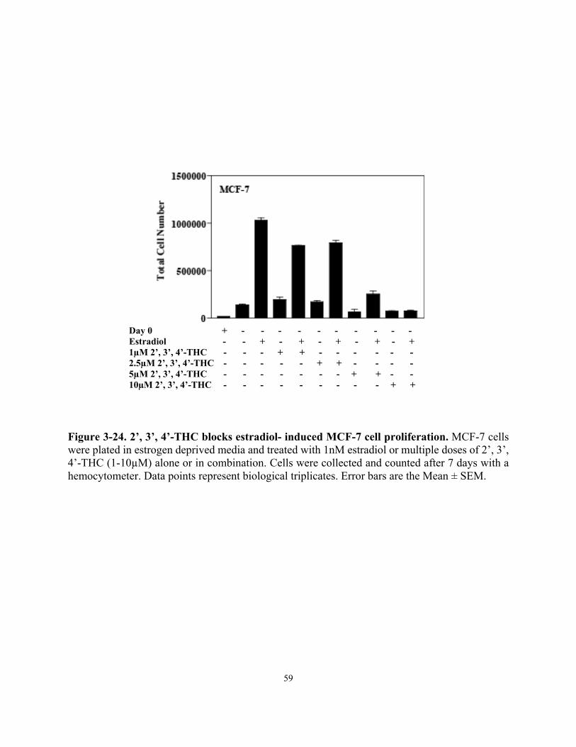
Inhibition of estradiol-induced MCF-7 cell proliferation was assessed by plating 50,000 cells per well of a 6-well tissue culture dish in phenol red-free DMEM/ F-12 supplemented with 5% stripped fetal bovine serum (Gemini Bio-products), 100U/mL of penicillin and streptomycin, 50μg/mL fungizone and 2mM glutamine. Cells were treated for 7 days with control ETOH, 1nM estradiol plus and minus increasing doses of 2’, 3’, 4’-THC (1, 2.5, 5 and 10µM). Cells were also treated with each dose of 2’, 3’, 4’-THC alone. Cells were counted using a hemocytometer and total cell number was compared to initial plating number on day 0 to control for cytotoxicity.
Cell Cycle Analysis using Flow Cytometry
MCF-7 cells were plated at a density of 200,000 cells per well in 6-well tissue culture dishes in phenol red- free DMEM/ F-12 supplemented with 5% stripped fetal bovine serum. Cells were treated for 24 hours with control ETOH, 10nM estradiol, and 5μM 2’, 3’, 4’-THC plus and minus estradiol. Cells were washed with room temperature 1X PBS, trypsinized with Trypsin-EDTA and spun down at 1500 rpm for 15 minutes. Media was aspirated off of the cell pellet and cells were washed once with ice cold 1X PBS and spun down. PBS was aspirated off each sample pellet and 500μL of 5μg/μl Propidium iodide solution was added to each pellet and left to sit in the dark for 20 minutes. Samples were run on a FC-500 using CXP software (Flow Cytometry Center, 491 LSA UC Berkeley, CA) and data was analyzed using FlowJo 7.6.5.
Mouse Purchase, Housing and Maintenance
Mice on a soy-free chow diet
8 week old C57BL/6J female ovariectomized mice were purchased from Jackson Laboratories, Sacramento, CA. Mice were housed and maintained according to OLAC standard procedures in the NAF facility at UC Berkeley, CA. All mice were fed a soy-free chow diet 2916.15 (Harlan Laboratories, Livermore, CA) starting one week before osmotic pump implantation. Mice were weighed once a week for the duration of the experiment.
Mouse osmotic pump preparation and implantation
Mini-Osmotic Pumps Model 2006 were purchased from Alzet and filled with vehicle, 1μg estradiol, 2mg of 2’, 3’, 4’-THC or the combination of 2’, 3’, 4’-THC and estradiol. All drugs were made using a laminar flow hood and dissolved in sterile vehicle consisting of 50% DMSO, 25% ETOH and 25% DI water. Pumps were handled with sterile gloves and filled using a 27 gauge filling tube and 1mL syringe. All pumps were placed in 1X PBS in 15mL sterile conical tubes and incubated overnight at 37˚. Pumps were surgically implanted into 8 week old C57BL/6J female ovariectomized mice (Jackson Laboratory, Sacramento, CA) posterior to the scapula and left for a duration of 4 weeks.
22
Determination of Mouse Tissue and Body Weight
The #2 mammary glands were dissected away from the subcutaneous tissue and weighed. All intraperatoneal gonadal fat was removed and weighed. Uterine tissue was collected, fluid drained and gonadal fat trimmed prior to weighing. Mouse body weight was measured in grams once a week.
RNA Extraction and Quantitative Real-Time PCR of Animal Tissues
Tissues were dissected and immediately frozen in liquid nitrogen. Before RNA isolation tissues were homogenized in PureZOL using the MP FastPrep-24 for 40 seconds. Total RNA was extracted and then treated with DNAse using the Aurum Total RNA Mini Kit for Fatty and Fibrous Tissue (Bio-Rad Laboratories, Hercules, CA). Reverse transcription reactions were performed using the iScript cDNA Synthesis Kit with 1 μg of total RNA according to manufacturer’s protocol. qPCR was performed with a Bio-Rad CFX96 Thermal Cycler System using SsoFast Eva Green Supermix (Bio-Rad). Mean ± SEM was calculated using Prism curve-fitting program (GraphPad Software, Inc., San Diego, CA).
Uterine Tissue Slide Preparation
Uterine tissue was removed and trimmed of excess adipose tissue. Tissues were fixed in formalin for 24 hours then transferred to 50% ETOH for 1 hour followed by 70% ETOH for another hour. After fixation tissues were sent to Histopathology Reference Laboratory (Hercules, CA) where they were paraffin embedded, sectioned and stained with hematoxylin and eosin for morphological examination.
23
REFERENCES
1. Tee, M.K., I. Rogatsky, C. Tzagarakis-Foster, A. Cvoro, J. An, R.J. Christy, K.R. Yamamoto, D.C. Leitman, Estradiol and Selective Estrogen Receptor Modulators Differentially Regulate Target Genes with Estrogen Receptors Alpha and Beta. Molecular Biology of the Cell, 2004. 15(3): pg. 1262-72.
2. Tzagarakis-Foster, C., R. Geleziunas, A. Lomri, J. An, D. C. Leitman, Estradiol represses human T-cell Leukemia Virus Type 1 Tax Activation of Tumor Necrosis Factor-Alpha Gene Transcription. Journal of Biological Chemistry, 2002. 277(47): pg. 44772-44777.
24
25
26
27
CHAPTER THREE
Results
28
Schematic of unique screening methods to identify new estrogens for use in MHT
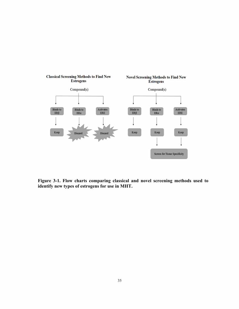
Figure 3-1 shows a model of classical screening techniques used to identify compounds with estrogenic activity compared to our approach to discover ERα coagonists. Classically, compounds which bind to ERα and/or which activate an estrogen response element are classified as estrogenic compounds and potentially harmful due to the role of ERα- mediated proliferation in reproductive tissues. Compounds that activate ERβ are considered safe, and therefore kept for further evaluation. We elected to test compounds that show little activity alone, but potentiate the effects of estradiol and potentially function as a coagonist, rather than an antagonist.
Synthetic chalcone compound 2’, 3’, 4’-THC synergizes estrogen-induced ERE activity in reporter assays
To identify ERα coagonists, we used U2OS cells which were cotransfected with ERα and ERE-tk luciferase and treated for 24 hours with 10nM estradiol in the absence or presence of 5µM of multiple di and trihydroxychalcone compounds (Figure 3-2). Estradiol produced a 40-fold induction of ERE-tk luciferase activity. 2, 4’ and 2, 2’, 5’ di and trihydroxychalcone had no estrogenic activity and did not change the activation of estradiol. 2’, 4’- dihydroxychalcone had estrogenic activity on its own, but did not change the overall estradiol response. 2, 2’, 4’-trihydroxychalcone displayed no estrogenic activity on its own, whereas it blocked estradiol induced luciferase activity. Only one compound, 2’, 3’, 4’-trihydroxychalcone displayed a unique estrogenic response. 2’, 3’, 4’-THC alone induced a 3-fold stimulation in luciferase activity and potentiated the activation by estradiol to 74-fold over control cells. This data demonstrates that 2’, 3’, 4’-THC exhibits properties consistent with an ERα coagonist. The synergistic activation of luciferase activity by 2’, 3’, 4’-THC was also observed in U2OS cells expressing ERβ (Figure 3-3).
To determine if the synergy occurs with other human estrogens, U2OS cells were cotransfected with ERα and ERE-tk luciferase and treated for 24 hours with 10nM estrone or estriol and 5µM 2’, 3’, 4’-THC alone or in combination (Figure 3-4). Estrone and estriol induced luciferase activity by 6.5 and 14-fold, respectively. 2’, 3’, 4’-THC alone caused a 2-fold activation of luciferase activity, whereas estrone produced a 6.5-fold activation. The combination of 2’, 3’, 4’-THC and estrone produced a synergistic 12-fold activation. Synergy was also seen by combining estriol and 2’, 3’, 4’-THC. Induction went from 14.1-fold with estriol alone to 27.5-fold with the estriol and 2’, 3’, 4’-THC combination. These studies demonstrate that 2’, 3’, 4’-THC can synergize the induction of luciferase activity not only in the presence of estradiol, but also with other endogenous estrogens through ERα.
2’, 3’, 4’-THC binds to the ligand binding domain of ERα and ERβ
To determine if 2’, 3’, 4’-THC was able to bind to ERα (Figure 3-5, A) and ERβ (Figure 3-5, B) the loss of specific bound [3H]-estradiol was used to look at the relative binding of 2’, 3’, 4’-THC in comparison to estradiol in U2OSα or U2OSβ cells. Competitive binding studies in intact cells found that 2’, 3’, 4’-THC binds to ERα and ERβ with an IC50 of 2.8μM and 3.9µM,
29
respectively. This data demonstrates that 2’, 3’, 4’-THC binds to both ERα and ERβ with similar affinity, but it displays much lower affinity than estradiol for each receptor.
2’, 3’, 4’-THC behaves as a unique coagonist on gene regulation in U2OSα cells
To determine if 2’, 3’, 4’-THC behaved as a coagonist or SERM on endogenous gene regulation, we examined the expression of known estrogen responsive gene KRT-19 and NKG2E, an estrogen and SERM regulated gene (Figure 3-6, 3-7). U2OS-ERα cells were treated for 24 hours with estradiol, tamoxifen or raloxifene, alone or in combination and then real-time PCR was done. Estradiol induced KRT-19 gene expression by 3.2-fold, whereas 2’, 3’, 4’-THC did not induce gene expression. The combination of 2’, 3’, 4’-THC plus estradiol synergized KRT-19 to 10.8-fold.
Tamoxifen and raloxifene did not regulate KRT-19 gene expression, whereas they blocked the estradiol-induced expression consistent with their well charcterized antagonist activity (Figure 3-6). Tamoxifen and raloxifene activated NKG2E expression by 14.3 and 3.9-fold (Figure 3-7, A, B). Estradiol activated NKG2E by 28-fold, whereas no effect was observed with 2’, 3’, 4’-THC. The combination of 2’, 3’, 4’-THC plus estradiol synergized NKG2E expression to 50-fold. In contrast to the synergistic action 2’, 3’, 4’-THC on NKG2E expression tamoxifen or raloxifene had an antagonist effect by decreasing gene expression to 20.4 and 5.1-fold. 2’, 3’, 4’-THC in combination with tamoxifen and raloxifene had an inhibitory effect by decreasing fold change to 11 and 4.5-fold. The combination of tamoxifen or raloxifene with 2’, 3’, 4’-THC plus estradiol blocked the synergistic gene induction of NKG2E seen with 2’, 3’, 4’-THC plus estradiol alone. The data demonstrates that 2’, 3’, 4’-THC behaves as a unique coagonist on endogenous gene regulation of both estradiol and SERM-induced genes.
To determine how long it takes for 2’, 3’, 4’-THC plus estradiol to induce gene synergy, U2OSα cells were treated with estradiol or 2’, 3’, 4’-THC alone or in combination for 1, 3, 6, 16 or 24 hours (Figure 3-8). The maximal synergy of KRT-19 gene expression was observed at 24 hours. At this time point estradiol produced a 6.1-fold increase in KRT-19 mRNA, whereas 2’, 3’, 4’-THC had no effect. The combination activated KRT-19 gene expression by 35-fold. (Figure 3-8, A). The synergy between estradiol and 2’, 3’, 4’-THC on the NKG2E was observed at 16 h and (Figure 3-8, B) maximal gene synergy was observed at 24 hours. These studies demonstrate that the maximal synergy for the KRT-19 and NKG2E genes occurs at 24 h.
To determine if 2’, 3’, 4’-THC behaved as an estradiol coagonist on genes other than KRT-19 and NKG2E microarray analysis was done with U2OSα cells treated for 24 hours based off of time course data (Figure 3-8) with 10nM estradiol or 5µM 2’, 3’, 4’-THC alone or in combination. Cells were treated with a 5,000 higher dose of 2’, 3’, 4’-THC relative to estradiol based off of the IC50 values determined by the competitive binding curves in U2OSα cells (Figure 3-5). At doses at around the IC50 a heteroligand configuration is more likely to occur, with the simultaneous binding of 2’, 3’, 4’-THC and estradiol to ERα. Estradiol regulated a total of 756 genes, whereas cells treated with the combination of 2’, 3’, 4’-THC plus estradiol regulated 1,358 genes. 2’, 3’, 4’-THC alone weakly regulated a total of 31 genes (Figure 3-9).
Three classes of regulated genes emerged from the microarrays. Class I represented genes were regulated by estradiol alone and antagonized by 2’, 3’, 4’-THC treatment. There were 24 up-
30
regulated and 5 down-regulated Class I genes. Class II represented genes regulated by estradiol and potentiated by the addition of 2’, 3’, 4’-THC. Class II contained 75 up-regulated and 20 down-regulated genes. Class III represented genes regulated only by 2’, 3’, 4’-THC and estradiol combination. These genes are considered to be newly regulated genes because no regulation was observed with E2 or 2’, 3’, 4’-THC alone. 327 news genes were activated and 268 new genes repressed by the combination (Table 3-1). There were 222 genes which were uniquely regulated by estradiol alone. 534 which were regulated by both estradiol and 2’, 3’, 4’-THC plus estradiol, and 824 genes regulated by the combination of 2’, 3’, 4’-THC and estradiol (Figure 3-10).
Three of the highest regulated class III genes were chosen to authenticate the microarray data by RT-PCR. FGR (Figure 3-11, A) KCNK6 (Figure 3-11, B) and K6iRS3 genes (Figure 3-11, C) were not regulated by estradiol alone, but activated by the 2’, 3’, 4’-THC/estradiol combination.
Class II and III gene regulation is ERα-dependent
To determine if the regulation of class II and III genes was mediated by ERα, U2OSα cells were treated for 24 hours with estradiol or 2’, 3’, 4’-THC alone or the combination in the absence or presence of the ERα antagonist, ICI. RT-PCR analysis showed that class II genes, KRT-19 (Figure 3-12, A) and NKG2E (Figure 3-12, B) were regulated 71 and 3.0-fold by estradiol alone and synergized by addition of 2’, 3’, 4’-THC to 632 and 52-fold, respectively. 2’, 3’, 4’-THC or ICI treatment alone did not induce gene expression of both genes. The addition of ICI with 2’, 3’, 4’-THC plus estradiol treatment blocked the induction of the KRT-19 and NKG2E genes. Class III genes, K6iRS3 (Figure 3-12, C) and FGR (3-12, D) were not regulated by estradiol, 2’, 3’, 4’-THC or ICI treatment alone. 2’, 3’, 4’-THC in combination with estradiol induced K6iRS3 and FGR gene expression by 35 and 44-fold. ICI blocked the activation of the K6iRS3 and FGR genes, demonstrating that class II gene synergy and class III gene regulation require ERα.
2’, 3’, 4’-THC and estradiol combination enhances recruitment of ERα and SRC-2 protein to the KRT-19 promoter
A model for the possible synergistic activation of KRT-19 gene expression in cells treated with 2’, 3’, 4’-THC plus estradiol is shown in Figure 3-14. To explore the mechanism of gene synergy the amount of ERα and SRC-2 protein recruitment to the KRT-19 promoter was determined by ChIP analysis of cells treated for 1 or 2 hours. At 1 hour of estradiol treatment, ERα was recruited to the KRT-19 promoter by 10-fold, whereas 2’, 3’, 4’-THC plus estradiol treatment induced a 34-fold recruitment. A similar synergistic recruitment of ERα occurred at 2 h (Figure 3-14, A). 2’, 3’, 4’-THC did not enhance estradiol recruitment of SRC-2 at 1 hour, but recruitment was increased from about 5.9 to 15-fold by 2 hours with both estradiol and 2’, 3’, 4’-THC (Figure 3-14, B). 2’, 3’, 4’-THC alone did not induce recruitment of either ERα or SRC-2 at any time point.
31
2’, 3’, 4’-THC plus estradiol treatment of U2OSα cells stabilizes ERα protein and its serine 118 phosphorylation state
One possible explanation of the increased ERα and SRC-2 recruitment is that the 2’, 3’, 4’-THC/estradiol combination enhances ERα protein stability by increasing the phosphorylation state, western blot analysis and RT-PCR was conducted looking at total amounts of ERα. Estradiol increased total ERα protein levels at 1 hour compared to control cells and caused phosphorylation of ERα at serine 118, which leads to a more transcriptionally active ERα. 2’, 3’, 4’-THC did not change overall ERα protein levels and did not induce phosphorylation of ERα at 1 hour. Total and phosphorylated ERα was the same between the estradiol and 2’, 3’, 4’-THC plus estradiol group at 1 hour. At 6 hours of estradiol treatment both total and phosphorylated ERα protein decreased and total ERα decreased with 2’, 3’, 4’-THC. Cells treated with 2’, 3’, 4’-THC plus estradiol had total protein levels similar to 1 hour, and had maintained the phosphorylation state. By 24 hours total and phosphorylated ERα protein were absent in the estradiol and 2’, 3’, 4’-THC treated cells. Cells that had been treated with 2’, 3’, 4’-THC plus estradiol had an elevated levels of total ERα protein and phosphorylated ERα (Figure 3-15, A). To examine if increase in total ERα protein was due to increased transcript levels, RT-PCR was run looking at ESR1 transcript levels over time. No statistically significant change in transcript levels was observed (Figure 3-15, B). These results demonstrate the 2’, 3’, 4’-THC/estradiol combination increases ser118 phosphorylation, which could contribute to enhanced recruitment of ERα and SRC-2.
The Heteroligand Hypothesis
Figure 3-16 displays a working model of 2’, 3’, 4’-THC and estradiol binding to ERα at the same time to create heteroligand bound ERα. The heteroligand likely causes a conformational change that is more stable than estradiol bound ERα. The increase in stability could allow for increase in total ERα protein over time as well increased phosphorylation of serine 118. More transcriptionally active ERα, as well as increased recruitment of ERα and SRC-2 could be a potential mechanism for the synergy. The recruitment of the estradiol/2’, 3’, 4’-THC bound ERα to different set genes compared to estradiol alone could potentially explain how new genes are regulated in U2OSα cells at 24 hours.
Mutational analysis of the ERα LBD does not confirm 2’, 3’, 4’-THC plus estradiol binding as a heteroligand
In order to determine if 2’, 3’, 4’-THC was binding to ERα at the same time as estradiol in the LBD or an allosteric site, specific amino acids in the LBD were mutated and U2OS cells were cotransfected with mutated ERα and ERE-tk luciferase and treated for 24 hours. Point mutation of amino acid 538 showed no change in response (Figure 3-17, A). Mutation of amino acid 539 showed a loss in estradiol-induced luciferase activity and a decrease in response with cells treated with 2’, 3’, 4’-THC plus estradiol (Figure 3-17, B). Point mutation of amino acid 540 resulted in a loss of estradiol-induced activity and a loss of synergy with 2’, 3’, 4’-THC plus estradiol, but a slight increase in activity with 2’, 3’, 4’-THC alone (Figure 3-17, C). Mutation
32
of 541 resulted in an overall increase in activity, whereas mutations 542 and 544 resulted in an overall decrease in activity (Figure 3-17, D, E, F).
Functional analysis elucidates heteroligand mechanism of action on gene expression for 2’, 3’, 4’-THC plus estradiol
To further explore the heteroligand hypothesis as mechanism of action for 2’, 3’, 4’-THC, we took advantage of our previous finding that one of the chalcones screened, 2, 2’, 4’-THC binds to ERα with a 10-fold greater affinity than 2’, 3’, 4’-THC, but with a 1000-fold less affinity compared to estradiol (Table 3-2). We reasoned that if 2’, 3’, 4’-THC forms a heteroligand with estradiol then 2, 2’, 4’-THC should be able to block the synergy by competing with 2’, 3’, 4’-THC, but the estradiol activation will be preserved because 2, 2’, 4’-THC will not compete with estradiol due to its lower affinity (Figure 3-18, A). U2OSα cells were treated for 24 hours with estradiol, 2’, 3’, 4’-THC and increasing doses of 2, 2’, 4’-THC (0.1-2µM) alone or in combination, and RT-PCR was done to examine KRT-19 gene expression (Figure 3-18, A). The addition of low doses of 2, 2’, 4’-THC in the presence of estradiol did not block estradiol-induced KRT-19 expression (Figure 3-18, B). In contrast, the synergy was blocked by the addition of 2, 2’, 4’-THC in a dose-dependent manner (Figure 3-18, B). These findings suggest that the synergy requires the formation of a heteroligand with estradiol and 2’, 3’, 4’-THC.
2’, 3’, 4’-THC lowers the EC50 of gene induction by estradiol
To determine if 2’, 3’, 4’-THC is able to shift the EC50 of gene induction by estradiol, U2OSα cells were treated with increasing doses of estradiol alone or in combination with 5µM 2’, 3’, 4’-THC. The activation of MSMB (Figure 3-19, A) FGR (Figure 3-19, B) KRT-19 (Figure 3-19, C) and K6iRS3 (Figure 3-19, D); class II and III genes was examined because they were among the genes that were regulated the most in microarray data. MSMB, FGR and K6iRS3 were not regulated by estradiol until 10-7 M, however, in combination with 2’, 3’, 4’-THC, MSMB and KRT-19 gene expression was induced with 10-9M and FGR and K6iRS3 with 10-
8M. 2’, 3’, 4’-THC did not regulate gene expression alone. These findings demonstrate that 2’, 3’, 4’-THC allows estradiol to regulate genes at a 10-100-fold lower dose.
2’, 3’, 4’-THC increases estradiol binding affinity for ERα and not ERβ
To determine if 2’, 3’, 4’-THC causes a change in binding affinity of estradiol for ERα or ERβ, U2OSα or U2OSβ cells were treated with 5nM [3H]-estradiol and 5µM 2’, 3’, 4’-THC. After the reaction had come to equilibrium, the amount of radioactivity inside the cell was counted and total binding was assessed. 2’, 3’, 4’-THC increased [3H]-estradiol binding by 2.1-fold (Figure 3-20, A). 2’, 3’, 4’-THC did not change [3H]-estradiol binding to ERβ (Figure 3-20, B). These findings demonstrate that 2’, 3’, 4’-THC enhances estradiol binding to ERα, but not ERβ.
33
2’, 3’, 4’-THC allows for estradiol to regulate new genes at physiological levels
The model depicted in figure 3-22 shows a potential mechanism of how 2’, 3’, 4’-THC plus estradiol regulate new genes based off of the EC50 shift in gene induction (Figure 3-19) and the increase in estradiol binding to ERα (Figure 3-20).
2’, 3’, 4’-THC binds to the LBD of ERα in MCF-7 cells
Estradiol plays a major role in ERα positive breast cancer cell proliferation, which suggest that the synergistic effects of 2’, 3’, 4’-THC on estradiol-regulated genes might lead to a greater stimulation of cell proliferation than estradiol alone (Figure 3-21). To determine if 2’, 3’, 4’-THC is able to bind to ERα in intact MCF-7 cells the loss of specific bound [3H]-estradiol was used to measure the relative binding affinity of 2’, 3’, 4’-THC in comparison to estradiol. 2’, 3’, 4’-Competitive radioligand studies found that THC binds to ERα with a relative affinity of 18µM (Figure 3-23), demonstrating that it binds to ERα in MCF-7 cells similar to U2OS cells.
2’, 3’, 4’-THC blocks estradiol-induced proliferation in ERα positive MCF-7 breast cancer cells
To determine if 2’, 3’, 4’-THC alters the proliferative effects of estradiol, MCF-7 cells were grown in serum stripped of estrogens and then treated with 1nM estradiol and increasing doses of 2’, 3’, 4’-THC (1µM-10µM) alone or in combination with estradiol. After seven days of treatment the cells were counted. Estradiol induced a 7.4-fold change in cell number whereas 2’, 3’, 4’-THC had no effect on proliferation. The fold induction in proliferation was blocked by 2’, 3’, 4’-THC in a dose-dependent manner. Maximal blocking of estradiol-induced proliferation was seen at 10µM of 2’, 3’, 4’-THC treatment with a 47% reduction in growth compared to estradiol alone (Figure 3-24). The data demonstrates that 2’, 3’, 4’-THC blocks proliferation in MCF-7 cells by blocking estradiol-induced proliferation.
2’, 3’, 4’-THC causes a G1 cell cycle arrest in MCF-7 cells
To determine how 2’, 3’, 4’-THC blocks cell proliferation, MCF-7 cells were stripped of estrogen and then treated for 24 hours with 10nM estradiol or 10µM 2’, 3’, 4’-THC alone or in combination. Cells were collected and nuclei were stained with Propidium iodide and flow cytometry was used to determine DNA content in the cell cycle. In control cells 79% were in G1, 12% in S phase and 6% in G2/M. Estradiol treatment caused the transition of cells from G1 phase to the S phase with 65% of gated cells in G1, 23% in S phase and 6% in G2/M. In contrast, 2’, 3’, 4’-THC did not stimulate DNA replication. 2’, 3’, 4’-THC produced a G1 cell cycle arrest in the presence of estradiol with 78% in G1 phase, 8% in S phase and 10% in G2/M (Figure 3-25, A). Figure 3-25, B shows G1/S ratio of treated cells. These data demonstrate that 2’, 3’, 4’-THC blocks estradiol- induced cell cycle transition from G1 to S phase and 2’, 3’, 4’-THC alone does not induce cellular proliferation.
34
2’, 3’, 4’-THC regulates genes in MCF-7 cells
To examine if 2’, 3’, 4’-THC blocked estradiol-induced gene expression, RT-PCR analysis was done in MCF-7 cells treated for 24 hours with 2’, 3’, 4’-THC alone or in combination with estradiol. Estradiol activated TFF1 (Figure 3-26, A) and GREB-1 (Figure 3-26, C) by 2-fold, which are known target genes in MCF-7 cells. 2’, 3’, 4’-THC was observed to also regulate genes TFF1 and GPX2 (Figure 3-26, B). The combination of estradiol with 2’, 3’, 4’-THC did not block estradiol-induction of gene regulation and increased expression of TFF1. These results indicate that 2’, 3’, 4’-THC causes tissue-specific regulation of genes.
2’, 3’, 4’-THC blocks estradiol-induced uterine weight gain in mice
To determine if 2’, 3’, 4’-THC was able to block estradiol-induced uterine weight gain in mice, 8 week old female ovariectomized mice were treated with 1µg (21ng/day) estradiol and increasing doses of 2’, 3’, 4’-THC alone or in combination for 4 weeks (Figure 3-27). Uterine tissue was dissected out and weighed. Mice treated for 4 weeks with control vehicle displayed an 11-fold increase compared to control mice. 2’, 3’, 4’-THC treatment alone increased uterine weight by 3-fold increase. The increase in uterine weight was blocked only when used at levels that were 2000-fold higher than estradiol, 2’, 3’, 4’-THC, 2’, 3’, 4’-THC can block estradiol-induced uterine weight gain in mice at levels that are consistent with the binding affinities of 2’, 3’, 4’-THC and estradiol.
2’, 3’, 4’-THC blocks estradiol-induced uterine weight and gene regulation without blocking effects in adipose tissue in mice on a soy-free chow diet
Whereas blocking uterine weight is a favorable property of 2’, 3’, 4’-THC, because endometrial cancer is an adverse effect of estrogens, it is important to determine if it produces unfavorable effects. 2’, 3’, 4’-THC had no effect on body weight or the weight of the mammary gland or adipose tissue in the absence or presence of estradiol (Figure 3-28). H&E staining shows that 2’, 3’, 4’-THC treated mice had one layer of epithelial cells lining the endometrial ducts (Figure 3-30, A, C), whereas, estradiol treated mice had multiple layers showing an increase in uterine proliferation status (Figure 3-30, B). Mice treated with 2’, 3’, 4’-THC plus estradiol displayed a similar pattern to control mice with one layer of epithelial cells surrounding the endometrial glands (Figure 3-30, D). RT-PCR of estradiol induced gene expression for Lactoferrin (Figure 3-29, A) LCN2 (Figure 3-29, B) S100A8 (Figure 3-29, C) and S100A9 (Figure 3-29, D) was examined and compared between estradiol treatment and 2’, 3’, 4’-THC plus estradiol treatment. 2’, 3’, 4’-THC alone did not induce expression of any examined genes. Estradiol produced a marked activation of these genes, which was blocked by 2’, 3’, 4’-THC. In adipose tissue, RT-PCR analysis was done to examine the expression of Aebp1 (Figure 3-31, A) LPL (Figure 3-31, B) FatP1 (Figure 3-31, C) and Saa3 (Figure 3-31, D). Aebp1, FatP1 and Saa3 were up-regulated by 2.2, 2.2 and 5.4-fold with estradiol treatment alone. Expression of both Aebp1 and LPL were up-regulated by addition of 2’, 3’, 4’-THC with estradiol to 4.2 and 1.7-fold, whereas the expression of Saa3 was down-regulated to 1.2-fold. 2’, 3’, 4’-THC alone regulated both Aebp1 and FatP1 by 3.4 and 2.9-fold.
35
Figure 3-1. Flow charts comparing classical and novel screening methods used to identify new types of estrogens for use in MHT.
36
Figure 3-2. Screening synthetic chalcone compounds for estrogenic activity through ERα. U2OS cells were cotransfected with ERα and ERE tk-luciferase. After transfection cells were treated with 5µM of multiple di and trihydroxychalcone compounds plus and minus 10nM estradiol for 24 hours. Cells were lysed and luciferase activity was measured using a luminometer. RLU is relative light units. Each data point represents biological triplicates. Error bars are the Mean ± SEM. (***, P-value < 0.001, t-test).
Figure 3-3. 2’, 3’, 4’-THC synergizes the estradiol induced transcriptional activity of ERE-tk luciferase in U2OS cells expressing ERα or ERβ. U2OS cells were cotransfected with ERE tk-luciferase and ERα or ERβ. After transfection cells were treated for 24 hours with 10nM estradiol and 5μM 2’, 3’, 4’-THC alone or in combination. Cells were lysed and luciferase activity was measured. RLU is relative light units. Each data point represents biological triplicates. Error bars are the Mean ± SEM. (**, P-values are < 0.01, t-test).
Estradiol + - + + - + 2’, 3’, 4’-THC - + + - + +
38
Figure 3-4. 2’, 3’, 4’-THC synergizes both estrone and estriol induced ERE-tk luciferase activity in a reporter assay. U2OS cells were cotransfected with ERα and ERE-tk luciferase and treated for 24 hours with 10nM estrone, 10nM estriol, 5µM 2’, 3’, 4’-THC alone or in combination. Cells were lysed and luciferase was measured using a luminometer. Data sets represent biological triplicates. Error bars are the Mean ± SEM. (**, P-value < 0.01, t-test).
Figure 3-5. 2’, 3’, 4’-THC binds to ERα and ERβ in a radioligand competitive binding assay in U2OS-ERα and β Cells. U2OS-ERα or β cells were treated with and without doxycycline for 24 hours to induce receptor expression. Cells were treated with 5nM [3H]-estradiol and increasing doses (2.5μM-150μM) 2’, 3’, 4’-THC for 1 hour at 37˚. Estrogen receptor bound [3H]-estradiol was counted using a liquid scintillation counter (LSC). Counts per minute (CPM) for specific binding was calculated by subtracting nonspecific binding from total binding. The loss of specific bound [3H]-estradiol was used to look at the relative binding of 2’, 3’, 4’-THC in comparison to estradiol. LogIC50 of 2’, 3’, 4’-THC for ERα and β = -4.4 (A) and -4.5 (B) approximately. Each data point is the average of quadruplet determinations ± SEM.
A B
-7 -6 -5 -4 -30
5000
10000
15000ER
[2', 3', 4'-THC]
CPM
-7 -6 -5 -4 -30
2500
5000
7500
10000ER
[2', 3', 4'-THC]
CPM
40
Figure 3-6. 2’, 3’, 4’-THC behaves as a unique coagonist on KRT-19 gene expression. U2OSα cells were treated for 24 hours with doxycycline to induce receptor expression. Cells were then treated for 24 hours with vehicle control, 10nM estradiol, 1µM 4-hydroxytamoxifen, 1µM raloxifene and 5µM 2’, 3’, 4’-THC alone or in combination. Each data point represents biological triplicates except data points for estradiol alone treatment, 2’, 3’, 4’-THC plus estradiol and tamoxifen plus estradiol represent n=2. Error bars are the mean ± SEM. (***, P-values are < 0.001).
Figure 3-7. 2’, 3’, 4’-THC behaves as a unique coagonist on SERM-regulated gene NKG2E. U2OSα cells were treated for 24 hours with doxycycline to induce receptor expression. Cells were treated with 10nM estradiol, 5µM 2’, 3’, 4’-THC, 1µM tamoxifen or raloxifene alone or in combination for 24 hours. Data points represent biological triplicates except estradiol and (tamoxifen plus 2’, 3’, 4’-THC plus estradiol) represents n=2. Error bars are ± SEM. (*, P-value ≤ 0.05).
Figure 3-8. The combination of 2’, 3’, 4’-THC plus estradiol causes synergy of both KRT-19 and NKG2E gene expression between 16 and 24 hours. U2OSα cells were treated for 24 hours with doxycycline to induce receptor expression. Cells were treated with 10nM estradiol and 5µM 2’, 3’, 4’-THC alone or in combination for 1, 3, 6, 16 or 24 hours. Data points represent biological triplicates except 2’, 3’, 4’-THC alone represents n=2. Error bars are ± SEM.
Figure 3-9. Total Regulated genes by estradiol, 2’, 3’, 4’-THC or 2’, 3’, 4’-THC plus estradiol in U2OSα cells at 24 hours. U2OSα cells were treated for 24 hours with doxycycline to induce receptor expression. Cells were then treated with 10nM estradiol or 5µM 2’, 3’, 4’-THC alone or in combination. Each data point represents total number of genes regulated (up or down) by each treatment according to microarray analysis with a cut off for regulation of 3 fold and a P-value of ≤ 0.05.
Estradiol + - +2’, 3’, 4’-THC - + +
44
Table 3-1. Three classes of genes are regulated by 2’, 3’, 4’-THC plus estradiol in U2OSα cells at 24 hours of treatment. U2OSα cells were treated for 24 hours with doxycycline to induce receptor expression. Cells were then treated with 10nM estradiol or 5µM 2’, 3’, 4’-THC alone or in combination. Class I genes represent genes which were antagonized by 2’, 3’, 4’-THC in the presence of estradiol. Class II genes represent genes whose expression was synergized by the combination of 2’, 3’, 4’-THC plus estradiol. Class III genes were unique to the 2’, 3’, 4’-THC plus estradiol treatment group whose expression was not regulated by estradiol or 2’, 3’, 4’-THC alone. Expression was assessed by up regulation or down regulation of 3 fold or more and a P-value ≤ 0.05.
Figure 3-10. Ven-diagram representation showing the total number of genes uniquely regulated by estradiol alone or 2’, 3’, 4’-THC plus estradiol. Estradiol uniquely regulated 222 genes. 2’, 3’, 4’-THC plus estradiol uniquely regulated 824 genes. Estradiol and 2’, 3’, 4’-THC plus estradiol both shared 534 genes in common. Expression was assessed by up regulation or down regulation of 3 fold or more and a P-value ≤ 0.05.
Estradiol Common 2’, 3’, 4’-THC+ Estradiol
45
Figure 3-11. RT-PCR conformation of microarray class III gene expression. U2OSα cells were treated for 24 hours with doxycycline to induce receptor expression. Cells were treated with multiple doses of estradiol (10-10, 10-9, 10-8, 10-7) alone or in combination with 5µM 2’, 3’, 4’-THC. Data points represent biological triplicates. Error bars are ± SEM.
-11 -10 -9 -8 -70
250
500
750
1000Estradiol
2', 3', 4'-THC + Estradiol
FGR
[ Estradiol ]
Fol
d C
han
ge (
mR
NA
)
-11 -10 -9 -8 -70
10
20Estradiol
2', 3', 4'-THC + Estradiol
K6iRS3
[ Estradiol ]
Fol
d C
hang
e (m
RN
A)
-11 -10 -9 -8 -70
5
10
15Estradiol
2', 3', 4'-THC + Estradiol
KCNK6
[ Estradiol ]
Fol
d C
hang
e (m
RN
A)
A B
C
46
Figure 3-12. Both microarray class II and III regulated genes require ERα. U2OSα cells were treated for 24 hours with doxycycline to induce receptor expression. Cells were treated with 10nM estradiol, 5µM 2’, 3’, 4’-THC or 1µM ICI alone or in combination for 24 hours. Data points represent biological triplicates. Error bars are the mean ± SEM.
Figure 3-13. Model showing the possible mechanism(s) of gene expression synergy of 2’, 3’, 4’-THC plus estradiol on the KRT-19 gene promoter.
48
Figure 3-14. 2’, 3’, 4’-THC plus estradiol increases recruitment of both ERα and SRC-2 receptors to the promoter of KRT-19. U2OSα cells were treated with doxycycline for 24 hours to induce receptor expression. Cells were treated with 10nM estradiol, 5µM 2’, 3’, 4’-THC alone or in combination for 1 or 2 hours. Proteins were crosslinked to DNA with formaldehyde and cell nuclear fractions were collected. ERα and SRC-2 proteins were immunoprecipitated and uncrosslinked from DNA. RT-PCR was run for KRT-19 promoter fragments showing ERα and SRC-2 recruitment. Each data point is a representative of either one (A) or two biological samples (B, 2 hour) that is a representative of 3 biological triplicates. 2’, 3’, 4’-THC alone did not recruit ERα or SRC-2. Error bars are Mean ± SEM.
A B
1 20
10
20
30
40
50Estradiol
2', 3', 4'-THC+ Estradiol
KRT-19
Treatment Time (hr)
ER
Rec
ruit
men
t
1 20
5
10
15Estradiol
2', 3', 4'-THC+ EstradiolKRT-19
Treatment Time (hr)S
RC
-2 R
ecru
itm
ent
49
Figure 3-15. 2’, 3’, 4’-THC plus estradiol stabilizes ERα total protein and transcriptionally active S118 phosphorylation state. U2OSα cells were treated for 24 hours with doxycycline to induce receptor expression. Cells were treated with 10nM estradiol, 5µM 2’, 3’, 4’-THC alone or in combination for 1, 3, 6 or 24 hours (A) or 1, 3, 6, 16 and 24 hours (B). Total ERα and serine 118 phosphorylated ERα levels were determined with Western Blot analysis. β-actin was used as a control for protein loading (A). Total ERα transcript levels were determined using RT-PCR (B). Data points represent one biological sample (A) or biological triplicates (B) and error bars are the Mean ± SEM (B).
Figure 3-16. Model of 2’, 3’, 4’-THC plus estradiol binding to ERα as a heteroligand complex.
Heteroligand Hypothesis of 2’, 3’, 4’-THC/Estradiol Mechanism
51
Figure 3-17. Transfection of point mutated amino acids of the ERα LBD looking for loss of synergistic activity on an ERE with 2’, 3’, 4’-THC plus estradiol treatment. U2OS cells were cotransfected with ERα LBD mutants and ERE-tk luciferase and treated for 24 hours with 10nM estradiol, 5µM 2’, 3’, 4’-THC alone or in combination. Cells were lysed and luciferase activity was measured using a luminometer. Data sets represent biological triplicates. Error bars are Mean ± SEM. (*, **, P-values are ≤ 0.05 and ≤ 0.01, t-test).
Table 3-2. Derived IC50 values for estradiol, 2, 2’, 4’-THC and 2’, 3’, 4’-THC competitive binding to ERα in U2OSα cells. IC50 values were calculated from biological quadruplets of a competitive [3H]-estradiol binding assay in U2OSα cells that had been treated with doxycycline for 24 hours followed by treatment with 5nM [3H]-estradiol and increasing doses of cold E2, 2, 2’, 4’-THC or 2’, 3’, 4’-THC. Cells were washed with BSA and lysed. CPMs were counted using a liquid scintillation counter. Specific binding = (Total CPM-Nonspecific CPM).
Compound IC50
Estradiol 2.3x10⁻⁹M
2, 2’, 4’-THC 5x10⁻⁶M
2’, 3’, 4’-THC 5x10⁻⁵M
53
Figure 3-18. Functional test for the heteroligand hypothesis. U2OSα cells were treated with doxycycline for 24 hours and then treated with increasing doses of 2, 2’, 4’-THC plus and minus 10nM estradiol (A) and the combination of estradiol plus 2’, 3’, 4’-THC with increasing doses of 2, 2’, 4’-THC (B). RT-PCR analysis was done to look at expression level of KRT-19. Data points represent biological triplicates. Error bars are the Mean ± SEM.
Figure 3-19. 2’, 3’, 4’-THC shifts the EC50 of estradiol induced gene expression. U2OSα cells were treated with doxycycline for 24 hours to induce receptor expression. Cells were then treated for 24 hours with increasing doses of estradiol (10-10, 10-9, 10-8, 10-7) plus and minus 5µM 2’, 3’, 4’-THC. Total RNA was isolated and cDNA was prepared. RT-PCR was run looking at gene expression of MSMB (A), FGR (B), KRT-19 (C) and K6iRS3 (D). Data points represent biological triplicates. Error bars represent the Mean ± SEM.
A B
C D
-11 -10 -9 -8 -7 -6
0
50
100
150Estradiol
2', 3', 4'-THC+ EstradiolMSMB
[Estradiol]
Fol
d C
han
ge (
mR
NA
)
-11 -10 -9 -8 -7 -6-500
0
500
1000
1500Estradiol
2', 3', 4'-THC+ EstradiolFGR
[Estradiol]
Fol
d C
han
ge (
mR
NA
)
-11 -10 -9 -8 -7 -6
0
500
1000
1500Estradiol
2', 3', 4'-THC+ EstradiolKRT-19
[Estradiol]
Fol
d C
han
ge (
mR
NA
)
-11 -10 -9 -8 -7 -6-10
0
10
20
30Estradiol
2', 3', 4'-THC+ EstradiolK6iRS3
[Estradiol]
Fol
d C
han
ge (
mR
NA
)
55
Figure 3-20. 2’, 3’, 4’-THC increases estradiol’s affinity for ERα and not ERβ. U2OSα cells were treated for 24 hours with doxycycline to induce receptor expression. After receptor expression induction cells were treated with increasing doses of [3H]-estradiol plus or minus 5µM 2’, 3’, 4’-THC. CPM were counted using a liquid scintillation counter. Data points represent biological triplicates. Error bars are Mean ± SEM. (***, P-Value < 0.001, t-test).
[3H]-Estradiol + + + + 2’, 3’, 4’-THC - + - +
56
Figure 3-21. Model of heteroligand gene regulation with physiological levels of estradiol.
57
Figure 3-22. The role of estrogen in breast cancer cell proliferation.
58
Figure 3-23. 2’, 3’, 4’-THC binds to ERα in MCF-7 cells. MCF-7 cells were treated with [3H]-estradiol and increasing concentrations of cold 2’, 3’, 4’-THC and allowed to come to equilibrium. Cells were washed with BSA, lysed and CPMs were counted with a liquid scintillation counter. Specific binding was calculated using the equation (Total binding-non-specific binding). Data points represent biological triplicates. Error bars are the Mean ± SEM.
-7 -6 -5 -4 -30
100
200
300
400 MCF-7
log of [CC7]
CPM
[2’, 3’, 4’-THC]
59
Figure 3-24. 2’, 3’, 4’-THC blocks estradiol- induced MCF-7 cell proliferation. MCF-7 cells were plated in estrogen deprived media and treated with 1nM estradiol or multiple doses of 2’, 3’, 4’-THC (1-10µM) alone or in combination. Cells were collected and counted after 7 days with a hemocytometer. Data points represent biological triplicates. Error bars are the Mean ± SEM.
Figure 3-25. 2’, 3’, 4’-THC blocks estradiol- induced MCF-7 cell proliferation by causing a G1 cell cycle arrest. MCF-7 cells were treated for 24 hours with 10nM estradiol or 10µM 2’, 3’, 4’-THC alone or in combination. Cells were collected and PI stained. Cell cycle analysis was done using FlowJo. Data sets represent biological duplicates. Error bars are the Mean ± SEM. (**, P-value ≤ 0.01, t-test).
Figure 3-26. RT-PCR confirmation of microarray data done in MCF-7 cells. MCF-7 cells were stripped of estrogen for 3 days and then treated with 10nM estradiol or 5µM CC7 alone or in combination for 24 hours. Total RNA was isolated, cDNA prepared and RT-PCR run looking at gene expression of TFF1 (A), GPX2 (B) and GREB-1 (C). Data points represent biological triplicates except for CC7 treatment in A, B represents n=2. Error bars are the Mean ± SEM.
A B
C
Estradiol + - + + - + 2’, 3’, 4’-THC - + + - + +
Estradiol + - +2’, 3’, 4’-THC - + +
62
Figure 3-27. 2’, 3’, 4’-THC blocks the estradiol- induced uterine weight gain in female mice at a ratio of 1:2000. 8 week old female ovariectomized mice were treated with vehicle control, 1µg (21ng/day) estradiol or 43µg/day 2’, 3’, 4’-THC alone or in combinations of estradiol: 2’, 3’, 4’-THC at ratios of 1:50, 1:100, 1:500, 1:1000 and 1:2000. Uterine tissue was collected and wet weight was measured. Data represents n=5 for control, estradiol, 1:50, 1:100, 1:500 and 1:000. 2’, 3’, 4’-THC represents n=3 and 1:2000 represents n=4. Error bars are the Mean ± SEM.
63
Figure 3-28. Tissue weights of mice treated for 4 weeks with estradiol, 2’, 3’, 4’-THC or 2’, 3’, 4’-THC plus estradiol. 8 week old female ovariectomized mice were treated with control vehicle, 1µg (21ng/day) estradiol or 43µg/day 2’, 3’, 4’-THC alone or in combination for 4 weeks. Overall body weight was measured (B) and uterine (A), mammary gland (C) and gonadal adipose tissue (D) was dissected out and weighed. Data points for uterine tissue and gonadal fat represent n=5. Data points for body weight represent n=4 except for estradiol treatment n=3. Data points for mammary gland weight represent n=5 except for 2’, 3’, 4’-THC plus estradiol n=4. Error bars represent Mean ± SEM.
Figure 3-29. 2’, 3’, 4’-THC blocks estradiol-induced uterine gene expression. 8 week old female ovariectomized mice were treated with control vehicle, 1µg (21ng/day) estradiol or 43µg/day 2’, 3’, 4’-THC alone or in combination for 4 weeks. Uterine tissue was dissected and total RNA was collected, cDNA prepared and RT-PCR run looking at gene expression on lactoferrin (A) LCN2 (B) S100A8 (C) and S100A9 (D). Data points represent biological triplicates except estradiol treatment for LF and S100A9 with n=2 and S100A8 with n=1. Error bars represent Mean ± SEM.
0
25
50
75Lactoferrin
Fol
d C
han
ge (
mR
NA
)
0
50
100
150S100A8
Fol
d C
han
ge (
mR
NA
)
0
250
500
750S100A9
**
Fol
d C
han
ge (
mR
NA
)
0
50
100
150LCN2
***
Fol
d C
han
ge (
mR
NA
)
A B
C D
Estradiol + - + + - +
2’, 3’, 4’-THC - + + - + +
65
Figure 3-30. 2’, 3’, 4’-THC blocks estradiol-induced endometrial cell proliferation in mice. 8 week old female ovariectomized mice were treated with control vehicle (A), 1µg (21ng/day) estradiol (B) or 43µg/day 2’, 3’, 4’-THC (C) alone or in combination (D) for 4 weeks. Uterine tissue samples were dissected, fixed in formalin and paraffin embedded and sectioned. Data represents one biological sample H&E stained as a representative for biological triplicates. Photos taken at 40X oil immersion.
66
Figure 3-31. Adipose tissue gene expression in mice treated with estradiol, 2’, 3’, 4’-THC or 2’, 3’, 4’-THC plus estradiol. 8 week old female ovariectomized mice were treated with control vehicle, 1µg (21ng/day) estradiol or 43µg/day 2’, 3’, 4’-THC alone or in combination for 4 weeks. Adipose tissue was dissected and total RNA was collected, cDNA prepared and RT-PCR run looking at gene expression of Aebp1 (A) LPL (B) FATP1 (C) and Saa3 (D). Data points for A, B represent n=5. Data points for FatP1 and Saa3 estradiol alone treatment represent n=4 and n=3. Error bars represent Mean ± SEM.
0
1
2
3
LPL*
Fol
d C
han
ge (
mR
NA
)
0
1
2
3
4
5
FatP1
Fol
d C
han
ge (
mR
NA
)
0
1
2
3
4
5
6
7Aebp1
*
Fol
d C
han
ge (
mR
NA
)
0.0
2.5
5.0
7.5
10.0
Saa3
* *F
old
Ch
ange
(m
RN
A)
A B
C D
Estradiol + - + + - +
2’, 3’, 4’-THC - + + - + +
67
CHAPTER FOUR
Discussion and Conclusion
68
Menopause is associated with multiple short-term symptoms, such as hot flashes and vaginal dryness and long-term diseases including osteoporosis, weight gain, and Type II diabetes. Estrogens are effective at reducing these symptoms and diseases, but are associated with an increase in breast and endometrial cancer and blood clots. Currently, estrogens are recommended for only short-term use, which prevents them from being used to combat chronic conditions associated with menopause. The proliferative effects of estrogens are mediated by ERα, which has made any estrogens that bind to ERα not appealing as alternatives to the current estrogens in hormone therapy. However, studies indicate that the beneficial effects of estrogens on the bone, weight gain and diabetes are also mediated by ERα. These observations have created a large dilemma to discover safer estrogens. We hypothesized that it might be possible to identify estrogens that bind to ERα along with estradiol to alter the gene expression, which could change the clinical effects of estradiol. The objective of these studies is to identify an ERα coagonist, which we define as a compound that binds to ERα simultaneous with estradiol to change the magnitude and pattern of genes expressed in cells. Our study shows that 2’, 3’, 4’-THC is a novel ERα coagonist, which can modulate estradiol on ERα regulated genes and physiological functions.
Transfection data was used to screen the estrogenic activity of multiple synthetic chalcone compounds. Our results showed that 2’, 3’, 4’-THC synergized estradiol-induced reporter activity on an ERE in cells overexpressing ERα without displaying estrogenic activity on its own. The other trihydroxychalcones (2, 2’, 4’-THC and 2, 2’, 5’-THC) tested also displayed no estrogenic activity on their own, but these chalcones antagonized the effects of estradiol. 2’, 3’, 4’-THC is the only examined chalcone that contains all hydroxyl groups on the prime ring and also has a 3’ hydroxyl group. Because the other two trihydroxychalcones also contain either a 2’ or 4’ hydroxyl position it is likely that the 3’ hydroxyl is important for coagonist activity. The two screened dihydroxychalcones did not display coagonist activity, however, it is interesting to note that 2, 4’-DHC had no activity, but when the 2 hydroxyl position was present in the 2’ hydroxyl group it displayed estrogenic activity on its own. The addition of a third hydroxyl group from 2’, 4’-DHC to 2, 2’, 4’-THC resulted in antagonistic activity. However, when the 2 hydroxyl group is moved to a 3’ hydroxyl position the chalcone now confers a coagonist activity. These findings demonstrate that by moving the position of the hydroxyl groups you can change the activity of the chalcone compounds. It is important to note that the molecular weight of 2’, 3’, 4’-THC is 256.26 which is similar to estradiol at 272.38. This observation indicates that 2’, 3’, 4’-THC can easily fit in the ligand binding pocket of the estrogen receptor similar to other chalcones including isoliquiritigenin, which has a molecular weight of 256.25 and is known to bind to both ERα and ERβ [1].
2’, 3’, 4’-THC was also inactive on its own in cells expressing ERβ, however, it synergized estradiol-induced reporter activity. This demonstrates that 2’, 3’, 4’-THC is not selective for ERα or ERβ. The fact that 2’, 3’, 4’-THC also acts synergistically with ERβ indicates that it could potentiate the known anti-proliferative effects mediated by ERβ [2], which will be an important clinical property [1, 3]. However, it is important to identify compounds that can also modify the activity of ERα, because the effects on the bone and adipose tissue are mediated by ERα. Therefore, an ideal compound would be one that activates ERβ, but can block the proliferative effects of ERα, while preserving the beneficial effects of ERα [4]. The transfections studies indicate that 2’, 3’, 4’-THC is a potential candidate to produce these effects.
69
Although estradiol is the main estrogen secreted from the ovaries, women at menopause have higher levels of circulating estrone from the conversion of adrenal androgens by aromatase in fat tissue. It is important to assess the synergistic activation of 2’, 3’, 4’-THC with other forms of estrogen. Similar to estradiol, 2’, 3’, 4’-THC also produced a synergistic activation of estrone and estriol. When comparing the magnitude of synergy observed with 2’, 3’, 4’-THC and estradiol with estrone and estriol, there was no statistical difference. This demonstrates that the synergistic activation is not specific to estradiol and it occurs with other circulating forms of estrogens.
Due to the fact a radiolabelled 2’, 3’, 4’-THC is not commercially available, we used a [3H]-estradiol competitive binding assay to determine if 2’, 3’, 4’-THC bound to ERα or ERβ. 2’, 3’, 4’-THC bound to both estrogen receptors with similar affinities. The binding affinity of 2’, 3’, 4’-THC was in the micromolar range compared to estradiol’s nanomolar range, displaying a difference of about 10,000 times less affinity. While the affinity of 2’, 3’, 4’-THC is much greater than estradiol it is in a similar range to some compounds that are used at pharmacological doses such as tamoxifen and raloxifene. It is important to note that because we calculated the affinity of 2’, 3’, 4’-THC by competing off estradiol, the binding affinity of 2’, 3’, 4’-THC is relative to estradiol. Another interesting observation was 2’, 3’, 4’-THC and estradiol had an affinity difference of approximately 1:10,000, the concentrations used of each ligand where synergy was observed on the transfection. We speculated that 1:10,000 ratio of estradiol to 2’, 3’, 4’-THC is important for creating a heteroligand configuration in ERα.
Known compounds which bind to estrogen receptors are classified as agonists or antagonists on gene regulation, so it was exciting to observe that 2’, 3’, 4’-THC behaves as an ERα coagonist. 2’, 3’, 4’-THC did not induce estradiol regulated gene KRT-19 on its own, yet it synergized with estradiol. Since our goal is to identify estrogens that retain agonistic activity in bone and adipose tissue we needed to make sure that 2’, 3’, 4’-THC does not act like tamoxifen or raloxifene on gene regulation. As demonstrated by a previous study in our lab, both tamoxifen and raloxifene induce NKG2E gene regulation [5]. In contrast, 2’, 3’, 4’-THC did not activate NKG2E, but instead it produced a synergistic activation of the estradiol response. This data demonstrated that 2’, 3’, 4’-THC does not behave as a classical SERM on endogenous gene regulation. A major indication for tamoxifen and raloxifene is for the prevention of breast cancer, which depends on their antagonist action in breast cancers. In MCF-7 cells tamoxifen and raloxifene antagonized the estradiol induction of the KRT-19 gene. The observation that 2’, 3’, 4’-THC actually potentiates the effects of estradiol, demonstrate that it does exhibit properties of the classical SERMs. Based on these findings it is apparent that the anti-proliferative effect that we observed in MCF-7 cells occurs through a different mechanism than the anti-proliferative actions of the SERMs. Clearly it will be important do determine if 2’, 3’, 4’-THC behaves similar to other SERM-regulated genes on a more global scale.
Although 2’, 3’, 4’-THC did not display SERM-like activity on endogenous gene regulation through ERα, this is expected when comparing their structures. Tamoxifen and raloxifene have larger molecular weights of 371 and 510 compared to 2’, 3’, 4’-THC at 256. These SERMs also contain bulky side chains which stick out of ERα’s binding pocket when bound [6, 7] preventing rotation of H12 over the LBD required for the formation of an active AF-2 for coactivator binding. The smaller size of 2’, 3’, 4’-THC would allow it to fit in the LBD of ERα alone, or at the same time with estradiol, and theoretically adopt the favorable estradiol conformation of H12. The binding of isoliquiritigenin, a chalcone compound that shares the same structural background as
70
2’, 3’, 4’-THC, demonstrates that chalcone-bound ER adopts an agonistic conformation [3]. The fact that 2’, 3’, 4’-THC in the presence of estradiol changes the activity of the estrogen receptor supports the idea that ERα is adopting a third conformation, a novel coagonist conformation distinct from the agonist conformation induce by estradiol and the antagonist conformation formed by the SERMs.
Microarray analysis provides additional data that 2’, 3’, 4’-THC behaves as an ERα coagonist, because it changes the estradiol-mediated gene expression profile. It is well established that tamoxifen or raloxifene block the regulation of genes by estradiol. 2’, 3’, 4’-THC is unique in that it itself only regulates 31 genes, but dramatically changes the profile of genes regulated by estradiol. The fact that 2’, 3’, 4’-THC changes the estradiol response of cells without behaving as an agonist or antagonist demonstrates that 2’, 3’, 4’-THC should be classified as a coagonist. Although 2’, 3’, 4’-THC binds to ERα directly, the synergistic response on estrogen gene expression and overall gene expression change occurs at 24 hours. The change in gene expression found on the microarray could be due to combined primary and secondary gene regulation. Regardless of the effects being due to primary or secondary events, the unique genes we examined require ERα because a loss of gene regulation occurred with ICI treatment prior to 2’, 3’, 4’-THC and estradiol.
The ability of 2’, 3’, 4’-THC to change the estradiol gene profile through ERα in ways distinct from estradiol and SERMs solidifies the existence of a new type of estrogen, an ERα coagonist. 2’, 3’, 4’- THC. Because we did not compare overall gene expression profiles induced by 2’, 3’, 4’-THC and estradiol in multiple cell lines, it is hard to assess whether or not the overall gene profile is a beneficial one, let alone a tissue- selective one. A previous publication from our lab showed that tamoxifen and raloxifene were active on their own and regulated a specific set of genes. It is well known that when tamoxifen and raloxifene are in combination with estradiol they behave as antagonists, blocking estradiol-induced gene expression while regulating genes on their own [8]. 2’, 3’, 4’-THC does not behave as an agonist or antagonist on estradiol gene regulation in U2OS cells, instead it changes the overall estradiol gene expression pattern, thereby demonstrating its unique coagonist activity. Furthermore, additional studies will need to done to determine if 2’, 3’, 4’-THC acts as an ERα coagonist in other cell types and in animals.
Although exploring the mechanism of action of 2’, 3’, 4’-THC plus estradiol on the newly regulated class III genes is interesting the location of estrogen response elements located within those genes is unknown. For this reason we decided to pursue exploring the mechanism of synergy utilizing the KRT-19 promoter as a model. The KRT-19 promoter has a primary ERE and is a gene which is regulated by estradiol and synergized by the addition of 2’, 3’, 4’-THC. It was previously published by our lab that estradiol enhanced the recruitment of ERα to the KRT-19 gene after treatment with estradiol for 2 hours [9]. The combination of 2’, 3’, 4’-THC and estradiol produced a synergistic recruitment of ERα and p160 coactivator SRC-2 to the promoter of estradiol-regulated gene KRT-19. These finding provide a potential mechanism of action of 2’, 3’, 4’-THC on estradiol gene synergy, but does not explain the mechanism for new gene regulation. Besides increasing recruitment of SRC-2, it is possible that there is recruitment of other p160 coactivators to the promoters of newly regulated genes. It is also possible that 2’, 3’, 4’-THC redirects ERα and p160 coactivators to regulatory elements present in newly regulated genes, accounting for new gene regulation observed on the microarray.
71
Another potential target for the 2’, 3’, 4’-THC/estradiol combination is ERα phosphorylation, because the phosphorylation of serine 118 is associated with increased transcriptional activity. We observed that the combination of 2’, 3’, 4’-THC and estradiol stabilizes S118 phosphorylation state of ERα over time. Previous publications have shown that the S118 phosphorylation state of the ER occurs when it is ligand-bound and is necessary for transcription [10]. Our data demonstrates that 2’, 3’, 4’-THC alone does not induce phosphorylation, however, in combination with estradiol stabilizes the S118 phosphorylation state of ERα. Another important observation is that overall total ERα protein levels increased over time with the combination of 2’, 3’, 4’-THC and estradiol without a statistically significant increase in transcript levels, suggesting the ERα is in more stable conformation when bound to 2’, 3’, 4’-THC and estradiol. Another possibility is that 2’, 3’, 4’-THC could be regulating a secondary factor which is decreasing the degradation or de-phosphorylation of ER. A recent publication utilizing modified ERα constructs determined that a heteroligand complex consisting of two monomers of ERα bound by two different ligands could be possible [11]. Our data suggests that 2’, 3’, 4’-THC and estradiol could be binding as a heteroligand complex either in the same binding pocket of ERα or on different monomers, which could enhance the stability of ERα.
Because a radiolabeled 2’, 3’, 4’-THC is not available it is difficult to perform studies to prove that 2’, 3’, 4’-THC/estradiol form a heteroligand with ERα. The observation that another chalcone, 2, 2’, 4’-THC had a higher affinity than 2’, 3’, 4’-THC provided an important reagent to investigate the heteroligand hypothesis. 2, 2’, 4’-THC has the same molecular weight and similar structure as 2’, 3’, 4’-THC, but binds ERα with 10 times greater affinity. We hypothesized that 2, 2’, 4’-THC would not be able to replace estradiol in the binding pocket of ERα because estradiol’s affinity was 1000 times greater, but could compete with 2’, 3’, 4’-THC due to its 10-fold greater affinity. Consistent with this hypothesis were the observations 2, 2’, 4’-THC did not alter estradiol activation of KRT-19 gene expression, and blocked the synergistic action of 2’, 3’, 4’-THC. This data functionally indicates the existence of an ERα heteroligand complex with 2’, 3’, 4’-THC and estradiol.
Although it has been published that an ERα heteroligand can exist in vitro, we show that a heteroligand can exist in intact cells. Although we show the existence of a heteroligand by functional analysis on gene regulation, it is not conclusive. Future experiments could be done to crystallize the structure of 2’, 3’, 4’-THC and estradiol and elucidate the binding site of 2’, 3’, 4’-THC. Although we show that functionally a heteroligand can exist with 2’, 3’, 4’-THC, Liu et al observed that it could exist with tamoxifen and raloxifene. Both Liu et al and our observations demonstrate that a heteroligand complex can exist with many different types of ligands, expanding our understanding of the vast dynamics of estrogen receptor gene regulation and providing new opportunities to develop alternative estrogens that produce different clinical outcomes.
RT-PCR analysis shows that many class III genes including MSMB, FGR and K6iRS3 require high non-physiological levels of estradiol to be induced, which could explain why they were not identified as estradiol regulated genes on the microarray. The addition of 2’, 3’, 4’-THC allows estradiol to regulate these genes at physiological estrogen levels, which led to the regulation of new genes. We observed that total binding of estradiol to ERα and not ERβ increased in the presence of 2’, 3’, 4’-THC, possibly explaining the decrease in the EC50 of gene expression. With less estradiol, but a higher affinity, it is possible to get regulation of genes that usually require much more estradiol due to a lower binding affinity in the absence of 2’, 3’, 4’-THC. A major
72
problem in women during menopause is that lower circulating estradiol levels lead to changes in tissue physiology, more than likely due to overall gene expression changes. Our findings indicate that 2’, 3’, 4’-THC could be administered to menopausal women alone with the goal of altering the gene expression profile and clinical effects of endogenous estrogens. By treating women with 2’, 3’, 4’-THC alone without exogenous estrogens there will be a much lower risk for breast and endometrial cancer.
Although 2’, 3’, 4’-THC synergizes with estradiol on gene expression in U2OSα cells, it binds and blocks estradiol induced proliferation in MCF-7 breast cancer cells. We observed that 2’, 3’, 4’-THC bound to endogenous ERα in MCF-7 cells, but it did not induce proliferation and was able to block estradiol-induced proliferation of cells. We determined that 2’, 3’, 4’-THC blocked the estradiol-induced G1 to S phase cell cycle transition in MCF-7 cells. Consistent with our findings in U2OS cells, 2’, 3’, 4’-THC did not exhibit antagonistic activity on estradiol regulated genes. 2’, 3’, 4’-THC did not block estradiol regulation of GPX2, TFF1 or GREB-1, and up-regulated the expression of GPX2 and TFF1 in the presence of estradiol. Although we were able to show that 2’, 3’, 4’-THC blocks estradiol-induced proliferation, we were unable to determine its mechanism by studying only a few estradiol regulated genes. It is probable that 2’, 3’, 4’-THC, or the combination of 2’, 3’, 4’-THC and estradiol are regulating a differential set of genes changing the overall proliferative profile of estradiol from a proliferative to an anti-proliferative profile. Further studies are needed to explore this possibility.
We observed that 2’, 3’, 4’-THC can change the estradiol induced gene profile in the mouse uterus and white adipose tissue, displaying a beneficial gene expression profile. Ovariectomized mice treated with estradiol had an increase in uterine weight and endometrial cell proliferation which was blocked by 2’, 3’, 4’-THC. 2’, 3’, 4’-THC also blocked estradiol-induced gene expression in the uterus. These observations suggest that 2’, 3’, 4’-THC behaves as an ERα antagonist in uterine tissue. Because it is known that ERα is responsible for mediating endometrial proliferation, it is possible that 2’, 3’, 4’-THC is behaving as an ERα antagonist. However, it is unlikely that at a 1:2000 ratio of estradiol to 2’, 3’, 4’-THC that 2’, 3’, 4’-THC could act as an antagonist by blocking estradiol binding. It is more likely that 2’, 3’, 4’-THC is not behaving as an antagonist but an ERα modulator that changes the overall gene expression profile which would not be elucidated by observing only a few estradiol-regulated genes. 2’, 3’, 4’-THC might also produce anti-proliferative effects in the uterus by activating ERβ, but this seems unlikely because the mouse uterus mainly expresses ERα. [12].
Our study also demonstrated that 2’, 3’, 4’-THC changed estradiol gene expression patterns in mouse white adipose tissue. It has been demonstrated in the ERα knockout mouse that the effects of estradiol on white adipose tissue are mediated by ERα [4]. We observed that estradiol regulated genes in white adipose tissue and that 2’, 3’, 4’-THC modulated the expression of those genes. Surprisingly, 2’, 3’, 4’-THC regulated Aebp1 and FatP1 and had a small, yet not significant, additive effect with estradiol. These results demonstrate that 2’, 3’, 4’-THC is active in adipose tissue and that it is able to modulate the gene expression pattern induced by estradiol. Taken together with its effects in the uterus, 2’, 3’, 4’-THC maintains estrogen effects in adipose tissue by increasing the expression of Aepb1, FatP1 and LPL and blocks proliferative effects in the uterus. Although we are unable to establish that these changes in estrogen gene expression are mediated through ERα, the promising profile of 2’, 3’, 4’-THC and estradiol warrants further investigation.
73
CONCLUSION
Transfection, binding, ChIP, Western Blot and gene expression studies in U2OSα cells, combined with proliferation studies in MCF-7 cells, demonstrate that 2’, 3’, 4’-THC has unique properties that distinguish it from the current agonist and antagonist used clinically. The activities of 2’, 3’, 4’-THC are best described as an ERα coagonist, because it has very little activity alone but it potentiates the effects of estradiol on some genes and causes estradiol to regulate a new set of genes at physiological levels. Our findings suggest that 2’, 3’, 4’-THC produces its coagonist effects by forming an ERα heteroligand complex and increasing the affinity of estradiol for ERα to cause a shift in the EC50 of estradiol regulated gene induction. 2’, 3’, 4’-THC and estradiol induces global estradiol gene expression changes in U2OSα cells distinct from estradiol alone without causing proliferation of MCF-7 breast cancer cells. Animal studies demonstrate that 2’, 3’, 4’-THC blocks estradiol-induced proliferation and endometrial cell growth at a ratio of 1:2000 in uterine tissue while modulating estradiol effects on gene regulation in adipose tissue. MHT has been used for over 60 years, but clinical studies indicate the risk exceed the benefits, particularly when used continuously for a long time to treat chronic conditions associated with menopause. Because much fewer eligible women are taking MHT there is a need to develop safer estrogens for long-term use. 2’, 3’, 4’-THC exhibits several properties that are potential favorable for MHT. First, 2’, 3’, 4’-THC might act as an ERα coagonist to inhibit the proliferation of breast cells by altering the pattern of genes expressed. Second, 2’, 3’, 4’-THC might be able to be used to increase the effects of low circulating estrogens, thereby decreasing the need for exogenous estrogens that are known to increase the risk of breast and endometrial cancer. Third, our results show that 2’, 3’, 4’-THC activates ERβ which might be a useful property to prevent the proliferative effects of estrogens in the breast, uterus, and on hot flashes. The findings in this proposal indicate ERα coagonists, such as 2’, 3’, 4’-THC might represent a new class of estrogens for MHT to treat chronic conditions associated with menopause.
74
REFERENCES
1. Hajirahimkhan, A., C. Simmler, Y. Yuan, J.R. Anderson, S.N. Chen, D. Nikolic, B.M. Dietz, G.F. Pauli, R.B. van Breemen, J.L. Bolton, Evaluation of Estrogenic Activity of Licorice Species in Comparison with Hops used in Botanicals for Menopausal Symptoms. Public Library of Science, 2013. 8(7): pg. 67947.
2. Paruthiyil, S., A. Cvoro, M. Tagliaferri, I. Cohen, E. Shtivelman, D.C. Leitman, Estrogen Receptor β Causes a G2 Cell Cycle Arrest by Inhibiting CDK1 Activity through the Regulation of Cyclin B1, GADD45A and BTG2. Breast Cancer Research and Treatment, 2011. 129(3): pg. 777-84.
3. Mersereau, J.E., N. Levy, R.E. Staub, S. Baggett, T. Zogovic, S. Chow, W.A. Ricke, M. Tagliaferri, I. Cohen, L.F. Bjeldenes, D. C. Leitman, Liquiritigenin is a Plant-derived Highly Selective Estrogen Receptor Beta Agonist. Molecular and Cellular Endocrinology, 2008. 283(1-2): pg. 49-57.
4. Heine, P.A., J.A. Taylor, G.A. Iwamoto, D.B. Lubahn, P.S. Cooke, Increased Adipose Tissue in Male and Female Estrogen Receptor-alpha Knockout Mice. Proceedings of the National Academy of Science U.S.A., 2000. 97: pg. 12729-12734.
5. Levy, N., X. Zhao, H. Tang, R.B. Jaffe, T.P. Speed, D.C. Leitman, Multiple Transcription Factor Elements Collaborate with Estrogen Receptor Alpha to Activate an Inducible Estrogen Response Element in the NKG2E Gene. Endocrinology, 2007. 148(7): pg. 3449-3458.
6. Dayan, G., M. Lupien, A. Auger, S. Anghel, W. Rocha, S. Croisetiere, J. Katzenellenbogen, S. Mader, Tamoxifen and Raloxifene Differ in Their Functional Interactions with Aspartate 351 of Estrogen Receptor α. Molecular Pharmacology, 2006. 70(2): 579-588.
7. Brzozowski, A.M., A. Pike, Z. Dauter, R. Hubbard, T. Bonn, O. Engstrom, L. Ohman, G. Greene, J.A. Gustafsson, M. Carlquist, Molecular Basis of Agonism and Antagonism in the Oestrogen Receptor. Nature, 1997. 389: pg. 753-758.
8. Ball, L.J., N. Levy, X. Zhao, C. Griffin, M. Tagliaferri, I. Cohen, W.A. Ricke, T.P. Speed, G.L. Firestone, D.C. Leitman, Cell Type and Estrogen Receptor-Subtype Specific Regulation of Selective Estrogen Receptor Modulator Regulatory Elements. Molecular Cellular Endocrinology, 2009. 299(2): 204-211.
9. Cvoro, A., C. Tzagarakis-Foster, D. Tatomer, S. Paruthiyil, M. Fox, D.C. Leitman, Distinct Roles of Unliganded and Liganded Estrogen Receptors in Transcriptional Repression. Molecular Cell, 2006. 21(4): pg. 555-564.
10. Chen, D., T. Riedl, E. Washbrook, P. Pacce, C. Coombes, J.M. Egly, S. Ali, Activation of Estrogen Receptor α by S118 Phosphorylation Involves a Ligand-Dependent Interaction with TFIIH and Participation of CDK7. Molecular Biology of the Cell, 2000. 6: pg. 127-137.
11. Liu, S., S.J. Han, C.L. Smith, Cooperative Activation of Gene Expression by Agonists and Antagonists Mediated by Estrogen Receptor Heteroligand Dimer Complexes. Molecular Pharmacology, 2013. 83(5): pg. 1066-1077.
12. Couse, J.F., J. Lindzey, K. Grandien, J.A. Gustafsson, K.S. Korach, Tissue Distribution and Quantitative Analysis of Estrogen Receptor-Alpha (ERα) and Estrogen Receptor-Beta
75
(ERβ) Messenger Ribonucleic Acid in the Wild-type and ERalpha-knockout Mouse. Endocrinology, 1997. 138: pg. 4613-4621.