OIMB GP 99.3 .H4 L3 STRUCTURE AND COMPOSITION OF THE HEMOCYANIN FROM THE PURPLE SHORE CRAB HEMIGRAPSUS NUDUS by KRISTIN LARSON A THESIS Presented to the Department of Biology and the Graduate School of the University of Oregon in partial fulfillment of the requirements for the degree of Master of Science 19 a2
Transcript
OIMBGP99.3.H4L3
STRUCTURE AND SUBu~IT COMPOSITIONOF THE HEMOCYANIN FROM
THE PURPLE SHORE CRABHEMIGRAPSUS NUDUS
byKRISTIN LARSON
A THESIS
Presented to the Department of Biologyand the Graduate School of the University of Oregon
in partial fulfillment of the requirementsfor the degree ofMaster of Science
r'~arch 19a2
iii
An Abstract of the Thesis of
Kristin Larson for the degree of Master of Science
in the Departmeht of Biology to be taken March 1982
Ti tIe: STRUCTURE AND SUBUNIT COfJIPOSITION OP THE HEr·mCYANIN
PROfv1 THE PURPLE SHORE CRAB HEMIGRAPSUS NUDUS
Approved:
Hemocyanins serve as oxygen carriers for diverse
numbers of molluscs and arthropods. Arthropod hemocyanins
are large, polymeric copper-containing proteins with
molecular weights ranging from 4.5 x 10⁴ to 9 x 10⁶ •
These giant respiratory proteins consist of subunits with
molecular weights of 70,000-80,000 and an s20 = 5.- ,w
Amongst various groups of arthropods, these 5S subunits
are found arranged in aggregates of six (~. 16S hexamer),
7. Elution profile of hemolymph fromadult H. nudus on BioGel A-5M(200-400 mesh), 0.05 ionic strength,Tris-HCl, pH 7.5, 0.1 M in NaCl,10 roM in MgC1 2 and 10 roM in CaC1
2• • • •• 28
Regular gel electrophoresis of H.nudus hemocyanin at pH 7.0•••
8.
9. Regular gel electrophoresis of H.nudus hemocyanin at pH 8.9•••• • • • • • 33
10. SDS gel electrophoresis of H. nudushemocyanin. • • • • • • • • • • • · . . . . 36
11. Results of cross-referencingexperiment between SDS gel andregular gel at pH 8.9 ••••• · . . . . . 38
Figure
12.
13.
14.
Proposed correspondence of proteinbands obtained by regular gelelectrophoresis at pH 8.9 withthe bands obtained by SDS gelelectrophoresis. • • • • • • • •
Peptide map of protein componentsobtained from regular gels atpH 8.9 •••••••••••••
Peptide map of protein componentsobtained from SDS gels • • • • •
· . . . '"
· . . . .
• • • • •
Page
40
43
45
xi
15.
16.
17.
18.
Chromotography of hemolymph from H.nudus adult and egg homogenateon BioGel A-5M (200-400 mesh),0.05 ionic strength Tris-HCl(pH 7.5),0.1 M NaCl, 10 mMMgC1 2 and 10 mM CaC1 2 • • • • • • •
Regular gel electrophoresis of adultand egg hemolymph of H. nudusat pH 7.0•••••••••••••
Regular gel electrophoresis of adultand egg hemolymph of H. nudusat pH 8.9•••••••••••••
SDS gel electrophoresis of adult andegg hemolymph of H. nudus. • • • •
· . . .
· . . .
· . . .
61
64
66
69
19. Peptide map of protein components ofegg homogenate obtained from SDS gels. . . 73
20. SDS gel electrophoresis of H. nuduszoea hemolymph • • • • • • • • • • · . . . 78
21. Peptide map of protein components of zoeahemolymph obtained from SDS gels • • • •• 80
1
INTRODUCTION
Hemocyanin is a copper-containing respiratory protein
which has been studied extensively (for reviews see:
Van Holde and Van Bruggen, 1971; Lontie and Witters, 1973;
Bonaventura et al., 1977; Antonini and Chiancone, 1977;
Bannister, 1977; Wood, 1980; Bonaventura and Bonaventura,
1980; Mangum, 1980; Lamy and Lamy, 1981; Mangum, 1981;
Van Holde and Miller, in press). These high molecular
weight, polymeric proteins are found freely dissolved
in the hemolymph of diverse numbers of molluscs and
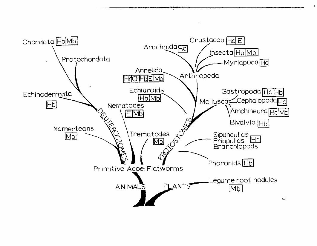
arthropods (Fig. 1). In the phylum Mollusca, hemocyanins
are found among the classes Gastropoda, Cephalopoda and
Polyplacophora. To date, none has been found in the
Monoplacophora, the Scaphopoda or in the Bivalvia. Among
the Arthropoda, hem~cyanin appears in the classes Crustacea,
Arachnida (spiders and scorpions) and Merostomata (horseshoe
crabs). No hemocyanin has been reported for the Progoneata
(millipedes), Chilopoda (centipedes) or Insecta, with the
possible exception of Scutigera longicornis (Chilopoda:
Myriapoda) "(Rajulu, 1969).
The name hemocyanin is somewhat misleading since the
molecule does not have heme as its prosthetic group.
2
Phylogenetic distribution of respiratoryproteins (modified from van Holde and Miller,in press). Mb = myoglobin, Hb = h~moglobin,
E = erthrocruorin, Ch = chlorocruorin,Hr = hemerythrin, Hc = hemocyanin.
Rather, it contains two copper atoms at the active site
which combine reversibly with an oxygen molecule. Hemocyanin
is named for the bluish color it displays upon oxygenation
(Predericq, 1878) which reflects a change in the oxidation
state of the copper atoms:
/ -2Cu (I) Cu (I) + 02~========~7 Cu (II) 02 Cu (II)
oxyhemocyaninblue
The mechanism for oxygen binding was first proposed by
Orgel (1958) and later confirmed using resonance spectro-
scopy (Loehr et al., 1974; !reedman et al., 1976).
Although the hemocyanins of arthropods and molluscs
appear homologous in terms of amino acid content (Ghiretti-
Magaldi et al., 1975), they differ radically at all other
structural levels. This is readily apparent when one
compares the quaternary structures of these hemocyanins.
In arthropod hemocyanins there are two copper atoms per
approximately 70,000 daltons of protein. This corresponds
to the molecular weight of the minimum polypeptide chain
or functional subunit. The subunits, which are observed
in vitro only, are obtained by raising the pH, lowering the
divalent cation concentration or by lowering the ionic
strength of the medium. The subunits are known to have a
sedimentation coefficient of about 5S (Eriksson-Quensel
and Svedberg, 1936; Van Holde and Van Bruggen, 1971).
rL;:'1'.,
5
Conversely, there are approximately 50,000 daltons of
protein per two copper atoms in molluscan hemocyanins and
this does not represent a minimal functioning subunit,
rather it is known as an oxygen binding domain (Lontie,
et al., 1973; Brouwer, et al., 1976). These domains are
linked together like a "string of pe arls" to form a large
, ~ subunit with as many as eight oxygen binding sites and
SO = 11 (Brouwer and Kuiper, 1973; Waxman, 1975).20,w
The molluscan hemocyanin sUbunits, as found in the native
proteins, are aggregated into giant cylindrical molecules
with molecular weights up to 9,000,000. These polYmers
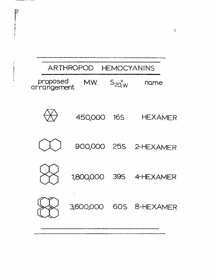
are composed of 20 subunits. In arthropods, the native
hemocyanins generally occur as six subunit aggregates
known as hexamers (S020,w = 16) and/or multiples thereof:
2-hexamers (25S), 4-hexamers (39S) and 8-hexamers (60S)
(Van Holde and Miller, in press) (Fig. 2).
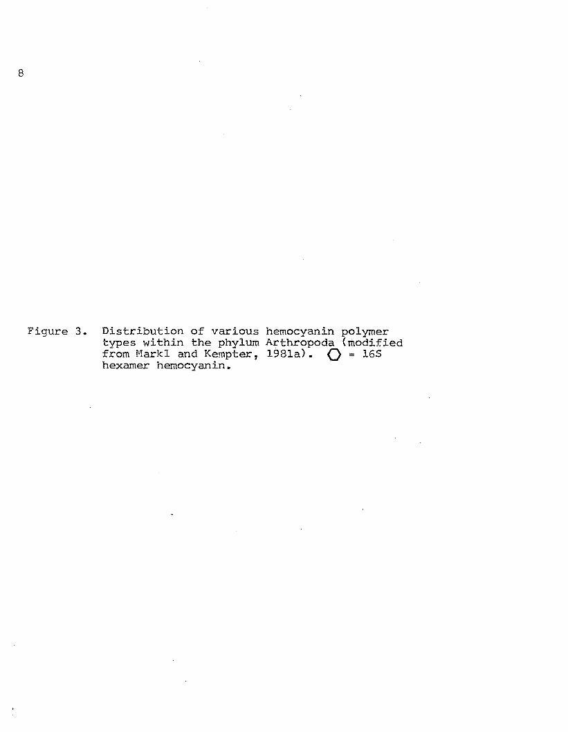
Markl (1979b) has suggested that the state of
aggregation of arthropod hemocyanin appears to be character-
istic for each species and may be specific to different
taxonomic groups (Pig. 3). A number of exceptions occur,
however. Por instance, the 60S molecule is found only
among the primitive horseshoe crabs and the 39S molecule
appears exclusively among some of the Thalassinid shrimps
and the more ancient Chelicora'tos ~ Host of the other
taxonomic groups possess e:l't;!ior the 255 2-hexamer or 165
6
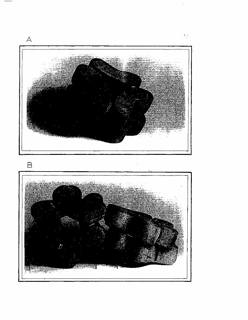
Figure 2. Structural characteristics of the arthropodhemocyanins.
pf",
7
ARTHROPOD 'HEMOCYANfNS
proposed M.W 520~W namearrangement
45QOOO 165 HEXAMER
900j OOO 255 2-HEXAMER
8
Figure 3. Distribution of various hemocyanin polymertypes within the phylum Arthropoda (modifiedfrom Mark1 and Kempter, 1981a). () = 165hexamer hemocyanin.
modern spiders 00 25SJ65o
merostomata 83 60S
raraChnida 83 355
more ancientchel icerates
395thalasSinid CX)shrimp CX)
00____-~~ crustacea 0 255)165
ANCESTRAL HEMOCYAN' N'0
10
single hexamer hemocyanin molecule or both.
As was noted, arthropod hemocyanins exist, in vivo,
as a small number of discreet molecular classes denoted
by sedimentation coefficients 16S, 25S, 39S, and 60S.
It is generally accepted that these molecular populations
are not in equilibrium; however, a few scattered exceptions
occur (cf. Arisaka and van Holde, 1979). The large
aggregates observed in arthropod hemocyanins can be
dissociated to individual polypeptide chains (subunits)
by raising the pH and/or removing divalent cations with
a chelating agent or lowering the ionic strength. In
other words, both H+ and Ca++ (Mg++) have stabilizing
effects on the large hemocyanin aggregates.
Formerly, it was assumed that the sUbunits obtained
by dissociation of the hemocyanin from a given organism
were all the same and represented the products of a single
gene. However, with the advent of high resolution probes
it has become clear that hemocyanins are composed of more
than one type of polypeptide chain or subunit. This
subunit heterogeneity is particularly striking in those
hemocyanins which form the larger aggregates. For example
the 60S molecule of Limulus is composed of 8-15 electro
phoretically distinct subunits (Markl et al., 1979a;
Brenowitz et al., 1981). Yet heterogeneity is also
observed in the smaller aggregates: the hemocyanin of
11
Palinurus which exists, in vivo, as a hexamer contains 6
different subunit types (Markl et al., 1979b).
It is difficult to make any generalization with
respect to the role played by these heterogeneous subunits
in arthropod hemocyanins, although the heterogeneity does
appear to have both functional (Sullivan et a1., 1974)
and structural (Lamy et a1., 1981a) implications. One
trend that has become increasingly apparent among hemo
cyanins in the decapod crustaceans is that in those species
where both 16S and 25S molecules are present in the native
hemocyanin, the 25S molecule contains more subunit types
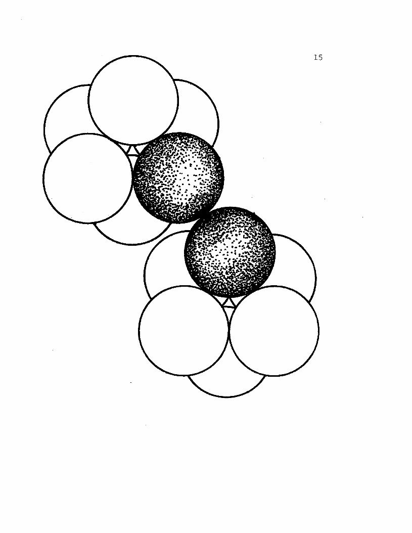
than the 16S molecule. Evidence now suggests that these
additional subunits are involved in linking the two hexamers
together (Fig. 4 and 5). In some instances, the inter
hexameric bond is a disulfide bridge (Jeffrey et a1.,
1978; Mark1 et a1., 1981), in others the "1inker bond"
shows a marked sensitivity to calcium (Morimoto and Kegeles,
1971; Terwilliger, in press), while in still others, it
is contended that polar and ionic interactions are primarily
responsible for the stability of the double hexamer
conformation (Herskovits et al., 1981b).
The recent studies of Markl and Kempter (198la, 1981b)
have added yet another dimension to our understanding of
subunit heterogeneity. Using immunochemical techniques
they have been able to divide the diverse numbers of
10 roM Tris-HCl (pH 6.3) and 0.01% bromphenol blue for 2
minutes at 1000 C. These samples were electrophoresed
into 7.5% slab gels with 3 cm stacking gels and run at
23
100 v for approximately 4 hours. The gels were stained in
Coomassie brilliant blue R (Fairbanks et al., 1971) and
destained in 10% acetic acid. Gel calibrants included
phosphorylase
(M = 68,000)r
A (M = 93,000), bovine serum albuminr
and ovalbumin (M = 43,000) (Sigma Chemicals).r
Cross-referencing of Polyacrylamide
Gel Protein Patterns
In order to determine the true number of different
polypeptide chains present in the hemocyanin of Hemi-
grapsus nudus, isolated protein bands from one gel system
were re-electrophoresed into a different system. Most
frequently cross-referencing was done between "native",
pH 8.9, gel protein bands and the SOS gel system. Following
electrophoresis in the first system, gels were removed
from the electrophoretic apparatus; stained and destained
rapidly, according to Cleveland et al. (1977). The-
protein band of interest was carefully excised and left
to soak in 0.5 ml of SOS incubation buffer at room tempera
ture for 30 minutes and then overnight at _20 0 C. After
thawing, the protein in the slice was electrophoresed
into an SOS gel as described previously and stained per
usual.
The method of cross-referencing protein patterns
24
was also employed between the regular gel systems. In
this case it is not possible to stain for protein because,
unlike the SDS system, there is no detergent present in the
regular gel incubation buffer to solublize the protein
after it is fixed. Rather a template of the protein
banding pattern was used to locate band positions in the
first unstained gel. Gel slices were removed from the
first system and incubated overnight at 4 0 C in 0.5 ml
of the upper electrode buffer appropriate to the second
gel system. Electrophoresis and staining techniques were
then carried out as described previously.
Peptide Maps
To confirm the unique nature of the subunit protein
chains present in the hemocyanin of H. nudus, peptide
maps of individual subunits were made. The procedure
involves the partial digestion of the proteins by a protease
in the presence of SDS <Cleveland et al., 1977).
Protein bands were cut from both SDS and pH 8.9 regular
gels and soaked for 30 minutes in SDS stacking gel buffer
at room temperature. Gel slices were then transferred
to a 15% SDS slab gel with a 4-5 cm stacking gel. The
wells of the slab gel, containing the excised slices, were
overlaid with 4 mg Staphlococcus aureus V8 protease
25
(Worthington). Electrophoresis was then initiated at 100 v
and allowed to proceed until the tracking dye met the
stacking gel/resolving gel interface. Digestion took
place in the stacking gel for 45 minutes. Electrophoresis
was then resumed at 100 v for 3 hours until the dye front
reached the bottom of the gel. Gels were stained in
Coomassie brilliant blue R (Pairbankset al., 1971) and
and destained in 10% acetic acid.
Copper Staining
Since hemocyanin is a copper-containing protein, its
presence in polyacrylamide gels can be detected by a
test specific to this metal. The test is based on the
quenching of fluorescence of bathocuproine sulfonate by
Cu+ (Bruyninckx ~ al., 1978). Regular gels at pH 7.0~c
rand 8.9 were pre-electrophoresed in the gel buffer~?:
appropriate to the pH system used, excluding tetramethyl-
ethylenediamine (TEMED) and ethylenediaminetetraacetic acid
(EDTA) and diluted 1:4, for 4 hours to remove free radicals
present in the gel. The gels were then loaded and run as
previously described, in the absence of EDTA. Immediately
following electrophoresis, gels were soaked for 1 minute
in 16 mM ascorbate in glacial acetic acid and then for 1
minute in 0.28 roM aqueous bathocuproine sulfonate solution.
26
The staining pattern on the gel was observed by UV light
illumination. Copper, where present, was indicated by
dark bands against a light field of fluorescence and
marked with pin holes and India ink. After staining
for copper the gels were stained for protein with Coomassie
brilliant blue R (Fairbanks et al., 1971) and destained
in 10% acetic acid.
~..•..
f...{r~
27
RESULTS
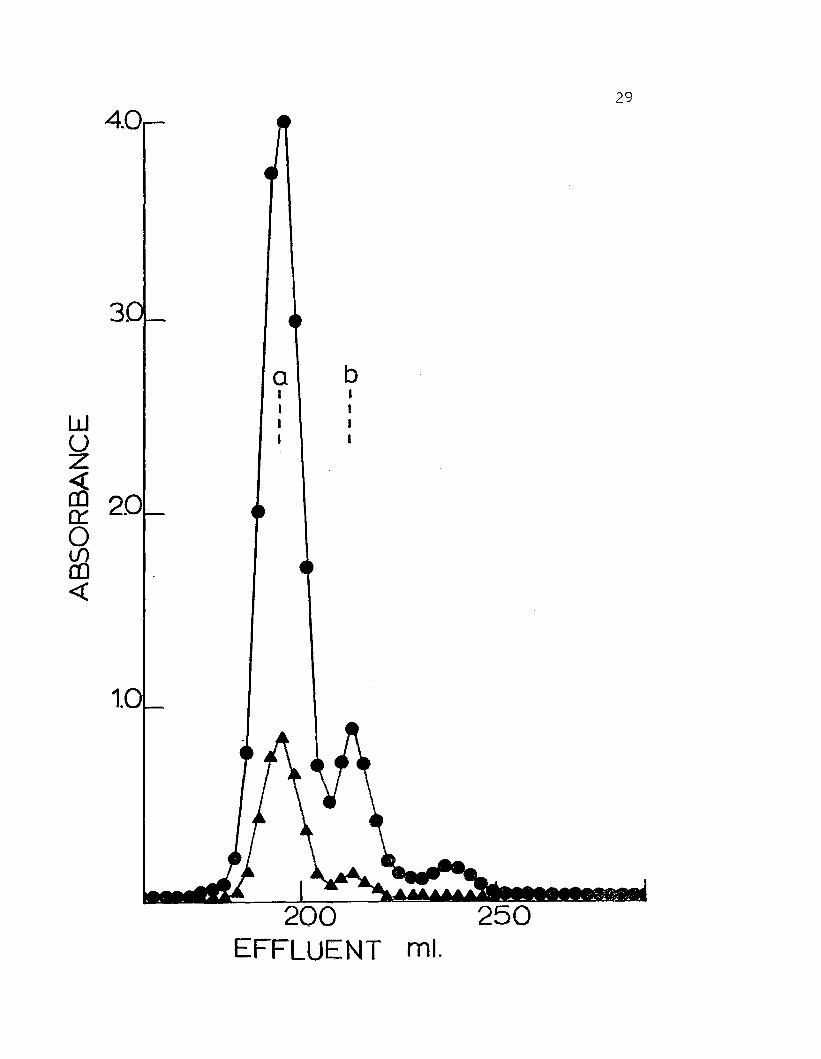
The hemocyanin of adult Hemiarapsus nudus chromato-
graphs on BioGel A-5M at pH 7.5 in the presence of divalent
cations as 2 peaks (Fig. 7). The material from the first
peak has an apparent molecular weight of 940,000. This
fraction comprises approximately 85% of the total protein
present. The second, minor peak has an apparent molecular
weight of 450,000 and makes up the remaining 15%. It
is assumed by analogy with numerous other crustacean
hemocyanins that peak I is 25S 2-hexamer hemocyanin and
peak II is predominantly l6S- single hexamer hemocyanin.
If fractions containing either the putative 25S or l6S
material are concentrated and re-chromatographed on the
same column they elute as a single peak corresponding to
peak I or II, respectively.
Absorption maxima are read at both 280 nm and 340 nm.
The ratio of these two absorption maxima does not vary
significantly for hemocyanin from the phylum Arthropoda
and can therefore give important information regarding
protein purity and degree of oxygenation of the hemocyanin.
Purified samples of 25S hemocyanin from H. nudus Hhich
28
Figure 7. Elution profile of hemolymph from Hemigrapsusnudus on a column of BioGel A-5M (200-400mesh). Column volume = 1.8 x 110 cm. Columnbuffer was 0.05 ionic strength Tris-HCl(pH 7.5), O.lM in NaCl, 10 roM in MgCl~ and10 ~1 in CaCl. Calibration proteins. a) .purified 25S gemocyanin from Cancer magister,b) purified l6S C. magister hemocyanin.Absorbances read at 280 nm ( .) and 340 nm( .).
4.0
3.
29
a bI II I
W I I
U I I
Z
~ 20~
0If)OJ<{
1.0
200EFFLUENT mL
30
have been scrubbed with oxygen give a 280 nm/340 nm ratio
of about 4.57. The ratio for 165 material is typically
near 6.85.
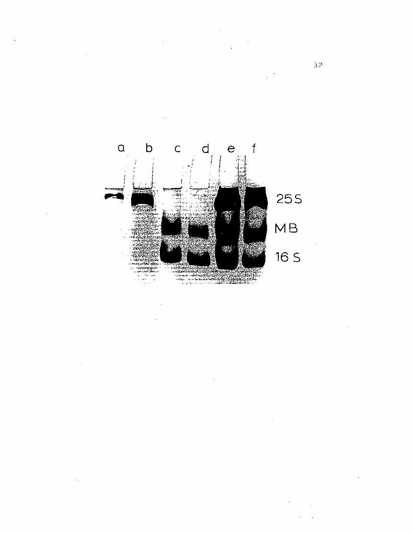
The pattern resulting from electrophoresis of purified
hemocyanin at either pH 7.0 or pH 7.4 is depicted in Figure
8. Under these conditions of pH and ionic strength, the
255 2-hexamer molecule remains intact and electrophoreses
as a single slow-moving band which stains strongly for
copper. Under the same conditions, the 165 fraction runs
as two distinct protein bands, a lower band and a central
band in between the other two bands. The lower protein
band from the 165 fraction stains very strongly for copper.
It is concluded that the lower band is 165 single-hexamer
hemocyanin by reference to the hemocyanin of Cancer
magister and the central or "mid-bandIT protein remains,
as yet, unidentified.
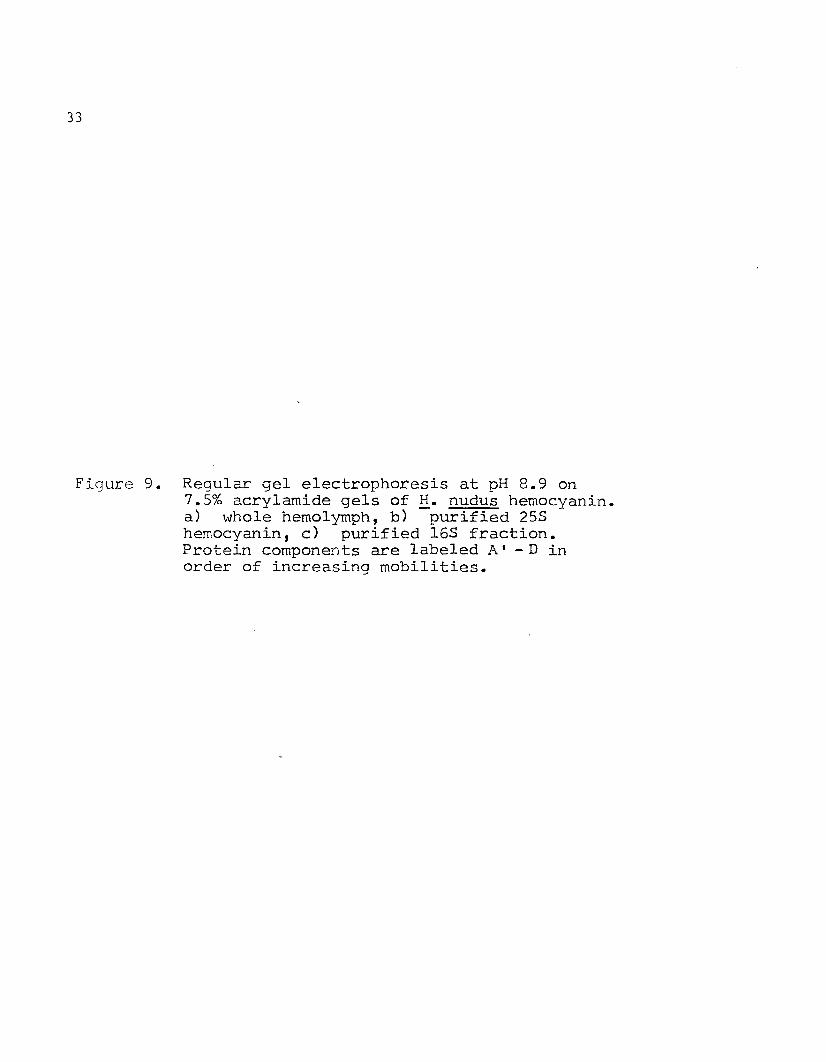
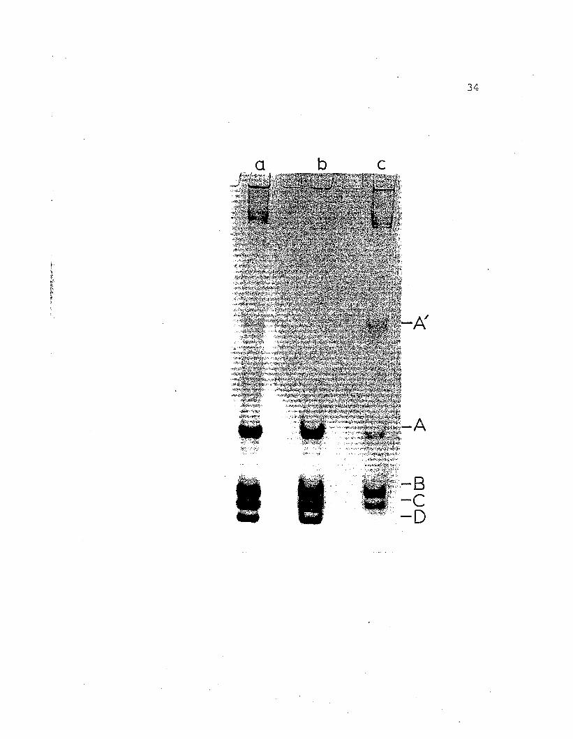
At pH 8.9 both the 255 and 165 aggregates dissociate
into 55 subunits which can be resolved on regular disc gels
at this pH (Fig. 9). The purified 255 fraction is resolved
as 4 protein bands labled A to D. The purified 165
fraction yields a similar pattern with two important
exceptions: A slow-moving band, Al is present only in
this fraction, and band D present in the 255 fraction
cannot be detected. All protein bands resolved on regular
gels at pH 8.9, with the exception of Al which was present
~.
. .~
Figure 8.. Regular gel electrophoresis at pH 7 .. 0 on7 .. 5% acry1amide gel of H. nudus hemocyanin ..a - b) purified 25S hemocyanin from majorpeak (Fig. 7), c - d) purified 16S fraction:mid-band protein (MB) and 165 hemocyanin,from minor peak (Fig .. 7), e - f) wholehemolYmph of g. nudus ..
Q
I: 1..
"".",..~........
b cI
. -l
d e f
255
MB
165
32
33
Figure 9. Regular gel electrophoresis at pH 8.9 on7.5% acrylamide gels of H. nudus hemocyanin.a) whole hemolYmph, b) purified 255hemocyanin, c) purified 165 fraction.Protein components are labeled A' -D inorder of increasing mobilities.
a
,1~1iAj"!t;a.;
'-~~';';~"
•""f5"::'f'~¥;' "".+
b c
A
;'-8~'~~-. ,
-c-D
34
35
in low concentrations,· stained for copper.
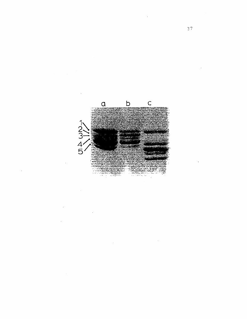
Purified samples of 25S and 16S hemocyanin were
denatured with SDS in the presence of 2-mercaptoethanol
and electrophoresed on SDS slab gels (Fig. 10). Five protein
bands of different staining intensity were resolved with
molecular weights ranging from 76,200 to 82,300. The
bands are labled 1-5 in order of descending size. Both
25S and l6S give indistinguishable banding patterns in
this system.
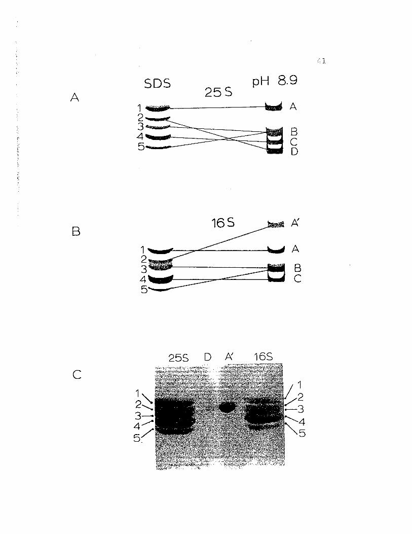
The results from cross-referencing experiments between
native gels at pH 8.9 and SDS gel electrophoresis are
shown in Figures 11 and 12. The important points gleaned
from these experiments are as follows: 1) Native protein
band B (pH 8.9) electrophoreses as 2 protein bands, 3 and
5, of differing molecular weight in the SDS system and,
2) Native band A' (pH 8.9), unique to the l6S fraction,
and native band D (pH 8.9) found only in the 25S fraction,
co-electrophoreses to the same position, band 2, in SDS.
From cross-referencing experiments betwee~ regular gel
systems it was found that the mid-band protein of the l6S
fraction observed at pH 7.0 and 7.5 electrophoreses to
position At on a pH 8.9 regular gel.
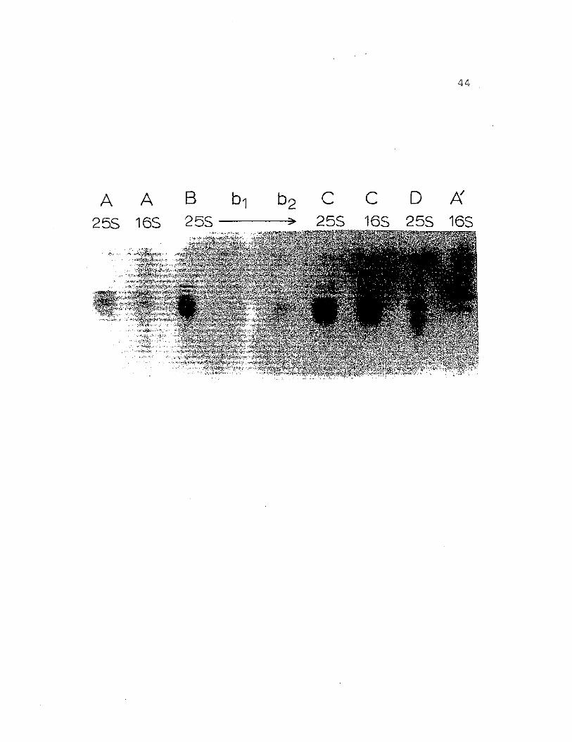
Further confirmation for the unique character of the
bands resolved in both native gels at pH 8.9 and those
observed in SDS was provided by peptide maps (Fig. 13 and
36
Figure 10. SDS gel electrophoresis of ~. nudushemocyanin on 7.5% acrylamide gels. a)purified l6S fraction, b) purified 25Shemocyanin, c) whole Cancer magisterhemocyanin. Protein components are labeled1 - 5 in order of decreasing apparent·molecular weights which range from 76,20082,300.
a b c
37
38
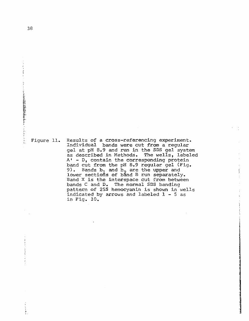
Figure 11. Results of a cross-referencing experiment.Individual bands were cut from a regulargel at pH 8.9 and run in the SDS gel systemas described in Methods. The wells, labeledAt - D, contain the corresponding proteinband cut from the pH 8.9 regular gel (Fig.9) • B~ds b l and b, are the upper andlower sections of band B run separately.Band X is the interspace cut from betweenbands C and D. The normal SDS bandingpattern of 25S hemocyanin is shown in wellsindicated by arrows and labeled I - 5 asin Fig. 10.
»
-E
o
39
40
Figure 12. Proposed correspondance of protein bandsobtained by regular gel electrophoresis atpH 8.9 with the bands obtained by SDS gelelectrophoresis as suggested by crossreferencing experiments (Fig. 11). A)255 hemocyanin, B) 165 fraction, C) resultof cross-referencing experiment whereslices containing the proteins from bandsD (255 only) and A' (165 only) wereelectrophoresed into the same well in 5D5.Note that both D and A' run to the sameposition (2) in SDS.
ASDS pH 8.9
25S1 4ijjjjfi#f b d A
!~~~~§
165
1 A
!;;;;=:~~
B
c255 D f:{. 16S
1,,2.........34 ..........5~
42
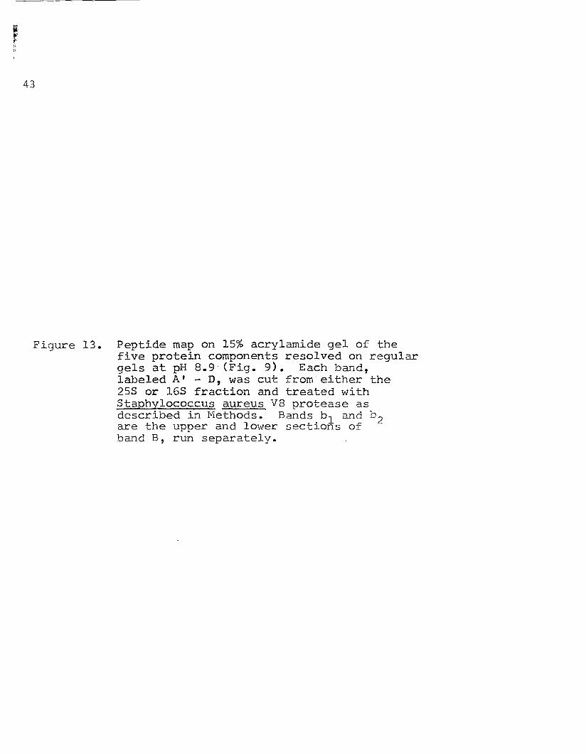
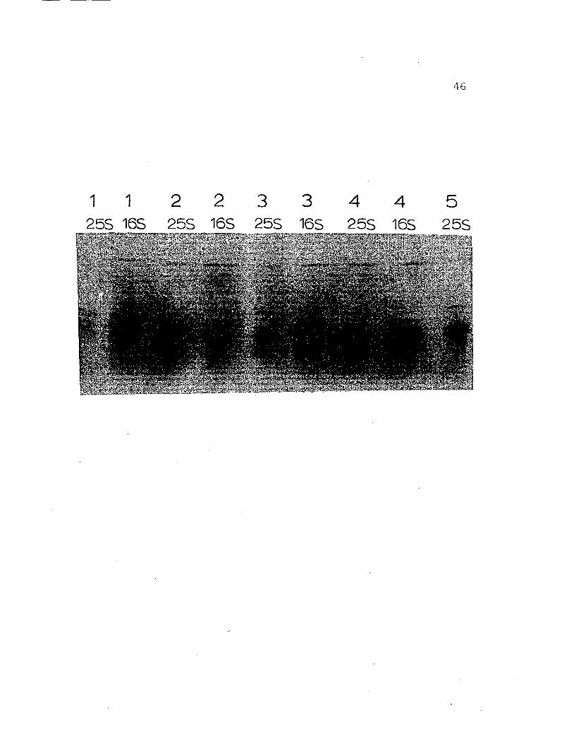
14). It was found that a given polypeptide chain will
give the same peptide map whether it is cut from regular
gels or from SDS gels. In the case of Hemigrapsus nudus
hemocyanin six distinct polypeptides exist. Of these,
five are certainly hemocyanin as judged by copper content
and apparent molecular weights. The exact identity of the
sixth protein, Alan pH 8.9 regular gels and mid-band
on pH 7.0 and 7.5 regular gels, remains unresolved.
The experiments just described were also performed
on whole hemolymph from both male and female crabs,
purified hemocyanin from the female crabs (with and without
eggs), on a freshly moulted individual and several other
individual crabs as well. The results of these experiments
did not differ from those just described for pooled
intermoult male hemocyanin.
43
Figure 13. Peptide map on 15% acrylamide gel of thefive protein components resolved on regulargels at pH 8.9 (Fig. 9). Each band,labeled AI - D, was cut from either the25S or l6S fraction and treated withStaphylococcus aureus V8 protease asdescribed in Methods. Bands b l and b 2are the upper and lower sections ofband B, run separately.
A A25S 165
44
B b1 b2 C eDt{.255 ----=!Ioo> 255 165 255 165
45
Figure 14. Peptide map on 15% acry1amide gel of thefive protein components resolved on SDSgels (Fig. 10). Each band, labeled 1 - 5,was cut from either the 25S or 16S fractionand treated with Staphylococcus aureus V8protease as described in Methods.
1 1255 165
2 2 3 3255 165 255 165
4 4255 165
46
5255
{,.
47
DISCUSSION
Quaternary Structure
The hemocyanin of Hemigrapsus nudus exists, in vivo,
as two distinct polymeric aggregates which can be separated
by exclusion chromatography. The major component of this
blood is a 25S 2-hexamer molec~le as jUdged by its
apparent molecular weight (940,000), copper-staining on
polyacrylamide gels and its 280 nm=340 nm absorbance ratio.
The average value observed in g. nudus hemocyanin for this
ratio is 4.57 which is well within the limits of 4 to 5
ascribed for pure hemocyanin (Markl, 1979b).
The second component, a 165 single-hexamer hemocyanin,
comprises up to 15% of the total protein present. However,
due to the presence of an additional unknown protein, the
exact quantity of 165 hemocyanin in this fraction has
proven difficult to measure with certainty. That an
additional protein is present is evidenced by the dispro-
portionately high 280 nm:340 nm ratio of 6.85. Additionally,
when purified 165 samples are resolved on a regular gel
at either pH 7.0 or 7.5 two bands of approximately equal
48
protein staining intensity are observed. Based on relative
mobilities of these two proteins in this electrophoretic
system in comparison to purified l6S hemocyanins of other
organisms, it is concluded that the lower-most band is l6S
hemocyanin and the mid-band is the unknown protein. This
view is supported by copper-staining and cross-referencing
experiments. Efforts to separate the unknown protein
from the l6S hemocyanin fraction have been unsuccessful.
The two hemocyanin species observed in Hemigrapsus
nudus (25S 2-hexamers and l6S hexamers) do not represent
a rapidly equilibrating system; rather they behave as a
mixture of discrete aggregates. This is shown by the
failure of purified hemocyanin solutions, either 25S
or l6S, to produce increased products of dissociation or
reassociation upon dilution. The lack of apparent
equilibrium or deviance from the mass action law among the
hemocyanins is attributed to the effects of heterogeneity
at the subunit level (Herskovitz et al., 1981a, 1981b).
Subunit Composition
The 25S and l6S aggregates of Hemigrapsus nudus
hemocyanin can be dissociated to six distinct subunits
whose apparent molecular weight in SDS range from 76,200
to 82,300. The presence of dissociation products with
49
apparently differing molecular weights suggests that the
hemocyanin of Hemigrapsus nudus is composed of a hetero-
geneous mixture of polypeptide chains. Yet, it can be
argued that the SDS gel method is not reliable for absolute
molecular weight determinations. Therefore, other lines
of evidence in support of the existence of microhetero-
geneity in H. nudus hemocyanin are proffered here and will
be the subject of the remainder of this discussion.
Using two different electrophoretic procedures it is
possible to dissociate the hemocyanin of Hemigrapsus nudus
into two different protein banding patterns. The first
pattern, obtained by regular gel electrophoresis at pH 8.9,
arises mainly as a result of charge differences among the
putative subunits. In this system each of the purified
aggregates, 25S and l6S, produces 4 major protein bands.
One of the bands (At) has a markedly retarded mobility
in this system. The other banding pattern, from SOS gel
electrophoresis, is the result of differences in apparent
molecular weights. In SOS both the 25S and 16S molecules
give identical patterns consisting of five bands each.
The fact that the SDS gel system resolves one band more
than observed in the regular gel system indicates that
two polypeptides are present which have similar charge
characteristics but different molecular weights in SOS.
This idea was confirmed by cross-referencing experiments;
50
band B of the regular gel syste~ migrates as bands 3 and
5 in SDS.
The cross-referencing experiments proved very instru-
mental in the determination of subunit numbers since it is~
~ unlikely that anyone component was concealed in both
t~ systems. The most revealing information came from ex-~
periments where proteins were cut from regular gels at
pH 8.9 and analyzed in the SDS system. Four of the
subunits obtained from regular gels were homogeneous in
both systems and all ran as single bands in SDS. Only
band B, which appeared homogeneous on regular gels, was
shown to be heterogeneous in SDS. On the other hand,
band 2 of the SDS system, which appeared homogeneous, was
actually composed of two proteins separable by regular
gel electrophoresis at pH 8.9. It is interesting to note~r that of the two proteins which migrate to position 2 on~
I.r SDS, one is observed only in the 25S fraction (D) and the~
f
other (AI) is unique to the 16S fraction such that only
when unpurified hemocyanin was run in the SDS gel system,
did band 2 really represent a heterogeneous mixture of
AI and D. Furthermore, on regular gels, at pH 8.9, band
AI migrates'as the slowest band; far behind the other
subunits, and band D migrates as the fastest band. In
other words, two seemingly different proteins with vastly
different mobilities in the regular gel system have the
51
same approximate molecular weights as determined by SDS
electrophoresis in the presence of reducing agents.
It was originally hypothesized that band D existed
as a single subunit only when it was a part of the 25S
2-hexameraggregate but dimerized when associated with the
16S fraction and thus ran as a slower band, AI. Upon
exposure to the disulfide-bridge reducing agent, 2-mercap-
toethanol, present in the SOS system, the dimer would
presumably dissociate and run as a single band to position
2 and become indistinguishable from its single chain
counterpart; O. Experiments with and without 2-mercap-
toethanol did not offer proof for this hypothesis.
Additionally it was found by cross-referencing gel slices
between pH 7.0 and 8.9 regular gels that band AI was also
the contaminating, mid-band protein observed on pH 7.0
(and 7.5) regular gels, and as such not likely to be
hemocyanin at all.
The cross-referencing experiments between regular.
and SDS gels argue strongly in support of heterogeneity
at the sUbunit-polypeptide level. Not only are multiple
protein bands observed in the SDS system, where proteolysis
for the observed heterogeneity, but multiple protein bands
are also detected on regular gels. Moreover, each band
isolated by the regular gel system corresponded neatly
52
to a protein band resolved by SDS gel electrophoresis.
The most unequivocal evidence for polypeptide
heterogeneity came from peptide maps obtained by limited
proteolysis of individual protein bands. Each of the
subunits isolated by SDS gel electrophoresis produced
a unique map. In particular, SDS band 1 produced the
same characteristic map regardless of whether it was cut
from a 25S fraction or a 16S fraction. This was also
true for SDS bands 3, 4 and 5. However, the map obtained
from SDS band 2 of 25S hemocyanin differed markedly from
the map obtained from SDS band 2 cut from a 16S fraction;
confirming the findings of the cross-referencing experiments
proposed in Figure 12 •
In addition, peptide maps obtained from subunits
isolated on regular gels at pH 8.9 correlated closely with
thOSe maps obtained from SDS and thus further verified
cross-referencing findings. For instance, the map of
band A (reg. gel) closely resembled the map of band 1
(SDS); band B (reg. gel) which was suspected to be a
doublet of SDS bands 3 and 5 gave a map that was a composite
of the maps from 3 and 5. Finally, a map of band AI
(reg. gel, 16S only) correlated with the map of 16S band
2 (SDS), while a map of band D (reg. gel, 25S) matched the
map of 25S band 2 (SDS).
In light of these findings it is possible to conclude
53
that the single hexamer, 165 molecules have at least 4
individual subunits which are also present in the 255
linked hexamers. Based on relative staining intensities,
it is proposed that subunits 1 and 4 (5D5) occur twice
in the 165 molecule making up the six subunits of the
165 hexamer. Furthermore, the 255 2-hexamer population
appears to have a subunit associated with ......1.L. that is not
present in the 165 fraction. Although no evidence exists
at present, it is possible that this subunit is necessary
for the assembly of subunits to a linked 255 2-hexamer
configuration. In the words of Markl and Kempter (1981a;
1981b), the protein isolated as band D on regular gels
at pH 8.9 may be a "conservative subunit" and as such,
act as a hexamer-hexamer link. 5uch a system has been
described in a number of arthropods, the simplest known
is that of the marine isopod Ligia pallassi (Terwilliger,
in press). The 165 hemocyanin of this organism is composed
of six subun-i ts of a single type, I11 , and the 255 2-hexaTT1.er
hemocyanin has 2 subunit types, M1 and M2 , in an apparent
ratio of 5:1. 5ince a dissociated 2-hexamer aggregate is
incompetent to reassociate in the absence of M2 subunits
it is suggested that the M2 subunit acts a linker in this
system.
Finally, it is concluded that genetic or sexual
polymorphisms can not account for the observed
54
heterogeneity since identical results were obtained from
individual crabs of both sexes.
Summary
The native hemocyanin of adult Hemigrapsus nudus
exists as two non-equilibrating populations of multi-
subunit aggregates: 255 2-hexamers and 165 hexamers.
The 2-hexamer molecule has a molecular weight of about
940,000 and comprises 85% of the protein present in
purified hemocyanin. The l6S fraction includes the
single hexamer hemocyanin molecule which has a molecular
weight of about 450,000 and makes up most of the remaining
15% of the protein. Also present in the fraction which
elutes at l6S is an additional protein of unknown identity.
Dialysis against an alkaline buffer, pH 8.9, in the
absence of divalent cations results in the dissociation
of the 25S and 165 aggregates to 5S subunits. The subunit
mixture was resolved by regular polyacrylamide gel
electrophoresis and gave characteristic patterns of four
protein bands each. The resulting patterns from the 255
and l6S molecules differed with respect to a slow-moving
band present only in the l6S fraction and a fast~moving
band found only in the 25S fraction.
Denaturation of the 25S and 163 molecules in the
55
presence of SDS and 2-mercaptoethanol yielded a pattern
of five protein bands with apparent molecular weights
ranging from 76,200 to 82,300. The banding patterns
produced by the 25S and 16S molecules are indistinguishable
from each other.
Through a series of cross-referencing experiments,
where isolated protein bands from the regular gel system
at pH 8.9 were analyzed in the SDS gel system, it was
possible to correlate the two patterns produced by these
electrophoretic systems. The results showed that all but
one of the bands resolved by regular gel electrophoresis
corresponded to distinct and separate bands in the SDS
system; the other band resolved as two bands in SDS.
In other cross-referencing experiments it was found
that the unidentified contaminant present in the l6S
fraction represented the slow-moving band observed on
regular gels at pH 8.9.
Peptide maps provided proof for the correlations
determined by cross-referencing experiments and lent
strong support to the idea that each band separated by
SDS represented a unique polypeptide chain.
It is concluded that the hemocyanin of adult Hemi
grapsus nudus is composed of five unique sUbunits, four of
which occur in both the 25S and 16S aggregates. The fifth
sUbunit, observed only in the 25S fractions, is thought
to act as a "linker molecule" and in this capacity aids
in the formation of the 255 2-hexamer configuration.
56
57
METHODS AND r1.ATERIALS
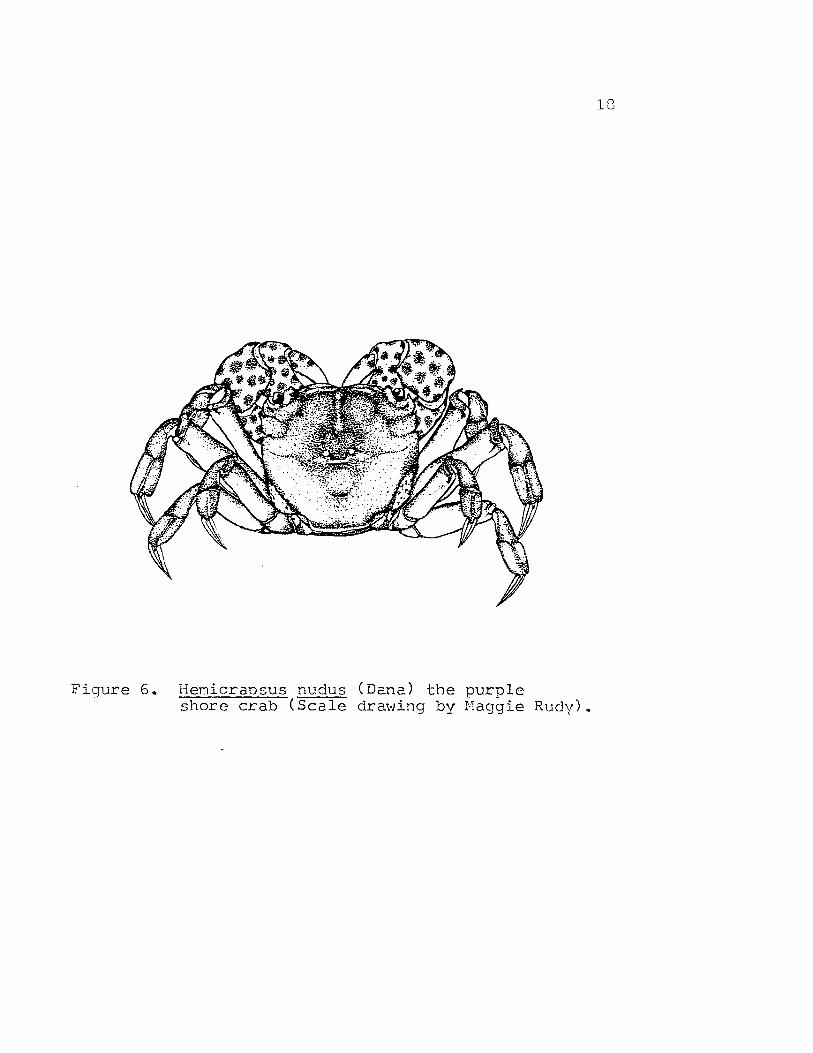
Ovigerous Hemigrapsus nudus (Dana) were collected
off Fossil Point, Coos Bay, Oregon. These animals were
kept in tanks of running seawater until needed.
Egg Hemocyanin
Eggs, both early and late in development, were obtained
by removing the berried pleopods and extracting the egg
masses from them. Following removal, the eggs were soaked
in filtered seawater in the cold for 1-2 hours. Eggs were
crushed in a tissue grinder to a fine homogenate in the
presence of a small amount of phenylmethylsulfonyl
fluoride (PMSF), a serine protease inhibitor. The extract
was then centrifuge9 at 12,000 x g for 15 minutes at 40 C
in a Sorvall RC-2B refrigerated centrifuge. The supernatant
was quickly passed through Whatman No. 1 filter paper to
remove the lipid layer and centrifuged again at 12,000 x g
for 10 minutes. The supernatant, which was brownish-orange,
was immediately applied to a column of Sephadex G-75
(1.8 x 32 cm) in equilibrium with a Tris-HCl buffer, pH 7.5,
58
that was 0.1 M in NaCl, 10 ~1 in MgC12
and 10 ~1 in CaC1 2 •
The void volume peak, which was a clear orange solution
and should contain hemocyanin if present was collected
and concentrated briefly on sucrose at 40 C and then applied
to a BioGel A-5f': column (1.8 x 110 cm) in equilibrium
with the same buffer. Further analyses were performed
as previously described for adult Hemigrapsus nudus.
Zoea Hemocyanin
The zoea of Hemigrapsus nudus were collected following
~ their release from ovigerous crabs held in running seawatert.'ft tanks. The zoea were placed in nylon netting and rinsedtrf with filtered seawater. Body fluid was extracted by
squeezing the animals in the nylon netting and collecting
the filtrate in a chilled beaker containing a few drops
of PMSF. The crushed animals were rinsed several times
with small quantitites of filtered seawater which were
also added to the contents of the chilled beaker. The
combined extract was then centrifuged at 12,000 x g for
15 minutes at 40 C in a Sorvall RC-2B refrigerated
centrifuge. A clear orange supernatant was removed and
concentrated briefly on sucrose at 40
C before application
to a BioGel A-5M column (1.8 x 110 cm) in equilibrium
with a Tris-HCl buffer, pH 7.5 that was 0.1 M in NaCl,
59
10 mM in MgC1 2 and 10 mM CaC1 2 • All subsequent analyses
were performed as previously described for adult Hemigrapsus
nudus.
60
RESULTS
Egg Hemocyanin
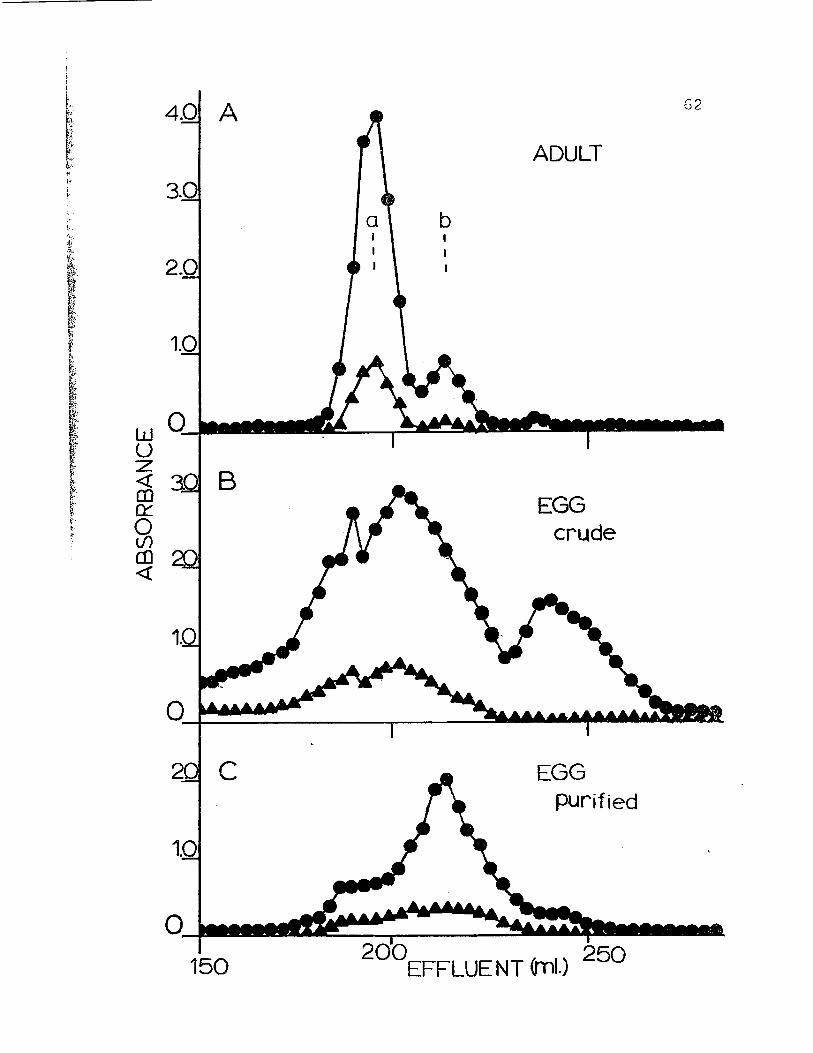
The crude homogenate obtained from the eggs of
Hemigrapsus nudus chromatographs as several protein peaks
(Fig. l5b). Rechromatography of samples with elution volumes
similar to those of adult hemocyanin yielded a single
peak with a shoulder on the leading edge (Fig. l5c). The
material from the main peak has an apparent molecular
weight of 450,000 daltons as judged by its elution volume.
The shoulder corresponds to an apparent molecular weight
of 940,000. It is assumed by analogy with adult Hemigrapsus
nudus hemocyanin and numerous other crustacean species
that the main peak is composed primarily of 16S single
hexamer hemocyanin and the shoulder is 25S 2-hexamer
hemocyanin. Ratios of absorption maxima read at both
280 nm and 340 nm give values of about 6 for the 255
shoulder and about 13 for the main 165 peak. These values
are high in contrast to those obtained from adult 255
and 16S hemocyanins (4.57 and 6.85, respectively),
suggesting the presence of additional proteins. Since
experiments with eggs both early and late in development
61
Figure 15. Chromatography of hemolymph from H. nudusadult (A), crude egg homogenate (B) andpurified egg (C) on a column of BioGel A-5N(200-400 mesh). Column volume = 1.8 x 110cm. Column buffer was 0.05 ionic strengthTris-HCl (pH 7.5), 0.1 M in NaCl, 10 ~~
in MgCl and 10 roM in caC1 2 • Calibrationmarkers? a) purified 25S hemocyanin fromCancer magister and b) purified 16S C.magister hemocyanin. Absorbances readat 280 nm. ( • ) and 340 nm. ( A ) •
4.0 A
3.0
ADULT
62
EGGcrude
EGGpurified
bIII
aII
I
200EFFLUENT (ml.) 250
c
2.0
150
1.0
1.0
w 0Ul _u~ B(!)0:::
~en«
63
gave the same results, they will not be distinguished
from each other in this report.
Electrophoretic analysis of purified egg hemocyanin
on regular gels at pH 7.0 yielded patterns similar to
the adult hemocyanin (Fig. 16); 25S material resolves
as a single slow moving protein band which stains strongly
for copper, and the 165 fraction runs as two distinct
protein bands. The lower band stains strongly for copper
and faint copper staining was detected for the mid-band
protein. It is concluded by reference to adult 16S
hemocyanin that the lower band, resolved in the egg 16S
fraction, is 165 hemocyanin. The egg hemocyanin as
resolved by this system differs from that of the adult in
two ways. First the so-called mid-band protein is present
in much higher relative concentration in the egg hemocyanin
than in the adult hemocyanin. Secondly there appears to
be considerably more 16S hemocyanin relative to 25S
hemocyanin in the egg preparations in comparison to the
adult hemocyanins.
At pH 8.9 both the 255 and 16S egg hemocyanins
dissociate into 5S subunits which can be resolved on
regular disc gels (Fig. 17). The purified 25S material
is resolved into three protein bands which apparently
correspond to bands B, C and D of the adult 25S hemocyanin.
The purified 16S material resolves as two protein bands
64
Figure 16. Regular gel electrophoresis at pH 7.0 on7.5% acrylamide gel of adult and egghemocyanins from H. nudus. a) purifiedegg 253 hemocyanin, b - e) samples of egg163 fraction taken at different pointsalong the major peak shown in Figure 15 C,f) whole adult hemolymph.
abc d e f
-255
-MB165
G5
Figure 17. Regular gel electrophoresis at pH 8.9 on7.5% acrylamide gel of adult and egghemocyanins from H. nudus. a) wholeadult hemolYmph, b) purified adult 25S,c) purified egg 25S hemocyanin, d)purified egg l6S fraction, e) purifiedadult l6S fraction, f) whole adulthemolymph.
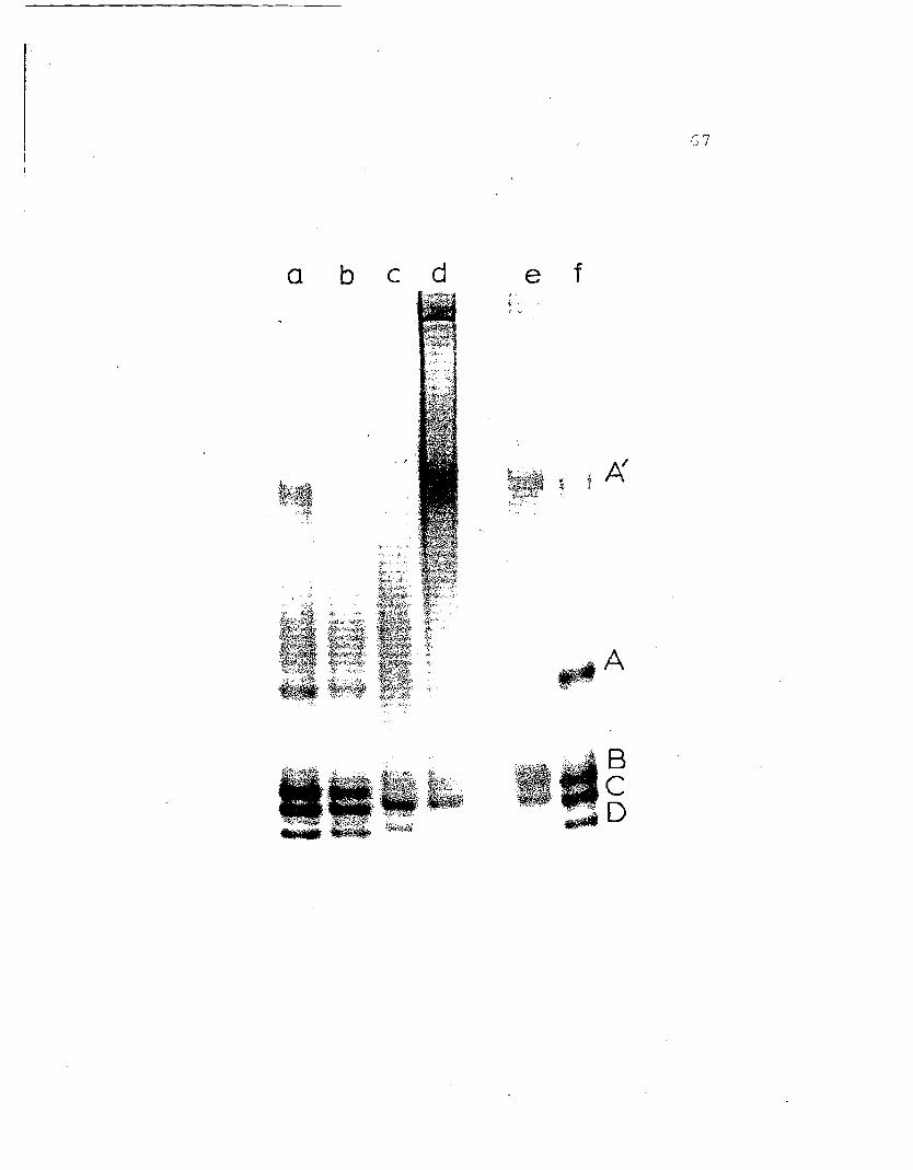
which correspond to adult bands Band C. High molecular
weight material is also present in the egg 16S fraction
including a band at position A'. Despite basic similarities
between the egg and adult hemocyanins as resolved in this
system two important differences occur: adult band A
can not be detected in either of the egg 25S or 16S
fractions and, although adult band B is observed in the
egg fractions it is .present in much lower relative
concentrations.
Purified samples of egg 25S and 16S hemocyanin were
denatured with SDS in the presence of 2-mercaptoethanol
and electrophoresed on SDS slab gels (Pig. 18). The puri-
fied 25S egg hemocyanin resolves as 3-4 protein bands
in this system which correspond to adult SDS bands 2, 3, 4
and trace quantities of 5. Adult band 1 is not detected
in egg 25S hemocyanin. Also present in the 25S egg
fraction were faster moving bands. These protein bands
might be the result of proteolytic degradation or are due
to slight contamination. by the mid-band protein. The
purified egg 16S hemocyanin resolves as 2 protein bands
in SDS which correspond to adult SDS bands 3 and 4. Adult
bands 1 and 5 are not detectable in the purified 16S
hemocyanin. When the mid-band protein is isolated and
run in the SDS system it resolves as four protein bands
two of which have molecular weights which correspond to
69
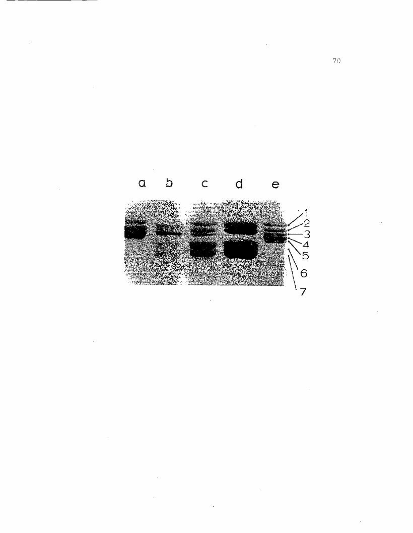
Figure 18. SDS gel electrophoresis on 7.5% acrylamidegel of adult and egg hemocyanins from H.nudus. a) whole adult hemolymph, b)purified egg 255 hemocyanin (with slightcontamination from mid-band protein), c)purified egg 165 fraction, d) mid-bandprotein (cut from pH 7.0 regular gel andrun in SDS system), e) purified adult 25Shemocyanin. The adult bands are labeled1 - 5 in order of decreasing molecularweights. Bands 6 and 7 have'molecularweights of 74,300 and 69,900, respectively.
a b c d e
71
adult bands 1 and 2, the other two have a higher mobility
than the adult subunits and are labled 6 and 7. The
molecular weight values for bands 6 and 7 as determined
by SDS gel electrophoresis are about 74,300 and 69,900,
respectively.
Through cross-referencing experiments it was possible
to correlate the adult hemocyanin protein bands as resolved
on regular gels (pH 8.9) with the 5 protein bands observed
in the SDS system. A similar cross comparison was carried
out on egg hemocyanins. Egg band B of pH 8.9 regular
gels electrophoresed to position 3 in SDS. Band C
electrophoresed to SDS position 4 and band D (25S only)
electrophoresed to position 2. The mid-band protein ran
to 4 positions; 1, 2, 6 and 7. The results from the
cross-referencing experiments with egg hemocyanins parallel
those obtained from similar experiments on adult hemocyanins
with 3 important exceptions. First, egg band B resolves
as only one major band in SDS (band 3), this is in contrast
to adult band B which resolves as two major protein bands
in SDS (3 and 5). Secondly, it was found that egg mid
band protein resolves as four bands on SDS (1, 2, 6 and 7)
while adult mid-band runs to SDS position 2 only. A
third difference which will be discussed later is the
conspicuous absence of band A from both 25S and 16S egg
hemocyanin and corresponding absence of subunit 1 in the
72
5D5 samples.
To confirm the relationship between subunits observed
in both 5D5 and on regular gels at pH 8.9, and to establish
the true relationship between adult and egg hemocyanins
peptide maps were made of individual egg proteins. Each
of the egg subunits isolated by 5D5 gel electrophoresis
produced a unique peptide map which corresponded closely
to the maps obtained from adult subunit bands (Fig. 19).
For instance, the map of egg band 4 from either the 255
or 165 fractions matched the peptide map of adult band 4.
In adult hemocyanin it was shown that the map of 255
band 2 differed from the map of 165 band 2; the adult 255
band 2 is a hemocyanin subunit and the 165 band 2 repre
sents the mid-band protein. The same situation is also
true of the egg hemocyanins. The peptide map of band 2
from the egg 255 fraction is different from that of the
egg 165 band 2. The map of egg 255 band 2 resembles the
maps of both adult bands 2 and 3. This can probably be
explained by cross-contamination between these neighboring
bands. The map of the egg 165 band 2 corresponds exactly
to the map of adult 165 band 2.
Additionally, the maps of egg bands 1 and 6 which
are also known to be components of the egg mid-band protein
produce peptide maps which are indistinguishable from the
map of adult 165 band 2. In other words, three of the
73
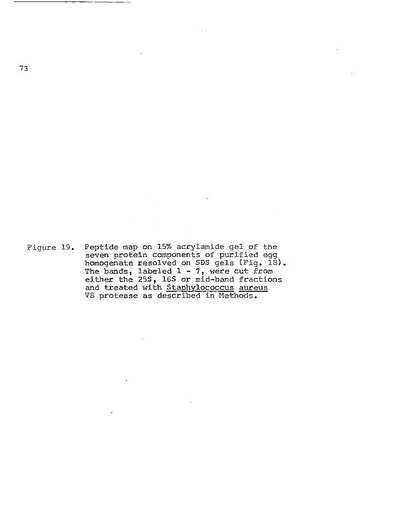
Figure 19. Peptide map on 15% acrylamide gel of theseven protein components of purified egghomogenate resolved on SDS gels (Fig. IS).The bands, labeled 1 - 7, were cut fromeither the 253, 163 or mid-band fractionsand treated with Staphylococcus aureusVS protease as described in Methods.
1122244MB 165 MB 255 165 255 165
7,1
6 7MB MB
75
components of the egg mid-band protein are the same as
the adult mid-band protein. The fact that a single protein
type runs as three different bands in SDS can probably
be attributed to heightened sensitivity of this egg protein
to proteolysis not observed in the adult protein. Another
possibility is that the mid-band protein components have
been chemically mOdified in such a way as to give them
slightly different mobilities in the SDS system. Egg
band 7, the fourth component of the mid-band protein,
produces a unique map and appears to be a protein that
is not present in the adult hemocyanin. An analysis of
egg band 3 is not included here since it was not possible
to obtain this subunit in high enough concentrations
for analysis by peptide maps.
Zoea Hemolymph
The results of experiments with the zoea are less
conclusive than those with egg hemocyanins. This is due
to the difficulty in obtaining sufficient quantity of zoea
hemolymph for a complete analysis. The results presented
here are tentative.
The hemolymph of the zoea of Hemigrapsus nudus
chrorn.~Loqraphs on BioGel A-5M at pH 7.5 in the presence
of rljv~l,ent cations, as a complicated curve not unlike
76
that of the crude egg homogenate shown in Figure lSb.
Rechromatography of samples with elution volumes similar
to those of adult hemocyanin yields a small peak of very
low concentration.
On regular gels at pH 7.0, the purified hemolymph
from the zoea resolves as two protein bands. The upper
band corresponds closely to adult 255 hemocyanin. The
second band which is considerably wider than the first
runs slightly behind the adult 165 hemocyanin band. Both
of the zoea protein bands resolved in this system stain
for copper.
Due to the low concentration of zoea hemolYmph
available it was not possible to resolve this protein
on regular gels at pH 8.9. Instead the protein was
lyophilized and run in the 5DS electrophoresis system in
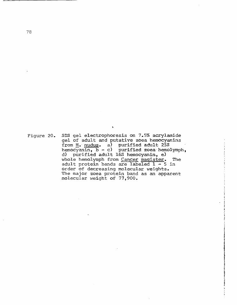
the presence of 2-mercaptoethanol (Fig. 20). A single
strongly staining protein band accompanied by several
faint bands was resolved by this system. The major band-
has an apparent molecular weight in SDS of 77,900 and will
be referred to as band 3.
Peptide maps were made of zoea protein bands (Fig. 21).
Each of the peptide maps produced from zoea hemolymph
proteins are the same. Furthermore they do not bear an
immediate resemblance to any of the adult hemocyanin
peptide maps.
77
At this point it is impossible to draw any conclusions
regarding the existence of hemocyanin in the zoea of
Hemigrapsus nudus. It can be stated, however, that the
hemolymph of this organism possesses at least one copper
containing protein which has an apparent molecular weight
similar to that of the adult hemocyanin. Also, this zoea
protein is composed of at least one subunit whose molecular
weight in SDS is within the range expected for hemocyanin
subunits. Since this study of zoea is not complete,
further discussion of this aspect of my research will not
be made in this thesis.
78
Figure 20. SDS gel electrophoresis on 7.5% acrylamidegel of adult and putative zoea hemocyaninsfrom H. nudus. a) purified adult 255hemocyanin, b - c) purified zoea'hemolymph,d) purified adult 165 hemocyanin, e)whole hemolymph from Cancer magister. Theadult protein bands are labeled 1 - 5 inorder of decreasing molecular weights.The major zoea protein band as an apparentmolecular weight of 77,900.
a b c d e
7')
80
Figure 21. Peptide map on 15% acrylamide gel of themajor and minor protein components of zoeahemolYmph resolved on SDS gels (Fig. 20).The bands; labeled 2 - 4 were cut from theSDS gel and treated with Staphylococcusaureus V8 protease as described in Methods.Three different concentrations of band 3were analyzed.
2' 3 3 3 4
21
82
DISCUSSION
The results of this experiment shed light on an area
of hemocyanin research that has long remained unexplored.
Two significant facts have emerged. First, the eggs and
possibly the zoea of Hemigrapsus nudus contain hemocyanin.
The existence of hemocyanin in either of these developmental
stages in arthropods has not been reported. It is suspected
however, that hemocy~in exists in the zoea of Cancer
magister CN.B. Terwilliger, personal communication).
Confirmation for the existence of 25S and l6S hemocyanin
in the eggs and zoea of Hemigrapsus nudus awaits electron
microscopy, however, the structural studies argue strongly
for this conclusion. This is particularly striking in
light of the structural similarities shared by the eggs,
zoea and adult hemocyanins. Putative hemocyanins from the
adult and egg stages elute as two protein peaks by molecular
exclusion chromatography and apparently correspond to the
25S 2-hexamer and l6S hexamer hemocyanins reported for
other crustacean hemocyanins (}1arkl, 1979b). Regular gel
electrophoresis at pH 7.0 and copper-staining experiments
further corroborate this finding. An additional protein
which elutes with the adult l6S fraction is also present
I I
I
33
in the egg 165 fraction and electrophoreses midway between
the 255 and 165 hemocyanins on regular gel at pH 7.0.
This protein has complicated the interpretation of the
data: Is it an apohemocyanin? Is it a hemocyanin subunit
with differing copper content? Is it a non-respiratory
protein associated with hemocyanin? The problem is even
more acute in the egg hemocyanins where the protein
dissociates and runs, as 4 separate protein bands in 5D5
as opposed to the single band observed for adult mid-band
protein. The solution to this problem awaits successful
separation of 165 hemocyanin from this additional unknown
protein.
The second outstanding fact to emerge from this study
is that the putative hemocyanins contained in the eggs
and zoea of Hemigrapsus nudus differ from those of the
adult. This conclusion is based on a comparative study
made between the developmental and adult hemocyanin subunit
compositions.
In the first part of this thesis it was demonstrated
that the hemocyanin molecules of adult H. nudus were
composed of 5 subunit types and that the 165 hexamer
lacked a subunit that was present in the 255 2-hexamer.
Different subunit types also exist in the egg hemocyanins,
but their numbers and relative concentrations differ from
those of the adult. For instance, a most obvious
84
difference is the absence of band A in the egg preparations.
In adult hemocyanins, band A is a relatively strong
staining protein band which electrophoreses to position
1 in SDS. The fact that purified l6S and 25S egg hemo
cyanins, when cut from pH 7.0 regular gels (excluding
mid-band) are also missing band 1, demonstrates further
that this particular subunit is not present in the egg
hemocyanins. Another difference observed between the adult
and egg hemocyanins is found in band B. If this protein
band i~ cut from an adult sample and run in SDS it separates
into two distinct sUbunits, 3 and 5. Conversely band B
of egg hemocyanin runs to position 3 only. In other words
egg band B represents a single unique subunit (instead of
two); this fact probably accounts for the lowered relative
staining intensity of band B observed in the egg prepara
tions. Finally, as mentioned, adult mid-band protein,
present in l6S samples only, electrophoreses to position 2
in SDS. Mid-band protein from egg 16S electrophoreses as
4 bands in SDS. Nat only does it resolve at position 2 but
to a position roughly equivalent to hemocyanin position 1
and two new positions, 6 and 7. Since it is unlikely that
mid-band protein represents a hemocyanin, it will not be
considered as a contributor to subunit heterogeneity in
the egg hemocyanins.
In the first part of this thesis it was demonstrated
85
that the 255 2- hexamer molecule of the adult hemocyanin
contained a subunit that was not present in the 165 single
hexamer fraction. The possible role of this subunit as a
hexamer-hexamer linker was discussed. The same phenomenon
is also observed in the egg hemocyanins. That is, band
D is present in the 255 hemocyanin of the eggs and missing
from the egg 165 fraction. But the two molecular popula
tions are the same in every other respect.
This finding ties in nicely with the ideas of Markl
and Kempter (1981a, 1981b) regarding variable and conserva
tive subunits. As defined, variable subunits have specific
conformations and as such probably specific roles in
hexamer assembly. In the case of Hemigrapsus nudus,
perhaps the subunits which appear and disappear throughout
ontogeny are the "variable subunits". These are the
components whose specific functions come and go as the
functional demands of the animal are altered. Conversely,
the conservative subunits are non-specific in their
conformation, they vary little from crustacean species to
species. This suggests that they have not differentiated
much during evolution. Markl and Kempter suggest that
these conservative subunits are involved in the formation
of double hexamers. Perhaps the subunit represented as
band D in both the egg and adult 255 hemocyanins of
HemiqraDsus nudus is a "conservative subunit". If this
86
is true, then this study shows that not only has the
subunit been conserved through evolution but throughout
ontogeny as well.
In summary, the hemocyanin observed in the eggs of
Hemigrapsus nudus represents a simplified version of the
adult hemocyanin. It appears that the egg l6S hexamer is
composed of two different subunit types and the 25S 2
hexamer consists of three subunit types; the third possibly
acting as a hexamer-hexamer link. The functional and
physiological implications of this simplification are not
known. Perhaps it reflects the less demanding needs of a
developing embryo for oxygen. Or maybe the expression of
only a portion of the hemocyanin genome has been acceler
ated at the egg stage and is awaiting some physiological
or environmental stimulus to complete its expression.
The answer to these questions and a cascade of others
have only recently been addressed. But jUdging from what
little has been learned, the study of ontogenic changes
in hemocyanins promises to be an exciting new area of
research.
87
BIBLIOGRAPHY
Antonini, E. and E. Chiancone (1977). Assembly of multisubunit respiratory proteins. Ann. Rev. Biophys.Bioeng. 6, 239-271.
Arisaka, F. and K.E. van Holde (1979). Allostericproperties and the association equilibria of hemocyaninfrom Callianassa californiensis. J. Mol. BioI. 134,
41-73.
Bannister, J.V. (Ed.) (1977). Structure and Function ofHaemocyanin. Berlin: Springer-Verlag. 295 pp.
Bonaventura, J., C. Bonaventura, and B. Sullivan (1977).Non-heme oxygen transport proteins. In 1I0xygen andPhysiological Function (Jobsis, F., ed). pp. 177-220,Professional Information Library, Dallas, Texas.
30naventura, J. and C. Bonaventura (1980a). HemocyaninsRelationships in their structure, function andassembly. Am. Zool. 20, 7-17.
Brenowitz, M., C. Bonaventura and J. Bonaventura, and E.Gianazza (1981). Subunit composition of a high molecularweight oligomer: Limu1us polyphemus hemocyanin. Arch.Biochem. Biophys. <In press).
Brouwer, M. and H.A. Kuiper (1973). Molecular weightanalysis of Helix pomatia ~-hemocyanin in guanidinehydrochloride, urea, and sodium dodecy1 sulfate. Eur.J. Biochem. 35, 428-435.
Brouwer, M., M. Wolters and E.F.J. van Bruggen (1976).Proteolytic fragmentation of Helix pomatia ~-hernocyanin:
Structural domains in the polypeptide chain. Biochemistry15, 2618-2623.
Bruyninckx, W.J., S. Gutteridge and H.S. Mason (1978).Detection of copper on polyacrylamide gels. Ana1yt.Biochem. 89, 174-177.
88
Cleveland, D.w., S.G. Fischer, M.W. Kirschner and U.K.Laemmli (1977). Peptide mapping by limited proteolysisin sodium dodecyl sulfate and analysis by gel electrophoresis. J. BioI. Chern. 252, 1102-1106.
Davis, B.J. (1964) Disc gel electrophoresis - II. Methodand application to human serum protein. Ann. N.Y.Acad. Sci. 121, 404-427.
Decleir, W. and A. Richard (1970). A study of the bloodproteins in Sepia officinalis L. with special referenceto embryonic hemocyanin. Comp. Biochem. Physiol. 34,203-211.
Decleir, W., J. Lemaire, and A. Richard (1971). Thedifferentiation of blood proteins during ontogeny inSepia officinalis L. Comp. Biochem. Physiol. 40B,923-930.
Ellerton, H.D., D.E. Carpenter and K.E. van Holde (1970).Physical studies of hemocyanins. V. Characterizationand subunit structure of the hemocyanin of Cancermagister. Biochemistry 9, 2225-2232.
Eriksson-Quensel, I.-B. and T. Svedberg (1936). Themolecular weights and pH stability regions of thehemocyanins. BioI. Bull. 71, 498-547.
Fairbanks, G., T.L. Steck, and D.F.H. Wallach (1971).Electrophoretic analysis of the human erythrocytemembrane. Biochemistry 10, 2606-2616.
Fredericq, M.L. (1878). Sur l'hemocyanine, substancenouvelle du sang de Poulpe (Octopus vulgaris). C.R.Acad. Sci. (Paris) 87, 996-998.
Freedman, T.B., J.S. Loehr and T.M. Loehr (1976). Aresonance Raman study of the copper protein hemocyanin.New evidence for the structure of the oxygen-bindingsite. J. Am. Chern. Soc. 98, 2809-2815.
Ghiretti-Magaldi, A., G. Tamino and B. Salvato (1975).The monophyletic origin of hemocyanins on the basisof the amino acid composition. Structural implications.Boll. Zool. 42, 167-179.
Herskovits, T.T., L.J. Erhunmwunsee, R.C. San George andA. Herp (1981a). Subunit structure and dissociationof Callinectes sapidus hemocyanin. Biochim. Biophys.Acta 667, 44-58.
89
Herskovits, T.T., R.C. San George and L.J. Erhunmwunsee(1981b). Light scattering investigation of the subunitdissociation of Homarus americanus hemocyanin. Effectsof salts and urease Biochemistry 20, 2580-2587.
Jeffrey, P.D., D.C. Shaw and G.B. Treacy (1978). Hemocyaninfrom the Australian freshwater crayfish Cherax destructor.Characterization of a dimeric subunit and its involvementin the formation of the 25S component. Biochemistry 17,3078-3084.
Laemmli, U.K. (1970). Cleavage of structural proteinsduring the assembly of the head of bacteriophage T4.Nature (London) 227, 680-685.
Lamy, J., M.C. Bijholt, p.-Y. Sizaret, J. Lamy and E.F.J.van Bruggen (1981a). Quaternary structure of scorpion(Androctonus australis) hemocyanin; Localization ofsubunits with immunological methods and electronmicroscopy. Biochemistry 20, 1849-1856.
Larson, B.A., N.B. Terwilliger and R.C. Terwilliger(1981). Subunit heterogeneity of Cancer magisterhemocyanin. Biochim. Biophys. Acta (In press).
Loehr, J.S., T.B. Freedman and T.M. Loehr (1974). Oxygenbinding to hemocyanin: A resonance Raman spectroscopicstudy. Biochem. Biophys. Res. Commun. 56, 510-515.
Lontie, R. and R. Witters (1973). Hemocyanin. In:Inorganic Biochemistry. G.L. Eichorn, Ed. Amsterdam:Elsevier. pp. 344-358.
Lontie, R., M. deLey, H. Robberecht and R. Witters (1973).Isolation of small functional subunits of Helix pomatiahaemocyanin after- subtilisin treatment. Nature NewBioI. 242, 180-182.
Mangum, C.P. (1980). Respiratory function of the hemocyanins. Amer. Zool. 20, 19-38.
~1angum, C.P. (1981a). Oxygen transport in the blood.In: Biology of Crustacea Vol. II. L.H. Mantel, Ed.New York: Academic Press. (In press).
r~arkl, J., A. Markl, W. Schartau and B. Linzen (1979a).Subunit heterogeneity in arthropod hemocyanins. I.Chelicerata. J. Comp. Physiol. 130, 283-292.
90
Markl, J., A. Hofer, G. Bauer, A. Markl, B. Kempter,M. Brenzinger and B. Linzen (1979b). Subunit heterogeneity in arthropod hemocyanins. II. Crustacea.J. Comp. Physiol. 133, 167-175.
Markl, J., H. Decker, W. Stocker, A. Savel, B. Linzen,W.G. Schutter and E.F.J. van Bruggen (1981). On therole of dimeric subunits in the quaternary structureof arthropod hemocyanins. Hoppe-Seylers Z. Physiol.Chern. 362, 185-188.
Markl, J. and B. Kempter (1981a). Subunit heterogeneityin arthropod hemocyanins. In: Invertebrate OxyqenBinding Proteins: Structure, Active Site, and Function.J. Lamy and J. Lamy, Eds. New York: Marcel Dekker.pp. 125-137.
Markl, J. and B. Kempter (1981b). Subunit heterogeneityin crustacean hemocyanins as deduced by two-dimensionalimmunoelectrophoresis. J. Comp. Physiol. 141, 495-502.
Morimoto, K. and G. Kegeles (1971). Subunit interactions.of lobster hemocyanin. I. Ultracentrifuge studies.Arch. Biochem. Biophys. 142, 247-257.
Rajulu, G.S. (1969). Presence of haemocyanin in the bloodof a centipede Scutigera longicornis (Chilopoda:Myriapoda). Curr. Sci. 38, 168-169.
Roxby, R., K.I. Miller, D. Blair and K.E. van Holde(1974). Subunits and association equilibria of Callianassa californiensis hemocyanin. Biochemistry 13,1662-1668.
Sullivan, B., J. Bonaventura and C. Bonaventura (1974).Functional differences in the multiple hemocyaninsof the horseshoe crab, Limulus polyphemus. Proc.Natl. Acad. Sci. USA 71, 2558-2562.-- -- --
Terwilliger, N.B. and R.C. Terwilliger (1980).mental changes in Cancer magister hemocyanin.~. Zool. 20, 907.
Develop(Abstract).
TheVol 5,New York:
91
Terwilliger, N. (1982). Effect of subunit compositionon quaternary structure of isopod (Ligia pallasii)hemocyanin. Biochemistry (In press>.
Terwilliger, N.B. and R.C. Terwilliger (1982). Changesin the subunit structure of Cancer magister hemocyaninduring larval development. J. Exp. Zool. (In press).
van Holde, K.E. and E.F.J. van Bruggen (1971).Hemocyanins. In: Biological MacromoleculesPt A. S.N. Timasheff and G.D. Fasman, Eds.Marcel Dekker. pp.1-55.
van Holde, K.E. and K.I. Miller (1982). The Hemocyanins.(In press).
Waxman, L. (1975). The structure of arthropod and molluschemocyanins. J. BioI. Chern. 250, 3796-3801.
Wood, E.J. (1980). The oxygen transport and storageproteins of invertebrates. Essays in Biochemistry 16,1-47.