Byoung Chul Cho, M.D., Ph.D. Yonsei Cancer Center Yonsei University College of Medicine JE-UK Laboratory of Molecular Cancer Therapeutics FALCON (Fight Against Lung Cancer Oncology Network) Fibroblast Growth Factor Receptor 1 Gene Amplification Is Associated with Poor Survival and Cigarette Smoking Dosage in Resected Squamous Cell Lung Cancer
Transcript
Byoung Chul Cho, M.D., Ph.D.
Yonsei Cancer Center
Yonsei University College of Medicine
JE-UK Laboratory of Molecular Cancer Therapeutics
FALCON (Fight Against Lung Cancer Oncology Network)
Fibroblast Growth Factor Receptor 1 Gene Amplification Is Associated with
Poor Survival and Cigarette Smoking Dosage in Resected Squamous Cell Lung Cancer
Lung Cancer Mutation ConsortiumIncidence of Driver Mutations in Adenocarcinoma
Mutation found in 54% (280/516) oftumors completely tested (CI 50-59%)
Kris et al ASCO 2011
ROS1
Squamous Cell Carcinoma of Lung
Lung squamous cell carcinoma (SqCC) accounts for ~30% of non-small cell lung cancer
Currently, lung SqCC lacks any druggable target
~90% are male smokers (Korean Cancer Registry)
Despite advances in personalized treatment of adenocarcinoma, effective targeted therapies for SqCC has remained elusive
Frequencies of Potential Driver Mutations inLung Squamous Cell Carcinoma
Lancet oncol 2011;12:175
Mutation Frequency (%)
EGFR <5
ALK <5
HER2 0
BRAF 0
KRAS <5
PIK3CA <5
AKT1 <5
MAP2K1 0
MET <5
~70% un-known
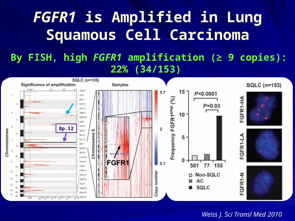
FGFR1 is Amplified in Lung Squamous Cell Carcinoma
Weiss J. Sci Transl Med 2010
8p.12
By FISH, high FGFR1 amplification (≥ 9 copies): 22% (34/153)
FGFR1 amplifications are associated with FGFR inhibitor activity
Weiss J. Sci Transl Med 2010
To investigate the frequency and the prognostic impact of FGFR1 amplification in surgically resected lung SqCC
To evaluate the association between smoking does and FGFR1-amplification
Study Purpose
Patient and Method
SqCC patients that underwent radical resection of a primary lung cancer at Severance Hospital between 1998 and 2009.
Selection criteria (n= 262): availability of tumor tis-sue from the primary lung cancer, smoking-data, and survival data
Construct a tissue-microarray with 2-mm diameter cores (3 cores per tumor)
FGFR1 FISH assay was performed on the tissue-mi-croarrays using FGFR1-probe that hybridizes to the band 8p12-8p11.23 with Spectrum Orange (red) and CEP 8 with Spectrum Green (Abbott Molecular®)
Prespecified Criteria1
“high-amplification” FGFR1 copy number ≥ 9 “low-amplification” FGFR1 copy number >2 or <9 “disomy” FGFR1 copy number = 2
Gene Copy Number
1Weiss J et al. Sci Transl Med 2010
FGFR1 protein & mRNA Expression
IHC analysis was performed using FGFR1 Ab (Epitomics, Burlingame, CA) Only clear membranous staining of the tumor cells was
considered positive and cytoplasmic or granular staining was considered negative or trace
Scoring system (0-400): % of positive tumor cells (0% to 100%) X dominant staining pattern (1: negative or trace, 2: weak, 3: moderate, 4: intense)
mRNA expression analysis was performed by QuantiGene Reagent Systems in FFPE tissue samples
Patient characteristics according to FGFR1 gene amplification by FISH
FGFR1 IHC staining & Gene Copy Number by FISH
34 (13%) 105 (40.1%) 123 (46.9%)
Association between FGFR1 GCN and FGFR1 protein & mRNA Expression
FGFR1 Amplification Is Associated with Poor Sur-vival in Resected Lung SqCC Patients