Page 1
1
PowerPoint® Lecture Slides prepared by Leslie Hendon University of Alabama, Birmingham
C H A P T E R
Copyright © 2011 Pearson Education, Inc.
5 The Integumentary System
Copyright © 2011 Pearson Education, Inc.
The Skin and the Hypodermis
• Skin—our largest organ • Accounts for 7% of body weight • Varies in thickness from 1.5–4.4mm • Divided into two distinct layers • Epidermis • Dermis
• Hypodermis—lies deep to the dermis • Composed of areolar and adipose tissues • Not part of the integumentary system, but
shares some of skin’s properties
Copyright © 2011 Pearson Education, Inc.
Skin Structure
Figure 5.1
Epidermis
Hair shaft
Dermis Reticular layer
Papillary layer
Hypodermis (superficial fascia)
Dermal papillae
Pore
Subpapillary vascular plexus
Appendages of skin Eccrine sweat gland Arrector pili muscle Sebaceous (oil) gland Hair follicle Hair root
Nervous structures Sensory nerve fiber Lamellar (Pacinian) corpuscle Hair follicle receptor (root hair plexus)
Dermal vascular plexus
Adipose tissue
Copyright © 2011 Pearson Education, Inc.
The Skin and Hypodermis
• Functions 1. Protection—cushions organs and protects from
bumps, chemicals, water loss, UV radiation 2. Regulation of body temperature---Capillary
network and sweat glands regulate heat loss 3. Excretion—urea, salts, and water lost through
sweat 4. Production of vitamin D---Epidermal cells use
UV radiation to synthesize vitamin D 5. Sensory reception—Contains sense organs
associated with nerve endings
Copyright © 2011 Pearson Education, Inc.
Figure 5.2 Gross structure of skin and underlying tissues.
Deep fascia
Muscle
Hypodermis
Dermis
Epidermis
Copyright © 2011 Pearson Education, Inc.
Epidermis • Is composed of keratinized stratified squamous
epithelium • Contains four main cell types
• Keratinocytes • Location—stratum spinosum; produce keratin a fibrous
protein • Melanocytes
• Location—basal layer; manufacture and secrete the pigment melanin
• Tactile epithelial cells (Merkel cells) • Location—basal layer; attached to sensory nerve endings
• Dendritic cells (Langerhans cells) • Location—stratum spinosum; part of immune system;
macrophage-like
Page 2
2
Copyright © 2011 Pearson Education, Inc.
Epidermis
• Keratinocytes—most abundant cell type in epidermis • Arise from deepest layer of epidermis • Produce keratin, a tough fibrous protein • Produce antibodies and enzymes • Keratinocytes are dead at skin's surface
Copyright © 2011 Pearson Education, Inc.
Layers of the Epidermis
• Stratum basale (stratum geminativum) • Stratum spinosum • Stratum granulosum • Stratum lucidum (only in thick skin) • Stratum corneum
Copyright © 2011 Pearson Education, Inc.
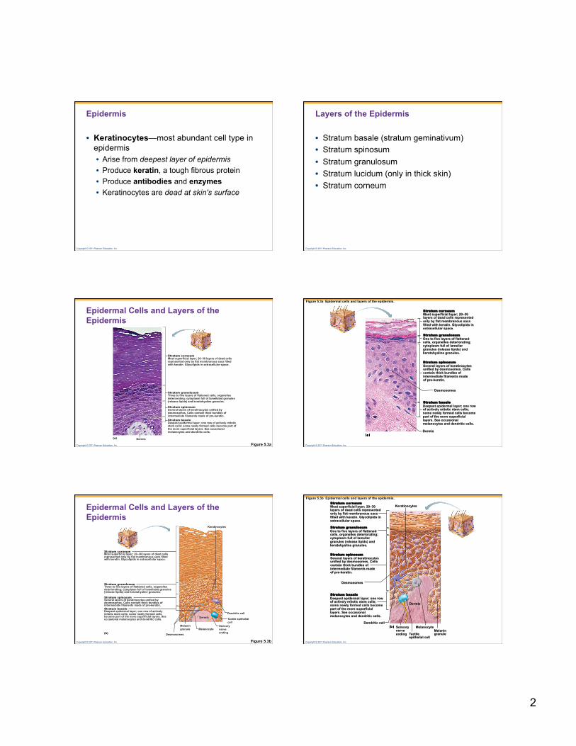
Epidermal Cells and Layers of the Epidermis
Figure 5.3a Dermis (a)
Stratum corneum Most superficial layer; 20–30 layers of dead cells represented only by flat membranous sacs filled with keratin. Glycolipids in extracellular space.
Stratum granulosum Three to five layers of flattened cells, organelles deteriorating; cytoplasm full of lamellated granules (release lipids) and keratohyaline granules.
Stratum spinosum Several layers of keratinocytes unified by desmosomes. Cells contain thick bundles of intermediate filaments made of pre-keratin. Stratum basale Deepest epidermal layer; one row of actively mitotic stem cells; some newly formed cells become part of the more superficial layers. See occasional melanocytes and dendritic cells.
Copyright © 2011 Pearson Education, Inc.
Figure 5.3a Epidermal cells and layers of the epidermis.
Stratum corneum
Stratum granulosum
Stratum spinosum
Stratum basale
Dermis
Desmosomes
Most superficial layer; 20–30 layers of dead cells represented only by flat membranous sacs filled with keratin. Glycolipids in extracellular space.
One to five layers of flattened cells, organelles deteriorating; cytoplasm full of lamellar granules (release lipids) and keratohyaline granules.
Several layers of keratinocytes unified by desmosomes. Cells contain thick bundles of intermediate filaments made of pre-keratin.
Deepest epidermal layer; one row of actively mitotic stem cells; some newly formed cells become part of the more superficial layers. See occasional melanocytes and dendritic cells.
Copyright © 2011 Pearson Education, Inc.
Epidermal Cells and Layers of the Epidermis
Figure 5.3b
Melanocyte Melanin granule
Tactile epithelial cell
Sensory nerve ending
Dendritic cell Dermis
Keratinocytes
Desmosomes (b)
Stratum corneum Most superficial layer; 20–30 layers of dead cells represented only by flat membranous sacs filled with keratin. Glycolipids in extracellular space.
Stratum granulosum Three to five layers of flattened cells, organelles deteriorating; cytoplasm full of lamellated granules (release lipids) and keratohyaline granules.
Stratum spinosum Several layers of keratinocytes unified by desmosomes. Cells contain thick bundles of intermediate filaments made of pre-keratin. Stratum basale Deepest epidermal layer; one row of actively mitotic stem cells; some newly formed cells become part of the more superficial layers. See occasional melanocytes and dendritic cells.
Copyright © 2011 Pearson Education, Inc.
Figure 5.3b Epidermal cells and layers of the epidermis. Stratum corneum
Stratum granulosum
Stratum spinosum
Stratum basale
Desmosomes
Most superficial layer; 20–30 layers of dead cells represented only by flat membranous sacs filled with keratin. Glycolipids in extracellular space.
One to five layers of flattened cells, organelles deteriorating; cytoplasm full of lamellar granules (release lipids) and keratohyaline granules.
Several layers of keratinocytes unified by desmosomes. Cells contain thick bundles of intermediate filaments made of pre-keratin.
Deepest epidermal layer; one row of actively mitotic stem cells; some newly formed cells become part of the more superficial layers. See occasional melanocytes and dendritic cells.
Keratinocytes
Dermis
Dendritic cell Sensory nerve ending
Melanocyte
Tactile epithelial cell
Melanin granule
Page 3
3
Copyright © 2011 Pearson Education, Inc.
Layers of the Epidermis • Stratum basale
• Deepest layer of epidermis • Attached to underlying dermis • Cells actively divide • Stratum basale contains
• Tactile epith. Cells/Merkel cells—associated with sensory nerve ending
• Melanocytes—secrete the pigment melanin • Stratum spinosum (spiny layer)
• “Spiny” appearance caused by: • Artifacts of histological preparation
• Contains thick bundles of intermediate filaments (tonofilaments) • Resist tension • Contain protein prekeratin
• Contains star-shaped dendritic cells • A type of macrophage • Function in immune system
Copyright © 2011 Pearson Education, Inc.
Layers of the Epidermis • Stratum granulosum
• Consists of keratinocytes and tonofilaments • Tonofilaments contain:
• Keratohyaline granules—help form keratin • Lamellated granules—contain a waterproofing glycolipid
• Stratum lucidum (clear layer) • Occurs only in thick skin
• Locations of thick skin—palms and soles • Composed of a few rows of flat, dead keratinocytes
• Stratum corneum (horny layer) • Thick layer of dead keratinocytes and thickened plasma
membranes • Protects skin against abrasion and penetration
Copyright © 2011 Pearson Education, Inc.
Figure 5.4 Thick skin.
Stratum corneum
Epidermis
Dermis
Stratum lucidum Stratum granulosum
Stratum spinosum
Stratum basale
Dermal papilla
Collagen fibers
Papillary dermis
Reticular dermis
Copyright © 2011 Pearson Education, Inc.
Dermis
• Second major layer of the skin • Strong, flexible connective tissue • Richly supplied with blood vessels (important role in
temperature control) and nerves • Has two layers
• Papillary layer—includes dermal papillae • Reticular layer
• Deeper layer—80% of thickness of dermis • Flexure lines
• Creases on palms
Copyright © 2011 Pearson Education, Inc.
The Two Regions of the Dermis
Figure 5.5
Dermis
(a) Light micrograph of thick skin identifying the extent of the dermis, (100×)
(b) Papillary layer of dermis, SEM (570×)
(c) Reticular layer of dermis, SEM (430×) Copyright © 2011 Pearson Education, Inc.
Friction ridges
(a) Friction ridges of finger tip (SEM 20×)
(b) Cleavage lines in the reticular dermis
(c) Flexure lines of the hand
Openings of sweat gland ducts
Flexion creases on digit Flexion creases on the palm
Dermal Modifications
Figure 5.6
Page 4
4
Copyright © 2011 Pearson Education, Inc.
Hypodermis
• Deep to the skin—also called superficial fascia or subcutaneous layer
• Contains areolar and adipose CT • Anchors skin to underlying structures • Helps insulate the body • Has different distribution in males and
females
Copyright © 2011 Pearson Education, Inc.
Skin Color
• Three pigments contribute to skin color • Melanin • Most important pigment—made from
tyrosine • Carotene • Yellowish pigment from carrots and
tomatoes • Hemoglobin • Caucasian skin contains little melanin • Allows crimson color of blood to show
through
Copyright © 2011 Pearson Education, Inc.
Nails
• Nails—scalelike modification of epidermis • Made of hard keratin • Parts of the nail • Free edge • Body • Root • Nail folds • Eponychium—cuticle
Copyright © 2011 Pearson Education, Inc.
Lateral nail fold
Lunule
(a)
Nail matrix
Root of nail Proximal nail fold
Nail bed Phalanx (bone of fingertip)
Eponychium (cuticle)
Body of nail
Free edge of nail
(b)
Structure of a Nail
Figure 5.7
Copyright © 2011 Pearson Education, Inc.
Appendages of the Skin
• Hair • Flexible strand of dead, keratinized cells • Hard keratin—tough and durable • Chief parts of a hair • Root—imbedded in the skin • Shaft—projects above skin's surface
Copyright © 2011 Pearson Education, Inc.
Appendages of the Skin
• Hair has three concentric layers of keratinized cells • Medulla—central core • Cortex—surrounds medulla • Cuticle—outermost layer
• Hair follicles • Extend from epidermis into dermis
• Hair bulb • Deep, expanded end of the hair follicle
• Root plexus • Knot of sensory nerves around hair bulb
Page 5
5
Copyright © 2011 Pearson Education, Inc.
Cross Section of a Hair
Figure 5.8a, b
Hair shaft
Arrector pili Sebaceous gland
Hair root
Hair bulb
(a) Diagram of a cross section of a hair within its follicle
Connective tissue root sheath
Follicle wall
Cuticle
Glassy membrane
Cortex Medulla
Internal epithelial root sheath
External epithelial root sheath
Hair
Hair shaft
Arrector pili Sebaceous gland
Hair root
Hair bulb (b) Photomicrograph of a cross section
of a hair and hair follicle (185×)
Connective tissue root sheath
Follicle wall
Cuticle
Glassy membrane
Cortex Medulla
Internal epithelial root sheath
External epithelial root sheath
Hair
Copyright © 2011 Pearson Education, Inc.
Hair shaft
Arrector pili Sebaceous gland
Hair root
Hair bulb
(c) Diagram of a longitudinal view of the expanded hair bulb of the follicle, which encloses the matrix
Internal epithelial root sheath
External epithelial root sheath
Connective tissue root sheath
Follicle wall
Hair matrix
Melanocyte
Hair papilla
Subcutaneous adipose tissue
Medulla Cortex Cuticle
Glassy membrane
Hair root
Longitudinal Section of Base of Follicle
Figure 5.8c, d
Hair shaft
Arrector pili Sebaceous gland
Hair root
Hair bulb
(d) Photomicrograph of longitudinal view of the hair bulb in the follicle (130×)
Internal epithelial root sheath
External epithelial root sheath
Connective tissue root sheath
Follicle wall
Hair matrix
Hair papilla
Subcutaneous adipose tissue
Medulla Cortex Cuticle
Glassy membrane
Hair root
Copyright © 2011 Pearson Education, Inc.
Appendages of the Skin
• Arrector pili muscle • Bundle of smooth muscle • Hair stands erect when arrector pili contracts
• Vellus hairs • Body hairs of women and children
• Terminal hairs • Hair of scalp • Axillary and pubic area (at puberty)
• Hair thinning and baldness • Due to aging • Male pattern baldness
Copyright © 2011 Pearson Education, Inc.
Sebaceous Glands
• Occur over entire body • Except palms and soles
• Secrete sebum—an oily substance • Simple alveolar glands • Holocrine secretion—entire cell breaks up to
form secretion • Most are associated with a hair follicle
• Functions of sebum • Collects dirt; softens and lubricates hair and
skin
Copyright © 2011 Pearson Education, Inc.
(a) Photomicrograph of a sectioned sebaceous gland (140×)
Sebaceous gland duct
Hair in hair follicle
Secretory cells
Dermal connective tissue
Sebaceous gland
Sweat pore
Eccrine gland
Sebaceous Glands
Figure 5.9a Copyright © 2011 Pearson Education, Inc.
Figure 5.8a Skin glands.
Photomicrograph of a sectioned sebaceous gland (90×)
Dermal connective tissue
Sebaceous gland duct
Hair in hair follicle
Secretory cells
Sebaceous gland
Eccrine gland
Sweat pore
Page 6
6
Copyright © 2011 Pearson Education, Inc.
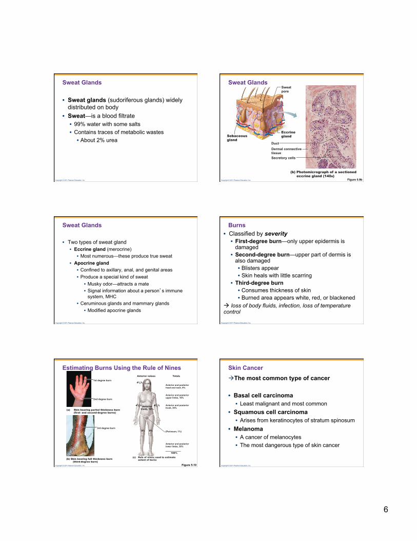
Sweat Glands
• Sweat glands (sudoriferous glands) widely distributed on body
• Sweat—is a blood filtrate • 99% water with some salts • Contains traces of metabolic wastes • About 2% urea
Copyright © 2011 Pearson Education, Inc.
(b) Photomicrograph of a sectioned eccrine gland (140×)
Secretory cells
Dermal connective tissue
Duct
Sebaceous gland
Sweat pore
Eccrine gland
Sweat Glands
Figure 5.9b
Copyright © 2011 Pearson Education, Inc.
Sweat Glands
• Two types of sweat gland • Eccrine gland (merocrine)
• Most numerous—these produce true sweat • Apocrine gland
• Confined to axillary, anal, and genital areas • Produce a special kind of sweat
• Musky odor—attracts a mate • Signal information about a person’s immune
system, MHC • Ceruminous glands and mammary glands
• Modified apocrine glands
Copyright © 2011 Pearson Education, Inc.
Burns • Classified by severity • First-degree burn—only upper epidermis is
damaged • Second-degree burn—upper part of dermis is
also damaged • Blisters appear • Skin heals with little scarring
• Third-degree burn • Consumes thickness of skin • Burned area appears white, red, or blackened
loss of body fluids, infection, loss of temperature control
Copyright © 2011 Pearson Education, Inc.
(b) Skin bearing full thickness burn (third-degree burn)
3rd degree burn
Estimating Burns Using the Rule of Nines
Figure 5.10
(a) Skin bearing partial thickness burn (first- and second-degree burns)
1st degree burn
2nd degree burn
Anterior and posterior head and neck, 9%
41⁄2% 41⁄2%
Anterior and posterior upper limbs, 18%
Anterior and posterior lower limbs, 36%
100%
(c) Rule of nines; used to estimate extent of burns
Totals Anterior values
Anterior and posterior trunk, 36% Anterior
trunk, 18%
9% 9% (Perineum, 1%)
41⁄2%
Copyright © 2011 Pearson Education, Inc.
Skin Cancer The most common type of cancer
• Basal cell carcinoma • Least malignant and most common
• Squamous cell carcinoma • Arises from keratinocytes of stratum spinosum
• Melanoma • A cancer of melanocytes • The most dangerous type of skin cancer
Page 7
7
Copyright © 2011 Pearson Education, Inc.
Skin Cancer
Figure 5.11 Copyright © 2011 Pearson Education, Inc.
The Skin Throughout Life
• Middle to old age • Skin thins and becomes less elastic • Shows harmful effects of environmental
damage • Skin inflammations become more common