Biosci. Rep. (2013) / 33 / art:e00016 / doi 10.1042/BSR20120105 C-terminal low-complexity sequence repeats of Mycobacterium smegmatis Ku modulate DNA binding Ambuj K. KUSHWAHA and Anne GROVE 1 Department of Biological Sciences, Louisiana State University, Baton Rouge, LA 70803, U.S.A. Synopsis Ku protein is an integral component of the NHEJ (non-homologous end-joining) pathway of DSB (double-strand break) repair. Both eukaryotic and prokaryotic Ku homologues have been characterized and shown to bind DNA ends. A unique feature of Mycobacterium smegmatis Ku is its basic C-terminal tail that contains several lysine-rich low-complexity PAKKA repeats that are absent from homologues encoded by obligate parasitic mycobacteria. Such PAKKA repeats are also characteristic of mycobacterial Hlp (histone-like protein) for which they have been shown to confer the ability to appose DNA ends. Unexpectedly, removal of the lysine-rich extension enhances DNA-binding affinity, but an interaction between DNA and the PAKKA repeats is indicated by the observation that only full-length Ku forms multiple complexes with a short stem-loop-containing DNA previously designed to accommodate only one Ku dimer. The C- terminal extension promotes DNA end-joining by T4 DNA ligase, suggesting that the PAKKA repeats also contribute to efficient end-joining. We suggest that low-complexity lysine-rich sequences have evolved repeatedly to modulate the function of unrelated DNA-binding proteins. Key words: DNA binding, electrophoretic mobility-shift assay, Ku protein, low-complexity repeats, non-homologous end-joining (NHEJ) Cite this article as: Kushwaha, A.K. and Grove, A. (2013) C-terminal low-complexity sequence repeats of Mycobacterium smegmatis Ku-modulate DNA binding. Biosci. Rep. 33(1), art:e00016.doi:10.1042/BSR20120105 INTRODUCTION Many proteins in both prokaryotes and eukaryotes have been identified that contain stretches of simple amino acid sequence repeats that have low information content due to biased amino acid composition and a lack of amino acid diversity. These seg- ments are referred to as LCRs (low-complexity regions). These sequences can either be homopolymers or they can be composed of a few different amino acids, often classified as intrinsically disordered regions [1]. Within a protein, these LCRs have been found to evolve more rapidly than flanking sequences such that their length and amino acid content may differ widely between homologues encoded by different species. These sequences are also characterized by a lack of identifiable three-dimensional structure and are therefore underrepresented in the protein data bank [2]. Because of compositional plasticity and lack of three- dimensional structure, the functional role of low-complexity se- ............................................................................................................................................................................................................................................................................................................ Abbreviations used: DSB, double-strand break; EMSA, electrophoretic mobility-shift assay; Hlp, histone-like protein; LCR, low-complexity regions; LigD, ligase D; NHEJ, non-homologous end-joining; PNK, polynucleotide kinase; TBE, Tris/borate/EDTA; TKu, truncated Ku. 1 To whom correspondence should be addressed (email [email protected]). quences is not properly understood. However, studies have sug- gested that position (terminal or central) of the LCRs within a protein sequence plays an important role in determining their function. Proteins with terminal LCRs are important in stress responses, translation and transport processes, and those with central LCRs have been implicated in transcription [3]. In vitro characterization of DNA-binding proteins that con- tain LCRs at either their N- or C-termini has shown that the LCRs modulate functional properties. For example, Deinococ- cus radiodurans HU contains proline-, alanine- and lysine-rich PAKKA repeats at its N-terminus that affect the binding-site size and mode of binding to four-way junction DNA [4,5]. Similar PAKKA repeats are present at the C-termini of HU homologues encoded by some members of the actinomycetes and by a mem- ber of the genus Kineococcus. In mycobacteria, the HU homo- logues, also referred to as Hlps (histone-like proteins), contain a particularly extensive C-terminal tail composed of the repeated PAKKA units. In vitro Mycobacterium smegmatis Hlp promotes c 2013 The Author(s) This is an Open Access article distributed under the terms of the Creative Commons Attribution Non-Commercial Licence (http://creativecommons.org/licenses/ by-nc/2.5/) which permits unrestricted non-commercial use, distribution and reproduction in any medium, provided the original work is properly cited 175
Transcript
Biosci. Rep. (2013) / 33 / art:e00016 / doi 10.1042/BSR20120105
C-terminal low-complexity sequence repeats ofMycobacterium smegmatis Ku modulate DNAbindingAmbuj K. KUSHWAHA and Anne GROVE1
Department of Biological Sciences, Louisiana State University, Baton Rouge, LA 70803, U.S.A.
SynopsisKu protein is an integral component of the NHEJ (non-homologous end-joining) pathway of DSB (double-strand break)repair. Both eukaryotic and prokaryotic Ku homologues have been characterized and shown to bind DNA ends. A uniquefeature of Mycobacterium smegmatis Ku is its basic C-terminal tail that contains several lysine-rich low-complexityPAKKA repeats that are absent from homologues encoded by obligate parasitic mycobacteria. Such PAKKA repeatsare also characteristic of mycobacterial Hlp (histone-like protein) for which they have been shown to confer theability to appose DNA ends. Unexpectedly, removal of the lysine-rich extension enhances DNA-binding affinity, but aninteraction between DNA and the PAKKA repeats is indicated by the observation that only full-length Ku forms multiplecomplexes with a short stem-loop-containing DNA previously designed to accommodate only one Ku dimer. The C-terminal extension promotes DNA end-joining by T4 DNA ligase, suggesting that the PAKKA repeats also contribute toefficient end-joining. We suggest that low-complexity lysine-rich sequences have evolved repeatedly to modulate thefunction of unrelated DNA-binding proteins.
Key words: DNA binding, electrophoretic mobility-shift assay, Ku protein, low-complexity repeats, non-homologousend-joining (NHEJ)
Cite this article as: Kushwaha, A.K. and Grove, A. (2013) C-terminal low-complexity sequence repeats of Mycobacterium smegmatisKu-modulate DNA binding. Biosci. Rep. 33(1), art:e00016.doi:10.1042/BSR20120105
INTRODUCTION
Many proteins in both prokaryotes and eukaryotes have beenidentified that contain stretches of simple amino acid sequencerepeats that have low information content due to biased aminoacid composition and a lack of amino acid diversity. These seg-ments are referred to as LCRs (low-complexity regions). Thesesequences can either be homopolymers or they can be composedof a few different amino acids, often classified as intrinsicallydisordered regions [1]. Within a protein, these LCRs have beenfound to evolve more rapidly than flanking sequences such thattheir length and amino acid content may differ widely betweenhomologues encoded by different species. These sequences arealso characterized by a lack of identifiable three-dimensionalstructure and are therefore underrepresented in the protein databank [2]. Because of compositional plasticity and lack of three-dimensional structure, the functional role of low-complexity se-
quences is not properly understood. However, studies have sug-gested that position (terminal or central) of the LCRs within aprotein sequence plays an important role in determining theirfunction. Proteins with terminal LCRs are important in stressresponses, translation and transport processes, and those withcentral LCRs have been implicated in transcription [3].
In vitro characterization of DNA-binding proteins that con-tain LCRs at either their N- or C-termini has shown that theLCRs modulate functional properties. For example, Deinococ-cus radiodurans HU contains proline-, alanine- and lysine-richPAKKA repeats at its N-terminus that affect the binding-site sizeand mode of binding to four-way junction DNA [4,5]. SimilarPAKKA repeats are present at the C-termini of HU homologuesencoded by some members of the actinomycetes and by a mem-ber of the genus Kineococcus. In mycobacteria, the HU homo-logues, also referred to as Hlps (histone-like proteins), contain aparticularly extensive C-terminal tail composed of the repeatedPAKKA units. In vitro Mycobacterium smegmatis Hlp promotes
Figure 1 Ku protein from M. smegmatis(A) Sequence alignment of the C-terminal protein sequence of various mycobacterial Ku homologues. Low complexityPAKKA repeats of M. smegmatis Ku protein are underlined in red. M_smg, M. smegmatis; M_gil, M. gilvum; M_JLS,Mycobacterium sp. JLS; M_KMS, Mycobacterium sp. KMS; M_bov, M. bovis; M_tub, M. tuberculosis; M_avi, M. avium;M_int, M. intracellulare; M_kan, M. kansasii; M_mar, M. marinum; M_ulc, M. ulcerans. The site of truncation is markedwith an arrow. (B) Schematic representation of the N-terminal domain (solid box) and the lysine rich C-terminal tail.Mutant protein lacking C-terminal PAKKA repeats is TKu. (C) Gel filtration analysis of Ku and TKu. Linear calibration curverepresents the logarithm of molecular mass as a function of elution volume. The elution volumes of dimeric Ku and TKuare indicated by arrows. Inset: Coomassie Brilliant Blue-stained SDS/PAGE gel (12 % gel) showing purified proteins. Lane1, molecular-mass markers in kDa; lanes 2–3, purified TKu and Ku, respectively.
DNA end-joining by T4 DNA ligase and this ability has beenattributed to the lysine-rich C-terminal domain [6]. Streptomycescoelicolor likewise encodes Hlp with PAKKA repeats, and its de-letion was shown to be associated with decreased heat resistanceand an expanded nucleoid [7]. What is particularly intriguingis that these proteins contain LCRs that resemble those foundwithin the C-terminus of eukaryotic histone H1; in the case ofhistone H1, the basic repeat region is important for chromatincondensation [8–11].
Ku protein encoded by M. smegmatis is another example ofa protein with LCRs composed of PAKKA units (Figure 1B).Ku is an important component of the NHEJ (non-homologousend-joining) DSB (double-strand break) repair pathway in euk-aryotes and select prokaryotes such as Bacillus, Mycobacteriumand Pseudomonas [12–18]. Mycobacterium tuberculosis Ku hasbeen speculated to play a major role in repairing DSBs inducedby genotoxic defence of human cells [12,19], and it has been re-
ported that Ku specifically interacts with the polymerase domainof the multifunctional LigD (ligase D) protein to facilitate DSBrepair by NHEJ thereby protecting M. smegmatis against DSBsaccumulating during stationary phase [16,17,20,21].
Eukaryotic Ku proteins are heterodimers consisting of twosubunits Ku70 and Ku80 that together form a functional unit[22]. In contrast, prokaryotic Ku proteins are homodimers andmuch smaller (30–40 kDa), being composed of just the centralcore domain of eukaryotic Ku [17,23]. The β-barrel structure ofthis core domain is conserved despite limited sequence conserva-tion. In vitro analyses of eukaryotic Ku have shown that it bindsnon-specifically to both blunt and cohesive DNA ends. Its bindingaffinity varies from picomolar to nanomolar and is independentof the sequence and the structure of DNA ends, but it is affectedby the length of DNA duplex [24–27]. Stoichiometric measure-ments of eukaryotic Ku have indicated that it requires 14–25 bp ofDNA for binding [25,28,29]. M. tuberculosis Ku also binds DNA
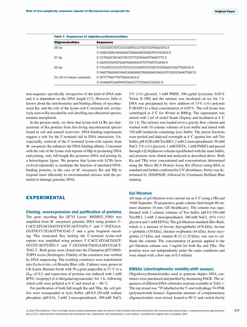
21/34 nt hairpin substrate 5′ -GTTTTTAGTTTATTGGGCGCG-3′
3′ -CAAAAATCAAATAATCGACCCTTTCGACCCGCGC-5′
non-sequence specifically irrespective of the kind of DNA endsand it is dependent on the DNA length [17]. However, little isknown about the stoichiometry and binding affinity of mycobac-terial Ku, and the role of the lysine-rich C-terminal tail, exclus-ively seen in Ku encoded by soil-dwelling mycobacterial species,remains unexplored.
In the present study, we show that lysine-rich LCRs are char-acteristic of Ku proteins from free-living mycobacterial speciesfound in soil and natural reservoirs. DNA-binding experimentssuggest a role for the C-terminal tail in DNA interaction. Un-expectedly, removal of the C-terminal lysine-rich repeats fromM. smegmatis Ku enhances the DNA-binding affinity. Consistentwith the role of the lysine-rich repeats of Hlp in promoting DNAend-joining, only full-length Ku promotes DNA end-joining bya heterologous ligase. We propose that lysine-rich LCRs haveevolved repeatedly to modulate the function of unrelated DNA-binding proteins, in the case of M. smegmatis Ku and Hlp torespond more efficiently to environmental stresses with the po-tential to damage genomic DNA.
EXPERIMENTAL
Cloning, overexpression and purification of proteinsThe gene encoding Ku (JCVI Locus: MSMEG_5580) wasamplified from M. smegmatis genomic DNA using primers 5′-CACCATGACGGGTGCGTCAGTTATG-3′ and 5′-TGCGAA-GGTGCCCTGAGTTACGAC-3′ and a gene fragment encod-ing TKu (truncated Ku) lacking the C-terminal lysine-richrepeats was amplified using primers 5′-CACCATGACGGGT-GCGTCAGTTATG-3′ and 5′-GCGGGCTAGGAATCCGACT-TGG-3′. Both genes were cloned into the Champion pET100/D-TOPO vector (Invitrogen). Fidelity of the constructs was verifiedby DNA sequencing. The resulting constructs were transformedinto Escherichia coli Rosetta Blue cells. Cultures were grown inLB (Luria–Bertani) broth with 50 μg/ml ampicillin at 37 ◦C to aD600 of 0.5, and expression of proteins was induced with 1 mMIPTG (isopropyl-β-D-thiogalactopyranoside) for 1 h, followingwhich cells were pelleted at 4 ◦C and stored at − 80 ◦C.
For purification of both full-length Ku and TKu, the cell pel-lets were resuspended in lysis buffer, pH 8.0 [50 mM sodiumphosphate (pH 8.0), 2 mM 2-mercaptoethanol, 300 mM NaCl,
5 % (v/v) glycerol, 1 mM PMSF, 300 μg/ml lysozyme, 0.05 %Triton X-100] and the mixture was incubated on ice for 1 h.DNA was precipitated by slow addition of 13 % (v/v) polyminP (BASF) to a final concentration of 0.05 %. The cell lysate wascentrifuged at 4 ◦C for 40 min at 8000 g. The supernatant wasmixed with 1 ml of nickel beads (Sigma) and incubated at 4 ◦Cfor 1 h. The mixture was loaded on to a gravity flow column andwashed with 10 column volumes of lysis buffer and eluted with150 mM imidazole-containing lysis buffer. The purest fractionswere pooled and dialysed overnight at 4 ◦C against low salt Trisbuffer, pH 8 [50 mM Tris/HCl, 2 mM 2-mercaptoethanol, 30 mMNaCl, 5 % (v/v) glycerol, 1 mM EDTA, 1 mM PMSF] and passedthrough a Q-Sepharose column equilibrated with the same buffer,and proteins were eluted and analysed as described above. BothKu and TKu were concentrated and concentrations determinedusing the Micro BCA Protein Assay Kit (Pierce) using BSA asstandard and further confirmed by UV absorbance. Purity was de-termined by SDS/PAGE, followed by Coomassie Brilliant Bluestaining.
Gel filtrationAll steps of gel filtration were carried out at 4 ◦C using a HiLoad16/60 Superdex 30 preparative grade column (bed length 60 cm,inner diameter 16 mm; GE Healthcare). The column was equi-librated with 2 column volumes of Tris buffer, pH 8.0 [50 mMTris/HCl, 2 mM 2-mercaptoethanol, 200 mM NaCl, 10 % (v/v)glycerol and 1 mM EDTA]. The gel filtration standard (Bio-Rad),which is a mixture of bovine thyroglobulin (670 kDa), bovineγ -globulin (158 kDa), chicken ovalbumin (44 kDa), horse myo-globin (17 kDa) and vitamin B-12 (1.35 kDa), was run to cal-ibrate the column. The concentration of protein applied to thegel filtration column was 1 mg/ml for both Ku and TKu. Theproteins were run independently under the same conditions andwere eluted with a flow rate of 0.5 ml/min.
EMSAs (electrophoretic mobility-shift assays)Oligodeoxyribonucleotides used to generate duplex DNA con-structs were purchased and purified by denaturing PAGE. The se-quences of different DNA substrates used are available in Table 1.The top strand was 32P-labelled at the 5′-end with phage T4 PNK(polynucleotide kinase). Equimolar amounts of complementaryoligonucleotides were mixed, heated to 90 ◦C and cooled slowly
to room temperature (22 ◦C) to form duplex DNA. The concen-trations of DNA were determined spectrophotometrically.
For binding assays under stoichiometric conditions, 40 or5 nM of 32P-labelled DNA was titrated with Ku or TKu, re-spectively, in a total reaction volume of 10 μl in binding buf-fer [25 mM Tris/HCl (pH 8), 50 mM NaCl, 0.1 mM Na2EDTA,0.05 % Triton X-100, 5 mM DTT (dithiothreitol) and 2 % (v/v)glycerol]. Reactions were incubated at room temperature for 1 h.A non-denaturing 8% polyacrylamide gel was prerun for 30 minat 175 V in 0.5× TBE [Tris/borate/EDTA (1×TBE = 45 mMTris/borate and 1 mM EDTA)] buffer [45 mM Tris borate(pH 8.3), 1 mM Na2EDTA], and samples were loaded withpower on. After electrophoresis, gels were dried, and protein–DNA complexes and free DNA were quantified by phosphor-imaging using software supplied by the manufacturer (ImageQuant 1.1). Percentage complex formation was plotted against[protein]/[DNA]. The stoichiometry of the protein–DNA com-plex was determined by algebraically calculating the value of xat the intersection of the tangents to the linear portions of thegraph. Experiments were performed in triplicate.
EMSAs for affinity determination were performed as de-scribed above, except that binding reactions contained 5 or0.5 nM of 32P-labelled DNA, titrated with Ku or TKu, respect-ively. For TKu, the binding buffer was modified to contain300 mM NaCl, keeping the concentration of other componentsthe same. Percentage complex formation was plotted as a functionof protein concentrations and fitted to the Hill equation:
f = fmax ([Ku]n/K nd )/(1 + ([Ku])n/K dn)
where [Ku] is the protein concentration, f is the fractionalsaturation, Kd reflects the apparent equilibrium dissociation con-stant and n is the Hill coefficient. All bands corresponding toprotein–DNA complexes, including the area between the fast-est migrating complex and the free DNA were considered ascomplex. Fits were performed using the program KaleidaGraph.The Kd value is reported as the mean +− S.D. Experiments wereperformed in triplicate.
End-joining assayPlasmid pUC18 was digested with EcoRI to obtain DNA withcohesive ends. Fifty nanograms of linear pUC18 was incubatedwith Ku or TKu at room temperature for 1 h. To this reaction,1 μl of 40 units/μl of T4 DNA ligase was added and incubatedat room temperature for 1 h. To one of the reactions, 1 μl ofexonuclease III (100 units/μl) was added and incubated at roomtemperature for 1 h. The reactions were terminated by adding 1 μlof stop buffer [5 mM EDTA, 1.1 % (v/v) glycerol and 0.2 mg/mlproteinase K] and 1 μl of 10 % (w/v) SDS. Samples were runon 0.8 % (w/v) TBE agarose gels and visualized by ethidiumbromide staining.
A 105 bp DNA duplex with cohesive ends was generated asdescribed [30]. The 105 bp DNA was labelled with 32P at the 5′-ends using T4 PNK. Five nanomolar 105 bp DNA was incubatedwith Ku or TKu at room temperature for 1 h. An aliquot (1 μl) ofT4 DNA ligase of concentrations 40 units/μl and 80 units/μl was
added to the reaction containing Ku and TKu, respectively, andincubated at room temperature for 1 h. Reactions were treatedwith exonuclease III and terminated as described above, follow-ing which they were phenol extracted and ethanol precipitated andloaded on a prerun 8 % polyacrylamide gel and electrophoresedusing 0.5 % TBE running buffer. Complexes were visualized byphosphorimaging.
RESULTS
M. smegmatis Ku contains a lysine-rich LCR at itsC-terminusSequence alignment of Ku proteins from mycobacterial speciesreveals very significant sequence conservation within the coredomain, but variation at the C-termini. Soil-dwelling mycobac-terial species such as M. smegmatis, M. gilvum, Mycobacteriumsp. JLS, Mycobacterium sp. KMS and other free-living mycobac-terial species such as M. avium, M. ulcerans, M. marinum andM. kansasii, which are found in natural reservoirs, encode Kuhomologues with low-complexity regions characterized by con-served lysine, alanine and proline residues. Strikingly, this LCRis entirely absent in Ku proteins from obligate parasites such asM. tuberculosis and M. bovis (Figure 1A), indicating that only Kuproteins encoded by free-living mycobacterial species inhabitingsoil or natural reservoirs contain these rapidly evolving LCRs.
Annotation of the M. smegmatis genome (JCVI) indicates thatKu consists of 358 amino acids, which includes several PAKKArepeats at the C-terminus (Figure 1A). To determine the role ofthe C-terminal LCR, M. smegmatis Ku and Ku truncated for theC-terminal region (TKu) were purified to apparent homogeneityas judged by Coomassie Brilliant Blue staining of SDS/PAGEgels (Figure 1C, inset); TKu was created by placing a stop codonafter residue 327 (Figures 1A and 1B). Analysis of Ku and TKu bygel filtration chromatography indicated that both proteins existas a homodimer in solution (Figure 1C); this observation wasfurther confirmed by glutaraldehyde cross-linking, which showedno trace of residual monomeric Ku or TKu (results not shown).
DNA binding by Ku and TKuWe expected the lysine-rich LCR to participate in DNA con-tacts based on its charge and the previous observation that sim-ilar repeats in HU and Hlp homologues modulate DNA binding.However, while Ku binds to 37 bp DNA with Kd = 8.6 +− 0.5 nM,TKu binds with much higher affinity (Kd = 4.2 +− 0.7 nM using abuffer with significantly higher ionic strength) (Figures 2A and2B, and Figures 3A and 3B). The Hill coefficients of 1.6 +− 0.1and 1.1 +− 0.1 for Ku and TKu, respectively, suggest modest pos-itive cooperativity of DNA binding for full-length Ku, reflectingpreferred binding of a second Ku protomer to the DNA. Con-sidering that Ku self-associates to bring together DNA ends, thisobservation can be readily reconciled with its normal function.No sequence preference of Ku and TKu is evident, as indicated
Role of low-complexity sequence repeats of Mycobacterium smegmatis Ku
Figure 2 Binding affinity and stoichiometry determination of TKu(A) Titration of TKu with 36 bp DNA in reaction mixture containing 300 mM NaCl and [DNA]<Kd. Lane 1, 36 bp DNA(0.5 nM) only; lanes 2–15, 36 bp DNA titrated with increasing concentrations (0.1–40 nM) of TKu. (B) Binding isotherm forTKu binding to 36 bp DNA. The best fit to the data were obtained using the Hill equation (R2 = 0.9883 and n = 1.1 +− 0.1).Error bars represent S.D. (C) Titration of TKu with 36 bp DNA in a reaction mixture containing 50 mM NaCl and [DNA]>Kd (stoichiometric conditions). Lane 1, 36 bp (5 nM) only; lanes 2–15, 36 bp titrated with increasing concentrations(1–120 nM) of TKu. (D) TKu-36 bp DNA binding stoichiometry plot. Percentage complex plotted against the ratio of TKuand 36 bp DNA concentrations. Gels contained 8 % acrylamide.
by the equivalent affinity for other 36 and 37 bp duplexes (resultsnot shown).
M. smegmatis Ku is a homodimer and removal of the C-terminal extension has no effect on oligomeric assembly (Fig-ure 1C). From EMSA performed under stoichiometric conditions,where proteins were titrated with 37 bp DNA, both TKu and Kuwere found to bind 37 bp DNA at a ratio of 4:1 (Figures 2C and2D, and Figures 3C and 3D), calculated by considering the mo-lecular weight of monomeric protein, which suggests that a dimerrequires ∼18 bp for binding and is consistent with eukaryotic Kuthat has been shown to require 14–25 bp of double-stranded DNAfor binding [25,28,29]. The formation of two discrete complexesis consistent with this interpretation.
As a further test of the duplex length required for optimalKu binding, we used a 21/34 nt hairpin DNA substrate that canaccommodate only one Ku dimer [31]. This DNA, which wasused for Ku–DNA structure determination, was designed to forma 14 bp duplex that is separated from 7 bp of duplex by a shortstem-loop that prevents Ku from sliding along the DNA; the 7 bpduplex is designed to be too short for stable complex formation,thus restricting Ku binding to the 14 bp segment (Figure 4). Abinding assay with this construct showed that TKu forms theexpected single complex, whereas Ku forms two complexes, mostprobably due to an interaction between the C-terminal tail of full-
length Ku with the 7 bp region of the hairpin substrate (Figure 4).That TKu fails to saturate this DNA construct even at 40 nMprotein suggests reduced affinity compared with the 37 bp DNA,perhaps reflecting that 14 bp is insufficient for optimal complexformation.
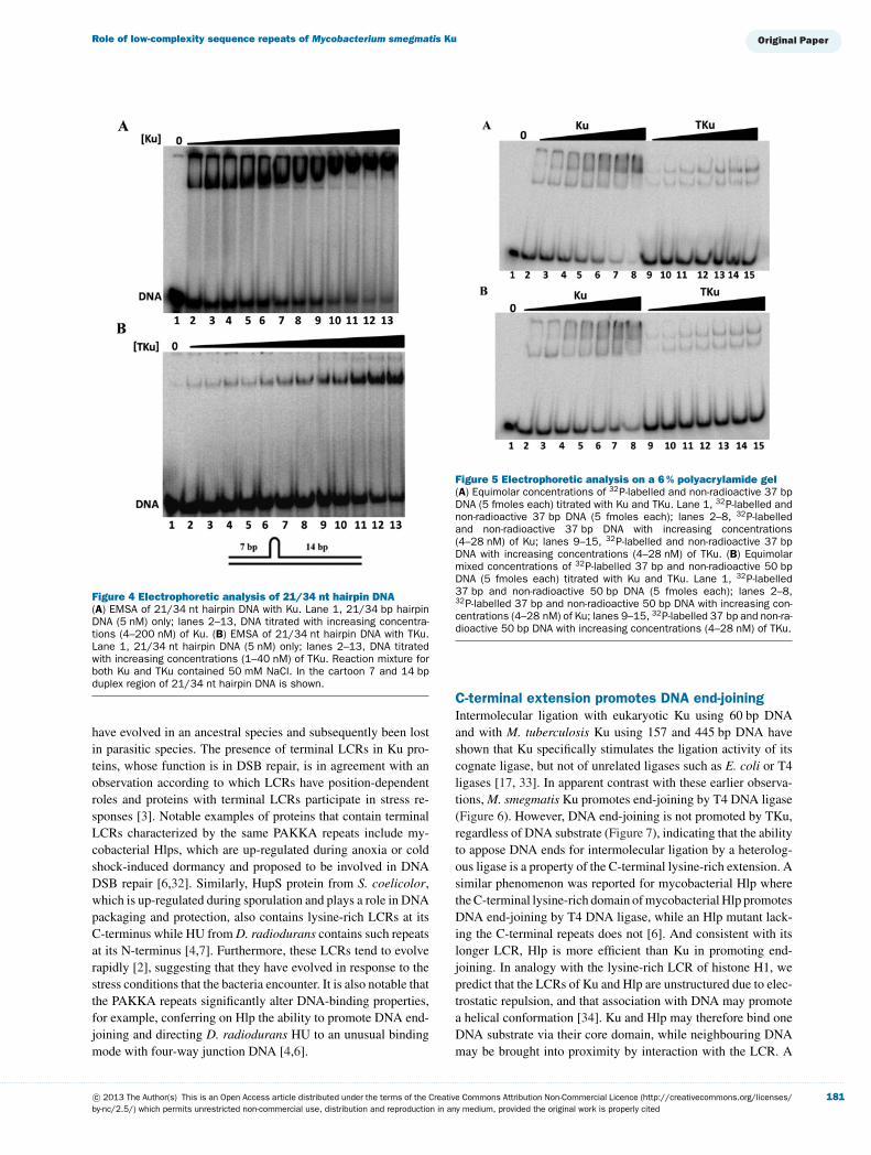
The inference that Ku may bind DNA shorter than 14 bpprompted us to investigate binding to 37 bp DNA using a 6 %polyacrylamide gel, which yields higher resolution. In this gelsystem, TKu still formed two complexes with 37 bp DNA, con-sistent with the estimated site size; however, three complexescould be detected with full-length Ku (Figure 5A). The detec-tion of a third complex is intriguing, and it might be a result ofprotein–protein interactions, leading to two Ku–DNA complexesassociating, or due to interaction of the C-terminal lysine-richtail with the DNA. To examine the presence of protein–proteininteraction, an assay was performed in which equimolar concen-trations of 32P-labelled 37 bp and non-radioactive 50 bp DNAwas mixed and titrated with increasing concentrations of Ku andTKu (Figure 5B) with the idea that the migration of a complexconsisting of two Ku–DNA complexes would be different if one37 bp DNA duplex is replaced with a 50 bp duplex. However,no such change in the mobility was observed, suggesting that ifKu–DNA complexes do associate in solution, such junctions arenot stable during electrophoresis. We therefore surmise that the
Figure 3 Binding affinity and stoichiometry determination of Ku(A) Titration of Ku with 37 bp DNA in a reaction mixture containing 50 mM NaCl and [DNA]<Kd. Lane 1, 37 bp DNA(5 nM) only; lanes 2–15, 37 bp DNA titrated with increasing concentrations (4–800 nM) of Ku. (B) Binding isotherm forKu binding to 37 bp DNA. The best fit to the data were obtained using the Hill equation (R2 = 0.9878 and n = 1.6 +− 0.1).Error bars represent S.D. (C) Titration of Ku with 37 bp DNA in a reaction mixture containing 50 mM NaCl and [DNA]>Kd (stoichiometric conditions). Lane 1, 37 bp (40 nM) only; lanes 2–15, 37 bp titrated with increasing concentrations(40–2800 nM) of Ku. (D) Ku-37 bp DNA binding stoichiometry plot. Percent complex plotted against the ratio of Ku and37 bp DNA concentrations. Gels contained 8 % acrylamide.
additional complex observed when full-length Ku interacts with37 bp DNA is due to interaction of its C-terminal tail with theDNA.
Deletion of the lysine-rich LCR results in loss ofDNA end-joining by T4 ligaseKu participates in NHEJ repair of DNA DSBs. Earlier studieshave reported that M. tuberculosis Ku specifically interacts withand stimulates the ligation activity of LigD protein from M. tuber-culosis and that it inhibits end-joining by T4 ligase, reflecting itspreferred binding to DNA ends [17]. In contrast, end-joiningassays with M. smegmatis Ku using linearized pUC18 or radiola-belled 105 bp DNA substrate showed that M. smegmatis Ku pro-motes end-joining by T4 ligase as can be seen by the appearanceof end-joined products with increasing concentration of Ku (Fig-ure 6). Treatment with exonuclease III digested the end-joinedproducts, which shows that Ku promotes formation of linear mul-timers and not circularization of the DNA (Figure 6A, lane 6). Incontrast to full-length Ku, TKu, at similar and even lower con-centrations, prevented the formation of end-joined products andalso protected DNA from exonucleolytic cleavage, most likely
reflecting its higher affinity binding (Figure 7). Taken together,these data show that while TKu is similar to M. tuberculosis Ku ininhibiting DNA end-joining by a heterologous ligase, full-lengthM. smegmatis Ku promotes such end-joining, implying that thisfeature is a property of the C-terminal extension.
DISCUSSION
LCRs in Ku encoded by free-living mycobacterialspeciesThe multiple sequence alignment of Ku from various mycobac-terial species revealed the presence of lysine-rich LCRs onlyin Ku encoded by soil-dwelling mycobacterial species suchas M. smegmatis, whereas Ku encoded by obligate parasitesincluding M. tuberculosis completely lack these LCRs (Fig-ure 1A, and Supplementary Figure S1 at http://www.bioscirep.org/bsr/033/bsr033e016add.htm). Considering the phylogeneticrelationship between mycobacterial species and the clusteringand reduced genome size of obligate parasites, the LCR may
Role of low-complexity sequence repeats of Mycobacterium smegmatis Ku
Figure 4 Electrophoretic analysis of 21/34 nt hairpin DNA(A) EMSA of 21/34 nt hairpin DNA with Ku. Lane 1, 21/34 bp hairpinDNA (5 nM) only; lanes 2–13, DNA titrated with increasing concentra-tions (4–200 nM) of Ku. (B) EMSA of 21/34 nt hairpin DNA with TKu.Lane 1, 21/34 nt hairpin DNA (5 nM) only; lanes 2–13, DNA titratedwith increasing concentrations (1–40 nM) of TKu. Reaction mixture forboth Ku and TKu contained 50 mM NaCl. In the cartoon 7 and 14 bpduplex region of 21/34 nt hairpin DNA is shown.
have evolved in an ancestral species and subsequently been lostin parasitic species. The presence of terminal LCRs in Ku pro-teins, whose function is in DSB repair, is in agreement with anobservation according to which LCRs have position-dependentroles and proteins with terminal LCRs participate in stress re-sponses [3]. Notable examples of proteins that contain terminalLCRs characterized by the same PAKKA repeats include my-cobacterial Hlps, which are up-regulated during anoxia or coldshock-induced dormancy and proposed to be involved in DNADSB repair [6,32]. Similarly, HupS protein from S. coelicolor,which is up-regulated during sporulation and plays a role in DNApackaging and protection, also contains lysine-rich LCRs at itsC-terminus while HU from D. radiodurans contains such repeatsat its N-terminus [4,7]. Furthermore, these LCRs tend to evolverapidly [2], suggesting that they have evolved in response to thestress conditions that the bacteria encounter. It is also notable thatthe PAKKA repeats significantly alter DNA-binding properties,for example, conferring on Hlp the ability to promote DNA end-joining and directing D. radiodurans HU to an unusual bindingmode with four-way junction DNA [4,6].
Figure 5 Electrophoretic analysis on a 6 % polyacrylamide gel(A) Equimolar concentrations of 32P-labelled and non-radioactive 37 bpDNA (5 fmoles each) titrated with Ku and TKu. Lane 1, 32P-labelled andnon-radioactive 37 bp DNA (5 fmoles each); lanes 2–8, 32P-labelledand non-radioactive 37 bp DNA with increasing concentrations(4–28 nM) of Ku; lanes 9–15, 32P-labelled and non-radioactive 37 bpDNA with increasing concentrations (4–28 nM) of TKu. (B) Equimolarmixed concentrations of 32P-labelled 37 bp and non-radioactive 50 bpDNA (5 fmoles each) titrated with Ku and TKu. Lane 1, 32P-labelled37 bp and non-radioactive 50 bp DNA (5 fmoles each); lanes 2–8,32P-labelled 37 bp and non-radioactive 50 bp DNA with increasing con-centrations (4–28 nM) of Ku; lanes 9–15, 32P-labelled 37 bp and non-ra-dioactive 50 bp DNA with increasing concentrations (4–28 nM) of TKu.
C-terminal extension promotes DNA end-joiningIntermolecular ligation with eukaryotic Ku using 60 bp DNAand with M. tuberculosis Ku using 157 and 445 bp DNA haveshown that Ku specifically stimulates the ligation activity of itscognate ligase, but not of unrelated ligases such as E. coli or T4ligases [17, 33]. In apparent contrast with these earlier observa-tions, M. smegmatis Ku promotes end-joining by T4 DNA ligase(Figure 6). However, DNA end-joining is not promoted by TKu,regardless of DNA substrate (Figure 7), indicating that the abilityto appose DNA ends for intermolecular ligation by a heterolog-ous ligase is a property of the C-terminal lysine-rich extension. Asimilar phenomenon was reported for mycobacterial Hlp wherethe C-terminal lysine-rich domain of mycobacterial Hlp promotesDNA end-joining by T4 DNA ligase, while an Hlp mutant lack-ing the C-terminal repeats does not [6]. And consistent with itslonger LCR, Hlp is more efficient than Ku in promoting end-joining. In analogy with the lysine-rich LCR of histone H1, wepredict that the LCRs of Ku and Hlp are unstructured due to elec-trostatic repulsion, and that association with DNA may promotea helical conformation [34]. Ku and Hlp may therefore bind oneDNA substrate via their core domain, while neighbouring DNAmay be brought into proximity by interaction with the LCR. A
Figure 6 End-joining assay with Ku(A) Lane 1, 5 nM 105 bp DNA only; lane 2, DNA and T4 DNA ligase(40 units/μl); lanes 3–5, DNA, T4 DNA ligase with increasing concen-trations (25, 200, 300, 400 nM) of Ku; lane 6, DNA, T4 DNA ligase,Ku (400 nM) and exonuclease III. The space between lanes 2 and 3reflects that lanes were removed to create the figure. (B) Lane 1, 50 ngof linear pUC18 DNA only; lane 2, DNA and T4 DNA ligase; lanes 3–6,DNA, T4 DNA ligase with increasing concentrations (200, 400, 600,800 nM) of Ku.
recent study by Grob et al. [35] on yeast and human Ku hasshown that Ku has a weak end-bridging activity contributing toend-to-end alignment during DSB repair by NHEJ. Our resultssuggest that the presence of PAKKA repeats in M. smegmatis Kumight enhance this activity. Moreover, the ability to bring distantDNA segments into proximity appears to be a shared feature ofproteins with C-terminal PAKKA-type repeats.
Removal of lysine-rich extension affectsDNA-binding affinityRemoval of the C-terminal lysine-rich repeats enhances the af-finity of Ku for DNA. This increase in affinity is also manifest inthe inability of both T4 DNA ligase and exonuclease III to accessthe TKu-bound DNA ends, which suggests that complexes with
Figure 7 End-joining assay with TKu(A) Lane 1, 5 nM of 105 bp DNA; lane 2, 105 bp DNA and T4 DNAligase (80 units/μl); lanes 3–8, 105 bp DNA, and T4 DNA ligase withincreasing concentrations (200, 400, 600, 800, 1000, 1200 nM) ofTKu; lane 9, 105 bp DNA, T4 DNA ligase, TKu (1200 nM) and exonuc-lease III. Note that a higher concentration of ligase is used comparedwith experiment in Figure 6 to obtain ligation products in absence ofprotein. (B) Lane 1, 50 ng of linear pUC18 DNA only; lane 2, DNA andT4 DNA ligase; lanes 3–7, DNA, T4 DNA ligase with increasing concen-trations (100, 200, 300, 400, 600 nM) of Ku; lane 8, DNA, T4 DNAligase, TKu (600 nM) and exonuclease III.
TKu fail to dissociate appreciably in solution during the time ofincubation. The gain of stable binding to DNA on truncation ofa C-terminal extension has also been reported for Pseudomonasaeruginosa Ku [13], which also contains an extended C-terminaltail, but it lacks PAKKA repeats.
The stoichiometry measurement suggests that the DNA-binding site size for both Ku and TKu is ∼18 bp (Figures 2Cand 2D, and Figures 3C and 3D). Consistent with the calculatedstoichiometry, TKu formed two complexes with 37 bp DNA ona 6 % polyacrylamide gel; in contrast, full length Ku formedthree complexes with 37 bp DNA on the same gel (Figure 5A).Also, binding to the 21/34 nt hairpin substrate, which has 7 and14 bp duplex regions separated by a hairpin structure, showedthat Ku forms two complexes, whereas TKu forms one complexonly with an apparent lower affinity compared with 37 bp DNAas evidenced by the failure to saturate this DNA construct (Fig-ure 4) [36]. For TKu, this suggests that its optimal site size is>14 bp. The differences in the binding properties of full-lengthKu and TKu could potentially be attributed to protein–protein
Role of low-complexity sequence repeats of Mycobacterium smegmatis Ku
interactions between DNA-bound Ku dimers or to the lysine-rich C-terminal LCRs interacting with DNA. Since the patternof complexes seen when Ku is mixed with equimolar concentra-tions of 32P-labelled 37 bp and non-radioactive 37 or 50 bp DNAis identical (Figure 5), we favour the latter interpretation. Interac-tion between the lysine-rich LCR and DNA would be expected torequire only a few base pairs, potentially allowing such interac-tion to occur with the 7 bp duplex region of the 21/34 nt hairpinconstruct or with residual base pairs within the 37 bp DNA notoccupied by Ku binding via its core DNA-binding motif.
In all, the lysine-rich C-terminus of M. smegmatis Ku signi-ficantly modulates DNA-binding properties and promotes DNAend-joining. Evidently, M. smegmatis Ku exhibits propertiesdistinct from those characteristic of M. tuberculosis Ku, prop-erties associated with its unique lysine-rich C-terminus. Low-complexity sequences, such as the PAKKA repeats found in M.smegmatis Hlp and Ku evolve rapidly and we suggest that M.smegmatis Ku has evolved in response to needs to cope withenvironmental stress such as desiccation.
AUTHOR CONTRIBUTION
Ambuj Kushwaha performed all of the experiments. Ambuj Kush-waha and Anne Grove contributed to experimental design and dataanalysis, and to writing the paper.
FUNDING
This work was supported by the National Science Foundation [grantnumbers MCB-0744240 and 1051610 (to A.G.)] and an LSU Eco-nomic Development Assistantship.
REFERENCES
1 Huntley, M. A. and Golding, G. B. (2002) Simple sequences arerare in the protein data bank. Proteins 48, 134–140
2 Haerty, W. and Golding, G. B. (2010) Low-complexity sequencesand single amino acid repeats: not just ‘junk’ peptide sequences.Genome 53, 753–762
3 Coletta, A., Pinney, J. W., Solis, D. Y., Marsh, J., Pettifer, S. R. andAttwood, T. K. (2010) Low-complexity regions within proteinsequences have position-dependent roles. BMC Syst. Biol. 4, 43
4 Ghosh, S. and Grove, A. (2006) The Deinococcusradiodurans-encoded HU protein has two DNA-binding domains.Biochemistry 45, 1723–1733
5 Ghosh, S. and Grove, A. (2004) Histone-like protein HU fromDeinococcus radiodurans binds preferentially to four-way DNAjunctions. J. Mol. Biol. 337, 561–571
6 Mukherjee, A., Bhattacharyya, G. and Grove, A. (2008) TheC-terminal domain of HU-related histone-like protein Hlp fromMycobacterium smegmatis mediates DNA end-joining. Biochemistry47, 8744–8753
7 Salerno, P., Larsson, J., Bucca, G., Laing, E., Smith, C. P. andFlardh, K. (2009) One of the two genes encodingnucleoid-associated HU proteins in Streptomyces coelicolor isdevelopmentally regulated and specifically involved in sporematuration. J. Bacteriol. 191, 6489–6500
8 Grove, A. (2011) Functional evolution of bacterial histone-like HUproteins. Curr. Issues Mol. Biol. 13, 1–12
9 Happel, N. and Doenecke, D. (2009) Histone H1 and its isoforms:contribution to chromatin structure and function. Gene 431, 1–12
10 Ellen, T. P. and van Holde, K. E. (2004) Linker histone interactionshows divalent character with both supercoiled and linear DNA.Biochemistry 43, 7867–7872
11 Bharath, M. M., Chandra, N. R. and Rao, M. R. (2002) Predictionof an HMG-box fold in the C-terminal domain of histone H1:insights into its role in DNA condensation. Proteins 49, 71–81
12 Della, M., Palmbos, P. L., Tseng, H. M., Tonkin, L. M., Daley, J. M.,Topper, L. M., Pitcher, R. S., Tomkinson, A. E., Wilson, T. E. andDoherty, A. J. (2004) Mycobacterial Ku and ligase proteinsconstitute a two-component NHEJ repair machine. Science 306,683–685
13 Zhu, H. and Shuman, S. (2010) Gap filling activities ofPseudomonas DNA ligase D (LigD) polymerase and functionalinteractions of LigD with the DNA end-binding Ku protein. J. Biol.Chem. 285, 4815–4825
14 Kobayashi, H., Simmons, L. A., Yuan, D. S., Broughton, W. J. andWalker, G. C. (2008) Multiple Ku orthologues mediate DNAnon-homologous end-joining in the free-living form and duringchronic infection of Sinorhizobium meliloti. Mol. Microbiol. 67,350–363
15 Wilson, T. E., Topper, L. M. and Palmbos, P. L. (2003)Non-homologous end-joining: bacteria join the chromosomebreakdance. Trends Biochem. Sci. 28, 62–66
16 Aniukwu, J., Glickman, M. S. and Shuman, S. (2008) The pathwaysand outcomes of mycobacterial NHEJ depend on the structure ofthe broken DNA ends. Genes Dev. 22, 512–527
17 Weller, G. R., Kysela, B., Roy, R., Tonkin, L. M., Scanlan, E., Della,M., Devine, S. K., Day, J. P., Wilkinson, A., d’Adda di Fagagna, F.et al. (2002) Identification of a DNA nonhomologous end-joiningcomplex in bacteria. Science 297, 1686–1689
18 Wright, D., DeBeaux, A., Shi, R., Doherty, A. J. and Harrison, L.(2010) Characterization of the roles of the catalytic domains ofMycobacterium tuberculosis ligase D in Ku-dependent error-proneDNA end joining. Mutagenesis 25, 473–481
19 Gong, C., Bongiorno, P., Martins, A., Stephanou, N. C., Zhu, H.,Shuman, S. and Glickman, M. S. (2005) Mechanism ofnonhomologous end-joining in mycobacteria: a low-fidelity repairsystem driven by Ku, ligase D and ligase C. Nat. Struct. Mol. Biol.12, 304–312
20 Pitcher, R. S., Green, A. J., Brzostek, A., Korycka-Machala, M.,Dziadek, J. and Doherty, A. J. (2007) NHEJ protects mycobacteriain stationary phase against the harmful effects of desiccation.DNA Repair 6, 1271–1276
21 Pitcher, R. S., Tonkin, L. M., Green, A. J. and Doherty, A. J. (2005)Domain structure of a NHEJ DNA repair ligase from Mycobacteriumtuberculosis. J. Mol. Biol. 351, 531–544
22 Downs, J. A. and Jackson, S. P. (2004) A means to a DNA end: themany roles of Ku. Nat. Rev. Mol. Cell. Biol. 5, 367–378
23 Aravind, L. and Koonin, E. V. (2001) Prokaryotic homologs of theeukaryotic DNA-end-binding protein Ku, novel domains in the Kuprotein and prediction of a prokaryotic double-strand break repairsystem. Genome Res. 11, 1365–1374
24 Blier, P. R., Griffith, A. J., Craft, J. and Hardin, J. A. (1993) Bindingof Ku protein to DNA. Measurement of affinity for ends anddemonstration of binding to nicks. J. Biol. Chem. 268, 7594–7601
25 Arosio, D., Cui, S., Ortega, C., Chovanec, M., Di Marco, S., Baldini,G., Falaschi, A. and Vindigni, A. (2002) Studies on the mode of Kuinteraction with DNA. J. Biol. Chem. 277, 9741–9748
26 Tuteja, N., Tuteja, R., Ochem, A., Taneja, P., Huang, N. W.,Simoncsits, A., Susic, S., Rahman, K., Marusic, L., Chen, J. et al.(1994) Human DNA helicase II: a novel DNA unwinding enzymeidentified as the Ku autoantigen. EMBO J. 13, 4991–5001
27 West, R. B., Yaneva, M. and Lieber, M. R. (1998) Productive andnonproductive complexes of Ku and DNA-dependent protein kinaseat DNA termini. Mol. Cell Biol. 18, 5908–5920
28 Yaneva, M., Kowalewski, T. and Lieber, M. R. (1997) Interaction ofDNA-dependent protein kinase with DNA and with Ku: biochemicaland atomic-force microscopy studies. EMBO J. 16, 5098–5112
29 Falzon, M., Fewell, J. W. and Kuff, E. L. (1993) EBP-80, atranscription factor closely resembling the human autoantigen Ku,recognizes single- to double-strand transitions in DNA. J. Biol.Chem. 268, 10546–10552
30 Ray, S. and Grove, A. (2009) The yeast high mobility group proteinHMO2, a subunit of the chromatin-remodeling complex INO80,binds DNA ends. Nucleic Acids Res. 37, 6389–6399
31 Walker, J. R., Corpina, R. A. and Goldberg, J. (2001) Structure ofthe Ku heterodimer bound to DNA and its implications fordouble-strand break repair. Nature 412, 607–614
32 Shires, K. and Steyn, L. (2001) The cold-shock stress response inMycobacterium smegmatis induces the expression of a histone-likeprotein. Mol. Microbiol. 39, 994–1009
33 Ramsden, D. A. and Gellert, M. (1998) Ku protein stimulates DNAend joining by mammalian DNA ligases: a direct role for Ku inrepair of DNA double-strand breaks. EMBO J. 17,609–614
34 Clark, D. J., Hill, C. S., Martin, S. R. and Thomas, J. O. (1988)Alpha-helix in the carboxy-terminal domains of histones H1 andH5. EMBO J. 7, 69–75
35 Grob, P., Zhang, T. T., Hannah, R., Yang, H., Hefferin, M. L.,Tomkinson, A. E. and Nogales, E. (2012) Electron microscopyvisualization of DNA-protein complexes formed by Ku and DNAligase IV. DNA Repair 11, 74–81
36 Andrews, B. J., Lehman, J. A. and Turchi, J. J. (2006) Kineticanalysis of the Ku-DNA binding activity reveals aredox-dependent alteration in protein structure that stimulatesdissociation of the Ku-DNA complex. J. Biol. Chem. 281,13596–13603
Received 23 October 2012/6 November 2012; accepted 20 November 2012
Published as Immediate Publication 20 November 2012, doi 10.1042/BSR20120105
Role of low-complexity sequence repeats of Mycobacterium smegmatis Ku
Figure S1 Sequence alignment of Mycobacterial Ku homologuesLow-complexity PAKKA repeats of M. smegmatis Ku protein are underlined in red. M_smg, M. smegmatis; M_gil, M. gilvum;M_JLS, Mycobacterium Sp. JLS; M_KMS, Mycobacterium sp. KMS; M_bov, M. bovis; M_tub, M. tuberculosis; M_avi, M.avium; M_int, M. intracellulare; M_kan, M. kansasii; M_mar, M. marinum; M_ulc, M. ulcerans.
Received 23 October 2012/6 November 2012; accepted 20 November 2012
Published as Immediate Publication 20 November 2012, doi 10.1042/BSR20120105


![Measure Repeats, Full-Measure In-Accords...151 Chapter 17 Measure Repeats, Full-Measure In-Accords General Introduction to Braille Repeats [T16, 16] One of the most notable differences](https://static.documents.pub/doc/80x56/5f64865f81c6606fce670a47/measure-repeats-full-measure-in-accords-151-chapter-17-measure-repeats-full-measure.jpg)


![Guaracha 2-3 C] (perc.) (piano) B (bass) Track (w/ repeats), then, … · Guaracha 2-3 C] (perc.) (piano) B (bass) Track (w/ repeats), then, D.C. al Coda (w/ repeats) Created Date:](https://static.documents.pub/doc/80x56/6063b007719805122e30aaa9/guaracha-2-3-c-perc-piano-b-bass-track-w-repeats-then-guaracha-2-3.jpg)


