C1-2 Puncture: A safe, efficacious, and potentially underutilized technique Wende Gibbs, MD, Department of Neuroradiology Paul Kim, MD, Department of Neuroradiology John Go, MD, Department of Neuroradiology Meng Law, MD, MBBS, Department of Neuroradiology University of Southern California, Keck School of Medicine Control # 179 eEdE-217
Transcript
C1-2 Puncture: A safe, efficacious, and potentially underutilized techniqueWende Gibbs, MD, Department of NeuroradiologyPaul Kim, MD, Department of NeuroradiologyJohn Go, MD, Department of NeuroradiologyMeng Law, MD, MBBS, Department of Neuroradiology
University of Southern California, Keck School of Medicine
Control # 1792eEdE-217
Disclosures Wende Gibbs: none
Paul Kim: none
John Go: none
Meng Law: Toshiba Grant Speakers Bureau; Bracco speaker and consultant; Guerbet Medical Advisory Board; Prism Stock; Fuji speaker and consultant
Purpose C1-2 puncture for CSF collection or contrast injection is perceived to
be more dangerous and difficult to perform than lumbar puncture, especially amongst younger radiologists who have little exposure to and training in the technique
There are relatively few reports of complications with lasting sequelae, all relating to puncture under fluoroscopic guidance, and most involving injection of metrizamide into the spinal cord
Certain patient populations require the C1-2 approach and it may be safer and more comfortable than the lumbar approach in others
Our purpose is to demystify the C1-2 puncture and justify the assertion that it should be more frequently utilized, for the benefit of patients, and in order to produce new radiologists capable of safely performing this important procedure
Approach
Briefly review the history of C1-2 puncture and current modes of practice
Outline indications and contraindications
Describe the technique using fluoroscopic and CT guidance
Review the literature describing complications of C1-2 puncture and discuss relevant vascular variants
Consider patient populations who require a cervical approach or would benefit from cervical puncture over lumbar approach
Introduction Lateral C1-2 puncture for CSF collection or cervical myelography is a safe
and useful alternative to lumbar puncture
The cervical approach is required in certain patient populations, including those who must remain supine, those with contraindications to lumbar approach, and in patients with myelographic block
Given the necessity of cervical puncture in certain cases, neuroradiology trainees must have adequate exposure and practice to safely and comfortably perform this procedure
While considered a safe procedure, C1-2 puncture is infrequently performed In a 2009 study, Yousem et al. polled neuroradiology program directors and found
that 14.3% (12/85) had not performed a C1-2 puncture for cervical myelography at their institution in the previous year1
47.6% (40/85) performed 1-5 per year 73% (59/81) stated that they trained fellows to perform the procedure Given the few cervical punctures performed, it may be that fellows do not perform
an adequate number to become competent and comfortable with this procedure
Introduction There have been scattered case reports and small series
describing the complications related to this approach: the overall number of complications relative to total cervical punctures performed is small2-7
With the exception of one report of 3 patients8, the literature describing the technique and complications is based on needle insertion under flouroscopic guidance
The increasing use of CT-guidance in radiology procedures has further decreased the risk of C1-2 puncture
As in all radiology procedures, thoughtful preparation, careful attention to technique, and solid knowledge of relevant anatomy and variants minimizes potential complications
C1-2 approach: History
1920: CSF sampled by cisternal puncture9
Neck flexed Needle blindly inserted in the
posterior midline below the occiput
Complications were not uncommon
1968: Neurosurgeons Kelley and Alexander10 described a lateral C1-2 approach for myelography as a safer, more easily performed route to the cervical subarchnoid space They utilized an 18g spinal needle and directed it anteriorly toward the
ventral subarachnoid space at the C1-2 level
1969: Radiologists used lateral C1-2 puncture for “painless gas myelography”, the alternative to Pantopaque myelography9
C1-2 approach: History
Historically, three different approaches have been used Oblique anterior approach with the needle
directed to the anterior 1/3 of the canal, lateral to the cord
Lateral approach with needle directed toward the cord to access the lateral subarachnoid space
Lateral approach with needle directed toward the posterior 1/3 of the canal
The third approach is used today: it is the safest trajectory in the presence of unsuspected variant vertebral artery anatomy (present in 1-2% of individuals3)
Orrison et al. Radiology 198311
C1-2 approach: Relevance The ubiquitous use of MRI to evaluate spinal pathology has markedly
decreased the use of contrast myelography
However, some patients cannot have an MR or will have suboptimal images MR incompatible medical devices Claustrophobia Spinal hardware
In addition, some neurosurgeons prefer myelograms or a combination of MRI and myelogram for surgical planning
Collection of CSF for diagnosis or access for intrathecal chemotherapy injection remains a common procedure performed with imaging guidance
For these reasons, spinal puncture, whether lumbar or cervical, is a necessary skill for the radiologist, and competence in both approaches is required
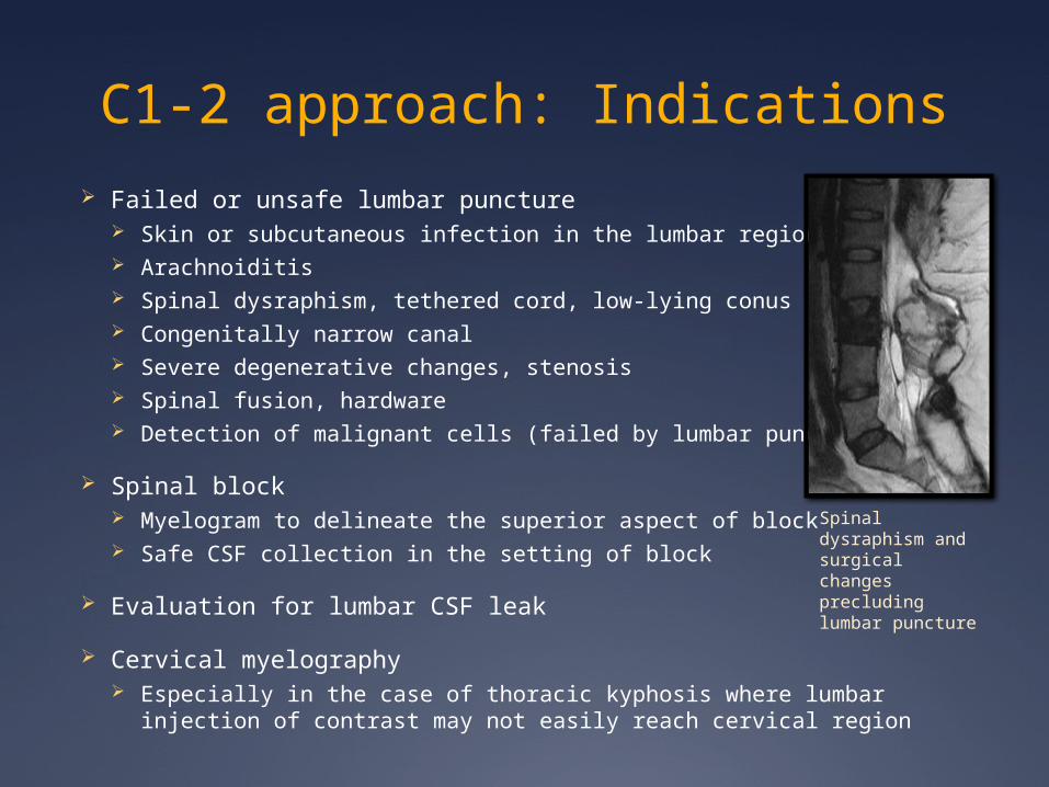
C1-2 approach: Indications Failed or unsafe lumbar puncture
Skin or subcutaneous infection in the lumbar region Arachnoiditis Spinal dysraphism, tethered cord, low-lying conus Congenitally narrow canal Severe degenerative changes, stenosis Spinal fusion, hardware Detection of malignant cells (failed by lumbar puncture)
Spinal block Myelogram to delineate the superior aspect of block Safe CSF collection in the setting of block
Evaluation for lumbar CSF leak
Cervical myelography Especially in the case of thoracic kyphosis where lumbar injection of contrast
may not easily reach cervical region
Spinal dysraphism and surgical changes precluding lumbar puncture
C1-2 Approach: Contraindications
Chiari malformation
Mass at C1-2 level
Acquired or congenital stenosis at C1-2
Ectatic vertebral arteries which extend to posterior 1/3 of canal (1-2%) May proceed cautiously if unilateral
PICA caudal loop extending to C1-2 level or PICA origin below C1 (extremely rare)
Contraindications common to cervical and lumbar puncture Increased intracranial pressure with impending
herniation Coagulopathy, platelets less than 50,000 (relative
contraindication)
C1-2 approach: Technique Prior imaging should be evaluated for Chiari malformation,
mass, narrowing at the C1-2 level, anomalous course of the vertebral arteries or PICA
Whether using fluoroscopic or CT guidance, the posterior 1/3 of the canal at the C1-2 level is the target for needle placement
The patient can be placed in any position (supine, prone, decubitus, oblique)
Fluoroscopic guidance has the advantages of speed and real time visualization of needle progress
CT guidance allows for clear visualization of the target and spinal cord
C1-2 approach: Technique The skin entry site is approximately 1cm posterior and
inferior to the mastoid process
This patient is in prone position on the CT gantry. The needle has been placed under CT guidance. After needle tip location was confirmed with good flow of CSF, tubing was connected for fluid collection.
C1-2 approach: Technique
Sagittal, axial, and coronal CTA images with reference lines at the C1-2 target level and location in the dorsal subarachnoid space. Note the typical location of the vertebral arteries at this level, far from the target. No vessels are seen along the anticipated course of the needle (yellow star and arrows).
Technique
Sagittal and axial T2-W images with reference line drawn at the C1-2 puncture level.
The axial image shows that there is nearly 6mm of dorsal subarachnoid space at the target level.
Technique: Fluoroscopic guidance
The patient is prepared in the same manner as for lumbar puncture with sterilization of the skin and draping
With the patient in a comfortable position, a scout image is obtained A true lateral projection is vital The C1 arch should appear as a single line,
endplates of C2-3 should overlap
Divide the canal into thirds: the needle target is in the posterior 1/3
Steadily advance the needle, removing the stylet to check for CSF as needed
On the AP projection the needle may reach the midline before CSF is obtained
AP
Technique: Fluoroscopic guidance The needle may “tent” the tough cervical dura
before penetration
The needle should be advanced with bevel oriented posteriorly until the dura is punctured, then turned 90 - 180 degrees for ease of CSF collection
If injecting contrast, watch contrast diffuse freely from needle tip If contrast pools, the needle may be subdural
or epidural If linear contrast is seen in the central canal,
consider injection into a syrinx Return the stylet and reposition
Flow of arterial blood from the hub, pain or parestesias in the extremities, or Lhermitte sign indicate that the procedure should be terminated
anterior
posterior
Case: Fluoroscopic guidance
65-year-old man with altered mental status.
The patient had a known history of severe arachnoiditis as well as extensive spinal hardware and bone graft. Cervical puncture was performed for CSF collection.
Note the proper position of the needle in the posterior 1/3 of the canal.
Technique: CT guidance After patient positioning on the CT gantry, a grid is
placed on the neck for puncture site localization, identical to a CT guided biopsy
After marking the desired entry site, the skin is sterilized and the patient is draped
Measuring the distance from skin surface to subarachnoid space target on the initial image can greatly aid in estimation of needle depth, decreasing the number of times CT confirmation or stylet removal must be performed
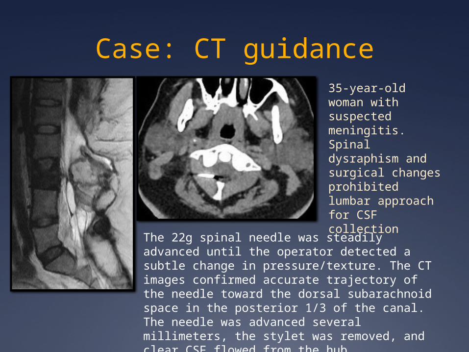
Case: CT guidance35-year-old woman with suspected meningitis. Spinal dysraphism and surgical changes prohibited lumbar approach for CSF collection
The 22g spinal needle was steadily advanced until the operator detected a subtle change in pressure/texture. The CT images confirmed accurate trajectory of the needle toward the dorsal subarachnoid space in the posterior 1/3 of the canal. The needle was advanced several millimeters, the stylet was removed, and clear CSF flowed from the hub.
Case: CT guidance18-year-old woman with history of candida spinal meningitis with extensive adhesions, loculated collections, arachnoiditis, and near complete spinal block.
A lumbar puncture was attempted in order to fill the distal thecal sac, but only trace contrast could be injected amidst the clumped nerve roots. Cervical puncture and contrast injection was performed to assess flow above the block.Note contrast in the
dependent subarachnoid space at the C1-2 level (patient had to be placed in decubitus position)
C1-2 approach: Complications Complications of C1-2 puncture are rare with proper adherence to technique,
solid knowledge of anatomy and relevant variants, and adequate training
Most complications and adverse events related to cervical puncture are identical to those of lumbar puncture Pain Anxiety Bleeding Infection Contrast allergy (in the setting of myelography) Subarachnoid hemorrhage (exceedingly rare) Epidural or subdural hemorrhage (very rare)
Complications unique to the cervical approach2-7 Accidental C0-C1 puncture (fluoroscopic guidance) Spinal cord puncture Spinal cord contrast injection Vascular injury (Variant anatomy of vertebral artery or PICA) Spinal cord hemorrhage (exceedingly rare)
C1-2 approach: Complications Review of the literature reveals case reports and small series
describing complications of C1-2 puncture All cases with complications were performed under fluoroscopic
guidance The majority of cases involved cervical myelography and reported as
complications adverse events related to neck hyperextension Thus, the number of reported complications related to “C1-2 puncture
for cervical myelography” overestimates the risks associated with the C1-2 approach in general
Robertson et al. performed a mail survey of neuroradiologists to determine modes of practice and major complications2
220 respondents reported 68 complications in an estimated total of 187,300 cervical myelograms
2/3 of complications related to patient positioning/hyperextension 1/3 related to the actual C1/2 puncture
1 epidural hematoma 1 recovery after surgical intervention
Modified from Robertson et al. Radiology 1990
C1-2 approach: Complications Katoh et al. reviewed 112 cases of lateral C1-2 puncture for cervical
myelography using fluoroscopic guidance3
In this study, 10 of 112 (9%) of the cases had technical complications 3 cases of cord puncture and injection of contrast 3 cases of inadvertent puncture between the occiput and C1 1 case of epidural venous plexus puncture 1 case of epidural space contrast injection
Of these ten cases, only one patient had neurologic deficits: a case of cord injection with metrizamide resulting in permanent left sided hypoalgesia below the C2 level
The authors conclude that the complications primarily resulted from two technical factors: Positioning of the neck Misdirection of the xray beam
C1-2 approach: Complications Neurologic deficits associated with intramedullary contrast injection
generally occur immediately and typically improve after the procedure
The severity of symptoms appears to be related to volume injected and contrast type: all significant complications were reported after metrizamide injection3,7,12
Simon et al. reported a case of intramedullary contrast injection and reviewed reported cases of this complication7
A total of 26 cases were found Pain during injection was reported in 6 cases (1 patient had no pain, 19 cases
did not report symptoms) Symptoms after the procedure included pain, hallucinations, meningisumus,
quadriparesis, paraparesis, arm weakness Outcomes were reported in 23 of the cases
10 patients had persistant neurologic deficits In 11 cases there was complete resolution of symptoms 2 patients died
C1-2 approach: Complications A small number of hemorrhagic complications are found as case reports in
the literature
Rogers (1983) reported the death of a patient from extensive cervical and intracranial subdural hemorrhage4
In this case, an anomalous vertebral artery was found The patient had received 5 cervical punctures without incident before his death after the
6th puncture
Mapstone (1983) reported a case of extensive multicompartment hemorrhage after cervical puncture of a coagulopathic child with leukemia13
Abla (1986) described a case of delayed subarachnoid hemorrhage 36 hours after C1-2 myelogram6
Emergent surgical decompression was performed The patient had mild long term neurologic sequelae
Aghi (2004) reported a case of subarachnoid hemorrhage, asceptic meningitis, and hydrocephalus after cervical puncture5
Emergent surgical decompression and clot evacuation was performed The patient recovered without neurologic sequelae
C1-2 approach: Relevant Anatomy
Two vascular variants put patients at increased risk of arterial laceration producing hematoma, spasm, thorombosis or rarely, subarchnoid hemorrhage with the cervical approach: Anomalous course of the vertebral artery Low-lying PICA loop or low PICA origin
Extradural origin of PICA, penetrating the dura between C1 and C2 (modified from Fine et al. J Neurosurgery 1999.)16
Typically, the entire course of the PICA is confined to the intracranial compartment
Siclari et al. report two cases of PICA origin at the C2 level14
Tokuda et al. found that 2 of 300 patients studied had PICA origin at C215
These PICAs did not traverse the posterior 1/3 of the canal, thus would not have been along the trajectory of a C1-2 puncture
Relevant Anatomy: PICA
Brinjikji and colleagues studied 346 PICAs in 211 patient cerebral angiograms in order to determine the frequency of PICA origin or descent below the C1 level17
2 of 346 PICAs evaluated (0.6%) extended below the C1 arch, thus at potential risk in the setting of C1-2 puncture
No PICA origins were found below C1
280/346 PICAs (80.9%) were entirely located above the foramen magnum
64/346 PICAs (18.5%) were located below the foramen magnum but above the inferior aspect of the posterior C1 arch
Relevant Anatomy: Vertebral artery
Katoh et al reviewed 164 vertebral artery catheter angiograms in order to determine the incidence of an anomalous course potentially dangerous in the setting of cervical puncture3
In 3/164 cases (1.9%) the vertebral artery passed over the posterior 1/3 of the canal: the target site for cervical puncture
Anterior to the canal 70.6% (113)
Anterior 1/3 of the canal 26% (45)
Mid canal 1.9% (3)
Posterior canal 1.9% (3)
Relevant Anatomy: Vertebral artery
A rare variation of the vertebral artery is partial duplication or fenestration
While nearly non-existent in American and European literature, Kowada et al. identified these anomalies in 1% (22/1685) of angiograms performed on Japanese patients18
Rogers’ report of death from subdural hemorrhage, described earlier, was hypothesized to have resulted from puncture of this very rare variant
Normal course
Vertebral artery duplication
Modified from Rogers, J Neurosurg 19834
C1-2 approach: expanded indications
In addition to indications described previously, these patient populations would benefit from CT guided cervical puncture as a first line approach Benefit from supine position: safety and/or comfort
Intubation Respiratory difficulties Recent abdominal or chest surgery Neck or shoulder pain
Minimize position changes Trauma, cervical collar, halo If myelography is performed, the patient remains in the same position for scanning
after contrast injection
Difficulty moving or maneuvering Elderly Obtunded
Lumbar stenosis with desired contrast destination in the cervical spine or intracranial compartment Contrast enhanced CT cisternogram for CSF leak
C1-2 approach: possible expanded indications
Would patients with high body mass index (who would require a 7 inch or longer spinal needle) benefit from a CT guided cervical approach? Fluoro-guided lumbar puncture in patients with high BMI takes longer,
is more difficult, and results in greater radiation exposure to patient and operator19
If the cervical approach is used, it is likely that a shorter needle could reach the target
Direct visualization of the target would allow estimation of needle depth and fewer confirmatory CT checks would be necessary, thus potentially decreasing radiation dose
Would cancer patients with significant lumbar stenosis benefit from cervical rather than lumbar puncture for instillation of intrathecal chemotherapy?
Summary C1-2 puncture is infrequently performed, and likely underutilized
Perceived to be more dangerous and difficult Younger radiologists and trainees have not received sufficient practice to become
comfortable and competent
Despite decreased use of myelography, cervical puncture for CSF collection and contrast or chemotherapy injection remains relevant, and required in certain situations
Careful review of the literature reveals only a small number of complications related to the cervical approach All studies used fluoroscopic guidance Nearly all complications related to injection of metrizamide into the cord CT guidance further decreases the risks
Increased utilization of this approach will benefit patients directly, with increased safety and comfort in some scenarios, and indirectly, as neuroradiology trainees develop competence and confidence to perform this important procedure
References1. Yousem DM, Gujar SK. Are C1-2 punctures for routine cervical myelography below the standard of care? AJNR Am J Neuroradiol. 2009;30(7):1360-3.2. Robertson HJ, Smith RD. Cervical myelography: survey of modes of practice and major complications. Radiology. 1990;174(1):79-83.3. Katoh Y, Itoh T, Tsuji H, Matsui H, Hirano N, Kitagawa H. Complications of lateral C1-2 puncture myelography. Spine. 1990;15(11):1085-7.4. Rogers LA. Acute subdural hematoma and death following lateral cervical spinal puncture. Case report. J Neurosurg. 1983;58(2):284-6.5. Aghi M, Coumans JV, Valery-coumans J, Brisman JL. Subarachnoid hematoma, hydrocephalus, and aseptic meningitis resulting from a high cervical myelogram. J Spinal Disord Tech. 2004;17(4):348-51.6. Abla AA, Rothfus WE, Maroon JC, Deeb ZL. Delayed spinal subarachnoid hematoma: a rare complication of C1-C2 cervical myelography. AJNR Am J Neuroradiol. 1986;7(3):526-8.7. Simon SL, Abrahams JM, Sean grady M, Leroux PD, Rushton SA. Intramedullary injection of contrast into the cervical spinal cord during cervical myelography: a case report. Spine. 2002;27(10):E274-7.8. Schick RM, Humphrey CC, Wang AM, Brooks ML, Rumbaugh CL. CT guided lateral C1-C2 puncture. J Comput Assist Tomogr. 1988;12(4):715-6.9. Heinz ER. Development of the C1-C2 puncture in neuroradiology: a historical note. AJNR Am J Neuroradiol. 2005;26(1):5-6.10. Kelly DL, Alexander E. Lateral cervical puncture for myelography. Technical note. J Neurosurg. 1968;29(1):106-10.11. Orrison WW, Eldevik OP, Sackett JF. Lateral C1-2 puncture for cervical myelography. Part III: Historical, anatomic, and technical considerations. Radiology. 1983;146(2):401-8.12. Servo A, Laasonen EM. Accidental introduction of contrast medium into the cervical spinal cord. A case report. Neuroradiology. 1985;27(1):80-2.13. Mapstone TB, Rekate HL, Shurin SB. Quadriplegia secondary to hematoma after lateral C-1, C-2 puncture in a leukemic child. Neurosurgery. 1983;12(2):230-1.14. Siclari F, Burger IM, Fasel JH, Gailloud P. Developmental anatomy of the distal vertebral artery in relationship to variants of the posterior and lateral spinal arterial systems. AJNR Am J Neuroradiol. 2007;28(6):1185-90.15. Tokuda K, Miyasaka K, Abe H, et al. Anomalous atlantoaxial portions of vertebral and posterior inferior cerebellar arteries. Neuroradiology. 1985;27(5):410-3.16. Fine AD, Cardoso A, Rhoton AL. Microsurgical anatomy of the extracranial-extradural origin of the posterior inferior cerebellar artery. J Neurosurg. 1999;91(4):645-52.17. Brinjikji W, Cloft H, Kallmes DF. Anatomy of the posterior inferior cerebellar artery: relevance for C1-C2 puncture procedures. Clin Anat. 2009;22(3):319-23.18. Kowada M, Yamaguchi K, Takahashi H. Fenestration of the vertebral artery with a review of 23 cases in Japan. Radiology. 1972;103(2):343-6.19. Boddu SR, Corey A, Peterson R, et al. Fluoroscopic-guided lumbar puncture: fluoroscopic time and implications of body mass index--a baseline study. AJNR Am J Neuroradiol. 2014;35(8):1475-80.