2
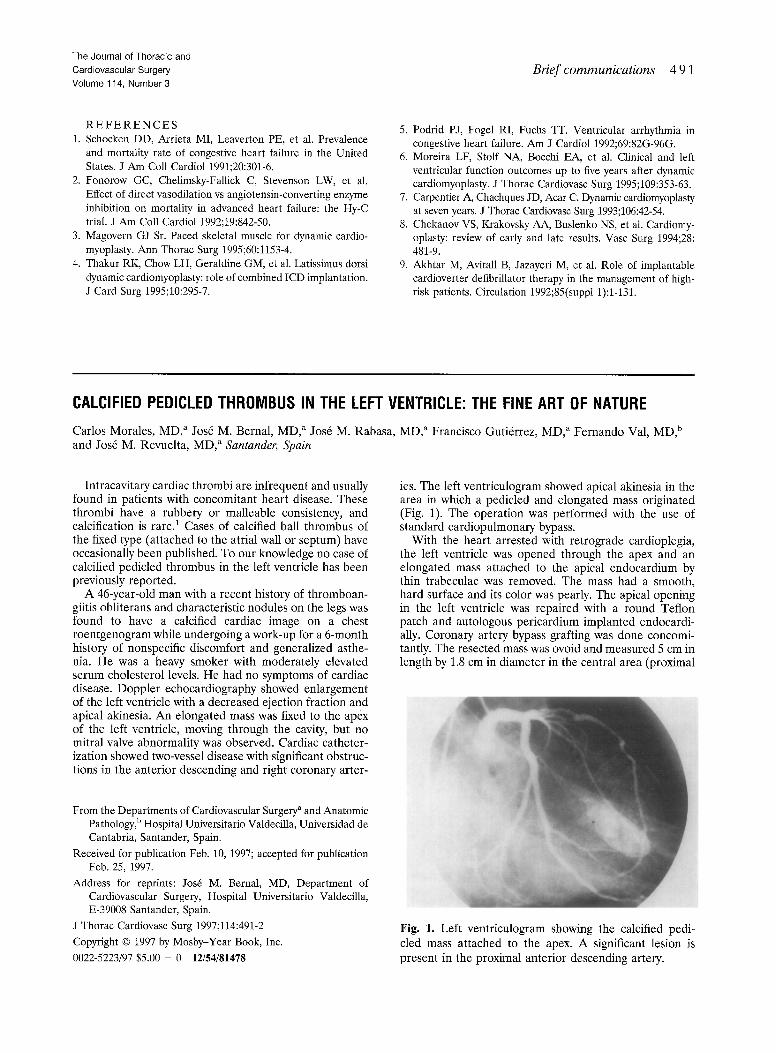
The Journal of Thoracic and Cardiovascular Surgery Volume 114, Number 3 Brief communications 4 9 1 REFERENCES 1. Schocken DD, Arrieta MI, Leaverton PE, et al. Prevalence and mortality rate of congestive heart failure in the United States. J Am Coll Cardiol 1991;20:301-6. 2. Fonorow GC, Chelimsky-Fallick C, Stevenson LW, et al. Effect of direct vasodilation vs angiotensin-converting enzyme inhibition on mortality in advanced heart failure: the Hy-C trial. J Am Coll Cardiol 1992;19:842-50. 3. Magovern GJ Sr. Paced skeletal muscle for dynamic cardio- myoplasty. Ann Thorac Surg 1995;60:1153-4. 4. Thakur RK, Chow LH, Geraldine GM, et al. Latissimus dorsi dynamic cardiomyoplasty:role of combined ICD implantation. J Card Surg 1995;10:295-7. 5. Podrid PJ, Fogel RI, Fuchs TT. Ventricular arrhythmia in congestive heart failure. Am J Cardiol 1992;69:82G-96G. 6. Moreira LF, Stolf NA, Bocchi EA, et al. Clinical and left ventricular function outcomes up to five years after dynamic cardiomyoplasty. J Thorac Cardiovasc Surg 1995;109:353-63. 7. Carpentier A, Chachques JD, Acar C. Dynamiccardiomyoplasty at seven years. J Thorac Cardiovasc Surg 1993;106:42-54. 8. Chekanov VS, KrakovskyAA, Buslenko NS, et al. Cardiomy- oplasty: review of early and late results. Vasc Surg 1994;28: 481-9. 9. Akhtar M, Avitall B, Jazayeri M, et al. Role of implantable cardioverter defibrillator therapy in the management of high- risk patients. Circulation 1992;85(suppl 1):1-131. CALCIFIED PEDICLEDTHROMBUS IN THE LEFT VENTRICLE: THE FINE ART OF NATURE Carlos Morales, MD, a Jos6 M. Bernal, MD, a Jos6 M. Rabasa, MD, a Francisco Gutidrrez, MD, a Fernando Val, MD, b and Jos6 M. Revuelta, MD, a Santander, Spain Intracavitary cardiac thrombi are infrequent and usually found in patients with concomitant heart disease. These thrombi have a rubbery or malleable consistency, and calcification is rare. 1 Cases of calcified ball thrombus of the fixed type (attached to the atrial wall or septum) have occasionally been published. To our knowledge no case of calcified pedicled thrombus in the left ventricle has been previously reported. A 46-year-old man with a recent history of thromboan- giitis obliterans and characteristic nodules on the legs was found to have a calcified cardiac image on a chest roentgenogram while undergoing a work-up for a 6-month history of nonspecific discomfort and generalized asthe- nia. He was a heavy smoker with moderately elevated serum cholesterol levels. He had no symptoms of cardiac disease. Doppler echocardiography showed enlargement of the left ventricle with a decreased ejection fraction and apical akinesia. An elongated mass was fixed to the apex of the left ventricle, moving through the cavity, but no mitral valve abnormality was observed. Cardiac catheter- ization showed two-vessel disease with significant obstruc- tions in the anterior descending and right coronary after- From the Departments of Cardiovascular Surgery a and Anatomic Pathology, b Hospital Universitario Valdecilla, Universidad de Cantabria, Santander, Spain. Received for publication Feb. 10, 1997; accepted for publication Feb. 25, 1997. Address for reprints: Jos6 M. Bernal, MD, Department of Cardiovascular Surgery, Hospital Universitario Valdecilla, E-39008 Santander, Spain. J Thorac Cardiovasc Surg 1997;114:491-2 Copyright © 1997 by Mosby-Year Book, Inc. 0022-5223/97 $5.00 + 0 12/54/81478 ies. The left ventriculogram showed apical akinesia in the area in which a pedicled and elongated mass originated (Fig. 1). The operation was performed with the use of standard cardiopulmonary bypass. With the heart arrested with retrograde cardioplegia, the left ventricle was opened through the apex and an elongated mass attached to the apical endocardium by thin trabeculae was removed. The mass had a smooth, hard surface and its color was pearly. The apical opening in the left ventricle was repaired with a round Teflon patch and autologous pericardium implanted endocardi- ally. Coronary artery bypass grafting was done concomi- tantly. The resected mass was ovoid and measured 5 cm in length by 1.8 cm in diameter in the central area (proximal Fig. 1. Left ventriculogram showing the calcified pedi- cled mass attached to the apex. A significant lesion is present in the proximal anterior descending artery.